Abstract
Feline α 1-acid glycoprotein (fAGP) increases during feline infectious peritonitis (FIP). We have recently identified a 29 kDa protein that we named feline AGP-related protein (fAGPrP) due to its cross-reactivity with an anti-human AGP monoclonal antibody. In this work we describe the tissue distribution of fAGPrP during FIP, and its relationship with feline coronavirus (FCoV) and myeloid cells. Tissues from five control cats and from 15 cats with FIP were examined by immunohistochemistry using monoclonal antibodies against human AGP, FCoV and myeloid antigens. Diffuse fAGPrP positivity within the lesions, likely due to vascular plasma leakage, endothelial and epithelial lining were detectable. Compared to controls, fAGPrP-expressing cells often increased in number and were diffusely distributed in lymph nodes, as usually occurs for IgM-producing plasma cells during early immune responses. These findings did not depend on the presence of FCoVs or of myeloid cells, suggesting that fAGPrP is not directly involved in the pathogenesis of FIP.
Introduction
α 1-acid glycoprotein (AGP), is an acute phase protein (APP) belonging to the lipocalin family (Lögdberg and Wester, 2000). It is characterized by low molecular weight (41–43 kDa), low pI (2.8–3.8) and high percentage of carbohydrates (45%). AGP is supposed to have an immunomodulatory and anti-inflammatory role, based on its ability to down-regulate neutrophil and lymphocyte responsiveness and to modulate the production of anti-inflammatory cytokines by peripheral blood mononuclear leukocytes (Bories et al., 1990;Vasson et al., 1994). Some of these anti-inflammatory and immunomodulatory activities depend on the glycosylation pattern of AGP (Shiyan and Bovin, 1997). In humans, the rate of sialylation has been suggested to increase the resistance to inflammation, even in viral infections such as HIV (Atemezem et al., 2001; Mackiewicz and Mackiewicz, 1995; Rabehi et al., 1995).
A similar mechanism might be involved, in cats, in the resistance to feline coronavirus (FCoV) infection: in fact, after exposure to the FCoV, feline AGP (fAGP) concentration transiently increases in blood from cats that did not develop feline infectious peritonitis (FIP) (Giordano et al., 2004). In contrast, high levels of fAGP are persistently detectable in blood from cats with FIP (Duthie et al., 1997).
During studies on the glycosylation pattern of fAGP we identified a second, different protein of about 29 kDa, that we named fAGP relatedprotein (fAGPrP) due to its cross-reactivity with a monoclonal anti-human AGP antibody. The concentration of fAGPrP in blood was not related to that of fAGP. A computational analysis (http://dove.embl-heidelberg.de/blast) of human AGP did not find protein homologues to AGP, suggesting that fAGPrP is a protein different from AGP. Moreover, the protein disappeared when AGP was purified to homogeneity. However, since the low isoelectric point of AGP is largely due to the glycan moiety of the protein (Shiyan and Bovin, 1997), it is also possible that a partial deglycosylated isoform of 29 kDa is not co-purified with the completely glycosylated AGP (Paltrinieri et al., 2003). We are now purifying and sequencing this protein to understand if it is an isoform of fAGP, a new APP or a protein with different function. In the meanwhile, we have examined fAGPrP distribution in blood and tissues from cats with different pathological conditions: fAGPrP was underexpressed in serum from cats with FIP and overexpressed in cats with FIV and purulent inflammations. Moreover, in healthy cats fAGPrP had an intrahepatocytic localization and the plasma positivity typical of APP (Ceciliani et al., 2002) but was also expressed on tissue cells which were particularly abundant in the lamina propria of small intestine and in perifollicular areas of lymphoid organs. During inflammation, fAGPrP showed an endothelial and an epithelial lining and the number of positive cells was very variable depending on the disease: this variability was particularly evident in the few cases of FIP examined (Paltrinieri et al., 2003).
In the present study we investigate the role of fAGPrP during FIP by examining its tissue distribution and its relationship with FCoVs and myeloid cells.
Material and methods
Tissue samples were taken from five cats without inflammatory disease (controls) and from 15 cats with FIP. Liver, spleen, lymph nodes, kidney, small intestine and lung were sampled from all the control cats. Small intestine and lungs were available for only 11 and 8 cats with FIP, respectively, while the other organs were sampled from all the cats with FIP.
Immunohistochemistry was performed on 5 μm thick sections obtained from formalin fixed and paraffin embedded samples. Monoclonal antibodies against human AGP (Sigma Diagnostic, St. Louis, MO, USA), Feline Coronavirus (kindly provided by Prof. N.C. Pedersen, Davis, USA), and myeloid cell antigens expressed on both granulocytes and macrophages (MAC387—DAKO, Glostrup, Denmark) were applied on serial sections at the final dilution of 1:5000. The Avidin Biotin Complex (ABC) method with a commercially available kit (Vectastain Elite, Vector Labs Inc, Burlingame, CA, USA) was used to detect the positive reaction, as previouslydescribed (Hsu et al., 1980), after inhibition of theendogenous peroxidase (H2O21% in methanol). Antigen unmasking was performed using microwave pretreatment (two cycles of 5 minutes in citrate-buffered solution, 0.01 M, pH 6.2) (Cattoretti et al., 1993). Diaminobenzidine served as chromogen for the reaction and the slides were counterstained with Mayer's haematoxyilin.
In each section, the number of cells positive for fAGPrP was evaluated and expressed as follows:
−=absent
+/−=rare (<2 per high power field)
+=mild (2–5 per high power field)
++=moderate (5–10 per high power field)
+++=abundant (>10 per high power field)
In the same way the intensity of vascular positivity for fAGPrP was evaluated and considered as faint (+), moderate (++) or strong (+++).
Results
Distribution of positive cells and intensity of vascular positivity
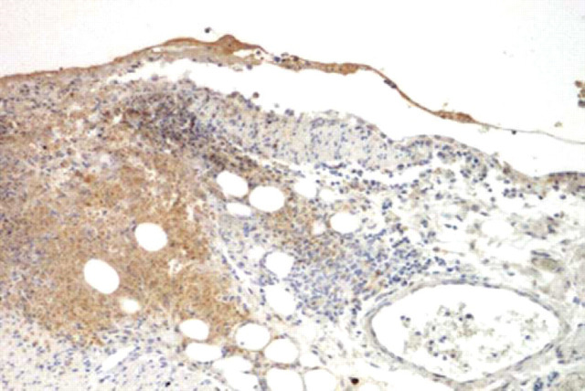
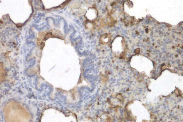
In controls, fAGPrP positivity was detected in plasma and within hepatocytes. fAGPrP-expressing cells were also detectable, particularly in the lamina propria of small intestine and in perifollicular areas of lymphoid organs. In cats with FIP a diffuse weak positivity within the perivisceral fibrin close to the pyogranulomatous foci, possibly due to plasma leakage from vessels, was detectable (Fig. 1). The number of cases on which lung, kidney and spleen showed moderate or strong plasmatic positivity was higher than in controls (Table 1). Moreover, the endothelium stained often positive in both the inflamed tissues and in tissues showing no signs of inflammation, particularly in kidney, and a strong positivity on epithelial surface of lung was detectable (Fig. 2). This finding was also detectable in lungs that did not show pyogranulomatous lesions or fibrinous pleuritis.
Figure 1.
Omentum. Diffuse fAGPrP positivity (brown color) close to a perivascular pyogranulomatous lesion. Immunohistochemistry, anti-human AGP antibody, Mayer's hematoxylin counterstain, 100×.
Table 1.
Distribution of cats with different intensity of plasmatic fAGPrP positivity
| + | ++ | +++ | ||
|---|---|---|---|---|
| Intestine | FIP | 3 | 7 | 1 |
| Controls | 1 | 3 | 1 | |
| Kidney | FIP | 3 | 4 | 8 |
| Controls | 1 | 3 | 1 | |
| Lung | FIP | 2 | 3 | 3 |
| Controls | 5 | 0 | 0 | |
| Lymph nodes | FIP | 6 | 7 | 2 |
| Controls | 2 | 3 | 0 | |
| Spleen | FIP | 4 | 6 | 5 |
| Controls | 2 | 3 | 0 |
+=faint positivity; ++=moderate positivity; +++=strong positivity.
Figure 2.
Lung. FAGPrP (brown color) is particularly abundant on bronchial and alveolar epithelium in a lung with a severe, diffuse chronic interstitial pneumonia. Immunohistochemistry, anti-human AGP antibody, Mayer's hematoxylin counterstain, 250×.
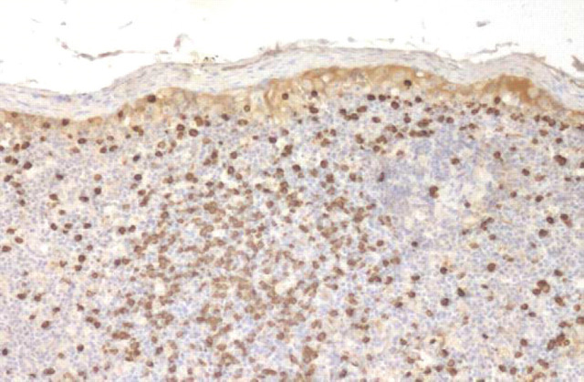
The number of positive cells increased in lung, lymph node and spleen compared to controls, although a strong variability among tissues was detectable (Table 2). Moreover positive cells were often diffusely distributed throughout the lymphoid organs, loosing the typical perifollicular localization (Fig. 3).
Table 2.
Distribution of cats with different number of fAGPrP-expressing cells
| - | +/- | + | ++ | +++ | ||
|---|---|---|---|---|---|---|
| Intestine | FIP | 1 | 3 | 4 | 3 | 0 |
| Controls | 1 | 1 | 2 | 1 | 0 | |
| Kidney | FIP | 3 | 6 | 4 | 2 | 0 |
| Controls | 1 | 2 | 1 | 1 | 0 | |
| Lung | FIP | 0 | 1 | 2 | 4 | 1 |
| Controls | 2 | 3 | 0 | 0 | 0 | |
| Lymph nodes | FIP | 1 | 2 | 5 | 5 | 2 |
| Controls | 0 | 1 | 3 | 1 | 0 | |
| Spleen | FIP | 0 | 3 | 6 | 5 | 1 |
| Controls | 1 | 1 | 2 | 1 | 0 |
−=absent; +/-=rare (<2 per high power field); +=mild (2–5 per high power field); ++=moderate (5–10 per high power field); +++=abundant (>10 per high power field).
Figure 3.
Lymph node. Numerous fAGPrP-expressing cells, characterized by a strong brown-stained cytoplasm, diffusely distributed throughout a follicular structure. Immunohistochemistry, anti-human AGP antibody, Mayer's hematoxylin counterstain, 100×.
Relationship among fAGPr, FCoV and myeloid cells
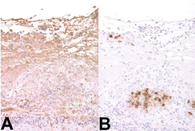
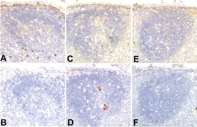
The amount of fAGPrP was not related to the amount of virus detectable in the lesions or to the presence of granulocytes or macrophages. In most of the cases the above described diffuse positivity in the fibrin was close to a small cluster of cells bearing the FCoV antigen (Fig. 4), while in other cases a diffuse positivity was found even in the absence of viruses. The endothelial lining detectable in small and medium vessels was often more intense toward foci containing FCoVs (Fig. 5). Positive cells were occasionally present within the fibrinous perivisceritis, independently on the presence of viruses. In the same way, intraparenchimatous foci were often rich of FCoV-positive cells with fAGPrP positivity confined to the central areas of necrosis, and myeloid cells peripherally distributed (Fig. 6). Also the above mentioned changes in both the number and distribution of fAGPrP positive cells were not dependent on the presence of parenchimal lesions or of viruses. Moreover, the presence of viral antigen in lymphoid organs did not influence the number and distribution of positive cells: in many cases, in fact, FCoV bearing cells were detectable within the germinal centers but few positive cells were detectable in perifollicular areas, while in other cases positive cells increased in number and were irregularly distributed compared to controls, even in absence of viral antigen (Fig. 7).
Figure 4.
Intestine. A: The fAGPrP positivity is detectable as a diffuse brown staining in perivisceral fibrin; B: FCoV antigen (brown color) is detectable within macrophage-like cells in pyogranulomatous foci. Immunohistochemistry, antibodies against human AGP (A) and FCoV (B), Mayer's hematoxylin counterstain, 100×.
Figure 5.
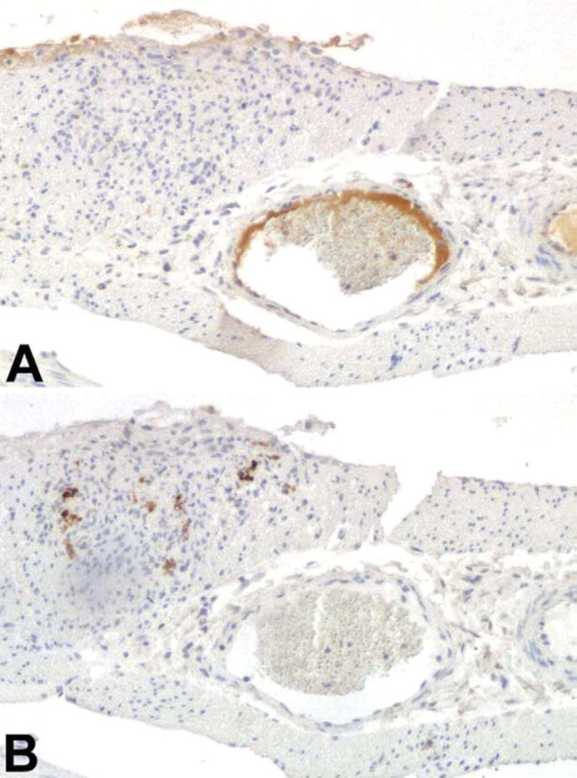
Intestine. A: Plasma fAGPrP is detectable asa strong brownish band localized on the vessel's side directed toward inflammatory foci; B: FCoV antigen (brown color) is detectable within the lesion. Immunohistochemistry, antibodies against human AGP (A) and FCoV (B), Mayer's hematoxylin counterstain, 100×.
Figure 6.
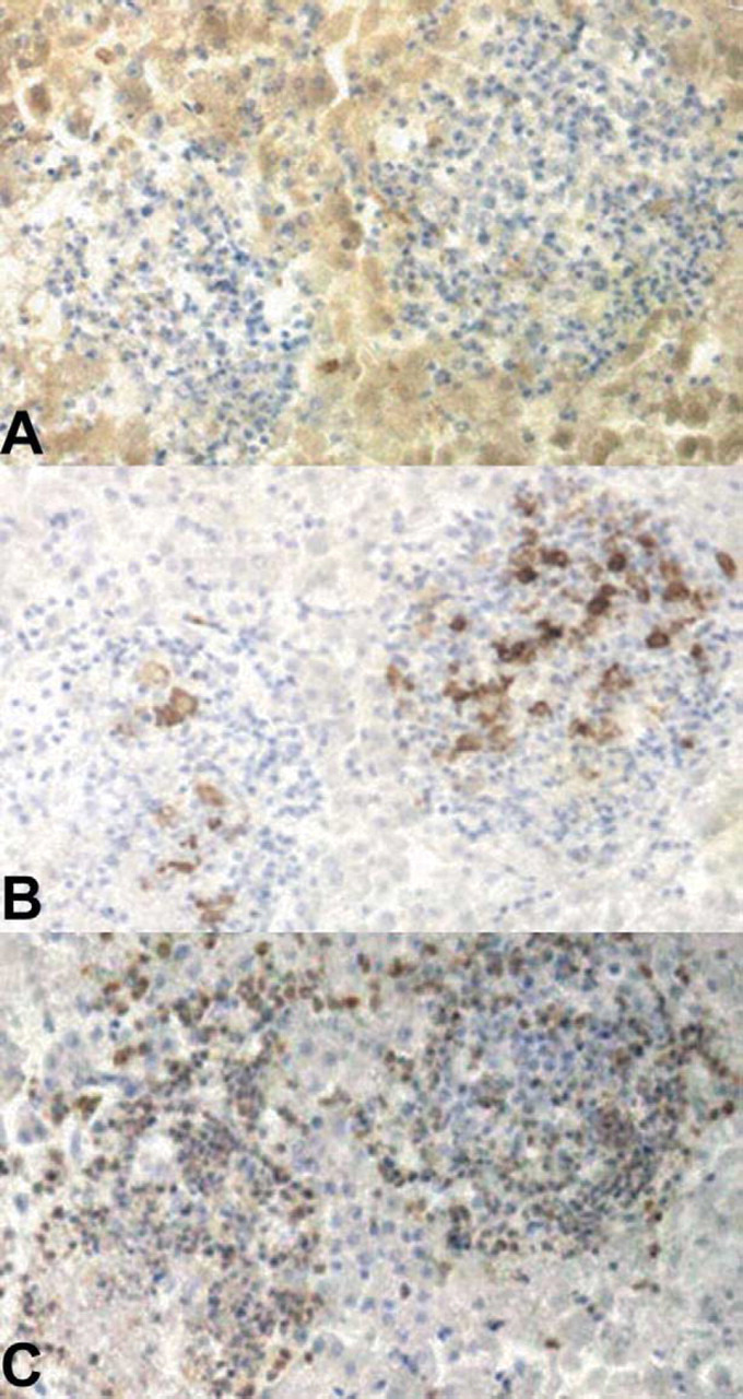
Liver. A: Positive fAGPrP staining is detectable as a diffuse brown staining in necrotic areas of pyogranulomatous foci; B: large amount of viral antigen (brown color) is detectable within the same foci; C: scattered myeloid cells (granulocytes and macrophages), characterized by a strong brownish cytoplasmic staining are detectable on the same lesions. Immunohistochemistry, antibodies against human AGP (A), FCoV (B), and myeloid antigens (C), Mayer's hematoxylin counterstain, 100×.
Figure 7.
Lymphnodes. Absence of correlation between fAGPrP-expressing cells and FcoV (both fAGPrP- and FCoVs-expressing cells have a brown-stained cytoplasm): fAGPrP-expressing cells can be normally present (A) around follicular structures without viruses (B), rare (C) in presence of FCoV (D) or virtually absent (E) in FCoV-free follicles (F). Immunohistochemistry, antibodies against human AGP (A, C, E) and FCoV (B, D, F), Mayer's hematoxylin counterstain, 100×.
Discussion
The aim of this work was to investigate the possible pathogenetic role of fAGPrP during FIP by analysing its tissue distribution and its relationship with both the FCoVs and the myeloid cells. Previous findings suggested that fAGPrP is down-regulated in the blood but variably distributed in tissues from cats with FIP (Paltrinieri et al., 2003). This variability was confirmed by the present study and the distribution of fAGPrP in the control cats was similar to that previously reported. Nevertheless, some of the results from the cats with FIP apparently contrast with the previous ones: the intensity of fAGPrP positivity in plasma was often stronger than in controls. However, this mainly occurred in tissues such as kidney and lung, in which vessels are normally so small as to be not easily detectable. It appears more likely than the strong positivity detected in these organs is due to increased amounts of plasma due to vasodilation rather than to an increased expression of fAGPrP. Moreover, the presence of extravasated fAGPrP close to pyogranulomatous foci or on pulmonary epithelium might explain the previously reported decrease of circulating fAGPrP (Paltrinieri et al., 2003).
Generally, a variable amount of positive cells on the lesions, and few differences in the number of cells among controls and cats with FIP, were detectable. Only in lung and in lymphoid organs were the number of positive cells higher than in controls. The increased number of cells in the lung might be due, again, to the low number of cells detectable in a normal lung: a non-specific inflammatory response might lead to an easily detectable increased number of “responsive” cells. These cells, in fact, are morphologically similar to IgM- or IgA-producing plasma cells (Waly et al., 2001). On this perspective it is not so surprising to find it within inflammatory foci, where plasma cells have been already identified (Kipar et al., 1998), or in lymphoid organs.
Our observations do not support the hypothesis of a direct relationship between the amount of FCoV or of myeloid cells and the presence of fAGPrP. The only immunohistochemical finding that suggests a possible interaction between fAGPrP and FCoVs is that positive plasma was often oriented toward inflammatory foci containing viral antigen. However, it should be considered that the amount of FCoVs per lesion has already been reported to be extremely variable and not related to the extent or to the histotype of the lesion (Tammer et al., 1995; Kipar et al., 1998): it cannot be excluded, thus, that the amount of fAGPrP might be related to other factors possibly involved in the histogenesis of the lesions (e.g. immune-complexes, cytokines).
The lack of relationship among fAGPrP positivity and myeloid cells is not surprising: the amount of MAC387-positive cells within the lesion, in fact, has been shown to increase when large necrotic areas are present, most likely due to the chemotactic effect exerted on myeloid cells by the necrosis itself (Paltrinieri et al., 1998).
The last observation regards the fAGPrP-expressing cells in lymph nodes: the presence of FCoV within germinal centers of lymphoid follicles has already been reported (Kipar et al., 1998) and it is thought to be associated to a different reactivity, characterized by increased circulating γ-globulins (Paltrinieri et al., 2001). The lack of relationship among FCoVs and fAGPrP-expressing cells is not surprising, since the latter are not thought to be IgG-producing plasma cells. In contrast, in some cats the increased number and the diffuse distribution of fAGPrP-expressing cells was consistent with the hypothesis that these cells might be IgM-producing plasma cells. During the early phase of the immune responses against FCoV, when circulating β-globulins, among which IgM migrate, increase (Paltrinieri et al., 2001), IgM-producing plasma cells might increase in lymphoid tissues. Unfortunately, our work has been performed on spontaneous cases of FIP: therefore we cannot know when the cats had the first contact with the virus or how long the presence of viruses was lasting. These speculations need to be supported by results from experimental infections, on which the changes in blood concentration of different proteins can be easily followed (Jacobse-geels et al., 1982;Stoddart et al., 1988).
In conclusion, our results support the hypotheses that fAGPrP is involved in inflammation and that fAGPrP-expressing cells are plasma cells. Nevertheless, fAGPrP do not seem to be directly involved in the histogenesis of the lesions during FIP or in the distribution of the virus throughout the body. The underexpression of fAGPrP previously recorded in blood from cats with FIP is most likely due to the extravasation from blood toward inflammatory foci or lung epithelium. However, these findings does not exclude that fAGPrP may play some role in protecting cats from the disease: in this regards further studies on the structure and glycosylation pattern of this protein in FCoV infected non-symptomatic cats are required.
Acknowledgments
This work was supported by the university grant FIRST 2001. The authors are very grateful to Dr Elisa Gabanti and to Dr Diane Addie.
References
- Atemezem A, Mbemba E, Vassy R, Slimani H, Saffar L, Gattegno L. Human alpha1-acid glycoprotein binds to CCR5 expressed on the plasma membrane of human primary macrophages, Biochemical Journal, 356, 2001, 121–128. [DOI] [PMC free article] [PubMed] [Google Scholar]
- Bories PN, Feger J, Benbernou N, Rouzeau JD, Agneray J, Durand G. Prevalence of tri- and tetraantennary glycans of human alpha 1-acid glycoprotein in release of macrophage inhibitor of interleukin-1 activity, Inflammation, 14, 1990, 315–323. [DOI] [PubMed] [Google Scholar]
- Cattoretti G, Pileri S, Parravicini C, Becker MH, Poggi S, Bifulco C, Key G, D'Amato L, Sabattini E, Feudale E, Reynolds F, Gerdes J, Rilke F. Antigen unmasking on formalin-fixed, paraffin-embedded tissue sections, Journal of Pathology, 171, 1993, 83–98. [DOI] [PubMed] [Google Scholar]
- Ceciliani F, Giordano A, Spagnolo V. The systemic reaction during inflammation: the acute phase proteins, Protein and Peptide Letters, 3, 2002, 211–223. [DOI] [PubMed] [Google Scholar]
- Duthie S, Eckersall PD, Addie DD, Lawrence CE, Jarrett O. Value of α1-acid glycoprotein in the diagnosis of feline infectious peritonitis, Veterinary Record, 141, 1997, 299–303. [DOI] [PubMed] [Google Scholar]
- Giordano A, Spagnolo V, Colombo A, Paltrinieri S. Changes in some acute phase protein and immunoglobulin concentrations in cats affected by feline infectious peritonitis or exposed to feline coronavirus infection, Veterinary Journal, 167, 2003, 38–44. [DOI] [PMC free article] [PubMed] [Google Scholar]
- Hsu SM, Raine L, Farger H. Use of avidin-biotin-peroxidase complex (ABC) in immunoperoxidase techniques: a comparison between ABC and unlabeled antibody (PAP) procedures, Journal of Histochemistry and Cytochemistry, 29, 1980, 577–580. [DOI] [PubMed] [Google Scholar]
- Jacobse-geels H.E.L., Daha M.R., Horzinek M.C. Antibody, immune complexes and complement activity fluctuations in kittens with experimentally induced feline infectious peritonitis, American Journal of Veterinary Research, 43, 1982, 666–670. [PubMed] [Google Scholar]
- Kipar A, Bellmann S, Kremendhal J, Reinacher M. Cellular composition, coronavirus antigen expression and production of specific antibodies in lesions in feline infectious peritonitis, Veterinary Immunology and Immunopathology, 65, 1998, 243–257. [DOI] [PMC free article] [PubMed] [Google Scholar]
- Lögdberg L, Wester L. Immunocalins: a lipocalin subfamily that modulates immune and inflammatory responses, Biochimica et Biophysica Acta, 1482, 2000, 284–297. [DOI] [PubMed] [Google Scholar]
- Mackiewicz A, Mackiewicz K. Glycoforms of serum alpha 1-acid glycoprotein as markers of inflammation and cancer, Glycoconjugate Journal, 12, 1995, 241–247. [DOI] [PubMed] [Google Scholar]
- Paltrinieri S, Parodi Cammarata M, Cammarata G, Mambretti M. Type IV hypersensitivity in the pathogenesis of FIPV-induced lesions, Journal of Veterinary Medicine B, 45, 1998, 151–159. [DOI] [PubMed] [Google Scholar]
- Paltrinieri S, Grieco V, Comazzi S, Parodi Cammarata M. Laboratory profiles in cats with different pathological and immunohistochemical findings due to feline infectious peritonitis (FIP), Journal of Feline Medicine and Surgery, 3, 2001, 149–159. [DOI] [PMC free article] [PubMed] [Google Scholar]
- Paltrinieri S, Ceciliani F, Gabanti E, Sironi G, Giordano A, Addie D. Expression patterns in feline blood and tissues of a1 acid glycoprotein (AGP) and of an AGP related protein (AGPrP), Comparative Clinical Pathology, 12, 2003, 140–146. [DOI] [PMC free article] [PubMed] [Google Scholar]
- Rabehi L, Ferriere F, Saffar L, Gattegno L. Alpha 1-acid glycoprotein binds human immunodeficiency virus type 1 (HIV-1) envelope glycoprotein via N-linked glycans, Glycoconjugate Journal, 12, 1995, 7–16. [DOI] [PubMed] [Google Scholar]
- Shiyan SD, Bovin NV. Carbohydrate composition and immunomodulatory activity of different glycoforms of alpha1-acid glycoprotein, Glycoconjugate Journal, 14, 1997, 631–638. [DOI] [PubMed] [Google Scholar]
- Stoddart ME, Whicher JT, Harbour DA. Cats inoculated with feline infectious peritonitis virus exhibit a biphasic acute phase plasma protein response, Veterinary Record, 123, 1988, 621–624. [PubMed] [Google Scholar]
- Tammer R, Evensen O, Lutz H, Reinacher M. Immunohistological demonstration of feline infectious peritonitis virus antigen in paraffin-embedded tissues using feline ascites or murine monoclonal antibodies, Veterinary Immunology and Immunopathology, 49, 1995, 177–182. [DOI] [PMC free article] [PubMed] [Google Scholar]
- Vasson MP, Arveiller M Roch, Couderc R, Baguet JC, Raichvarg D. Effects of alpha-1 acid glycoprotein on human polymorphonuclear neutrophils: influence of glycan microheterogeneity, Clinica Chimica Acta, 224, 1994, 65–71. [DOI] [PubMed] [Google Scholar]
- Waly N, Gruffyd-Jones TJ, Stokes CR, Day MJ. The distribution of leococytes subsets in the small intestine of healthy cats, Journal of Comparative Pathology, 124, 2001, 172–182. [DOI] [PubMed] [Google Scholar]


