

Abstract
The average life span reported in laboratory and lay literature for the domestic rabbit is 5 to 10 years. The author and other veterinarians are now regularly seeing rabbits living to 9 or 10 years, the oldest reported in the author's practice being 14 years. Rabbits are herbivorous prey species with continually growing (elodont) teeth. This feature allows the geriatric rabbit to possess teeth that are essentially “new”, a distinct advantage over geriatric carnivores. Expanded longevity, while generally desirable, necessarily accompanies an increase in geriatric disorders. This article examines the spectrum of disease that can affect the geriatric rabbit as well as crucial factors concerning the clinical management of the animal up to the end of its life. An improved understanding of geriatric disorders in pet rabbits allows early recognition and the opportunity to improve quality of life.
Keywords: Rabbit, Geriatric, Arthritis, Dental disease
Exotic companion mammal veterinarians are seeing an extension of life spans of pet rabbits seen in practice. The average life span reported in laboratory and lay literature for the domestic rabbit is 5 to 10 years. The author and others are now regularly seeing rabbits living to 9 or 10 years, the oldest reported in the author's practice being 14 years.
Rabbits are herbivorous prey species with continually growing (elodont) teeth.1 This feature allows the geriatric rabbit to possess teeth that are essentially “new,” a distinct advantage over geriatric carnivores. In the wild, longevity is not naturally achieved by prey species. Expanded longevity is generally desirable; however, it necessarily accompanies an increase in geriatric disorders. An improved understanding of geriatric disorders in pet rabbits allows early recognition and the opportunity to improve quality of life.
Well care for geriatric rabbits
Opinions vary as to recommended frequency of examinations for apparently well older rabbits. The author prefers to begin biannual examinations with analysis of the complete blood count and biochemistry panel at age 5 years, with examinations increasing as age and condition indicate. The stress of presentation for examination and phlebotomy should be weighed against the advantage of frequent evaluation.
Chronic renal failure
Chronic renal failure is common in rabbits, and is often recognized during an episode of acute failure.2 Whereas acute renal failure (sudden onset of filtration failure characterized by accumulation of uremic toxins and fluid/electrolyte and acid/base imbalance) can be characterized as pre-, post-, and intrinsic renal, most acute failures in older rabbits are an acute episode of a chronic intrinsic renal failure.2 Clinical signs and symptoms may be subtle, but may include dehydration, polyuria/polydypsia, weight loss, failure to groom, anorexia, and depression. There are numerous causes of chronic renal disease in rabbits, but some are primarily identified only in laboratory animals, for example, feeding of diets with excessive vitamin D and calcium. Other causes include urolithiasis, bacterial infections, neoplasia, and nephrocalcinosis.2
Encephalitozoon cuniculi (ECUN) has been demonstrated to produce mild to severe interstitial nephritis. A recent study of histopathologic examinations of pet rabbits that died or were euthanized showed that of 48 rabbits with ECUN spores detected in the brain, 39 also had interstitial nephritis; 89.6% of renal lesions were described as chronic, whereas the rest were acute. However, the same study showed that the degree of severity of lesions did not always correlate with the degree of clinical symptoms.3 Antemortem diagnosis of ECUN as a cause of renal failure in rabbits is difficult, but can be supported with serology and protein electrophoresis.4
Management of acute renal failure is similar to that for other species.5 Management of chronic renal failure is supportive. Some cases benefit from long-term administration of subcutaneous fluids and hand feeding. The GIF Tube Implant kit is a silicone catheter designed for long-term implantation in the subcutis for administration of fluids (GIF-Tube, PractiVet, Phoenix, AZ). Though designed for dogs and cats, the author and others have used this product in rabbits with chronic renal failure requiring longer-term at-home subcutaneous administration of fluid (see Hospice and end-of-life issues later in this article) (Fig. 1 ). The manufacturer provides a demonstration CD of implantation instructions for practitioners and at-home use for owners.
Fig. 1.

The GIF Tube Implant kit.
Erythropoietin has been administered to rabbits with secondary anemia.2
The author has encountered numerous rabbits experiencing good quality of life for many months, despite muscle wasting and persistently elevated blood urea nitrogen and creatinine.
Cardiovascular disease
Cardiovascular disease is gaining recognition in exotic companion mammals as the level of veterinary care increases and patients age. The rabbit is a model for atherosclerosis in humans, as lesions are readily produced by feeding a diet high in fat.6 However, little is known about naturally occurring disease in the pet rabbit.
The thoracic cavity and lungs of the rabbit are exceptionally small in comparison with those of other similar-sized animals. Severe reduction in pulmonary mass (as in mediastinal tumors) does not seem to produce symptoms until late in the course of the disease. Therefore, it is likely the rabbit may not exhibit cardiac-related respiratory symptoms until late in the course of the disease. Symptoms can include depression, exercise intolerance, and increased respiratory rate and effort, which in the author's experience may be absent at rest and significantly worse with exercise.
Diagnosis of heart disease is enhanced with radiography and cardiac ultrasonography, which is important to better characterize heart disease and determine treatment options. Normal parameters for echocardiography have been described.7
Cardiac conditions reported in the rabbit include valvular disease (endocardiosis) and cardiomyopathy. Several diseases have been reported to cause cardiomyopathy in the laboratory rabbit, but incidence in pet rabbits is unknown, and likely to be low. These include nutritional deficiencies (vitamin E deficiency), viral (coronavirus), bacterial (salmonellosis or pasteurellosis), or protozoal (ECUN) infections, and toxins.7, 8
Excessive stress may produce heart disease. Stress causes catecholamine release in rabbits, and when sustained can result in coronary vessel constriction with ischemic cardiomyopathy.7
Treatment of congestive heart failure relies on use of drugs traditionally used in canine medicine, and are based on anecdotal reports of success.2 Drugs used by the author for management of cardiac disease are listed in Table 1 . Drug dosages should be adapted to clinical response and improvement of echocardiographic results. The use of taurine has been shown to improve cardiovascular function in laboratory rabbits with induced cardiac failure (see Table 1).
Table 1.
Drug dosages for geriatric rabbits
| Drug | Dosage (mg/kg) | Comments |
|---|---|---|
| Meloxicam28 | 0.2–0.3 IM, PO SID | Some report higher dosages required for more chronic pain; monitor renal values throughout treatment21 |
| Ketoprofen28 | 3 every 24 h IM | |
| Carprofen28 | 4–5 every 24 h PO | |
| Furosemide29 | 1–4 every 4–6 h IM | |
| Enalapril7 | 0.5 every 12–24 h PO | |
| Digoxin29 | 0.005–0.01 every 24–48 h PO | |
| Pimobendam7 | 0.1–0.3 every 12–24 h PO | |
| Taurine30 | 100 SID PO | Has demonstrated improvement in cardiac function in rabbits with artificially induced heart failure |
Consider dose reduction in rabbits with renal and/or hepatic disease.
Abbreviations: BSAVA, British Small Animal Veterinary Association; IM, intramuscular; PO, by mouth; SID, once a day.
Vascular disease can produce hypertension. Indirect blood pressure is determined using an ultrasonic Doppler and pediatric cuff (width 30%–40% of the diameter of the limb circumference) usually placed proximal to the carpus. Acquiring an audible pulse with the Doppler requires significant practice; initial difficulty encountered should not discourage the practitioner from developing this skill. The author and others normally note blood pressures in normal patients between 110 and 180 mm Hg using this method.
Arthritis/degenerative joint disease
Osteoarthritis and vertebral spondylosis are commonly encountered in older rabbits. Associated signs and symptoms include reluctance to move, urine/fecal staining due to inability to clean and properly direct the urine stream, and lameness.
The rabbit is a laboratory model for trauma-induced arthritis and response to drug therapy. In rabbits whose legs were immobilized with plaster casts, those receiving intra-articular injections of 0.3 mL hyaluronic acid showed significant reduction of cartilage degeneration when compared with rabbits receiving a saline injection.9 Similar effects (retardation of progression of osteoarthritis) were seen in rabbits undergoing anterior cruciate ligament transection that were injected with an intra-articular mixture of glucose or dextrose, amino acids, and ascorbic acid 5 times weekly.10 Other drugs shown to reduce degenerative changes in rabbit osteoarthritis models include intra-articular administered sodium hyaluronate and orally administered glucosamine hydrochloride, 100 mg by mouth daily.11, 12, 13
Several studies exist on the use of joint health products in dogs, and include glucosamine, chondroitin, P54FP (Indian and Javanese tumeric extract), green-lipped mussels, and ω-3 fatty acids.14 Results are variable; however, a systematic review of the literature showed that there is moderate evidence that some joint health products (JHPs), including green-lipped mussels products, P54FP, a combination of chondroitin sulfate, glucosamine hydrochloride, and manganese ascorbate, provide some benefit.14 The author is unaware of studies on the effects of JHPs in rabbits with naturally occurring disease.
Product recommendation can be difficult, and dosing is extrapolated from other species. The ACCLAIM system has been proposed as a means for veterinarians to evaluate the label claims and quality of specific JHPs (Table 2 ).15
Table 2.
ACCLAIM system for rapid evaluation of joint health product labels15
| A | A name you recognize? | Products manufactured by an established company that provides educational materials for veterinarians or other consumers are prerable to joint health products manufactured by a new company |
| C | Clinical experience | Companies that support clinical research and have their products used in clinical trials that are published in peer-reviewed journals to which veterinarians have access are more likely to have a quality product |
| C | Contents | All ingredients should be clearly indicated on the label |
| L | Label claims | Label claims that sound too good to be true probably are. Products with realistic label claims based on results of scientific studies, rather than testimonials, are more likely to be reputable. Products with illegal claims (claim to diagnose, treat, cure, or prevent a disease) should be avoided |
| A | Administration recommendations | Dosing instructions should be accurate and easy to follow; it should be easy to calculate the amount of active ingredient administered per dose per day |
| I | Identification of lot | A lot identification number or some other tracking system indicates that a premarket or postmarket surveillance system exits to ensure product quality. In addition, companies that have voluntarily instituted current good manufacturing practices and other quality-control or quality-assurance techniques (eg, tamper-resistant packaging of identification of individual tablets or capsules) provide evidence of long-term investment in the product and company |
| M | Manufacturer information | Basic company information should be clearly stated on the label. Preferably, this should include a Web site or details for contacting customer support |
From Oke S. Oral joint supplements. The Horse 2008; May 1; with permission.
Housing should be optimized for rabbits with spondylosis or joint disease. Nonslip, soft surfaces are beneficial, as the force produced by hopping is much higher than that produced by walking. Lowering one side of the litter box allows easier access. Owners must be instructed to clean the perineum daily (see later discussion on perineal dermatitis).
Dental disease
Dental disease is not necessarily associated with aging in rabbits. Many older rabbits manage to avoid acquired dental disease and possess essentially normal, continually renewing teeth well into old age. Other older rabbits may develop varying patterns of dental disease apparently related to slowing or cessation of tooth growth, which may be due to attrition of the alveolus. Severe acquired dental disease, however, is primarily a disease of younger rabbits; in these patients, evidence of dental disease is often apparent before 3 years of age.1 Accurate diagnosis and excellent owner compliance can result in adequate disease management and acquisition of normal expected life span; in many cases these patients eventually die of diseases unrelated to those of dentition. The author and others frequently see geriatric rabbits that have had years of regular dental care. In some cases all teeth are eventually lost, and patients survive with good to excellent quality of life on a diet of liquid Critical Care (Oxbow Animal Health, Murdoch, NE). Diagnosis and treatment of dental disease is presented in great detail elsewhere.1
Splay leg
Several practitioners have reported unilateral or bilateral abduction of the thoracic limbs in older, often larger breed rabbits (Fig. 2 ). The condition seems to be associated with muscle wasting and is generally progressive. Housing on nonslick surfaces is helpful.
Fig. 2.
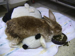
Older rabbit with a history of severe acquired dental disease, retrobulbar abscess, and enucleation of the right eye. The rabbit survived with good quality of life for several years on Oxbow Critical Care and soaked pellets. Worsening splay leg of the thoracic limbs resulted in decreased mobility. The owner used towels and a stuffed animal to support the pet during brief hospice care at home before ultimately choosing euthanasia.
Muscle wasting
Though not a primary disease disorder, muscle wasting and moderate to severe weight loss is a common feature in aged rabbits. Causes are varied, and can include chronic renal failure and acquired dental disease. A thorough approach is required to discover the underlying etiology.
Supplemental feeding is often beneficial. Critical Care can be offered in a dish or via syringe feeding. The manufacturer provides detailed instructions for product use.
Ocular lesions
Age-related ocular lesions, including cataract formation, are common in rabbits. Current literature suggests a high percentage of ocular lesions may be caused by ECUN.
A 2005 study describing histologic features of ECUN-induced ocular lesions in rabbits demonstrated intraocular locally extensive pyogranulomatous infiltration of the posterior chamber with disruption of the anterior lens capsule. Spores were identified via immunohistochemical staining in all cases.16 Another study described ocular lesions as phacoclastic uveitis. All samples from abnormal eyes (n = 10) obtained via enucleation or phacoemulsification were positive for ECUN via the polymerase chain reaction.17
Chronic otitis
Stenosis of the ear canal is a common finding in aged lop-eared rabbits. In the author's experience, all older lop-eared rabbits have some degree of stenosis, which predisposes to otitis. Disease is difficult to manage medically due to abnormal anatomy. Lateral ear canal resection, ostectomy, and total ear canal ablation may be required in selected cases.18
Ulcerative pododermatitis and perineal dermatitis
Pododermatitis can be encountered in rabbits of any age, and is often a result of improper husbandry (wire-bottom cages, inadequate cleaning); however, any condition impairing ability to groom normally and exposure to urine/feces due to decreased mobility can contribute. Inability to groom also leads to perineal accumulation of feces and urine and dermatitis, or scalding.
It is important to address the underlying cause of decreased mobility. Pododermatitis is treated with local wound management (debridement, flushing with or without bandaging), housing on soft surfaces, and antibiotics plus analgesics. Underlying immunocompromise and wasting can delay or prevent healing in some cases. Severe cases with pus and involvement of tendons, ligaments, and joints may require surgical intervention, including amputation, which should be considered carefully in the geriatric rabbit.
Perineal dermatitis is treated the same as any other infected wound, with careful clipping of the hair, cleansing and application of antibiotics, and use of soothing ointments or products designed to promote healing once infection has resolved and granulation has begun. Some elderly rabbits benefit from regular preventative shaving of the perineum in order to allow the owner to cleanse more effectively. The author prefers dilute chlorhexidine and application of silver sulfadiazine cream (SSD, Par Pharmaceutical, Shreveport, LA), Zinc products (Zn7 Derm, Addison Biological Laboratory Inc, Fayette, MO), or healing products (Heal-X Soother Plus Cream or Spray, Zoological Education Network, Lake Worth, FL).
Neoplasia
Neoplasia occurs in rabbits, and incidence of all neoplasms increases with age. The most commonly encountered neoplasm is uterine adenocarcinoma. Uterine adenocarcinoma can produce few signs until the disease is advanced. The most commonly reported sign is hematuria, and uterine masses are often palpable. The abnormal uterus may be palpable, and metastasis, especially to liver and lungs, can occur in some cases.19
The most common clinical presentation of thymoma is bilateral exophthalmos, which may worsen with exercise or excitation. Respiratory symptoms usually occur later in the course of the disease. Treatment options include surgery and radiation.20
Modification of therapeutics in geriatric rabbits
In human medicine, drug dosages are often adjusted for geriatric patients due to assumed decrease in renal and hepatic function.21 Although specific recommendations for geriatric rabbits are unavailable, drug dosage modification should be considered, especially when organ disease has been positively identified.
Chronic renal failure impacts drug metabolism; therefore, adjustments in drug dosages should be considered in patients with chronic renal failure. In humans, various strategies for estimation of glomerular filtration rate (GFR) are used for “adjusting the dosage of medications excreted by glomerular filtration.”21 For example, in human patients with impaired clearance, dosage of butorphanol is adjusted to half the normal dose. Although estimation of GFR is unavailable for rabbit patients, serious consideration should be given to lowering dosages of all drugs, in particular, any drugs metabolized by glomerular filtration in rabbits with suspected renal insufficiency. Drugs more commonly used in rabbits metabolized at least partially via glomerular filtration include sulfonamides such as trimethoprim sulfamethoxazole.
While procaine penicillin is excreted via tubular secretion, impaired renal function causes delayed excretion in humans and other species. Meloxicam is metabolized by the liver. Use is not recommended in humans and tested animal patients that are dehydrated, or have liver or renal disease (due to reduction of blood flow to the kidneys).22
Sedation and anesthesia for the geriatric rabbit
Human anesthesiologists routinely modify protocols and drug dosages for the elderly. A geriatric rabbit otherwise in good health may not necessarily represent a serious anesthetic risk; however, clinicians must assume some alteration of cardiovascular and renal health with advancing age.
Certain drugs should be avoided in older patients. Medetomidine (Pfizer Animal Health, New York, NY) is listed frequently as a choice for pre-anesthesia and anesthesia in the rabbit.23 A few studies have demonstrated use in rabbits, but use in elderly patients has not been investigated. It should be noted that medetomidine and dexmedetomidine are contraindicated in dogs or cats with cardiovascular disease, respiratory disease, liver or kidney disease, or in any debilitated patient.24 Therefore, use in the geriatric rabbit should be avoided or used cautiously.
The author and others prefer a balanced approach to sedation and anesthesia for all exotic animal patients, including pre-anesthesia, analgesia, local/regional analgesia, and anesthesia if required. Use of a balanced approach allows for reduction of any single agent, including inhalant agents, thus increasing patient safety. Many brief procedures producing minimal discomfort can be accomplished with sedation only, for example, radiography, placement of an intravenous catheter, and clipping or minor wound care.25 In geriatric patients, dosages of all drugs are reduced depending on patient condition.
For geriatric patients in which the need for sedation/anesthesia for diagnostic or therapeutic procedures outweigh the risks, the author has had great success with the combination of midazolam with an opioid (butorphanol, buprenorphine, hydromorphone, fentanyl), with the addition of low-dose ketamine if required (Table 3 ). The addition of local analgesia with lidocaine, 1 mg/kg as a local or regional block is extremely beneficial. When additional anesthesia is required, inhalant isoflurane or sevoflurane can be added at minimal effective concentrations.
Table 3.
Anesthetic and analgesic drugs used by the author in geriatric rabbits
| Drug | Dosage (mg/kg) | Comments |
|---|---|---|
| Midazolam | 0.25 IM | |
| Butorphanol | 0.10–0.20 IM | |
| Buprenorphine | 0.04 IM | |
| Hydromorphone | 0.10 IM | |
| Ketamine | 1–10 IM | |
| Etomidate | 1–2 IV | Must be combined with benzodiazepine to prevent muscle tremors |
Consider further dose reduction in rabbits with renal and/or hepatic disease.
Abbreviations: IM, intramuscular; IV, intravenous.
An alternative is the use of injectable etomidate combined with midazolam. Etomidate is commonly used in geriatric and high-risk human patients, and is apparently unaffected by impaired renal function.26 Etomidate must be injected intravenously, and combined with a benzodiazepine to prevent temporary tetany and seizures. Onset of action is immediate, and duration is approximately 5 to 7 minutes in rabbits and other mammals. The author's experience with this drug has been overwhelmingly positive, even in patients with advanced disease conditions. However, death occurred in a single geriatric rabbit with suspected advanced cardiac disease.
Hospice and end-of-life issues
Significant attention has been directed toward hospice, or palliative end-of-life care for companion animals. A recent Internet search revealed numerous businesses offering hospice services for traditional pets. The concept of hospice care can be applied to aged rabbits as well.
In general, the hospice setting should be clean, quiet, and stress-free. If possible, ill or dying rabbits should not be separated from bonded companions, and the author is unaware of situations in which bullying or harassment has occurred. Bedding should provide traction, be soft, absorbent, and easy to clean. Hospital absorbent pads are ideal (Wings Maxima Disposable Underpads, Tyco Healthcare) (Fig. 3 ). Food and water bowls and bottles are placed within easy reach. Caloric and fluid needs can be supplied through syringe feeding of Critical Care formula as per manufacturer's instructions. Nasogastric tube feeding can be considered for those rabbits unable or unwilling to eat; however, stress associated with this procedure should strongly be considered. Nasogastric feeding should be considered a temporary measure only.
Fig. 3.

Ill rabbit hospitalized on absorbent human hospital “underpads.” Pads help prevent urine accumulation and scalding.
Some rabbits benefit from administration of fluids, either subcutaneously or intravenously. Willing owners can be instructed to do either in the home setting. In selected cases, an implanted subcutaneous catheter is beneficial (see earlier section on Chronic renal failure).
Daily care also includes gentle cleaning of the perineum to prevent scalding. In rabbits with chronic epiphora, accumulations of secretions should be removed frequently to avoid dermatitis. Rabbits unwilling or unable to groom may benefit from shaving of the hair of the perineum in order to reduce fecal and urine accumulation and allow owners to more effectively cleanse the area.
Analgesia for hospice patients should emphasize optimal pain control, with lesser regard for untoward systemic effects.
Techniques for euthanasia
Choosing the time for euthanasia is difficult in most circumstances, and is particularly complicated in rabbits due to their inherent tendency to hide signs of illness. A significant change in routine, and unwillingness to eat or accept treats or groom are clear indications of distress in rabbits. In situations whereby their condition is unlikely to improve, euthanasia should be considered.
Humane, stress-free euthanasia requires careful planning and implementation, especially when the owners wish to be present. The American Veterinary Medical Association Guidelines on Euthanasia for rabbits suggest the following: barbiturates, CO2, CO, or potassium chloride in conjunction with general anesthesia. Other listed acceptable techniques such as cervical dislocation are unacceptable to most veterinary staff and owners.27 The author prefers the following technique, especially when owners wish to be present: induction with medetomidine at 50 μg/kg, and ketamine, 30 mg/kg administered intramuscularly. Owners are encouraged to hold their pet during induction as comfort level allows. Smaller doses may be repeated if necessary. Once deep anesthesia is achieved with no response to toe pinch, euthanasia solution is administered intravenously or by cardiac puncture.
Owners should be given options regarding disposition of the pet after euthanasia. Frequently chosen options include home burial (local ordinances permitting), and private or group cremation. Most small animal cremation companies are willing to provide services to owners of rabbits and other exotic pets (Fig. 4 ).
Fig. 4.

Decorative urn designed by a pet cremation company for a rabbit.
References
- 1.Capello V., Gracis M. In: Rabbit and rodent dentistry. Lennox A.M., editor. Wiley-Blackwell (Formerly Zoologic Education Network); Hoboken (NJ): 2005. [Google Scholar]
- 2.Pare J.A., Paul-Murphy J. Disorders of the reproductive and urinary systems. In: Quesenbery K.E., Carpenter J.W., editors. Ferrets, rabbits and rodents, clinical medicine and surgery. Saunders; St. Louis (MO): 2004. pp. 183–193. [Google Scholar]
- 3.Csokai J., Grube A., Kunzel F. Encephalitozoonosis in pet rabbits (Oryctolagus cuniculus): pathohistological findings in animals with latent infection versus clinical manifestation. Parasitol Res. 2009;104(3):629–635. doi: 10.1007/s00436-008-1239-2. [DOI] [PubMed] [Google Scholar]
- 4.Cray C., Arcia G., Kelleher S. Application of ELISA and protein electrophoresis in the diagnosis of Encephalitozoon cuniculi infection in rabbits. Am J Vet Res. 2009;70(4):478–482. doi: 10.2460/ajvr.70.4.478. [DOI] [PubMed] [Google Scholar]
- 5.Paul-Murphy J. Critical care of the rabbit. Vet Clin Exot Anim. 2007;10:437–461. doi: 10.1016/j.cvex.2007.03.002. [DOI] [PubMed] [Google Scholar]
- 6.Finking G., Hanke H. Nikolaj Nikolajewitsch Antischkow established the cholesterol-fed rabbit as a model for atherosclerosis research. Atherosclerosis. 1997;135:1–7. doi: 10.1016/s0021-9150(97)00161-5. [DOI] [PubMed] [Google Scholar]
- 7.Pariaut R. Cardiovascular physiology and diseases of the rabbit. Vet Clin Exotic Anim. 2009;12:135–144. doi: 10.1016/j.cvex.2008.08.004. [DOI] [PubMed] [Google Scholar]
- 8.Harcourt-Brown F. Textbook of rabbit medicine. Elsevier Science Limited; London: 2002. Cardiorespiratory disease; pp. 324–334. [Google Scholar]
- 9.Liang M.H., Shang H., Sun L. Preventive effect of hyaluronic acid on degerative articular cartilage of immobilized rabbit knees. J Clin Rehab Tissue Engineering Research. 2007;11(45):9043–9046. [Google Scholar]
- 10.Park Y.S., Lim S.W., Lee I.H. Intra-articular injection of a nutritive mixture solution protects articular cartilage from osteoarthritic progression induced by anterior cruciate ligament transection in mature rabbits: a randomized controlled trial. Arthritis Research Therapy. 2007;9:R8. doi: 10.1186/ar2114. [DOI] [PMC free article] [PubMed] [Google Scholar]
- 11.Zhang S.Z., Wu Y.H. The effect of sodium hyaluronate on the rabbit osteoarthrosis. Shanghai Kou Qiang Yi Xue. 2003;12(3):187–190. [PubMed] [Google Scholar]
- 12.Wang S.X., Laverty S., Dumitriu M. The effects of glucosamine hydrochloride on subchondral bone changes in an animal model of osteoarthritis. Arthritis Rheum. 2007;56(5):1527–1548. doi: 10.1002/art.22574. [DOI] [PubMed] [Google Scholar]
- 13.Harcourt-Brown F. Textbook of rabbit medicine. Elsevier Science Limited; London: 2002. Skin diseases; pp. 224–248. [Google Scholar]
- 14.Aragon C.L., Hofmeister E.H., Budsberg S.C. Systematic review of clinical trials of treatments for osteoarthritis in dogs. J AmVet Med Assoc. 2007;250:514–521. doi: 10.2460/javma.230.4.514. [DOI] [PubMed] [Google Scholar]
- 15.Oke S. Indications and contraindications for the use of orally administered joint health products in dogs and cats. J Am Vet Med Assoc. 2009;234(11):1393–1397. doi: 10.2460/javma.234.11.1393. [DOI] [PubMed] [Google Scholar]
- 16.Giordano C., Weigt A., Vercelli A. Immunohistochemical identification of Encephalitozoon cuniculi in phacoclastic uveitis in four rabbits. Vet Opthal. 2005;8(4):271–275. doi: 10.1111/j.1463-5224.2005.00394.x. [DOI] [PubMed] [Google Scholar]
- 17.Csokai J., Joachim A., Gruber A. Diagnostic markers for encephalitozoonosis in pet rabbits. Vet Parsitol. 2009;163(1–2):18–26. doi: 10.1016/j.vetpar.2009.03.057. [DOI] [PubMed] [Google Scholar]
- 18.Capello V. Surgical treatment of otitis externa and media in pet rabbits. ExoticDVM. 2004;6(3):15–21. [Google Scholar]
- 19.Harcourt-Brown F. Textbook of rabbit medicine. Elsevier Science Limited; London: 2002. Urinogenital diseases; pp. 335–351. [Google Scholar]
- 20.Sanchez-Migallon D.G., Mayer J., Gould J. Radiation therapy for the treatment of thymoma in rabbits. J Exotic pet Med. 2006;15(2):138–144. [Google Scholar]
- 21.Spruill W.J., Wade W.E., Cobbii H.H. Estimating glomerular filtration rate with a new equation:application to pharmacy and drug dosing. Am J Health Syst Pharm. 2007;64(9):916. doi: 10.2146/ajhp060239. [DOI] [PubMed] [Google Scholar]
- 22.Turner P.V., Chen H.C., Taylor W.M. Pharmacokinetis of meloxicam in rabbits after single and repeat oral dosing. Comp Med. 2006;56(1):63–67. [PubMed] [Google Scholar]
- 23.Grint N.J., Murison P.J. A comparison of ketamine-midazolam and ketamine-medetomidine combinations for induction of anesthesia in rabbits. Vet Anaesth Analg. 2008;35(2):113–121. doi: 10.1111/j.1467-2995.2007.00362.x. [DOI] [PubMed] [Google Scholar]
- 24.Dexdomitor veterinary product information www.drugs.com/vet/dexdomitor.html Available at: Accessed May 5, 2009.
- 25.Lennox AM. It's great to sedate. Proceedings North Am Vet Conference, Orlando FL, 2009.
- 26.Etomidate Veterinary Product Information www.drugs.com/vet/etomidate.html Available at: Accessed June, 2009.
- 27.AVMA guidelines on euthanasia. 2007 American Veterinary Medical Association. Available at: www.avma.org/issues/animal_welfare/euthanasia.pdf. Accessed May 13, 2009.
- 28.Meredith A., Crossley D.A. Rabbits. In: Meredith A., Redrobe S., editors. BSAVA manual of exotic pets. 4th edition. British Small Animal Medical Association; Gloucester (UK): 2002. pp. 76–92. [Google Scholar]
- 29.Carpenter J.W. Exotic animal formulary. Elsevier; St. Louis (MO): 2005. Rabbits; pp. 411–442. [Google Scholar]
- 30.Takihaa K., Azuma J., Awata N. Beneficial effect of taurine in rabbits with chronic congestive heart failure. Am Heart J. 1986;112(6):1278–1284. doi: 10.1016/0002-8703(86)90360-1. [DOI] [PubMed] [Google Scholar]