Case summary
An 18-year-old woman presented to the clinic with painless bleeding of her gums upon brushing her teeth. The bleeding stopped spontaneously, and there was no other bleeding tendency. On further questioning, the patient had irregular menses and was taking the oral contraceptive pills (OCP) Diane-35ED® to regulate her menses. She had been on this medication for four months. She was not on any other medication and had no chronic illness.
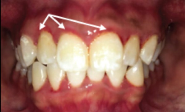
Upon examination, she appeared healthy. On extraoral examination, her face and neck were normal and symmetrical. The submandibular and sublingual lymph nodes were not palpable. Intraorally, her marginal gingiva was erythematous and slightly edematous but was non-tender upon palpation (Figure 1). The rest of the gingiva appeared normal in color, size and contour. The permanent dentition showed white spots on the dental enamel, a sign of hypomineralization or fluorosis. Oral hygiene was unsatisfactory, as dental plaque was visible along the marginal gingiva and interdental papillae.
Figure 1. Clinical picture showing erythematous and oedematous marginal gingiva.

Questions
What is the most likely diagnosis?
What are the symptoms and signs of this condition?
What are the risk factors of this condition?
What is the treatment needed for this condition?
Answers:
The most likely diagnosis is plaque-induced gingivitis. A differential diagnosis is non-plaque-induced gingivitis, which can be due to trauma, an allergic reaction, or an infection, such as viral infection (herpetic gingivostomatitis).
Symptoms and signs of gingivitis include bad breath (halitosis), gum bleeding during brushing or flossing, erythema, swelling, and tenderness of the gums.
Risk factors of gingivitis include dental plaque accumulation, plaque retentive areas (calculus and defective restorations), hormonal changes (during puberty, menopause, pregnancy, or OCP intake), systemic diseases (diabetes and HIV), drugs, smoking, aging, low vitamin C intake, as well as a family history of gingivitis.
Treatment for gingivitis includes oral hygiene instruction (OHI) and full mouth scaling. This patient was also advised to return for regular follow-ups for every three to four months. OCP was continued as this condition can be managed by the removal of the dental biofilm.
Discussion
Gingivitis is defined as an inflammation of the gums. It occurs when microbial plaque (bacteria) accumulates on the tooth surface as a result of ineffective tooth brushing. Therefore, effective tooth brushing is crucial to ensure adequate removal of food debris, which prevents further development of the plaque. Gingivitis is classified as localized when approximately 30% or less of the gingival tissue bleeds upon periodontal probing, and generalized if it is more than 30%.1 In gingivitis, there is no evidence of periodontal tissue destruction and loss of tooth attachment observable from x-ray film.1 Thus, gingivitis is reversible and preventable with proper oral hygiene practices.2
Patients with gingivitis can present with halitosis and painless gum bleeding, either spontaneously or during tooth brushing.1 However, to diagnose gingivitis, a thorough examination of gingival changes such as color, consistency, texture, and size should be performed.3 The inflamed gum will appear erythematous and edematous, and bleeds upon probing.4 In contrast, normal and healthy gingivae look pale pink and are tightly adapted, with knife-edge margins.5 As gingivitis progresses, the gum will become fluctuant and pointed with purulent exudates.6
Various factors have been recognized as gingival inflammation triggers. The local risk factors are dental plaque and plaque retentive factors such as calculus, overhanging retention, tooth anatomic factors (e.g., enamel pearl) and few others. Systemic diseases and specific malnutrition are also commonly associated with gingival inflammation and hypertrophy. These include hematological malignancy (leukemia), poorly controlled diabetes mellitus, smoking, and low intake of calcium and vitamin C.4,7
Sex hormone fluctuation is also a recognized risk factor for gingivitis, especially during puberty, menstruation, and pregnancy.4,7 Apart from that, several medications are well known to implicate gingival tissue overgrowth. These include calcium channel blockers (nifedipine), immunosuppressants (cyclosporine), antiepileptics (phenytoin), and OCR8–10
OCP is one of the most common culprits behind gingivitis in women. It has been reported that OCP-induced gingivitis can occur after only one month of OCP usage, leading to gum bleeding and swelling, particularly at the anterior mandibular segment of gum.10 A comparison between non-OCP and OCP users revealed that OCP usage is associated with increased gingival sulcus bleeding and probing pocket depth.11 OCP users also have poorer oral hygiene and more severe gingival inflammation than non-OCP users.12
How does OCP aggravate plaque-induced gingivitis, as it has in this case? The increased level of steroid hormones exaggerates the inflammatory reaction upon exposure to the existing dental biofilm.11 It was found that estrogen and progesterone receptors are present in gingival and periosteal tissue, which is capable of metabolizing these hormones.9 Progesterone stimulates vasodilatation and increases the permeability of the blood vessels in the gingiva, while estrogen promotes the proliferation of gingival fibroblasts and blood vessels, triggers the development of gingival connective tissue, and increases gingival inflammation even without plaque accumulation.9
This patient's oral hygiene was poor, with a visible accumulation of dental plaque. The bacteria in the built-up plaque on the tooth surface will then enter the gingival tissue, especially the gingival sulcus, and cause the marginal area to become susceptible to microbial infection. Microbial species typically involved in gingivitis are Streptococcus sp., Fusobacterium sp., Actinomyces sp., Veilonella sp., Treponema sp., and a few others.2 If left untreated, gingivitis can progress to periodontitis, which can cause irreversible damage not only to the gum but also to the surrounding bone that supports the teeth.
Treatment for patients with gingivitis varies depending on the type of gingivitis. For allergy-induced gingivitis, avoiding the allergen is the primary mode of treatment. For plaque-induced gingivitis, the principal aim of treatment is to reduce the dental biofilm and to eliminate the inflammation. Thus, prompt removal of all dental biofilm or plaque from the tooth surface and gingival sulcus is essential. Mild plaque, tartar and stain can be removed by effective toothbrushing. The patient should be taught how to perform effective toothbrushing techniques to ensure good personal plaque control and maintain optimal oral hygiene. The harder deposits might require scaling of the teeth performed by a dentist, followed by a series of appointments to assess and review the gum condition after treatment. The use of mouthwash is also useful in preventing the development of plaque and gingivitis.13 Following treatment, the patient should be assessed regularly at follow-up appointments every 3–4 months to review the progress of the treatment.14
A patient with mild drug-induced gingivitis can be treated with the non-surgical approach described above.14 Those with more severe presentations might require cessation of the offending drug. Patients with significant gingival hypertrophy might need corrective surgery to recontour the surface of gingival tissue.14 As this patient had mild gingivitis with no significant gingival hypertrophy, the OCP can be continued as long as the patient can maintain good oral hygiene.
This patient was also found to have dental fluorosis, which appeared as white spot lesions on the tooth surface. Fluorosis is characterized by hypomineralization of tooth enamel due to excessive ingestion of fluoride during childhood. The treatment of this condition is dependent on its severity. A mild case, such as this patient's, does not require specific treatment unless desired for cosmetic reasons. For moderate to severe cases, the options of treatment are bleaching, microabrasion, composite restorations, and, lastly, restoration of the teeth using ceramic veneers.15
How does this paper make a difference to general practice?
Gingivitis is a prevalent gum disease, and patients could come to a primary care clinic to seek treatment. This paper provides crucial knowledge to primary care physicians, enabling them to make a definitive diagnosis and refer the patient to a dentist early during the condition. Early referral allows patients to receive prompt treatment to prevent progression to periodontitis.
A primary care physician would have an overview of common aggravating factors in gingivitis, as well as several medications that could lead to gingival inflammation and hypertrophy, which may require modification of the patient's drugs.
References
- 1.Trombelli L, Farina R, Silva CO, Tatakis DN. Plaque-induced gingivitis: case definition and diagnostic considerations. J Periodontol [Internet] 2018 Jun;89(1):S46–73. doi: 10.1002/JPER.17-0576. http://doi.wiley.com/10.1002/JPER.17-0576 Available from. [DOI] [PubMed] [Google Scholar]
- 2.De Vries K. Gingivitis. Australian Journal of Pharmacy. Vol. 96. Australian Pharmaceutical Publishing Company Ltd; 2015. pp. 64–7. p. [Google Scholar]
- 3.Preshaw PM. Detection and diagnosis of periodontal conditions amenable to prevention. BMC Oral Health [Internet] 2015 Dec 15;15(S1):S5. doi: 10.1186/1472-6831-15-S1-S5. https://bmcoralhealth.biomedcentral.com/articles/10.1186/1472-6831-15-S1-S5 Available from. [DOI] [PMC free article] [PubMed] [Google Scholar]
- 4.Murakami S, Mealey BL, Mariotti A, Chapple ILC. Dental plaque-induced gingival conditions. J Periodontol [Internet] 2018 Jun;89(Suppl 1):S17–27. doi: 10.1002/JPER.17-0095. http://doi.wileycom/10.1002/JPER.17-0095 Available from. [DOI] [PubMed] [Google Scholar]
- 5.Highfield J. Diagnosis and classification of periodontal disease. http://doi.wiley.com/10.1111/j.1834-7819.2009.01140.x. Aust Dent J [Internet] 2009 Sep;54:S11–26. doi: 10.1111/j.1834-7819.2009.01140.x. Available from. [DOI] [PubMed] [Google Scholar]
- 6.Rodriguez DS, Sarlani E. Decision making for the patient who presents with acute dental pain. AACN Clin Issues. 2005;16(3):359–72. doi: 10.1097/00044067-200507000-00009. [DOI] [PubMed] [Google Scholar]
- 7.AlJehani YA. Risk factors of periodontal disease: review of the literature. Int J Dent [Internet] 2014. pp. 1–9.http://www.hindawi.com/journals/ijd/2014/182513/ Available from. [DOI] [PMC free article] [PubMed] [Retracted]
- 8.Samudrala P, Chava V, Chandana T, Suresh R. Drug-induced gingival overgrowth: A critical insight into case reports from over two decades. J Indian Soc Periodontol [Internet] 2016;20(5):496. doi: 10.4103/jisp.jisp_265_15. http://www.jisponline.com/text.asp?2016/20/5/496/207052 Available from. [DOI] [PMC free article] [PubMed] [Google Scholar]
- 9.Nandini DB. Oral contraceptives and oral health: An insight. Int J Med Dent Sci [Internet] 2016 Jul 1;5(2):1297–303. http://ijmds.org/wp-content/uploads/2016/06/1297-1303-ReV-OCP.pdf Available from. [Google Scholar]
- 10.Mahajan A, Sood R. Oral contraceptives induced gingival overgrowth – a clinical case report. POJ Dent Oral Care [Internet] 2017 Jul 13;1(1):1–5. https://proskolar.org/wp-content/uploads/2017/08/Oral-Contraceptives.pdf Available from. [Google Scholar]
- 11.Domingues RS, Ferraz BFR, Greghi SLA, Rezende MLR de, Passanezi E, Sant'Ana ACE. Influence of combined oral contraceptives on the periodontal condition. J Appl Oral Sci [Internet] 2012 Apr;20(2):253–9. doi: 10.1590/S1678-77572012000200022. http://www.scielo.br/scielo.php?script=sci_arttext&pid=S1678-77572012000200022&lng=en&tlng=en Available from. [DOI] [PMC free article] [PubMed] [Google Scholar]
- 12.Smadi L, Zakaryia A. The association between the use of new oral contraceptive pills and periodontal health: A matched case–control study. J Int Oral Heal [Internet] 2018;10(3):127. http://www.jioh.org/text.asp?2018/10/3/127/234518 Available from. [Google Scholar]
- 13.American Academy of Pediatric Dentistry Treatment of plaque-induced gingivitis, chronic periodontitis, and other clinical conditions. J Periodontol. 2004. [PubMed]
- 14.Scottish Dental Clinical Effectiveness Programme Prevention and treatment of periodontal diseases in primary care dental clinical guidance. 2014.
- 15.El Mourad AM. Aesthetic rehabilitation of a severe dental fluorosis case with ceramic veneers: a step-by-step guide. Case Rep Dent [Internet] 2018 Jun 6;2018:1–4. doi: 10.1155/2018/4063165. https://www.hindawi.com/journals/crid/2018/4063165/ Available from. [DOI] [PMC free article] [PubMed] [Google Scholar]


