Abstract
Aims
To monitor the performance of services for developmental dysplasia of the hip (DDH) in Northern Ireland and identify potential improvements to enhance quality of service and plan for the future.
Methods
This was a prospective observational study, involving all infants treated for DDH between 2011 and 2017. Children underwent clinical assessment and radiological investigation as per the regional surveillance policy. The regional radiology data was interrogated to quantify the use of ultrasound and ionizing radiation for this population.
Results
Evidence-based changes were made to the Northern Ireland screening programme, including an increase in ultrasound scanning capacity and expansion of nurse-led screening clinics. The number of infant hip ultrasound scans increased from 4,788 in 2011, to approximately 7,000 in 2013 and subsequent years. The number of hip radiographs on infants of less than one year of age fell from 7,381 to 2,208 per year. There was a modest increase in the treatment rate from 10.9 to 14.3 per 1,000 live births but there was a significant reduction in the number of closed hip reductions. The incidence of infants diagnosed with DDH after one year of age was 0.30 per 1,000 live births over the entire period.
Conclusion
Improving compliance with the regional infant hip screening protocols led to reduction in operative procedures and reduced the number of pelvic radiographs of infants. We conclude that performance monitoring of screening programmes for DDH is essential to provide a quality service.
Cite this article: Bone Joint J 2020;102-B(4):495–500.
Keywords: Hip, Dysplasia, Development, Screening, Surveillance, DDH, Radiation
Introduction
The early diagnosis and treatment of developmental dysplasia of the hip (DDH) is important as better outcomes are achieved with less invasive treatment.1 Undiagnosed DDH leads to gait abnormalities, chronic pain and accelerated osteoarthritis as well as increased surgical intervention.2
However, there is wide geographical variation in the policies and screening programmes used to diagnose DDH. For example, most centres in the UK use a selective screening programme. These programmes are based on risk factors and clinical examination which includes assessment of hip stability using Barlow3 and Ortolani4 tests, as well as observation for leg length discrepancy and assessment of hip abduction. UK guidelines from the Newborn and Infant Physical Examination group (NIPE) recommend that neonates with a positive examination should undergo ultrasound scanning (USS) within two weeks of life and that infants at risk, but exhibiting no clinical abnormalities, should undergo USS within the first six weeks of life.5
The policy in Northern Ireland differs slightly from the rest of the UK, in that we have retained an additional examination by a health visitor at four months.6 Furthermore, orthopaedic referrals are managed centrally by one team using a standardized treatment algorithm.7
Many European healthcare systems, however, have moved to a universal screening programme for DDH.8-10 The reported benefits of universal screening are a reduction in late diagnosis, with decreased need for surgical intervention. Woolacott et al,11 however, have reported that universal screening results in an increased rate of treatment. Universal screening also requires expenditure on increased USS as well as clinical staff, resulting in cost-benefit analyses of universal versus selective screening protocols being contested.12-14
Previous audits in Northern Ireland found that delays in treatment, due to limited capacity, led to an increase in the numbers of pelvic radiographs being taken for surveillance. However, in 2013, additional funding was made available throughout the province to expand USS screening as well as for nursing support in diagnosing DDH.
It was anticipated that these changes would increase the number of patients treated in a Pavlik harness and decrease the closed reduction rates as a result of earlier diagnosis. The effect on very late presenters was expected to be minimal, as these patients had not been referred to the orthopaedic service. It was also anticipated that these changes would reduce infant exposure to radiograph examination.
Methods
This is a prospective study of all children treated for DDH who were born in Northern Ireland between 1 January 2011 and 31st December 2017. There were 170,580 live births (83,008 female, 48.7%) and of these infants, 2,213 were diagnosed and treated for DDH (1,911 female, 86.4%) giving an overall treatment rate of 13.0 per 1,000 live births. The 2017 birth cohort was excluded from the analysis of those presenting over one year of age, to ensure adequate follow-up at the time of analysis.
Completeness of capture was assured by checking clinic lists, operating logs, and the Northern Ireland Picture Archiving and Communication System (NIPACS; Sectra Group, Linköping, Sweden). The study was registered as an audit by the Standards, Quality and Audit department of the Belfast Health and Social Care Trust (Reference no. 5082).
Data collected included demographics, birth information, and risk factors. Clinical findings of the referring clinician were noted and the initial radiological status of the hip was recorded. All procedures, both operative and nonoperative, were registered along with any complications the infant suffered. Children not born in Northern Ireland and those with teratological hips were excluded. In reporting the findings, patients are categorized by their birth year. Data capture was completed on 31st March 2019 using a custom Microsoft Access database (version 16.0; Microsoft, Redmond, Washington, USA). This database was structured to allow comparison of data against definitions that had been used in other studies. All infants were treated according to a standardized treatment protocol.7
The regional radiology system was interrogated to determine the number of infants less than one year of age undergoing radiological assessment of hips during the study period. This system includes all services treating children with DDH and has been in place since 2010. As the search included all images of hips, a small number were included which had been ordered for other reasons. This was evaluated further by examining the pelvic radiographs carried out over the course of four randomly sampled weeks during the study period. Of the 354 radiographs performed during these sampled weeks, only four were for indications other than DDH.
Statistical analysis
Data was analyzed using Microsoft Excel (version 15.0; Microsoft, Redmond, Washington, USA). Primary analysis was conducted through linear regression modelling, with subsequent Bayesian analysis under the guidance of a medical statistician. Statistical significance was set at p < 0.05.
Results
The DDH treatment rate increased from 10.9 to 14.3 per 1,000 live births between 2011 and 2017. The number and rates of late presentation from 2011 to 2017 are shown in Table I. There was a major reduction in the rate of presentation after three months, most markedly between six months and one year. The reductions in those presenting between three months and one year were shown to be statistically significant on linear regression modelling and Bayesian analysis (p < 0.01). A total of 44 infants born in Northern Ireland from 2011 to 2016 were diagnosed after one year. Of these, 38 presented between their first and second birthdays. The rate of diagnosis above one year was 0.30 per 1,000 live births. There was no statistically significant change in this rate during this study.
Table I.
Late presentation rates for developmental dysplasia of the hip in Northern Ireland 2011 to 2017.
| Year | Births | Rate of late presentation per 1,000 live births | ||
|---|---|---|---|---|
| > 3 mths | > 6 mths | > 12 mths | ||
| 2011 | 25,273 | 5.90 (n = 149) | 1.94 (n = 49) | 0.24 (n = 6) |
| 2012 | 25,269 | 5.62 (n = 142) | 2.06 (n = 52) | 0.47 (n = 12) |
| 2013 | 24,277 | 3.91 (n = 95) | 0.99 (n = 24) | 0.25 (n = 6) |
| 2014 | 24,394 | 3.73 (n = 91) | 1.31 (n = 32) | 0.20 (n = 5) |
| 2015 | 24,215 | 3.84 (n = 93) | 1.12 (n = 27) | 0.45 (n = 11) |
| 2016 | 24,076 | 2.87 (n = 69) | 0.75 (n = 18) | 0.17 (n = 4) |
| 2017 | 23,076 | 3.21 (n = 74) | 0.65 (n = 15) | Not calculated (n = 4) |
The median age that treated infants were first seen decreased from 105 days in 2011 to 57 days in 2017. The 95th centile for the age first seen moved from 319 days in 2011 to 158 days in 2017.
Of the neonates referred and treated because of clinical abnormalities, 43% (9/21) were seen by two weeks of age in 2011, rising to 66% (41/62) in 2017. Of those neonates referred with risk factors and treated, only 10.7% (12/112) had a USS by six weeks in 2017. However, in many infants the USS was performed just after six weeks and 62% (70/112) were performed by eight weeks. This percentage had risen from 40% (31/78) in 2011.
In 2011 there were 143 infants who were first seen between three and 12 months, which by 2017 had reduced to 70. For these infants the median delay to be seen from the time of referral was 42 (0 to 170) days in 2011 and in 2017 this was reduced to 33 (2 to 126) days.
There was an increase in the use of Pavlik harness as the sole treatment, from 5.5 per 1,000 live births to 13.1 per 1,000. In all, two infants developed avascular necrosis (AVN) of the hip out of a total of 2,027 hips treated exclusively in a Pavlik harness. Both involved decentred hips for which treatment was initiated late, at 12 and 16 weeks. One was classified as Bucholz and Ogden type 1 and one type 2.15 AVN was not found in dysplastic but located hips or in normal contralateral hips.
There were 15 incidents of femoral nerve palsy, all of which resolved upon removal of the Pavlik harness, however the median age of initiation of treatment was 11 weeks (interquartile range 8 to 16 weeks) for this group. There was only one femoral nerve palsy in the 1,422 infants treated in a Pavlik harness for a centred hip.
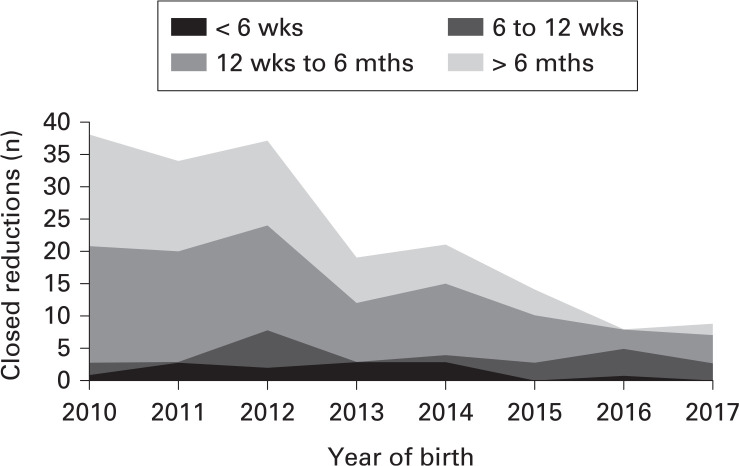
In this study, 126 hips in 117 infants were treated by closed reduction, (0.67 per 1,000 live births). There was a decrease in the number of closed reductions, from 1.2 per 1,000 live births in 2011, to 0.27 per 1,000 live births in 2017 (Figure 1). This reduction was statistically significant (p < 0.01) on linear regression modelling and Bayesian analysis and can be accounted for by the reduction in the number of patients presenting after 12 weeks of age.
Fig. 1.
Chart showing the number of closed reductions performed per year in Northern Ireland and their age at presentation.
There were 124 open reductions in 99 infants in this study, giving an overall open reduction rate of 0.58 per 1,000 live births. 45% (45/99) presented at six months of age or later and 16% (16/99) presented within the first six weeks of life.
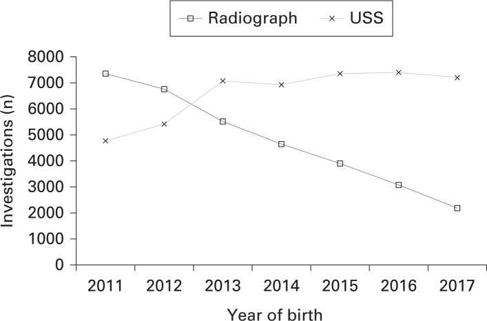
In 2011, 6,560 infants in Northern Ireland under one year had a total of 7,381 pelvic radiographs. During the same year, 4,203 infants underwent a total of 4,788 hip USSs. In 2014 there were 2,169 more USSs and 2,702 fewer pelvic radiographs. In subsequent years, there was no further increase in USSs but the number of pelvic radiographs continued to decline (Figure 2).
Fig. 2.
Chart showing the number of hip ultrasound scans (USS) and pelvic radiographs carried out in Northern Ireland for patients less than one year of age.
In 2017 there were 1,977 infants who had 2,208 radiographs. This reduction from 2011 to 2017 was statistically significant (p < 0.01). The number of infants who had a single radiograph of the pelvis before one year of age, without a previous USS of the hips, decreased from 5,014 in 2011 to 775 in 2017, (p < 0.01). The proportion of all infants, including those under treatment, who had a radiological follow-up after USS, fell from 43.8% to 24.5%.
The proportion of infants in Northern Ireland undergoing radiograph and USS imaging of the hips was 36% in 2011 (9,225 of 25,273 live births) and reduced to 32% by 2017 (7,402 of 23,076 live births) and the proportion of these infants primarily having an USS rose from 45.5% to 82.9% (4,203 to 6,139).
Discussion
As the data was collected prospectively and confirmed with the radiological images, we are confident that there is little that has not been captured in our study. Also, the population of Northern Ireland is stable with low migration levels. We have quantified the delays encountered by those who have been treated, but this may not reflect the pathway of other infants who have been assessed but not treated. Our data on pelvic radiographs includes examinations made for conditions such as trauma and infection. However, we estimate that the absolute number of these was small (≈ 1%) and likely to have been relatively constant over the years.
Following the introduction of increased screening for DDH in Northern Ireland the treatment rate has risen from 10.9 per 1,000 live births to 14.3 per 1,000 live births. Increase in treatment, however, is commonly seen with an increased level of the use of USS,16 although the Northern Ireland treatment rate is lower compared to the Austrian universal screening programme, which is 35 per 1,000 live births.8
The increased screening capacity in Northern Ireland has helped reduce the delay in assessment for at-risk patients. Further development of this service is needed to meet NIPE standards. Our data demonstrates that only 10.7% of infants had hip USS within six weeks. A target of six weeks or more could be obtained however, with relatively few additional resources. Early commencement of treatment is important for successful outcomes for DDH8 and performance needs to be closely monitored in any population screening system, regardless of the screening philosophy.
From our observations, the improvements made to the Northern Ireland screening service have had a demonstrable effect on treatment modality within the patient population. The decreased numbers of infants undergoing closed reduction was largely a result of reducing the number who were first seen after 12 weeks of life. In our study, very few infants who were seen at fewer than six weeks ultimately had a successful closed reduction. This was because the small number of these children who were not successfully treated with a Pavlik harness generally did not have hips that were amenable to closed reduction.
Thallinger et al8 explored the incidence of open reduction in Austria to assess the efficacy of screening in Austrian-born children and reported a rate of 0.12 per 1,000 live births. Biedermann et al10 assessed the delivery of the service in one region of Austria and found an open reduction rate of 0.04 per 1,000 live births. However, their closed reduction rate at 0.86 per 1,000 live births was slightly higher than the current rate in Northern Ireland. Our open reduction surgery rate was 0.58 per 1,000 live births. Half of these children presented beyond six months of age, which often precluded closed reduction, resulting in a higher rate of open reduction compared with a universal screening programme. It would therefore be difficult for our selective screening service to match this and get below a rate of 0.4 per 1,000 unless there was a marked improvement in the rate of detection among the wide range of different clinical examiners. This objective may be challenging to achieve, given the subjectivity of clinical signs such as ‘clicking’ hips.17
With further improvement in our programme, the rate of closed reductions could fall further. As it is relatively simple to capture the number of closed reductions performed, we believe that closed reduction is a useful indicator of performance of a screening service. For a population with an incidence of DDH similar to ours we would suggest that a target of less than 0.3 per 1,000 births is attainable.
The rate of diagnosis after one year of age in England has been recently reported as 1.28 per 1,000 live births.18 This is significantly higher than the rates in our study. As we have individually checked the records and radiology for every patient, we believe that our findings reflect the true incidence in our population. Broadhurst et al18 used large national databases which were interrogated for diagnostic codes. The use of large datasets, which were not designed for the purpose of identifying late diagnosis of hip dysplasia, has the potential to introduce errors unless there is rigorous application of coding standards.
It is possible that there may be a small number of infants in our study who are as yet undiagnosed and will present after 12 months of age. Extrapolating from the English data on age distribution, we expect 98% of this cohort to have presented to orthopaedic services by the end of our data capture. Even so, such an adjustment would only increase our late presentation rate from 0.30 to 0.31 per 1,000 live births, which is similar to the better rates reported from clinical surveillance.19
Using a definition of late diagnosis as an infant who is diagnosed six months or more and who required surgery, we have reported a rate of 1.14 per 1,000 live births from 1983 to 1987 and 0.59 per 1,000 live births in the 1991 to 1997 cohort.20 However, our performance on this measure had deteriorated to 1.11 in the 2003 birth cohort. We believe this deterioration was a consequence of a higher referral rate exceeding our ultrasound capacity and therefore creating delays. In 2011, this rate was 0.71 per 1,000 live births, and has improved to 0.21 per 1,000 live births by 2016. However, using this measure our results do not reach the level achieved in Malmo with clinical screening alone.21
In Northern Ireland, a clinical examination by a health visitor at four months of age was retained due to the finding in 2003 that a significant number of late presentations were identified at this time. This assessment was removed in the Hall 4 guidance adopted elsewhere in the UK.6 However, making a diagnosis at that time may be too late to allow for treatment by splintage. The fall in the number of closed reductions was due to fewer presenting after 12 weeks, indicating a better performance by our surveillance system during the earlier assessments.
There have been several studies which have demonstrated an increased childhood cancer risk in those undergoing postnatal radiological investigations.22-24 It has been suggested that effective absorbed doses of radiation are higher in younger patients, due to less attenuation of the radiation beam.25 Lifetime cancer risk has been shown to be elevated in scoliosis patients under radiological surveillance,26 and Simony et al27 have reported a five-fold increase in lifetime risk for these patients, particularly for breast and endometrial cancer. These findings suggest that the infant DDH population, which is predominantly female, may be at risk from cancer and should be protected whenever possible.
Between the first and last years of the study there were 2,421 more USSs but 5,173 fewer radiographs. It is possible to account for just over half of this reduction by assuming the use of ultrasound as a direct replacement for radiological investigations. Furthermore, there would be a further reduction in the use of pelvic radiograph in patients who had already undergone USSs. However, in our study this only accounted for about 600 fewer radiographs. The remaining reduction was due to a decrease in the ordering of radiographs from primary care, reflecting the increasing confidence in the Northern Ireland screening service as well as from feedback to referring clinicians over what was considered best practice.
The treatment for DDH with a Pavlik harness may not be entirely innocuous, with potential complications including AVN28 and femoral nerve palsy. Our findings though are in agreement with the existing literature, which suggests that AVN in normal or located but dysplastic hips is very rare.29,30 Murnaghan et al31 reported an incidence of femoral nerve palsy of 2.5% in a population of 1,218 patients, all these infants regaining femoral nerve function.31 Our incidence of femoral nerve palsy was lower and all our infants also recovered. Therefore, it is our view that femoral nerve palsy does not appear to be a significant issue, provided there is careful monitoring while the infant is in splintage.
Although it was not our intention to give details on the financial cost and benefits of screening, we found that with 2,000 extra hip USSs, there were 5,000 fewer radiographs and 30 fewer operative closed reductions each year. These improvements have the potential to provide benefits to many children with less exposure from anaesthesia, radiation, and complications such as AVN and dysplasia.32
In conclusion, there have been concerns that current screening programmes are not fit for purpose.33 However, our data demonstrates that continuous monitoring leads to improved outcomes and the reduced need for surgical intervention, with the possible risk of poor long-term outcomes.32
Take home message
- Although selective screening will not detect all cases of DDH, it can be made more effective by monitoring service performance.
- Significant improvements in patient outcome can be achieved with relatively modest levels of targeted funding.
Author contributions
D. J. Milligan: Wrote the paper, Performed the statistical analysis, Performed the literature search, Redrafted the paper.
A. P. Cosgrove: Wrote the paper, Performed the statistical analysis, Performed the literature search, Redrafted the paper.
Funding statement
No benefits in any form have been received or will be received from a commercial party related directly or indirectly to the subject of this article.
ICMJE COI statement
None declared
Acknowledgements
The authors would like to thank Sister B. Trainor, Sister P. Haugh, Sister G. Thomson, and Sister R. Hamill for assistance in data capture, Ms J. Moss and Mr K. Porter for assistance with radiology data, and the enthusiasm of the ultrasonographers in providing the service. We would also like to thank Dr Mark James Thompson for his help with detailed statistical analysis.
Open access statement
This is an open-access article distributed under the terms of the Creative Commons Attribution Non-Commercial No Derivatives (CC BY-NC-ND 4.0) licence, which permits the copying and redistribution of the work only, and provided the original author and source are credited. See https://creativecommons.org/licenses/by-nc-nd/4.0/.
Trial registration number
This study was registered as an audit by the Standards, Quality and Audit department of the Belfast Health and Social Care Trust (Reference no. 5082).
This article was primary edited by S. P. F. Hughes.
References
- 1.Sewell MD, Rosendahl K, Eastwood DM. Developmental dysplasia of the hip. BMJ. 2009;339:b4454. [DOI] [PubMed] [Google Scholar]
- 2.Furnes O, Lie SA, Espehaug B, et al. Hip disease and the prognosis of total hip replacements. A review of 53,698 primary total hip replacements reported to the Norwegian arthroplasty register 1987-99. J Bone Joint Surg Br. 2001;83:579–586. [DOI] [PubMed] [Google Scholar]
- 3.Barlow T. Early diagnosis and treatment of congenital dislocation of the hip. J Bone Joint Surg. 1962;44-B:292–301. [Google Scholar]
- 4.Ortolani M. Un segno poco noto e sua importanza per la diagnosi precoce di prelussazione congenital dell’anca. Pediatria (Napoli). 1937;45:129–136. [Google Scholar]
- 5.No authors listed . Newborn and infant physical examination (NIPE) screening programme handbook. GOV.UK, 2019. https://www.gov.uk/government/publications/newborn-and-infant-physical-examination-programme-handbook/newborn-and-infant-physical-examination-screening-programme-handbook (date last accessed 29 January 2020).
- 6.Hall DMB, Elliman D. Health for All Children. Revised Fourth Ed. Oxford: OUP, 2006. [Google Scholar]
- 7.Donnelly KJ, Chan KW, Cosgrove AP. Delayed diagnosis of developmental dysplasia of the hip in Northern Ireland: can we do better? Bone Joint J. 2015;97-B(11):1572–1576. [DOI] [PubMed] [Google Scholar]
- 8.Thallinger C, Pospischill R, Ganger R, et al. Long-term results of a nationwide general ultrasound screening system for developmental disorders of the hip: the Austrian hip screening program. J Child Orthop. 2014;8(1):3–10. [DOI] [PMC free article] [PubMed] [Google Scholar]
- 9.Ihme N, Altenhofen L, von Kries R, Niethard FU. [Hip ultrasound screening in Germany. Results and comparison with other screening procedures]. Orthopade. 2008;37(6):541–546. 548–549. [DOI] [PubMed] [Google Scholar]
- 10.Biedermann R, Riccabona J, Giesinger JM, et al. Results of universal ultrasound screening for developmental dysplasia of the hip: a prospective follow-up of 28 092 consecutive infants. Bone Joint J. 2018;100-B(10):1399–1404. [DOI] [PubMed] [Google Scholar]
- 11.Woolacott NF, Puhan MA, Steurer J, Kleijnen J. Ultrasonography in screening for developmental dysplasia of the hip in newborns: systematic review. BMJ. 2005;330(7505):1413. [DOI] [PMC free article] [PubMed] [Google Scholar]
- 12.Gray A, Elbourne D, Dezateux C, et al. Economic evaluation of ultrasonography in the diagnosis and management of developmental hip dysplasia in the United Kingdom and Ireland. J Bone Joint Surg Am. 2005;87-A(11):2472–2479. [DOI] [PubMed] [Google Scholar]
- 13.Rosendahl K, Markestad T, Lie R, Sudmann E, Geitung JT. Cost-effectiveness of alternative screening strategies of developmental dysplasia of the hip. Arch Pediatr Adolesc Med. 1995;149(6):643–648. [PubMed] [Google Scholar]
- 14.Grill F, Müller D. [Results of hip ultrasonographic screening in Austria]. Orthopade. 1997;26(1):25–32. [DOI] [PubMed] [Google Scholar]
- 15.Bucholz RW, Ogden JA. Patterns of ischemic necrosis of the proximal femur in nonoperatively treated congenital hip disease. The Hip: Proceedings of the Sixth Open Scientific Meeting of the Hip Society. St Louis, MO: Mosby, 1978:43–63. [Google Scholar]
- 16.Roovers EA, Boere-Boonekamp MM, Castelein RM, Zielhuis GA, Kerkhoff TH. Effectiveness of ultrasound screening for developmental dysplasia of the hip. Arch Dis Child Fetal Neonatal Ed. 2005;90(1):F25–F30. [DOI] [PMC free article] [PubMed] [Google Scholar]
- 17.Humphry S, Thompson D, Price N, Williams PR. The ‘clicky hip’: to refer or not to refer? Bone Joint J. 2018;100-B(9):1249–1252. [DOI] [PubMed] [Google Scholar]
- 18.Broadhurst C, Rhodes AML, Harper P, et al. What is the incidence of late detection of developmental dysplasia of the hip in England?: a 26-year national study of children diagnosed after the age of one. Bone Joint J. 2019;101-B(3):281–287. [DOI] [PubMed] [Google Scholar]
- 19.Bjerkreim I, Johansen J. Late diagnosed congenital dislocation of the hip. Acta Orthop Scand. 1987;58(5):504–506. [DOI] [PubMed] [Google Scholar]
- 20.Verzin EJ, McLean J, Cosgrove AP. Developmental dysplasia of the hip in Northern Ireland: Audit of the 2003 birth cohort. J Bone Joint Surg. 2008;90-B(Suppl III):522. [Google Scholar]
- 21.Düppe H, Danielsson LG. Screening of neonatal instability and of developmental dislocation of the hip. A survey of 132,601 living newborn infants between 1956 and 1999. J Bone Joint Surg Br. 2002;84-B(6):878–885. [DOI] [PubMed] [Google Scholar]
- 22.Polhemus DW, Koch R. Leukemia and medical radiation. Pediatrics. 1959;23(3):453–461. [PubMed] [Google Scholar]
- 23.Hartley AL, Birch JM, McKinney PA, et al. The Inter-Regional Epidemiological Study of Childhood Cancer (IRESCC): past medical history in children with cancer. J Epidemiol Community Health. 1988;42(3):235–242. [DOI] [PMC free article] [PubMed] [Google Scholar]
- 24.Infante-Rivard C, Mathonnet G, Sinnett D. Risk of childhood leukemia associated with diagnostic irradiation and polymorphisms in DNA repair genes. Environ Health Perspect. 2000;108(6):495–498. [DOI] [PMC free article] [PubMed] [Google Scholar]
- 25.Linet MS, Kim KP, Rajaraman P. Children’s exposure to diagnostic medical radiation and cancer risk: epidemiologic and dosimetric considerations. Pediatr Radiol. 2009;39(1 Suppl 1):S4–S26. [DOI] [PMC free article] [PubMed] [Google Scholar]
- 26.Ronckers CM, Doody MM, Lonstein JE, Stovall M, Land CE. Multiple diagnostic X-rays for spine deformities and risk of breast cancer. Cancer Epidemiol Biomarkers Prev. 2008;17(3):605–613. [DOI] [PubMed] [Google Scholar]
- 27.Simony A, Hansen EJ, Christensen SB, Carreon LY, Andersen MO. Incidence of cancer in adolescent idiopathic scoliosis patients treated 25 years previously. Eur Spine J. 2016;25(10):3366–3370. [DOI] [PubMed] [Google Scholar]
- 28.Madhu TS, Akula M, Scott BW, Templeton PA. Treatment of developmental dislocation of hip: does changing the hip abduction angle in the hip spica affect the rate of avascular necrosis of the femoral head? J Pediatr Orthop B. 2013;22(3):184–188. [DOI] [PubMed] [Google Scholar]
- 29.Pap K, Kiss S, Shisha T, et al. The incidence of avascular necrosis of the healthy, contralateral femoral head at the end of the use of Pavlik harness in unilateral hip dysplasia. Int Orthop. 2006;30(5):348–351. [DOI] [PMC free article] [PubMed] [Google Scholar]
- 30.Kruczynski J. Avascular necrosis of the proximal femur in developmental dislocation of the hip. Incidence, risk factors, sequelae and MR imaging for diagnosis and prognosis. Acta Orthop Scand Suppl. 1996;268:1–48. [PubMed] [Google Scholar]
- 31.Murnaghan ML, Browne RH, Sucato DJ, Birch J. Femoral nerve palsy in Pavlik harness treatment for developmental dysplasia of the hip. J Bone Joint Surg Am. 2011;93-A(5):493–499. [DOI] [PubMed] [Google Scholar]
- 32.Pollet V, Van Dijk L, Reijman M, Castelein RMC, Sakkers RJB. Long-term outcomes following the medial approach for open reduction of the hip in children with developmental dysplasia. Bone Joint J. 2018;100-B(6):822–827. [DOI] [PubMed] [Google Scholar]
- 33.Perry DC, Paton RW. Knowing your click from your clunk: is the current screening for developmental dysplasia of the hip fit for purpose? Bone Joint J. 2019;101-B(3):236–237. [DOI] [PubMed] [Google Scholar]


