Figure 2.

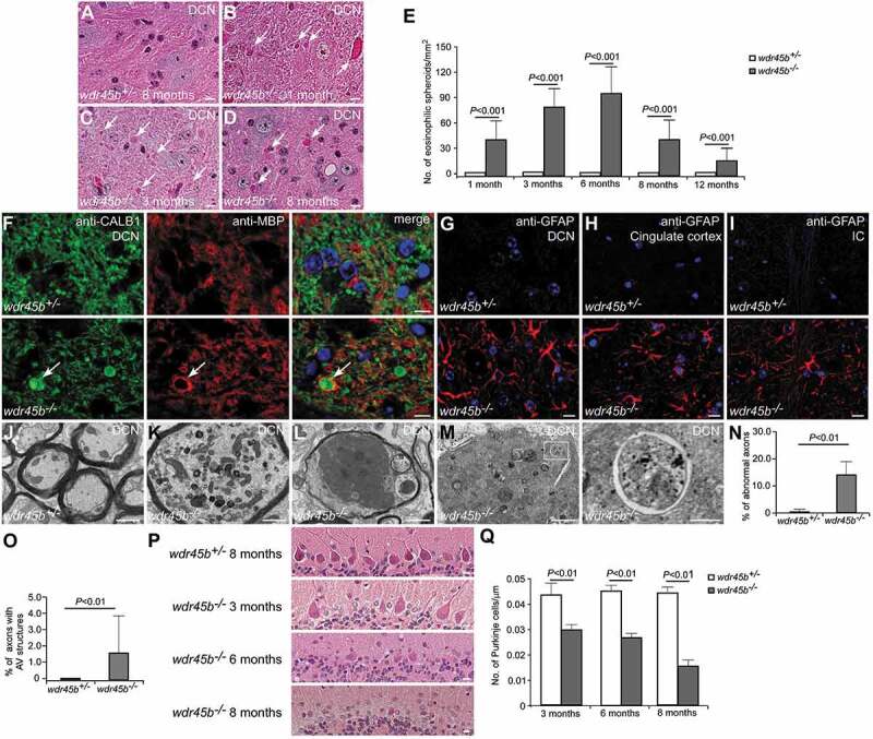
Axon swelling and cerebellar atrophy in wdr45b KO mice. (A-D) H&E staining shows eosinophilic spheroids (arrows) in the DCN of wdr45b-/- mice at 1, 3 and 8 months (B-D). No spheroids are found in control mice (A). Bars: 10 μm. (E) The number of eosinophilic spheroids in the DCN of wdr45b+/- and wdr45b-/- mice at 1, 3, 6, 8 and 12 months is shown as mean ± SD (n = 3). (F) Immunostaining with CALB1 (green) and MBP (red) antibodies shows that CALB1-positive swollen axons (arrows) are enwrapped by MBP-labeled myelin in the DCN regions of wdr45b-/- mice. (G-I) Reactive astrogliosis is found in the DCN region (G), cingulate cortex (H) and IC (I) of wdr45b-/- mice. IC, internal capsule. Bar: 10 μm (F-I). (J-M) EM images show normal myelinated axons in the DCN of wdr45b+/- mice at 5 months (J), while those in wdr45b-/- mice are swollen and some contain large numbers of mitochondria (K), dark axoplasm (L) and double-membrane autophagosomes (M). The right panel shows enlargement of the boxed area in the left panel in M. Bars: 1 μm (J, K, L and left panel in M); 200 nm (right panel in M). (N and O) The percentages of the axons that are abnormal (N) or contain autophagosome structures (O) in wdr45b+/- mice and wdr45b-/- mice are shown as mean ± SD (n = 50 micrographs of 548 μm2). AV, autophagosome. (P and Q) Purkinje cell numbers gradually decrease with age in wdr45b-/- mice (P). Mean ± SD (n = 3) is shown in Q. Bars: 10 μm.
