Figure 3.

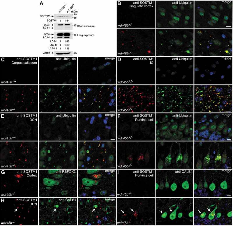
wdr45b-/- mice display autophagy defect. (A) Immunoblotting results show that levels of SQSTM1, LC3-I, LC3-II and LC3-II:I are increased in the cerebella of wdr45b-/- mice compared to wdr45b+/- mice. Quantification of SQSTM1, LC3-I and LC3-II levels normalized by ACTB levels and LC3-II:I ratios is shown. (B-F) Accumulation of SQSTM1 (red) and ubiquitin (green) positive signals in the cingulate cortex (B), corpus callosum (C), IC (D), DCN (E) and Purkinje cells (F) of wdr45b-/- mice. IC, internal capsule. (G) Costaining of SQSTM1 (red) and RBFOX3 (green) shows that SQSTM1 aggregates accumulate in neurons in the cortex of wdr45b-/- mice. (H and I) Costaining of SQSTM1 (red) and CALB1 (green) shows that SQSTM1 accumulates in the swollen axons (H) and cell bodies (I) of Purkinje cells in wdr45b-/- mice, as indicated by arrows. Bars: 10 μm (B-I).
