Abstract
Background
Leishmaniasis is a human and animal disease caused by parasites of the genus Leishmania, which is now divided into four subgenera, Leishmania, Viannia, Sauroleishmania and Mundinia. Subgenus Mundinia, established in 2016, is geographically widely dispersed, its distribution covers all continents, except Antarctica. It consists of 5 species; L. enriettii and L. macropodum are parasites of wild mammals while L. martiniquensis, L. orientalis and an unnamed Leishmania sp. from Ghana are infectious to humans. There is very little information on natural reservoir hosts and vectors for any Mundinia species.
Methods
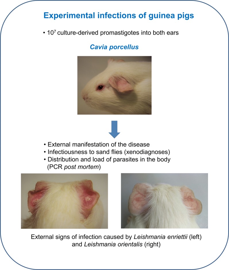
Experimental infections of guinea pigs with all five Mundinia species were performed. Animals were injected intradermally with 107 culture-derived promastigotes into both ear pinnae. The courses of infections were monitored weekly; xenodiagnoses were performed at weeks 4 and 8 post-infection using Lutzomyia migonei. The distribution of parasites in different tissues was determined post-mortem by conventional PCR.
Results
No significant differences in weight were observed between infected animals and the control group. Animals infected with L. enriettii developed temporary lesions at the site of inoculation and were infectious to Lu. migonei in xenodiagnoses. Animals infected with L. martiniquensis and L. orientalis developed temporary erythema and dry lesions at the site of inoculation, respectively, but were not infectious to sand flies. Guinea pigs infected by L. macropodum and Leishmania sp. from Ghana showed no signs of infection during experiments, were not infectious to sand flies and leishmanial DNA was not detected in their tissue samples at the end of experiments at week 12 post-inoculation.
Conclusions
According to our results, guinea pigs are not an appropriate model organism for studying Mundinia species other than L. enriettii. We suggest that for better understanding of L. (Mundinia) biology it is necessary to focus on other model organisms.
Keywords: Leishmania, Mundinia, Guinea pig, Leishmania enriettii, Leishmania martiniquensis, Leishmania orientalis, Leishmania macropodum, Animal model
Background
Leishmaniases are vector-borne diseases whose etiological agents are protozoan parasites of the genus Leishmania (Kinetoplastida: Trypanosomatidae). Known previously as the L. enriettii complex, the subgenus Mundinia was established recently and currently contains 5 species: L. enriettii, L. macropodum, L. orientalis, L. martiniquensis and an unnamed Leishmania sp. from Ghana [1–3]. According to phylogenetic analyses, this subgenus is the first to branch from the other Leishmania subgenera, indicating that species of this subgenus are likely to represent the most ancient and divergent group of species within the Leishmania [2, 4]. The geographical distribution of Mundinia species covers all continents except Antarctica, which can be explained by the formation of individual species from their common ancestor after the breakup of Gondwana [2].
Many important details of the biology of these parasites are unknown. The identity of the insect vectors responsible for transmission of L. (Mundinia) has not been confirmed for any species yet. It has been assumed that these parasites, similar to other Leishmania, would be transmitted by sand flies of the genus Phlebotomus and/or Sergentomyia in the Old World and Lutzomyia in the New World (Diptera: Phlebotominae), and this may be the case. Recently, however, Forcipomyia (Lasiohelea) biting midges (Diptera: Ceratopogonidae) were reported as likely vectors of L. macropodum in Australia [5], and laboratory experiments have revealed a high susceptibility of Culicoides sonorensis to L. enriettii [6]. The observations raise the possibility of non-sand fly vectors for at least some of the Mundinia.
Similarly, there is little current information on the natural mammalian reservoir hosts for these parasites. Leishmania enriettii, is a parasite that has only ever been found in domestic guinea pigs (Cavia porcellus) in Brazil, first isolated in the 1940s [7]. The natural host of L. enriettii is not known although often assumed to be a wild rodent of some kind. Leishmania macropodum is a parasite first isolated from red kangaroos in Australia, but from a game park in a region where these animals are not found [5]. There is evidence of L. macropodum infection in three other species of Australian macropods, which are more likely to be the true host(s) of this parasite [7]. On the other hand, human cases have been described with L. martiniquensis, L. orientalis and Leishmania sp. from Ghana. Leishmania martiniquensis was first isolated from a HIV positive man on Martinique Island in 1992 [8]. According to recent findings, this Leishmania species has a worldwide distribution with single or multiple cases reported from various continents where these parasites were isolated from various hosts such as horses, cows and humans [9–12]. Leishmania orientalis was formally described in 2018 [13]; in the past it was reported as “L. siamensis” [9, 14] but this name is a nomen nudum and should not be used anymore. Leishmania sp. from Ghana is a species causing cutaneous leishmaniasis in the Volta region in Ghana [4]. The last two species were not isolated from any mammalian species, except humans, and the identity of their reservoir hosts remains enigmatic.
Since very little is known about biology of these neglected parasites, the aim of our study was the establishment of model host organisms, which would enables testing their behaviour and properties in a mammalian host. Here we present results of experimental infections in guinea pigs with all five known L. (Mundinia) species.
Methods
Parasites and guinea pigs
Leishmania enriettii (MCAV/BR/45/LV90), L. macropodum (MMAC/AU/2004/AM-2004), Leishmania sp. from Ghana (MHOM/GH/2012/GH5), L. orientalis (MHOM/TH/2014/LSCM4) and two strains of L. martiniquensis (MHOM/MQ/1992/MAR1 and MHOM/TH/2011/CU1) were used. Parasites were maintained at 23 °C in M199 medium supplemented with 10% fetal calf serum (Gibco, Prague, Czech Republic), 1% BME vitamins (Sigma-Aldrich, Prague, Czech Republic), 2% sterile urine and 250 μg/ml amikacin (Amikin, Bristol-Myers Squibb, Prague, Czech Republic). In our laboratory both strains were maintained in a cryobank with 2–3 sub-passages in vitro before experimental infections of guinea pigs and no passages in animals were performed. Before experimental infection, parasites were washed by centrifugation (6000×g for 5 min) and resuspended in saline solution.
Female guinea pigs (Dunkin-Hartley) originating from AnLab (Prague, Czech Republic) were maintained in groups of 2 specimens in T4 boxes (58 × 37 × 20 cm); Velaz (Prague, Czech Republic) equipped with bedding (German Horse Span; Pferde, Prague, Czech Republic), breeding material (Woodwool) and hay (Krmne smesi Kvidera, Spalene Porici, Czech Republic), provided with a feed mixture V2233 Ms-H Guinea Pig maintenance (AnLab) and water ad libitum, with a 12 h light/12 h dark photoperiod, temperature of 22–25 °C and relative humidity of 40–60%. At the beginning of experiments the average weight of animals was 499 g and average age was 7 weeks.
Infection and xenodiagnoses of guinea pigs
Eighteen guinea pigs (Cavia porcellus) anaesthetized with ketamin/xylazin (37.5 mg/kg and 1.5 mg/kg, respectively) were injected with 107 stationary-stage promastigotes in 5 µl of sterile saline intradermally into the ear pinnae of both ears. The course of infection was recorded weekly. Three animals inoculated with the same volume of saline solution were used as a control for external signs of infection.
Xenodiagnoses were performed at weeks 4 and 8 post-infection (pi) using the permissive vector Lutzomyia migonei [15]. Five to six-day-old Lu. migonei were placed into plastic vials covered by fine nylon mesh and allowed to feed on the ear pinnae of anaesthetized animals. Engorged individuals were maintained for two days at 25 °C and then stored in tissue lysis buffer (Roche, Prague, Czech Republic) at -20 °C in pools of 5 females for subsequent PCR. Altogether 192 sand fly pools were tested.
At the end of the experiments, 12 weeks post-infection (pi), the hosts were euthanized, dissected and tissues from ears, paws, ear-draining lymph nodes, spleens, livers and blood were stored at − 20 °C for subsequent PCR.
Conventional PCR
DNA extraction from vectors and animal tissues was performed using the High Pure PCR Template Preparation Kit (Roche) according to the manufacturer’s instructions. The total DNA was used as a template for PCR amplification with the primers for a 246 bp long ITS1 sequence (forward primer 5′-AGA TTA TGG AGC TGT GCG ACA A-3′ and reverse primer 5′-TAG TTC GTC TTG GTG CGG TC-3′). Reactions were performed using EmeraldAmp® GT PCR Master Mix and cycling conditions were as follows: step 1, 94 °C for 3 min 30 s; step 2, 94 °C for 30 s; step 3, 60 °C for 30 s; step 4, 72 °C for 20 s; step 5, 72 °C for 7 min; followed by cooling at 12 °C. Steps 2–4 were repeated 35 times. Samples were analysed using 1% agarose gels.
Statistical analysis
Statistical analyses were carried out using R software ((http://cran.r-project.org/). Correlation of animal weight in different groups (infected and non-infected) and time was tested by co-variance analysis.
Results
No significant differences in weight were observed between infected animals and the control group (L. macropodum, P = 0.70; L. enriettii, P = 0.12; L. martiniquensis MAR1, P = 0.77; L. martiniquensis CU1, P = 0.12; Leishmania sp. from Ghana, P = 0.20; L. orientalis, P = 0.11; see Additional file 1: Table S1).
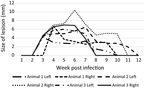
Development of dry lesions was observed in animals infected with L. enriettii. The lesions appeared on ear pinnae (the site of inoculation) by week 2–3 pi, increased in size through to week 5–6 pi and then healed, completely disappearing between weeks 8–12 pi (Figs. 1, 2a). Animals were efficiently infectious to sand flies on week 4 pi (9/16 positive pools) while infectiousness was reduced by week 8 pi (1/16 positive pools).
Fig. 1.

Development of lesions in guinea pigs infected with L. enriettii
Fig. 2.

Dry lesions observed in animals infected with L. enriettii (a) and nodules transforming to dry lesions in animals infected by L. orientalis (b); both at week 5 pi
In animals infected with L. martiniquensis (MAR1), temporary erythema was observed at the site of inoculation between weeks 4–8 pi, but no lesions developed and animals were not infectious to sand flies (0/32 positive pools).
Erythema at the site of inoculation was also observed in guinea pigs infected with L. orientalis. The erythematic spot appeared by week 3–4 pi. The spot nodulated, and the nodules subsequently transformed into dry lesions surrounded by a purple skin macula (Fig. 2b, Additional file 2: Table S2). The lesions healed by week 7–8 pi. However, animals were not infectious to sand flies (0/32 positive pools) and no leishmanial DNA was detected in any of the tested tissue samples.
Guinea pigs infected with L. macropodum, Leishmania sp. from Ghana and L. martiniquensis (CU1) did not show any external signs of infection during the whole experiment, animals were not infectious to sand flies (0/96 positive pools consisting of 5 blood-fed females each), and leishmanial DNA was not detected in any of tissue samples collected by the end of experiment on week 12 pi.
Discussion
Leishmaniases of men and animals caused by L. (Mundinia) species are emerging all over the world. The diseases in humans are characterized by symptoms varying from self-healing skin lesions [2, 12] to visceral forms. The latter prevail in HIV-positive patients [8, 16] but was also observed in immunocompetent humans [12, 17]. Very little is known about the life-cycle of these ancient and neglected species and an appropriate animal model is necessary for closer understanding of their biology.
Guinea pigs were chosen for the experimental model in our study as they are the only known non-human mammalian hosts of L. (Mundinia) species, except for kangaroos, cows and horses [6, 7, 9, 10, 18], which are not practicable for most laboratory investigations. Leishmania enriettii was repeatedly isolated from domestic guinea pigs from various localities in Brazil [6, 19]. Interestingly, individual cases were separated by long time periods, which does not agree with the fact that guinea pigs are popular pets, and according to several studies, they are very susceptible to infection [5, 20, 21]. We suggest that this rare incidence may have two different explanations. First, the prevalence of infection is actually much higher, but the owners of infected guinea pigs do not take them for veterinary checks, therefore, parasites are not isolated. Alternatively, guinea pigs are only incidental hosts and the primary reservoir hosts (and primary insect vectors) are not present in close vicinity to households. In this case, secondary vectors and/or reservoirs may be temporarily involved in transmission to domestic localities and domestic guinea pigs.
Our experiments confirmed the susceptibility of guinea pigs to L. enriettii. All infected animals showed development of typical ear lesions and the animals were infectious to sand flies. The numbers of positive sand flies were significantly higher at week 4 pi than at the later time interval, week 8 pi. This decrease of infectivity was also observed previously by Seblova et al. [6]. At week 12 pi, the animals did not show any more external signs of infection and Leishmania DNA was not detected in any of the examined tissue samples. Spontaneous healing of lesions was observed also by Paranaiba et al. [22]. In their experiments initiated by intradermal inoculation of 105 promastigotes, a different virulence between the two strains used was observed. The Cobaia strain did not develop any lesions, while strain L88 developed lesions that were growing by weeks 4–6 pi, and then diminution of lesions was observed until the end of the experiment. The authors also described development of larger lesions in groups where sand fly salivary glands were added to the inoculum.
However, the virulence of the L. enriettii parasite strain and the presence of sand fly salivary glands are not the sole factors influencing the degree of pathogenicity for guinea pigs. The outcome of infections is also dependent on the method of their initiation, i.e. on parasite numbers and stages (amastigotes vs promastigotes) used as an inoculum. Thomaz-Soccol et al. [23] described the development of serious symptoms of disease, such as the dissemination of parasites and subsequent death of all tested animals, when the inoculum consisted of amastigotes of a strain identical to L88 (according to isoenzyme analyses). Wide dissemination of L. enriettii in animals was also observed by Paraense et al. [20], who infected guinea pigs with amastigotes from lesion homogenates, and by Seblova et al. [6] who infected animals with 107 culture derived promastigotes.
Development of L. enriettii has also been tested in hamsters (Mesocricetus auratus), where infections were characterized by the development of temporary lesions at the site of inoculation and their subsequent healing [5, 24]. In experiments with wild guinea pigs (Cavia aperea), rhesus macaques and dogs [7], no animals showed any signs of infection, so domestic guinea pigs remain the best laboratory model for L. enriettii at present.
Here, we compared the susceptibility of guinea pigs to four other L. (Mundinia) species. The infections were lost in animals infected with L. macropodum, Leishmania sp. from Ghana and L. martiniquensis strain CU1. Animals infected with a second L. martiniquensis strain, MAR1, and with L. orientalis developed only temporary changes on the ears and the animals were not infectious to sand flies. However, PCR analysis showed no presence of leishmanial DNA by week 12 pi in any of the tested samples. We suggest that L. martiniquensis and L. orientalis are capable of temporary survival at the site of inoculation, but they cannot disseminate to other tissues of guinea pigs.
We suggest that for a better understanding of L. (Mundinia) biology it is necessary to focus on other model host organisms. The first choice could be BALB/c mice or hamsters as the most common animal models for research used with many L. (Leishmania) and L. (Viannia) species. Infections of these standard laboratory animals with their controlled genetic background may bring valuable information. Alternatively, genetically polymorphic models like wild rodents mimicking natural hosts could be used. These less common models can allow a better understanding of the dynamics of infection and host-parasite relationships related more closely to the situation in the wild [24]. On the other hand, when infected with L. enriettii and L. orientalis, guinea pigs could serve as a potential model for spontaneous healing, which could be informative for the design of vaccines.
Conclusions
Experimental infections showed that guinea pigs are not a good animal model for the subgenus Mundinia, with exception of L. enriettii. All other Mundinia species studied, L. orientalis, L. martiniquensis, L. macropodum and L. (Mundinia) sp. from Ghana, were not able to develop infections transmissible to sand flies.
Supplementary information
Additional file 1: Table S1. Weight gain of guinea pigs during the experiment.
Additional file 2: Table S2. External signs of infection on ears of infected guinea pigs during the experiment. Abbreviations: E, erythema; N, nodulus; DL, dry lesion.
Acknowledgements
We would like to thank to Tatiana Spitzova for help with statistical analysis and Tereza Lestinova for help with animal infections.
Abbreviations
- PCR
polymerase chain reaction
- pi
post-infection
Authors’ contributions
TB and JS carried out the experimental infections of animals and xenodiagnostic experiments. Molecular analysis was done by TB. JS and PV substantially contributed to conception and design of experiments. Article was drafted by TB and JS. Parasites were provided and manuscript was revised by PS and PB. All authors read and approved the final manuscript.
Funding
This study was funded by the Czech Science Foundation (GACR) (Grant number 17-01911S) and the ERD Funds, project CePaViP (CZ.02.1.01/0.0/0.0/ 16_019/0000759).
Availability of data and materials
All the data are included within the article and its additional files.
Ethics approval and consent to participate
Animals were maintained and handled in the animal facility of Charles University in Prague in accordance with institutional guidelines and Czech legislation (Act No. 246/1992 and 359/2012 coll. on Protection of Animals against Cruelty in present statutes at large), which complies with all relevant European Union and international guidelines for experimental animals. All the experiments were approved by the Committee on the Ethics of Laboratory Experiments of Charles University in Prague and were performed under permission no. MSMT-1373/2016-5 of the Ministry of the Environment of the Czech Republic. Investigators are certificated for experimentation with animals by the Ministry of Agriculture of the Czech Republic.
Consent for publication
Not applicable.
Competing interests
The authors declare that they have no competing interests.
Footnotes
Publisher's Note
Springer Nature remains neutral with regard to jurisdictional claims in published maps and institutional affiliations.
Contributor Information
Tomas Becvar, Email: becvart@natur.cuni.cz.
Padet Siriyasatien, Email: Padet.S@chula.ac.th.
Paul Bates, Email: p.bates@lancaster.ac.uk.
Petr Volf, Email: volf@cesnet.cz.
Jovana Sádlová, Email: JovanaS@seznam.cz.
Supplementary information
Supplementary information accompanies this paper at 10.1186/s13071-020-04039-9.
References
- 1.Espinosa OA, Serrano MG, Camargo EP, Teixeira MMG, Shaw JJ. An appraisal of the taxonomy and nomenclature of trypanosomatids presently classified as Leishmania and Endotrypanum. Parasitology. 2016;145:430–442. doi: 10.1017/S0031182016002092. [DOI] [PubMed] [Google Scholar]
- 2.Barratt J, Kaufer A, Peters B, Craig D, Lawrence A, Roberts T, et al. Isolation of novel trypanosomatid, Zelonia australiensis sp. nov. (Kinetoplastida: Trypanosomatidae) provides support for a Gondwanan origin of dixenous parasitism in the Leishmaniinae. PLoS Negl Trop Dis. 2017;11:1–26. doi: 10.1371/journal.pntd.0005215. [DOI] [PMC free article] [PubMed] [Google Scholar]
- 3.Jariyapan N, Daroontum T, Jaiwong K, Chanmol W, Intakhan N, Sor-Suwan S, et al. Leishmania (Mundinia) orientalis n. sp. (Trypanosomatidae), a parasite from Thailand responsible for localised cutaneous leishmaniasis. Parasit Vectors. 2018;11:3–11. doi: 10.1186/s13071-018-2908-3. [DOI] [PMC free article] [PubMed] [Google Scholar]
- 4.Kwakye-Nuako G, Mosore MT, Duplessis C, Bates MD, Puplampu N, Mensah-Attipoe I, et al. First isolation of a new species of Leishmania responsible for human cutaneous leishmaniasis in Ghana and classification in the Leishmania enriettii complex. Int J Parasitol. 2015;45:679–684. doi: 10.1016/j.ijpara.2015.05.001. [DOI] [PubMed] [Google Scholar]
- 5.Dougall AM, Alexander B, Holt DC, Harris T, Sultan AH, Bates PA, et al. Evidence incriminating midges (Diptera: Ceratopogonidae) as potential vectors of Leishmania in Australia. Int J Parasitol. 2011;41:571–579. doi: 10.1016/j.ijpara.2010.12.008. [DOI] [PubMed] [Google Scholar]
- 6.Seblova V, Sadlova J, Vojtkova B, Votypka J, Carpenter S, Bates PA, et al. The biting midge Culicoides sonorensis (Diptera: Ceratopogonidae) is capable of developing late stage infections of Leishmania enriettii. PLoS Negl Trop Dis. 2015;9:e0004060. doi: 10.1371/journal.pntd.0004060. [DOI] [PMC free article] [PubMed] [Google Scholar]
- 7.Muniz J, Medina H. Cutaneous leishmaniasis of the guinea pig, Leishmania enriettii n. sp. Hospital (Rio J) 1948;33:7–25. [PubMed] [Google Scholar]
- 8.Dedet JP, Roche B, Pratlong F, Caies-Quist D, Jouannelle J, Benichou JC, et al. Diffuse cutaneous infection caused by a presumed monoxenous trypanosomatid in a patient infected with HIV. Trans R Soc Trop Med Hyg. 1995;89:644–646. doi: 10.1016/0035-9203(95)90427-1. [DOI] [PubMed] [Google Scholar]
- 9.Müller N, Welle M, Lobsiger L, Stoffel MH, Boghenbor KK, Hilbe M, et al. Occurrence of Leishmania sp. in cutaneous lesions of horses in Central Europe. Vet Parasitol. 2009;166:346–351. doi: 10.1016/j.vetpar.2009.09.001. [DOI] [PubMed] [Google Scholar]
- 10.Lobsiger L, Müller N, Schweizer T, Frey CF, Wiederkehr D, Zumkehr B, et al. An autochthonous case of cutaneous bovine leishmaniasis in Switzerland. Vet Parasitol. 2010;169:408–414. doi: 10.1016/j.vetpar.2010.01.022. [DOI] [PubMed] [Google Scholar]
- 11.Reuss SM, Dunbar MD, Calderwood Mays MB, Owen JL, Mallicote MF, Archer LL, et al. Autochthonous Leishmania siamensis in horse, Florida, USA. Emerg Infect Dis. 2012;18:1545–1547. doi: 10.3201/eid1809.120184. [DOI] [PMC free article] [PubMed] [Google Scholar]
- 12.Pothirat T, Tantiworawit A, Chaiwarith R, Jariyapan N, Wannasan A, Siriyasatien P, et al. First isolation of Leishmania from northern Thailand: case report, identification as Leishmania martiniquensis and phylogenetic position within the Leishmania enriettii complex. PLoS Negl Trop Dis. 2014;8:e3339. doi: 10.1371/journal.pntd.0003339. [DOI] [PMC free article] [PubMed] [Google Scholar]
- 13.Sukmee T, Siripattanapipong S, Mungthin M, Worapong J, Rangsin R, Samung Y, et al. A suspected new species of Leishmania, the causative agent of visceral leishmaniasis in a Thai patient. Int J Parasitol. 2008;38:617–622. doi: 10.1016/j.ijpara.2007.12.003. [DOI] [PubMed] [Google Scholar]
- 14.Guimarães VCFV, Pruzinova K, Sadlova J, Volfova V, Myskova J, Filho SPB, et al. Lutzomyia migonei is a permissive vector competent for Leishmania infantum. Parasit Vectors. 2016;9:159. doi: 10.1186/s13071-016-1444-2. [DOI] [PMC free article] [PubMed] [Google Scholar]
- 15.Liautaud B, Vignier N, Miossec C, Plumelle Y, Kone M, Delta D, et al. First case of visceral leishmaniasis caused by Leishmania martiniquensis. Am J Trop Med Hyg. 2015;92:317–319. doi: 10.4269/ajtmh.14-0205. [DOI] [PMC free article] [PubMed] [Google Scholar]
- 16.Boisseau-Garsaud AM, Cales-Quist D, Desbois N, Jouannelle J, Jouannelle A, Pratlong F, et al. A new case of cutaneous infection by a presumed monoxenous trypanosomatid in the island of Martinique (French West Indies) Trans R Soc Trop Med Hyg. 2000;94:51–52. doi: 10.1016/S0035-9203(00)90435-8. [DOI] [PubMed] [Google Scholar]
- 17.Rose K, Curtis J, Baldwin T, Mathis A, Kumar B, Sakthianandeswaren A, et al. Cutaneous leishmaniasis in red kangaroos: isolation and characterisation of the causative organisms. Int J Parasitol. 2004;34:655–664. doi: 10.1016/j.ijpara.2004.03.001. [DOI] [PubMed] [Google Scholar]
- 18.Machado MI, Milder RV, Pacheco RS, Silva M, Braga RR, Lainson R. Naturally acquired infections with Leishmania enriettii Muniz and Medina 1948 in guinea-pigs from São Paulo, Brazil. Parasitology. 1994;109:135–138. doi: 10.1017/S0031182000076241. [DOI] [PubMed] [Google Scholar]
- 19.Paraense WL. The spread of Leishmania enriettii through the body of the guinea pig. Trans R Soc Trop Med Hyg. 1953;47:556–560. doi: 10.1016/S0035-9203(53)80008-8. [DOI] [PubMed] [Google Scholar]
- 20.Bryceson AD, Bray RS, Wolstencroft RA, Dumonde DC. Cell mediated immunity in cutaneous leishmaniasis of the guinea-pig. Trans R Soc Trop Med Hyg. 1970;64:472. doi: 10.1016/0035-9203(70)90047-7. [DOI] [PubMed] [Google Scholar]
- 21.Paranaiba LF, Pinheiro LJ, Macedo DH, Menezes-Neto A, Torrecilhas AC, Tafuri WL, et al. An overview on Leishmania (Mundinia) enriettii: biology, immunopathology, LRV and extracellular vesicles during the host–parasite interaction. Parasitology. 2017;145:1265–1273. doi: 10.1017/S0031182017001810. [DOI] [PubMed] [Google Scholar]
- 22.Thomaz-Soccol V, Pratlong F, Langue R, Castro E, Luz E, Dedet JP. New isolation of Leishmania enriettii Muniz and Medina, 1948 in Paraná State, Brazil, 50 years after the first description, and isoenzymatic polymorphism of the L. enriettii taxon. Ann Trop Med Parasitol. 1996;90:491–495. doi: 10.1080/00034983.1996.11813074. [DOI] [PubMed] [Google Scholar]
- 23.Belehu A, Turk JL. Establishment of cutaneous Leishmania enriettii infection in hamsters. Infect Immun. 1976;13:1235–1241. doi: 10.1128/IAI.13.4.1235-1241.1976. [DOI] [PMC free article] [PubMed] [Google Scholar]
- 24.Loría-Cervera EN, Andrade-Narváez FJ. Review: animal models for the study of leishmaniasis immunology. Rev Inst Med Trop Sao Paulo. 2014;56:1–11. doi: 10.1590/S0036-46652014000100001. [DOI] [PMC free article] [PubMed] [Google Scholar]
Associated Data
This section collects any data citations, data availability statements, or supplementary materials included in this article.
Supplementary Materials
Additional file 1: Table S1. Weight gain of guinea pigs during the experiment.
Additional file 2: Table S2. External signs of infection on ears of infected guinea pigs during the experiment. Abbreviations: E, erythema; N, nodulus; DL, dry lesion.
Data Availability Statement
All the data are included within the article and its additional files.


