Figure 1.
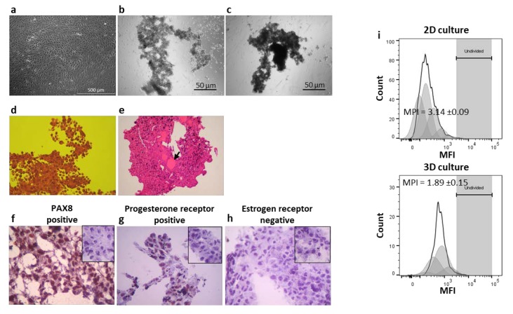
Analysis of morphological and histological features of the CAISMOV24 cells in 3D culture. Phase contrast microscopy of CAISMOV24 cells growing as (a) monolayer in 2D culture or growing as cell aggregates in 3D culture at (b) 24 h and (c) at 72 h. (d and e) Brightfield microscopy of histological cuts representative of CAISMOV24 cell aggregates (n = 8); (d) Histological cut showing papillary morphology of the cell aggregate (obj. 40×; H&E staining) and (e) the presence of focal acinar arrangement with secreted material (arrow; obj. 10×; H&E staining). Immunohistochemistry analysis of cryosections of 3D-cultured CAISMOV24 cells (n = 6 cell aggregates) showing nuclear expression of PAX8 (f) and progesterone receptor (g) in brown, as well as, absence of estrogen receptor (h) compared with their respective negative controls (insets); cells are counterstained with hematoxylin. (i) Proliferation profile of CAISMOV24 cells assessed by flow cytometry on day 5, following cell labeling with violet proliferation dye 450 (VPD450); shaded areas represent each of the new cell generations, which retained approximately half of the VPD450 fluorescence intensity of their parent cells. The mean proliferation index of CAISMOV24 cells was significant lower (p < 0.0001, t-student test) in 3D culture (n = 7 experimental repetitions) than in the 2D culture (n = 3). MPI = mean proliferation index.

