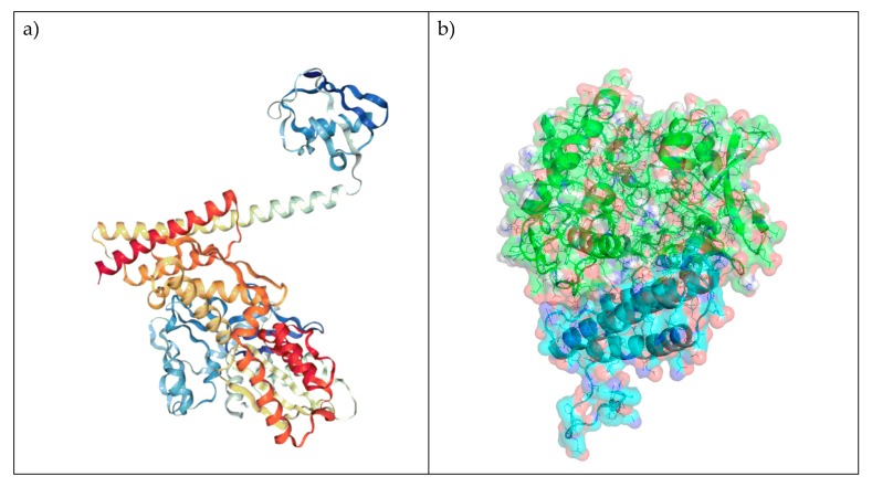
Figure 1.
Modelled interaction of BAG3 with HSP70. (a) Crystal structure of the homologue BAG1 binding to HSP70 (PDB 4HWI). (b) Homology model of BAG3 (cyan) binding to HSP70 (green). (c) Superimposition of BAG1 (magenta) and BAG3 (cyan) in complex with HSP70 (green). (d) Two-dimensional and three-dimensional depiction of plausible key interactions observed by visual inspection of the modelled interaction, BAG3 residues are coloured in light blue and HSP70 residues in green.


