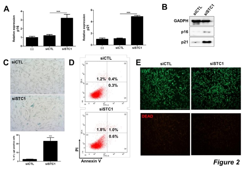
Figure 2.
Induction of TMSC senescence by STC1 inhibition. After three days of siSTC transfection, the expressions of cyclin dependent kinase inhibitors in TMSCs were determined in mRNA level by qPCR (A) and protein level by immunoblotting (B). Cellular senescence was assessed by β-gal staining and the number of β-gal positive cells compared to control group was counted (C). Annexin V and PI were stained in untreated or siSTC-treated TMSCs and analyzed for apoptosis by flow cytometry (D). Cell viability was evaluated by Live/Dead staining (E). Results are three technical replicates of TMSC from one donor. Representative results from two different TMSCs with similar tendency were presented. ***P < 0.001. Scale bar = 500 μm. Results are shown as mean ± SD.

