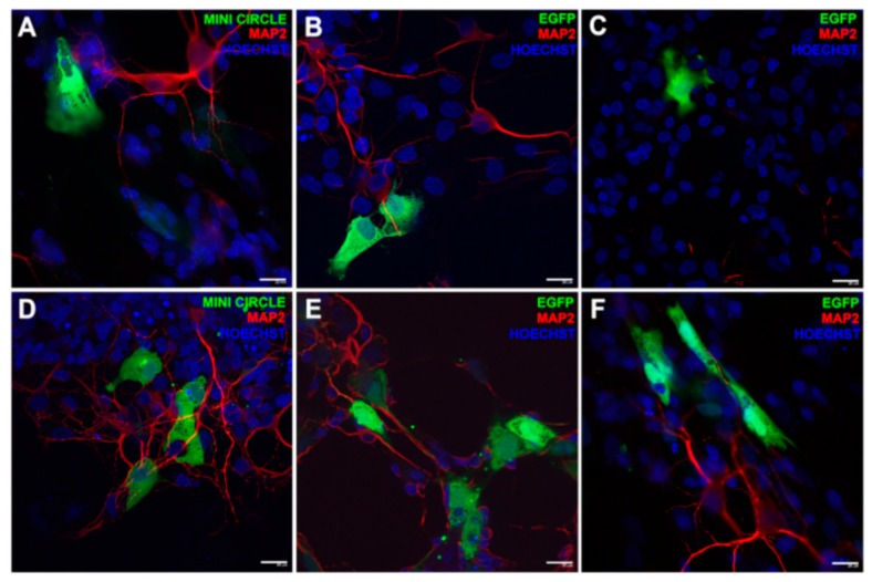
Figure 5.
Fluorescence immonostaining by GFP expression in embrionary rat retinal primary cells. The green signal was referred to the transfection event of DST20 nioplexes with MC-GFP, (B) pGFP 3.5 kb, or (C) pEGFP 5.5 kb. (D–F) Positive controls incubated with Lipofectamine™ 2000. Nuclei were stained with Hoechst 33,342 (blue) and neuronal dendrites with MAP2 (red). Scale bars: 20 μm. Reprinted from [116] Copyright (2019), with permission from Elsevier.

