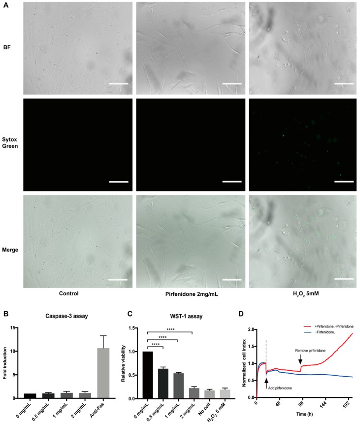
Figure 2.
Pirfenidone does not induce cell death and reversibly suppresses the proliferation of human primary intestinal fibroblasts. (A) Bright field (top panels), Sytox Green nucleic staining (middle panels) and overlay images (bottom panels) of p-hIFs exposed for 72 h to 2 mg/mL pirfenidone or for 24 h to 5 mmol/L H2O2 (positive control for necrosis) showing that less than 1% of pirfenidone-exposed p-hIFs were necrotic (representative image of n = 3). (B) Pirfenidone did not induce caspase-3 activity compared to anti-Fas-exposed (1 ug/mL for 9 h) p-hIFs (positive control for apoptotic cell death) (n = 3). (C) Water Soluble Tetrazolium Salt-1 (WST-1) assay showed pirfenidone dose-dependently suppressed the total metabolic activity of p-hIFs as proxy for total cell number (**** p < 0.0001, n = 3). (D) Pirfenidone (2 mg/mL for 72 h)-induced inhibition of p-hIF proliferation was reversible even refreshing the cells with pirfenidone-free medium, as analyzed in real-time in the xCELLigence. Scale bars = 50 μm.

