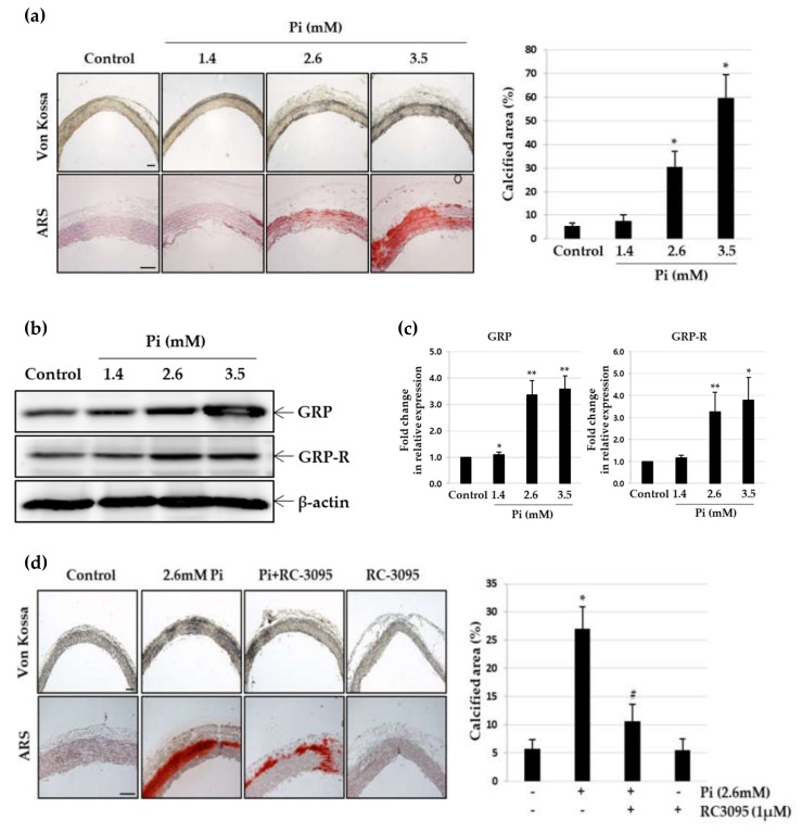
Figure 4.
Effect of RC-3095 on Pi-induced vascular calcification in cultured explants of aorta. Pieces of rat aorta were cultured in calcification medium (2.6 mM) for 10 days. (a) The calcified lesions were examined by von Kossa and ARS staining (left). Scale bar: 50μm. The percent calcified area was calculated using Calcification Analyzer Ver2 (right). * p < 0.01 vs. control. (b and c) Levels of GRP or GRP-R proteins and their mRNA were estimated by western blot analysis and real-time RT-PCR, respectively. * p < 0.05; ** p < 0.01 vs. control. Pieces of rat aorta were incubated in calcification medium (2.6 mM) in the presence or absence of RC-3095 (1 μM) for 10 days. (d) The calcified lesions were examined by von Kossa staining (left). Scale bar: 50μm.The percent calcified area was calculated using Calcification Analyzer Ver2 (right). * p < 0.01 vs. control, # p < 0.01 vs. 2.6 mM Pi, Data shown are the mean ± SD, obtained from at least three independent experiments. (e,f) Western blots were individually probed with antibodies against Runx2, calponin, Bcl2, Bad or β-actin.


