Figure 3.
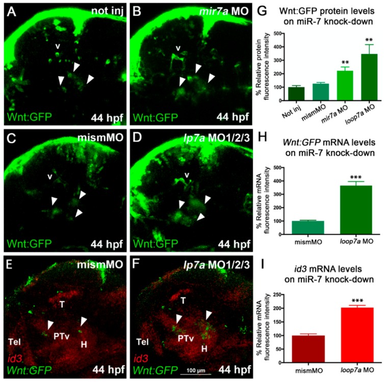
miR-7 knockdown increases Wnt signaling in the zebrafish diencephalon. (A,B,C) Morpholino-mediated knock-down of miR-7 (mature form) increases Wnt-reporter GFP (Green Fluorescent Protein) in the diencephalon (arrowheads) of miR-7 morphants (B) compared with not injected (A) and mismatched MO-injected (C) controls. Increased Wnt-reporter GFP is confirmed in loop7a1/2/3 (immature miR-7) morphants (D). (E,F) Two-color in situ hybridization for id3 (landmark) and Wnt:gfp mRNAs shows that most of the diencephalic Wnt-responsive cells (arrowheads) are located in the ventral posterior tuberculum (PTv) and hypothalamic (H) regions of 44 hpf embryos. Loop7a1/2/3 morphants display increased id3 and gfp expression, compared with controls. All panels show 44 hpf embryonic heads in lateral view, from anterior to the left. Tel: telencephalon; T: tectum; v: vessels. (G,H,I) Quantitative analysis of Wnt reporter in the zebrafish ventral diencephalon. Chart G refers to A–D experimental series; charts H and I refer to E–F experiments. Error bars represent ± SEM. The experiments were repeated thrice. Sample size n = 5 for quantification using Volocity software. Unpaired t-test results are indicated by (**) p < 0.01 and (***) p < 0.001. All panels zoom on the dorsal diencephalic (dd) region of 2 dpf embryos in lateral view, from anterior to the left. Displayed images represent the average phenotype from batches of n = 20 embryos per condition; the experiment was performed in duplicate.

