PLOS ONE logoLink to PLOS ONE
. 2020 Apr 8;15(4):e0230930. doi: 10.1371/journal.pone.0230930

Comprehensive histochemical profiles of histone modification in male germline cells during meiosis and spermiogenesis: Comparison of young and aged testes in mice

Misako Tatehana 1, Ryuichi Kimura 1, Kentaro Mochizuki 1,2, Hitoshi Inada 1, Noriko Osumi 1,*
Editor: Suresh Yenugu3
PMCID: PMC7141650  PMID: 32267870

Abstract

Human epidemiological studies have shown that paternal aging as one of the risk factors for neurodevelopmental disorders, such as autism, in offspring. A recent study has suggested that factors other than de novo mutations due to aging can influence the biology of offspring. Here, we focused on epigenetic alterations in sperm that can influence developmental programs in offspring. In this study, we qualitatively and semiquantitatively evaluated histone modification patterns in male germline cells throughout spermatogenesis based on immunostaining of testes taken from young (3 months old) and aged (12 months old) mice. Although localization patterns were not obviously changed between young and aged testes, some histone modification showed differences in their intensity. Among histone modifications that repress gene expression, histone H3 lysine 9 trimethylation (H3K9me3) was decreased in the male germline cells of the aged testis, while H3K27me2/3 was increased. The intensity of H3K27 acetylation (ac), an active mark, was lower/higher depending on the stages in the aged testis. Interestingly, H3K27ac was detected on the putative sex chromosomes of round spermatids, while other chromosomes were occupied by a repressive mark, H3K27me3. Among other histone modifications that activate gene expression, H3K4me2 was drastically decreased in the male germline cells of the aged testis. In contrast, H3K79me3 was increased in M-phase spermatocytes, where it accumulates on the sex chromosomes. Therefore, aging induced alterations in the amount of histone modifications and in the differences of patterns for each modification. Moreover, histone modifications on the sex chromosomes and on other chromosomes seems to be differentially regulated by aging. These findings will help elucidate the epigenetic mechanisms underlying the influence of paternal aging on offspring development.

Introduction

Autism spectrum disorder (ASD) is a neurodevelopmental disorder, and it has been reported to show a pandemic rise in recent decades. The most recent survey in the United States shows that one in 59 eight-year-old children has been diagnosed with ASD [1]. The reason for the rise might include changes in diagnosis, although paternal aging has been suggested as one of the biological factors. For example, meta-analyses of 5,766,794 children born from 1985–2004 in five countries have revealed a significant association between advanced paternal age and ASD risk in children [2].

Aging induces de novo mutations in the genome of sperm, and most of the mutations in autistic children are reported to be inherited from fathers [3]. However, a study using a mathematical model has demonstrated that the risk of psychiatric illness from de novo mutations derived from advanced paternal age has been estimated as 10–20% [4]. Therefore, some factors other than genetic mutations can influence the health and disease of offspring.

One possible mechanism for paternal aging effects on offspring is changes to the epigenetic information encoded in sperm. Previously, we have reported that sperm derived from aged mice have more hypomethylated regions in their genome that possibly affect offspring’s behavior [5]. However, the age-related alteration of histone modifications, another major epigenetic regulation, remains unclear. Sperm is produced through dynamic and complicated processes, termed spermatogenesis, which includes meiosis (in spermatocytes) and morphological changes to produce mature sperm. Considering frequent chromatin remodeling during spermatogenesis, alteration of histone modifications would have a high impact on spermatogenic processes. Additionally, some histone modifications remain in sperm after the histone-to-protamine replacement [69]. For example, it has been reported that histones remain in genomic regions that function in early developmental stages [10], and that alteration of histone retention sites in sperm can be inherited by the next generation [11]. Therefore, features of histone moiety in spermatogenesis would provide essential clues for understanding gene regulation after fertilization and subsequent development of offspring. Nevertheless, little is known about the localization patterns of histone modifications in male germline cells and the changes they undergo during aging.

Here, we first produced comprehensive histological profiles of major histone modifications, including histone H3 lysine 9 trimethylation (H3K9me3) and H3K27 acetylation (ac), during murine spermatogenesis, and the individual epigenetic modifications showed dynamic and distinct localization changes. We further examined the influence of aging on histone modifications in each step of spermatogenesis, and we found that the levels of histone modification during spermatogenesis were differentially affected by aging. We also noticed the accumulation of H3K79me3 on sex chromosomes. Further elucidation of how aging-induced epigenetic changes contribute to altered gene regulation in the offspring would provide insight into the influence of paternal aging on the health and disease state of the next generation.

Materials and methods

Animals

C57BL/6J mice at 3 months old were purchased from a breeder (Charles River Laboratories, Japan) and were raised to 12 months old at the Institute for Animal Experimentation at the Tohoku University Graduate School of Medicine; they were then used for histological and semiquantitative analyses. All animals were housed in standard cages in a temperature- and humidity-controlled room with a 12-hour light/dark cycle (light on at 8 am), and they had free access to standard food and water. All experimental procedures were approved by the Ethics Committee for Animal Experiments of the Tohoku University Graduate School of Medicine (#2017- MED210), and the animals were treated according to the National Institutes of Health guidance for the care and use of laboratory animals.

Histological analyses of histone modifications

Histological analysis was performed based on a previous our report [12]. Mice were anesthetized with isoflurane and perfused with PBS (pH 7.4) to remove blood. After perfusion, the testes were dissected, immersed in 4% PFA in PBS, and cut in half to be postfixed in 4% PFA overnight at 4°C. After washing in PBS, the testes were cryoprotected by being immersion in 5% and 10% sucrose in PBS solution for 1 hour each and then 20% sucrose in PBS solution overnight; then, the tissues were embedded in OCT compound (Sakura Finetek, Japan). Frozen testis sections were cut at a thickness of 10-μm on a cryostat (Leica, CM3050).

Next, 0.01 M citric acid (pH 6.0) solution was heated using a microwave and kept between 90°C and 95°C on a heat plate with gentle stirring. The sections were submerged in the solution for 10 minutes for antigen retrieval. The solution and sections were left to cool to 40–50°C and were then washed in 1×Tris-based saline containing 0.1% Tween 20 (TBST) (pH 7.4). Then, the sections were blocked by treatment with 3% bovine serum albumin and 0.3% Triton X-100 in PBS. The sections were incubated at 4°C overnight with the following primary antibodies at a dilution of 1/500: rabbit anti-H3K4me2 (07–030, Millipore), rabbit anti-H3K4me3 (ab8580, Abcam), rabbit anti-H3K9me3 (ab8898, Abcam), rabbit anti-H3K27me2 (ab24684, Abcam), rabbit anti-H3K27me3 (07–449, Millipore), rabbit anti-H3K27ac (ab4729, Abcam), rabbit anti-H3K79me2 (ab3594, Abcam), rabbit anti-H3K79me3 (ab2621, Abcam). Together with the antibody against each histone modification, the antibody mouse anti-SCP3 (ab97672, Abcam), a spermatocyte marker, was applied for easy evaluation for staging the testis tubes. Information on the primary antibodies is summarized in Table 1. Subsequently, either a Cy3- or Alexa488-conjugated secondary antibody (1:500) was applied together with 4’6-diamino-2-phenylindole (DAPI), and the sections were incubated at RT for one hour in a humid chamber. After washing in TBST at room temperature, specimens were mounted in Vectashield (Vector Laboratories, H-1200) or ProLong Diamond Antifade Mountant (Invitrogen, P36965). Images were captured using a confocal laser-scanning microscope (LSM780, Carl Zeiss, Jena, Germany).

Table 1. List of primary antibodies used in this study.

Antibody name Source Dilution Company (Catalogue) Reference
anti-H3K4me2 rabbit polyclonal 1/500 Millipore (07–030) [1315]
anti-H3K4me3 rabbit polyclonal 1/500 Abcam (ab8580) [1618]
anti-H3K9me3 rabbit polyclonal 1/500 Abcam (ab8898) [1921]
anti-H3K27ac rabbit polyclonal 1/500 Abcam (ab4729) [2224]
anti-H3K27me2 rabbit polyclonal 1/500 Abcam (ab24684) [25, 26]
anti-H3K27me3 rabbit polyclonal 1/500 Millipore (07–449) [24, 2729]
anti-H3K79me2 rabbit polyclonal 1/500 Abcam (ab3594) [3032]
anti-H3K79me3 rabbit polyclonal 1/500 Abcam (ab2621) [3335]
anti-SCP3 mouse monoclonal 1/500 Abcam (ab97672) [3638]
anti-α-Tubulin (S2 Fig) mouse monoclonal 1/500 Sigma-Aldrich (T6199) [39, 40]

All antibodies are commercially available, and their specificity has been reported in previous studies.

Semi-quantitative analyses of histone modifications

Confocal images of testis sections were semiquantitatively analyzed using ImageJ. SCP3 signals were used to select meiotic cells during spermatogenesis. First, we selected the “Region of Interest (ROI)” by identifying the nuclear area stained with DAPI. Then, the fluorescent intensity of DAPI and histone modifications in the ROI were measured, and the mean intensity was calculated for each signal intensity (total signal intensity/nuclear area). According to a previous literature [41], the mean intensity of histone modifications was subsequently normalized to the mean intensity of the DAPI signal (mean intensity of antibody signal/mean intensity of DAPI signal). We measured the integrated intensity of DAPI signal at each stage to show that DAPI staining was stable and reflects changes of DNA amount (S1 Fig). Compared to the intensity of pachytene spermatocytes, the intensity of round spermatid (after meiosis) was about half. The DNA amount was variable in the preleptotene and metaphase spermatocytes, where DNA synthesis and chromosomal segregation occurs, respectively. Finally, the mean of young group was set as 1, and the intensity of the aged group was plotted as a relative score. We analyzed male germline cells in several tubules from four different young (3 months old) or aged (12 months old) mice, yielding 50 cells to be analyzed for each stage and each histone moiety (50 cells x 12 types of cells were analyzed for each box plot in young group and aged group, respectively). We categorized the normalized intensity of the young testis as–(not detected), + (normalized intensity <0.4), ++ (0.4–0.6), +++ (0.6–0.8), ++++ (0.8–1.0), and +++++ (1.0<), as shown in Table 3. The graphs were drawn using the R [42] and ggplot2 [43] packages. The line on the box indicates the median. Scores of individual cells from young and aged testes are shown as black and red dots, respectively, over box plots in Figs 3-1 to 4-3.

Table 3. Summarized histone modification levels during spermatogenesis and their aging-associates changes.

pL(VIII) L (X) Z(XII) P(I) P(V) P(VIII) P(X) M(XII) R(I) R(V) R(VIII) E(X) Specific note
H3K4me2 Young ++++ +++++ ++++ +++ +++ + ++ ++++ +++++ +++++ ++++ +++++ Accumulation on XY body
Aged ↓↓** ↓↓** ↓↓** ↓↓** ↓↓** ↓↓** ↓↓** ↓↓** ↓↓** ↓↓** ↓↓** ↓↓**
H3K4me3 Young - - - - - - - ++++ ++++ +++++ ++++ ++++  
Aged - - - - - - - ↓↓**
H3K27ac Young +++++ + + + ++ ++ ++ + ++ ++ ++ +++++ Accumulation on XY body and sex chromosomes
Aged ↓↓** ↓** * ↓** ↓↓** ** **
H3K79me2 Young - - - - - + + ++ ++ +++++ +++++ ++++ Accumulation on sex chromosomes
Aged - - - - - ** * **
H3K79me3 Young - - - - - - - ++++   ++ ++++ +++++ Accumulation on sex chromosomes
Aged - - - - - - - ** - ↓* ↓**
H3K9me3 Young ++ +++ +++ +++ +++ + + + + + ++ ++ Accumulation on sex chromosomes and heterochromatin
Aged ↓↓** ↓↓** ↓↓** ↓↓** ↓↓** ↓↓** ↓↓** ↓↓** ↓* * ↓*
H3K27me2 Young - + + + ++ ++++ ++ ++ ++ +++ ++ ++ Accumulation on heterochromatin
Aged - ** ** ** ** ↓* ** ** ** **
H3K27me3 Young - +++ ++ ++ ++ ++ ++++ +++ +++ ++++ +++++ +++++ Accumulation on heterochromatin
Aged - ↓↓** ** ** ** ** ↓↓** ** **

Accumulation levels of histone modifications that activate (red) and inhibit (blue) gene expression are shown. Each histone modification level in male germline cells derived from young mice is indicated as–(not detected), + (normalized intensity <0.4), ++ (0.4–0.6), +++ (0.6–0.8), ++++ (0.8–1.0), and +++++ (1.0<) (Actual values for intensity young male germline cells are shown in S1 Table). Arrows show histone modification levels of male germline cells derived from aged mice compared to that of young mice. Single up/down arrows indicate that a signal was increased/decreased by less than 20% of the mean intensity compared to the signal from young animals. Double up/down arrows indicate that the signal was increased/decreased by more than 20% compared to the signal from young animals. Right arrows indicate that there were no significant differences between young and aged animals (*p < 0.05, **p < 0.01). Specific localization or accumulation is described in the specific notes section.

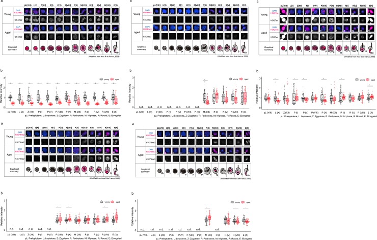
Fig 3. Comparison of localization patterns and intensity of H3K4me2 (3–1), H3K4me3 (3–2), H3K27ac (3–3), H3K79me2 (3–4) and H3K79me3 (3–5) in male germline cells in young (3M) and aged (12M) testes.

Fig 3

(a) Representative confocal images of histone marks in male germline cells in young and aged testes. In each group, upper panels are merged images of histone marks (magenta) and DAPI (blue), and lower panels show the grayscale images for histone marks. An illustrated overview shows subcellular localization of each histone modification (magenta) in male germline cells in young and aged testes. The numbers indicate individual cells at specific stages during spermatogenesis, as shown in Table 2. (b) Semiquantitative analysis of histone marks in male germline cells in young and aged testes. Gray boxes show the intensity of aged cells relative to young cells (**p < 0.01, *p < 0.05, n.d., not detected). Black and red dots show relative intensity of individual cells from young and aged testes, respectively. Each number in parenthesis represents the stage of spermatogenesis (see Table 2.).

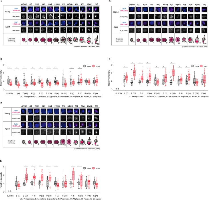
Fig 4. Comparison of localization patterns and intensity of H3K9me3 (4–1), H3K27me2 (4–2) and H3K27me3 (4–3) in male germline cells in young (3M) and aged (12M) testes.

Fig 4

(a) Representative confocal images of histone marks in male germline cells in young and aged testes. In each group, the upper panels are merged images of histone marks (magenta) and DAPI (blue), and the lower panels show grayscale images for histone marks. An illustrated overview shows subcellular localization of each histone modification (magenta) in male germline cells in young and aged testes. The numbers indicate individual cells at specific stages during spermatogenesis, as shown in Table 2. (b) Semiquantitative analysis of histone marks in male germline cells in young and aged testes. Gray boxes show the intensity of aged cells relative to young cells (**p < 0.01, *p < 0.05, n.d., not detected). Black and red dots show relative intensity of individual cells from young and aged testes, respectively. Each number in parenthesis represents the stage of spermatogenesis (see Table 2.).

Statistical analysis

Wilcoxon rank-sum tests were used to determine statistical significance between the young and aged groups at each stage using the statistical analysis software JMP® Pro 14 (SAS Institute Inc., Cary, NC, USA). Values of *p < 0.05 and **p < 0.01 were considered statistically significant.

Results

1. Morphological profiles showing histone modification patterns during spermatogenesis

Male germline cells drastically change their morphology during spermatogenesis. Previous studies have shown the localization of H3K79me2/3 in spermatocytes and spermatids as well as the accumulation of H3K4me, H3K9me and H3K27me during transformation from spermatogonia to elongated spermatids [34, 35, 4447]; however, comprehensive detailed subcellular localization patterns have not been fully examined. Here, we first investigated the localization of major histone modifications from preleptotene spermatocytes to elongated spermatids according to 12 stages based on a previous report [48]. This staging helped us to morphologically follow each step of spermatogenesis without any marker staining.

(1) Histone modifications that activate gene expression

First, we examined active marks for gene expression, i.e., H3K4me2, H3K4me3, H3K27ac, H3K79me2 and H3K79me3.

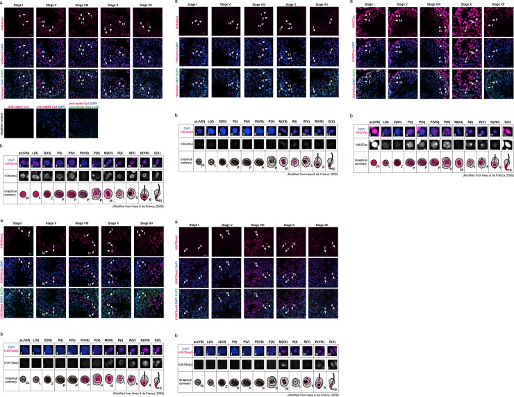
H3K4me2 signal was observed in all stages of seminiferous tubules. During stage VIII, in which the earliest meiotic spermatocytes (i.e., preleptotene cells) are located near the periphery of the tubule, H3K4me2 was detected in the whole nucleus of preleptotene cells, though the signal was not found in constitutive heterochromatin (Fig 1-1A, arrowhead 1). The signal persisted throughout meiosis (Fig 1-1A, arrowheads 2–6). In the pachytene cells of stage X, a strong signal was detected on the XY body (Fig 1-1A, arrowhead 7). The accumulation of H3K4me2 on the XY body disappeared in the M-phase spermatocytes at stage XII, and only a weak signal was observed in the chromosomes (Fig 1-1A, arrowhead 8). In round spermatids from stage I to VIII and in elongated spermatids at stage X, the signal was widely detected in the nucleus but not in the chromocenter (constitutive heterochromatin) (Fig 1-1A, arrowheads 9–12). Thus, the H3K4me2 modification seemed to indicate that it was first present in preleptotene cells, and then it accumulated on the XY body in late pachytene cells. Through spermatogenesis, H3K4me2 was not detected in the chromocenter (Fig 1-1B).

Fig 1. Localization of H3K4me2 (1–1), H3K4me3 (1–2), H3K27ac (1–3), H3K79me2 (1–4), and H3K79me3 (1–5) in male germline cells in the young (3 M) testis.

Fig 1

(a) Representative confocal images of histone marks (magenta) and SCP3 (green) in the seminiferous epithelium in stages I, V, VIII, X and XII. Nuclei were counterstained with DAPI (blue). The numbers with an arrowhead indicate an individual cell at the specific stage during spermatogenesis, as shown in Table 2. Scale bar: 20 μm. Negative controls (without primary antibodies) are shown in Fig 1–1. (b) Magnified images of the cells indicated by arrowheads in (a), and subcellular localization of each histone modification in the young testis is shown in magenta in a graphical summary. The upper panels are merged images of histone marks (magenta) and DAPI (blue), and the lower panels are grayscale images for the histone marks. Each parenthesis represents the stage of spermatogenesis (see Table 2.).

Table 2. The cells and stages indicated by each number and their abbreviations.
Number Cell and stage Abbreviation (Stage)
1 preleptotene spermatocyte at stage VIII pL (VIII)
2 leptotene spermatocyte at stage X L (X)
3 zygotene spermatocyte at stage XII Z (XII)
4 pachytene spermatocyte at stage I P (I)
5 pachytene spermatocyte at stage V P (V)
6 pachytene spermatocyte at stage VIII P (VIII)
7 pachytene spermatocyte at stage X P (X)
8 M phase spermatocytes at stage XII M (XII)
9 round spermatid at stage I R (I)
10 round spermatid at stage V R (V)
11 round spermatid at stage VIII R (VIII)
12 elongated spermatid at stage X E (X)

While H3K4me2 was observed beginning in preleptotene cells (Fig 1-1B), the H3K4me3 signal was not detected between preleptotene cells to pachytene cells (Fig 1-2A, arrowheads 1–7). H3K4me3 was first detected in M-phase spermatocytes in stage XII seminiferous tubules (Fig 1-2A, arrowhead 8). The signal was broadly distributed on condensed chromosomes (Fig 1-2B, arrowhead 8). After meiosis II, the signal was detected in round spermatids at stages I to VIII, though there was no signal in the chromocenter (Fig 1-2A, arrowheads 9–11). In elongated spermatids, the signal was weak and widely detected in the whole nucleus (Fig 1-2A, arrowhead 12). Thus, localization of the H3K4me3 modification appeared later in meiosis, and the signal was weak compared to the H3K4me2 modification (Fig 1-2B).

H3K27ac signal was observed in all stages of seminiferous tubules. At stage VIII, in which the earliest meiotic cells (i.e., preleptotene cells) were located near the periphery of the tubule, strong H3K27ac signals were detected in whole nuclei of preleptotene cells (Fig 1-3A, arrowhead 1). While the signal continued throughout meiosis, the intensity dramatically decreased when preleptotene cells differentiated into leptotene cells (Fig 1-3A, arrowhead 2–5). In stage VIII pachytene cells, a strong signal was detected on the XY body (Fig 1-3A, arrowhead 6). The accumulation of H3K27ac on the XY body disappeared in the pachytene cells of stage X and in the M-phase spermatocytes of stage XII, and only a weak signal was observed in the whole nucleus or chromosomes (Fig 1-3A, arrowheads 7–8). In round spermatids from stage I to V, the signal was widely detected in the nucleus, especially in the peri-chromocenter (putative sex chromosome) (Fig 1-3A, arrowheads 9–10). The strong signal at the peri-chromocenter disappeared in stage VIII round spermatids (Fig 1-3A, arrowhead 11). The H3K27ac signal increased again in elongated spermatids, suggesting that H3K27ac is required for spermiogenesis (Fig 1-3A, arrowhead 12). Thus, localization of the H3K27ac modification seemed to indicate that it was first present in preleptotene cells, and then it accumulated on the XY body in late pachytene cells and on the putative sex chromosome during early to middle stages of round spermatids (Fig 1-3B).

In pachytene spermatocytes from stage VIII to stage X, the H3K79me2 signal was widely detected in the nucleus (Fig 1-4A, arrowheads 6 and 7). In M-phase spermatocytes at stage XII, the signal was broadly observed on the condensed chromosomes (Fig 1-4A, arrowhead 8). In round spermatids at stage I, a weak H3K79me2 signal was observed in most of the nucleus, with the exception of the chromocenter (Fig 1-4A, arrowhead 9). Interestingly, a strong H3K79me2 signal was observed in the peri-chromocenter (putative sex chromosome) in round spermatids from stage V to stage VIII (Fig 1-4A, arrowheads 10 and 11), while no signal was observed on the chromocenter itself. In elongated spermatids at stage X, the strong signal on the putative sex chromosome disappeared (Fig 1-4A, arrowhead 12). Thus, localization of the H3K79me2 modification seemed to indicate that it was first present in the late pachytene spermatocytes and to accumulate on the putative sex chromosome during most stages of round spermatids (Fig 1-4B).

While H3K79me2 was observed in late pachytene cells (Fig 1-4B), the H3K79me3 signal was not detected in any pachytene cells. H3K79me3 was first detected in M-phase spermatocytes in stage XII seminiferous tubules (Fig 1-5A, arrowhead 8). Unlike H3K79me2, the H3K79me3 signal was not distributed on all condensed chromosomes; rather, it was found on specific chromosomes (Fig 1-5A, arrowhead 8). After meiosis II, the signal was not detected in stage I round spermatids (Fig 1-5A, arrowhead 9). However, in round spermatids from stage V to stage VIII, a broad H3K79me3 signal was observed again in the nucleus, and a strong signal was detected in the peri-chromocenter (putative sex chromosome) (Fig 1-5A, arrowheads 10–11). In stage X elongated spermatids, the signal was widely observed in the nucleus (Fig 1-5A, arrowhead 12). Thus, the H3K79me3 modification appeared in the late stage of meiosis and accumulated on the putative sex chromosome of round spermatids. A specific area of condensed chromosomes showed a strong H3K79me3 signal during the M-phase (Fig 1-5B).

(2) Histone modifications that inhibit gene expression

Next, we examined repressive marks: H3K9me3, H3K27me2 and H3K27me3.

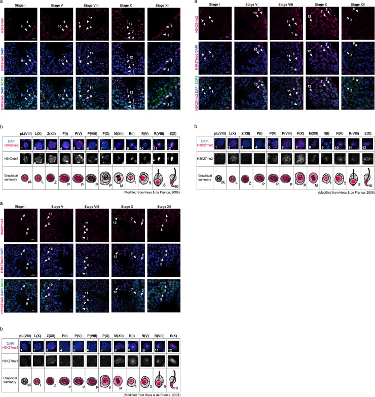
H3K9me3 was detected in all cell types examined in this study. Broad nuclear signals were observed from preleptotene spermatocytes at stage VIII to pachytene spermatocytes at stage V with strong punctate signals in the periphery of the nucleus (Fig 2-1A, arrowheads 1–5). In pachytene spermatocytes at stage VIII and stage X, only dot-like signals were detected (Fig 2-1A, arrowheads 6–7). These dotted signals matched with intense DAPI signals, indicating areas of heterochromatin. Consistent with a previous report [49], H3K9me3 was especially localized on the sex chromosome of stage XII M-phase spermatocytes (Fig 2-1A, arrowhead 8). In round spermatids of stage I through elongated spermatids of stage X, the signal was detected on the chromocenter and peri-chromocenter (putative sex chromosome) (Fig 2-1A, arrowheads 9–12). Thus, localization of H3K9me3 modification seemed to indicate that it was first present in preleptotene spermatocytes, and then it accumulated on the sex chromosome in M-phase spermatocytes, where it stayed until late round spermatids (Fig 2-1B).

Fig 2. Localization of H3K9me3 (2–1), H3K27me2 (2–2) and H3K27me3 (2–3) in male germline cells in the young (3 M) testis.

Fig 2

(a) Representative confocal images of histone marks (magenta) and SCP3 (green) in the seminiferous epithelium at stages I, V, VIII, X and XII. Nuclei were counterstained with DAPI (blue). The numbers with an arrowhead indicate individual cells at specific stages during spermatogenesis, as shown in Table 2. Scale bar: 20 μm. (b) Magnified images of the cells indicated by arrowheads in (a), and subcellular localization of each histone modification in the young testis is shown in magenta in a graphical summary. The upper panels are merged images of histone marks (magenta) and DAPI (blue), and the lower panels are grayscale images showing the histone marks. Each parenthesis represents the stage of spermatogenesis (see Table 2.).

H3K27me2 was detected in the nucleus of stage X leptotene cells through stage X pachytene cells in a salt-and-pepper manner (Fig 2-2A, arrowheads 2–7). In M-phase stage XII spermatocytes, the signal was broadly observed on all chromosomes (Fig 2-2A, arrowhead 8). In round stage I spermatids, strong dot-like signals were observed on the chromocenter, and there were also weak and broad signals in the nucleus (Fig 2-2A, arrowhead 9). The dot-like signals were observed until stage VIII round spermatids (Fig 2-2A, arrowheads 10–11), and they disappeared in elongated spermatids at stage X (Fig 2-2A, arrowhead 12). Thus, localization of the H3K27me2 modification was first observed in leptotene cells, and it accumulated on the chromocenter of all stages of round spermatids; however, the signal was relatively weak through spermatogenesis (Fig 2-2B).

H3K27me3 was detected in all cell types examined in this study except for preleptotene cells. In stage X leptotene cells through stage X pachytene cells, the signal was broadly but faintly detected in the nucleus (Fig 2-3A, arrowheads 2–7). In M-phase stage XII spermatocytes, the signal was widely observed on all chromosomes (Fig 2-3A, arrowhead 8). In stage I round spermatids through stage X elongated spermatids, strong dot-like signals were observed on the chromocenter in addition to the weak and broad signals in the nucleus (Fig 2-3A, arrowhead 12), while strong dot-like signals of H3K27me2 disappeared in elongated spermatids at stage X (Fig 2-3A, arrowhead 12). Thus, localization of the H3K27me3 modification seemed to indicate that it was first present in leptotene cells, which is the same timing as H3K27me2, and then H3K27me3 accumulated on the chromocenter in early round spermatids (Fig 2-3B).

The above localization patterns of histone modifications during spermatogenesis are shown in Table 3. In summary, H3K4me2 was detected in preleptotene cells through elongated spermatids, and H3K27ac was detected in leptotene cells through elongated spermatids. These two modifications were detected at the early stage of spermatogenesis, while other activation marks were detected at the later stage of spermatogenesis. All repressive marks examined in this study were detected at the early stages of spermatogenesis. All activation marks except H3K4me3 showed specific accumulation on the XY body in pachytene cells or on a sex chromosome in round spermatids, while all repressive makers accumulated on the chromocenter (i.e., constitutive heterochromatin). Only H3K9me3 accumulated on both sex chromosomes and the chromocenter.

2. Semi-quantitative analyses of histone modifications in young and aged testes

We next performed semiquantified analyses to examine whether the intensity of histone modifications is different between young and aged testes. We used 12-month-old mice for the aged group because we previously confirmed that the fertility of >12-month-old male mice was not changed, nor was the litter size of offspring derived from fathers at this age different, yet these offspring showed a significant difference in behavior [5, 50]. Moreover, these results are consistent with data from other papers reporting alterations of social behaviors in adult offspring derived from aged fathers [51, 52]. The histone modification signals were measured by ImageJ. The intensity of histone modifications was normalized to that of DAPI in the nucleus or chromosomes. The intensity of individual cells was plotted to show their individual changes because only one sperm cell will eventually be fertilized to develop a new individual.

(1) Histone modifications that activate gene expression

No difference was observed in the localization patterns of H3K4me2/3, H3K27ac, and H3K79me2/3 in young and aged testes (Fig 3-1A, 3-2A, 3-3A, 3-4A and 3-5A). However, semiquantitative analyses revealed differences in intensity levels among the observed histone modifications.

The intensity levels of H3K4me2 in the cells were lower at all specific stages of spermatogenesis in the aged testis than in the young testis (Fig 3-1B). However, the intensity levels of H3K4me3 were not significantly different in most of the cells. Only in M-phase spermatocytes at stage XII was H3K4me3 intensity lower in the aged testis than in the young testis (Fig 3-2B). The intensity levels of H3K27ac were lower in leptotene spermatocytes at stage X, zygotene spermatocytes at stage XII, pachytene spermatocytes at stages V and X in the aged testis than in the young testis (Fig 3-3B). However, higher intensity was detected in pachytene spermatocytes at stage I, round spermatids at stage I and elongated spermatids at stage X in the aged testis. In other cells, there were no significant differences. Compared with the young testis, higher intensity of H3K79me2 was detected in stage X pachytene spermatocytes, stage VIII round spermatids, and stage X elongated spermatids (Fig 3-4B) of the aged testis. However, other cells showed no difference in the intensity level. A higher intensity of H3K79me3 was detected in M-phase spermatocytes at stage XII of the aged testis than in the young testis, while a lower intensity was observed in round spermatids at stage VIII and elongated spermatids at stage X (Fig 3-5B).

Interestingly, H3K79me3 signal was strongly detected on specific chromosomes in M-phase spermatocytes at stage XII (S2A Fig). According to previous report, H3K79me3 accumulates on sex chromosomes in diakinesis to M-phase spermatocytes [35]. Therefore, we suspected that the chromosome on which H3K79me3 accumulated were sex chromosomes. To identify the sex chromosome that H3K79me3 associated with, we took an advantage of combining immunostaining with fluorescence in situ hybridization (FISH) on the same slide. First, immunostaining was performed to detect H3K79me3 signals in M-phase matocytes (S2B Fig). After images were captured using confocal microscopy, FISH was performed using probes that recognize X and Y chromosomes, which was followed by capturing FISH images of the same slides to obtain three signals (S2C Fig). Signals of H3K79me3 overlapped with those of X and Y chromosomes at the same position within the nucleus of M-phase spermatocytes. Finally, we confirmed that H3K79me3 specifically accumulated on the sex chromosomes in M-phase spermatocytes at stage XII.

(2) Histone modifications that inhibit gene expression

No differences were observed in the localization patterns of H3K9me3 and H3K27me2/3 in young and aged testes (Fig 4-1A,4-2A and 4-3A). However, semiquantitative analyses revealed differences in intensity levels among the histone modifications observed.

The intensity levels of H3K9me3 were lower in almost all the cells of the aged testis than it was in the young testis (Fig 4-1B). However, higher intensity was detected in stage V round spermatids in the aged testis. There were no significant differences in elongated spermatids at stage X. In contrast to H3K9me3, the intensity levels of H3K27me2 were higher in almost all cells of the aged testis, and only pachytene cells at stage VIII showed lower intensity in the aged testis than in the young testis (Fig 4-2B). Similar to the results for H3K27me2, almost all stages of cells showed higher intensity of H3K27me3 in the aged testis than in the young testis (Fig 4-3B). Only in stage X leptotene spermatocytes and stage X pachytene spermatocytes were lower intensity detected in the aged testis. There were no significant differences in other cell types.

The above intensity of histone modifications in the young and aged testes is shown in Table 3. In summary, histone modifications were altered by aging, and the alteration patterns were different per the type of each histone modification. Among the repressive marks, H3K9me3 levels were decreased in the aged testis, while H3K27me2 and H3K27me3 levels were increased during spermatogenesis. On the other hand, the intensity level of one activation mark, H3K4me2, was drastically decreased in spermatogenic cells of the aged testis. Other histone modifications also showed differences at some stages.

Discussion

In this study, we have made a catalog showing the transition of histone modification localization during spermatogenesis and its aging-induced changes. Since our ultimate goal is to understand molecular mechanisms that underlie risks for neurodevelopmental diseases of children from the paternal side, we focused on age-related alteration of histone modification in individual male germline cells. Individual histone modifications showed distinct localization patterns and different timing in terms of the start of their accumulation. Even among the histone modifications that have similar functions, different localization patterns were observed. Moreover, the intensity of histone modifications was altered by aging; aged testes showed different increased/decreased signals of histone modifications at different stages. There were some outliers in certain types of cells and stages; it might be possible that the cells/stages with several outliers could be critical timings vulnerable to alteration of histone modification during spermatogenesis.

We also identified a chromosome on which H3K79me3 accumulated in M-phase spermatocytes. H3K79me3 accumulated on sex chromosomes in M-phase spermatocytes and round spermatids. Morphologically, it is revealed that not only H3K79me3 but also other histone modifications, such as H3K79me2, H3K27ac and H3K9me3, accumulated on putative sex chromosomes or on the XY body. The intensity of these histone modifications specifically accumulated on the putative sex chromosomes or XY body was also altered by aging at some stages. Therefore, the epigenetic regulation of histone modifications may be different between sex chromosomes and other chromosomes, which may also be affected by aging.

A previous study reported that H3K4me2, a gene expression activation mark, showed dynamic alterations during spermatogenesis. H3K4me2 started to show a strong signal at the preleptotene spermatocytes, became weak in zygotene spermatocytes, and returned stronger again beginning in round spermatids in young mice [45]. Such dynamic alteration of H3K4me2 was also observed in our data; however, the level of H3K4me2 was decreased significantly through spermatogenesis in the aged testis compared to the young testis. It has been reported that overexpression of human KDM1A histone lysine 4 demethylase during spermatogenesis in mice caused a decrease in H3K4me2, leading to various phenotypic abnormalities such as craniofacial and skeletal defects, edema, hemorrhagic gut, extra digits, and missing eyes in E18.5 offspring [53]. The report further shows that the age-related decrease of H3K4me2 may possibly affect the spermatogenic process and that epigenetic alterations can be inherited by the next generation. Considering that H3K4 methylation may play a critical role in spermatogenesis and oogenesis [44, 54], the age-related decrease in H3K4me2 might lead to potential harmful effects in the offspring.

Another example of a histone modification involved in the inhibition of gene expression is H3K9me3. We observed its accumulation in spermatogonia, pachytene spermatocytes, round spermatids and elongated spermatids, and it accumulated on the X chromosome in round spermatids, as reported in previous studies [5558]. At the pachytene stage of meiosis I, genes on sex chromosomes are transcriptionally shut down by meiotic sex chromosome inactivation (MSCI) and are reactivated after meiosis [59, 60], to which H3K9me3 is reported to be involved [55]. It was also revealed that if there was no enrichment of H3K9me3, remodeling and silencing of sex chromosomes failed and resulted in germ cell apoptosis [49]. In postmeiotic spermatids, 87% of X-linked genes remain suppressed, while autosomes were largely active [61]. Moreover, spermatids form a distinct postmeiotic sex chromatin (PMSC) area that is enriched with H3K9me3 [61, 62]. Thus, H3K9me3, a repressive mark, has various roles during meiosis and in the postmeiotic phase. Our data showed that H3K9me3 appeared first in preleptotene spermatocytes and was observed through elongated spermatids, and then its levels drastically decreased in the aged testis during meiosis and in most of the stages of round spermatids. It is assumed that in the aged testis, failure of correct chromosome segregation during meiosis or leaky expression of X-linked genes might occur in the postmeiotic spermatids, even though the male germline cells safely proceed through meiosis.

Another repressive mark, H3K27me3, was present from leptotene cells to elongated spermatids. A study has reported that H3K27ac may act as an antagonist toward Polycomb-mediated silencing through inhibition of H3K27me3 [63]. H3K27ac signals were observed in all examined stages of male germline cells, especially in preleptotene cells and elongated spermatids. It is very interesting that H3K27ac and H3K27me3 showed exclusive localization in round spermatids; the former accumulated on the sex chromosome, while H3K27me3 accumulated on the chromocenter, a heterochromatic region. Therefore, the absence of H3K27ac (an activation mark) and the presence of H3K27me3 (a repressive mark) in the chromocenter may be reasonable with regard to gene regulation. We found that H3K27me3 was dramatically increased in the aged testis, while H3K27ac showed a slight decrease at certain stages. H3K27me3 inhibits transcription by inducing a compact chromatin structure [64, 65]. Thus, gene expression influenced by H3K27me3 might be more repressed in the aged testis than in the young testis.

Although there are limitations in our semiquantitative analyses using immunostaining signals, our data showing accumulation of H3K9me3, H3K27ac and H3K79me3 on sex chromosomes are considered to correspond to previous ChIP-seq data [66]. We were able to observe detailed dynamics of histone modifications during spermatogenesis by precisely distinguishing stages and cell types, and such segregation of cell types may be difficult in the preparation of samples for ChIP-seq analyses. A single-cell analysis would thus be interesting to perform in the future.

In a parallel study, we noticed that there were more hypomethylated genome regions in sperm derived from aged mice than there were in sperm from young mice [5]. Here, we observed a drastic decrease of H3K4me2 (an active mark), which seems at first to be contradictory. However, DNA hypomethylation has been observed more frequently in the gene body, not in promoter regions [5]. DNA hypomethylation does not always activate transcription, and methylation in the gene body is reported to be related to transcriptional activation [67]. Therefore, our results seem to be consistent. Moreover, we showed a drastic decrease of H3K4me2 (an active mark) and an increase of H3K27me2/3 (a repressive mark), yet we also noticed a decrease of H3K9me3 (a repressive mark). This seems reasonable because it has gradually been indicated that H3K27me2/3 and H3K9me3 are mutually exclusive [68]. It would be possible that individual histone modifications have different roles and that levels of histone modifications are determined by the balance among chromatin readers, writers and erasers. Therefore, further investigation is needed to determine which genome regions are targets of individual histone modifications and how aging changes these chromatin regulators in male germline cells.

It is not easy to elucidate the function of histone modifications and their changes through aging. However, progeria models can be used to determine aging-induced phenotypes and their underlying mechanisms. Hutchinson-Gilford progeria syndrome (HGPS) patients show premature aging, and cultured cells derived from HGPS patients exhibit increased levels of another repressive mark, H4K20me3, and a reduction of both H3K9me3 and H3K27me3 [69], i.e., histone modifications are inducing regions of heterochromatin [70]. Although this result seems to be rather puzzling based on our general knowledge about parallel regulation of H3K9me3 and H4K20me3, it might be considered that there are fewer heterochromatic regions in HGPS patients. Indeed, loss of heterochromatin is considered to be one of the mechanisms involved in aging [71, 72]. However, H3K27me2/3 was increased at most stages in the aged testis. A similar increase of H3K27me3 due to aging was observed in quiescent muscle stem cells [73]. Therefore, it may be reasonable to assume that the increase/decrease of H3K27me3 may depend on the tissue or cell type. One possible reason for these differences can be explained by the activities of histone methyltransferases or demethylases, some of which have tissue- or cell line-specific functions [74, 75]. Polycomb-repressive complex 2 (PRC2), which catalyzes the methylation of H3K27, transcriptionally represses somatic-specific genes and facilitates homeostasis and differentiation during mammalian spermatogenesis [76]. This specificity can be associated with different alteration patterns of histone modification in each tissue or cell line. Although it is known that PRC2’s methyltransferase subunit, EZH1, establishes and maintains H3K27 methylation that is essential for spermatogenesis [77], it is unclear how the accumulation level of H3K27 methylation is increased and affects spermatogenesis in aging. Thus, it might be interesting to investigate the dynamics of histone modification writers and erasers involved in H3K27 methylation and influence spermatogenesis in aged mice.

The sex chromosomes seemed to be unique in their histone modifications. In this study, we confirmed the accumulation of H3K79me3 on the sex chromosomes in spermatocytes at M phase. A similar localization of H3K9me3 has been shown in a previous study [49]. Moreover, H3K27ac accumulated on the XY body and sex chromosomes in spermatocytes and in round spermatids, respectively. H3K79me2 also showed a signal on the sex chromosomes in round spermatids. We found that the intensity of these histone modifications specifically accumulated on the sex chromosomes and/or XY body were also affected by aging at certain stages. What do those results mean?

According to the database of genes associated with an autism risk (SFARI at June 25, 2019), the X chromosome has 81 risk genes, which is comparable to 95 and 89 risk genes on the larger chromosomes 1 and 2, respectively. It may be reasonable to assume that these three chromosomes have a large number of autism risk genes depending on their size. However, the X chromosome has 16 “syndromic” genes, whereas chromosomes 1 and 2 have only four and five, respectively. Enrichment of the autism risk genes, especially syndromic genes, on the X chromosome might explain more severe symptoms in female patients because the X chromosome of the father is unavoidably transmitted only to a daughter. Therefore, more detailed analyses would be interesting to focus on target genes of histone modifications that are located on the X chromosome.

Our results demonstrated aging-related alterations of histone modifications in spermatogenesis. In the literature, the possibility for the effect of paternal epigenetic factors on offspring’s health is suggested [7882]. Currently, there is no report explaining how the paternal epigenome is actually passed on to offspring. This will be a next important question regarding paternal impact on the physical conditions of offspring. Although most histone proteins are replaced with protamines during spermatogenesis, some histone modifications in sperm remain at promoter regions that are known to regulate embryonic development [10], and altered histone retention sites in sperm can be inherited by the next generation [11]. Further studies are needed to reveal the impact of paternal aging on epigenetic mechanisms that may have a transgenerational impact on the health and disease state of progeny.

Supporting information

S1 File. DNA fluorescence in situ hybridization (DNA-FISH) followed by immunofluorescence staining.

(DOCX)

S1 Table. Mean intensity of histone modification in male germline cells derived from young mice.

(DOCX)

S1 Fig. Integrated intensity of DAPI signal in young mice.

Integrated intensity of DAPI signal stained with histone modification signal was measured. Each parenthesis represents the stage of spermatogenesis (see Table 2.). Error bars show S.D.

(TIFF)

S2 Fig. H3K79me3 localization on sex chromosomes in the spermatocyte at M phase.

(a) An illustrated overview shows subcellular localization of H3K79me3 (magenta). Lower panels show representative confocal images of H3K79me3 (magenta) and α-tubulin (green) in M phase cells (red square in the summarized illustration) in young and aged testes. Nuclei were counterstained with DAPI (blue). Each magnified image of the cells is indicated by the number shown in the box. (b) Representative confocal images of H3K79me3 (magenta) in M phase cells. Nuclei were counterstained with DAPI (blue). M phase cells are indicated with dotted lines and arrowheads. (c) Representative images of FISH show X (red) and Y (green) chromosomes in M phase cells on the same section as (b). Nuclei were counterstained with Hoechst (blue). The same M phase cells as seen in (b) are indicated with dotted lines and arrowheads.

(TIFF)

Acknowledgments

The authors thank Prof. Yasuhisa Matsui and Dr. Yoshio Wakamatsu for providing insightful comments and assistance, and Ms. Sayaka Makino for animal care. We are also grateful to all members of our laboratory for fruitful discussions and advice.

Data Availability

All relevant data are within the manuscript and its Supporting Information files.

Funding Statement

This work was supported by JSPS KAKENHI Grant Number JP16H06530 to N.O. (https://kaken.nii.ac.jp/ja/grant/KAKENHI-PLANNED-16H06530/) and JSPS KAKENHI Grant Number JP19K18627 to R.K. (https://kaken.nii.ac.jp/ja/grant/KAKENHI-PROJECT-19K18627/). The funders had no role in study design, data collection and analysis, decision to publish, or preparation of the manuscript.

References

  • 1.Baio J, Wiggins L, Christensen DL, Maenner MJ, Daniels J, Warren Z, et al. Prevalence of Autism Spectrum Disorder Among Children Aged 8 Years—Autism and Developmental Disabilities Monitoring Network, 11 Sites, United States, 2014. Morbidity and mortality weekly report Surveillance summaries (Washington, DC: 2002). 2018;67(6):1–23. [DOI] [PMC free article] [PubMed] [Google Scholar]
  • 2.Sandin S, Schendel D, Magnusson P, Hultman C, Suren P, Susser E, et al. Autism risk associated with parental age and with increasing difference in age between the parents. Molecular psychiatry. 2016;21(5):693–700. 10.1038/mp.2015.70 [DOI] [PMC free article] [PubMed] [Google Scholar]
  • 3.Kong A, Frigge ML, Masson G, Besenbacher S, Sulem P, Magnusson G, et al. Rate of de novo mutations and the importance of father's age to disease risk. Nature. 2012;488(7412):471–5. 10.1038/nature11396 [DOI] [PMC free article] [PubMed] [Google Scholar]
  • 4.Gratten J, Wray NR, Peyrot WJ, McGrath JJ, Visscher PM, Goddard ME. Risk of psychiatric illness from advanced paternal age is not predominantly from de novo mutations. Nature genetics. 2016;48(7):718–24. 10.1038/ng.3577 [DOI] [PubMed] [Google Scholar]
  • 5.Yoshizaki K, Koike T, Kimura R, Kikkawa T, Oki S, Koike K, et al. Paternal age affects offspring's behavior possibly via an epigenetic mechanism recruiting a transcriptional repressor REST. bioRxiv. 2019. [Google Scholar]
  • 6.Ooi SL, Henikoff S. Germline histone dynamics and epigenetics. Current opinion in cell biology. 2007;19(3):257–65. 10.1016/j.ceb.2007.04.015 [DOI] [PubMed] [Google Scholar]
  • 7.Chu DS, Liu H, Nix P, Wu TF, Ralston EJ, Yates JR, 3rd, et al. Sperm chromatin proteomics identifies evolutionarily conserved fertility factors. Nature. 2006;443(7107):101–5. 10.1038/nature05050 [DOI] [PMC free article] [PubMed] [Google Scholar]
  • 8.Gardiner-Garden M, Ballesteros M, Gordon M, Tam PP. Histone- and protamine-DNA association: conservation of different patterns within the beta-globin domain in human sperm. Molecular and cellular biology. 1998;18(6):3350–6. 10.1128/mcb.18.6.3350 [DOI] [PMC free article] [PubMed] [Google Scholar]
  • 9.Wykes SM, Krawetz SA. The structural organization of sperm chromatin. The Journal of biological chemistry. 2003;278(32):29471–7. 10.1074/jbc.M304545200 [DOI] [PubMed] [Google Scholar]
  • 10.Hammoud SS, Nix DA, Zhang H, Purwar J, Carrell DT, Cairns BR. Distinctive chromatin in human sperm packages genes for embryo development. Nature. 2009;460(7254):473–8. 10.1038/nature08162 [DOI] [PMC free article] [PubMed] [Google Scholar]
  • 11.Ben Maamar M, Sadler-Riggleman I, Beck D, Skinner MK. Epigenetic Transgenerational Inheritance of Altered Sperm Histone Retention Sites. Scientific reports. 2018;8(1):5308 10.1038/s41598-018-23612-y [DOI] [PMC free article] [PubMed] [Google Scholar]
  • 12.Kimura R, Yoshizaki K, Osumi N. Dynamic expression patterns of Pax6 during spermatogenesis in the mouse. Journal of anatomy. 2015;227(1):1–9. 10.1111/joa.12318 [DOI] [PMC free article] [PubMed] [Google Scholar]
  • 13.Custer LM, Snyder MJ, Flegel K, Csankovszki G. The onset of C. elegans dosage compensation is linked to the loss of developmental plasticity. Developmental biology. 2014;385(2):279–90. 10.1016/j.ydbio.2013.11.001 [DOI] [PMC free article] [PubMed] [Google Scholar]
  • 14.Shpargel KB, Sengoku T, Yokoyama S, Magnuson T. UTX and UTY demonstrate histone demethylase-independent function in mouse embryonic development. PLoS genetics. 2012;8(9):e1002964 10.1371/journal.pgen.1002964 [DOI] [PMC free article] [PubMed] [Google Scholar]
  • 15.Ma P, Pan H, Montgomery RL, Olson EN, Schultz RM. Compensatory functions of histone deacetylase 1 (HDAC1) and HDAC2 regulate transcription and apoptosis during mouse oocyte development. Proceedings of the National Academy of Sciences of the United States of America. 2012;109(8):E481–9. 10.1073/pnas.1118403109 [DOI] [PMC free article] [PubMed] [Google Scholar]
  • 16.Cai Q, Niu H, Zhang B, Shi X, Liao M, Chen Z, et al. Effect of EZH2 knockdown on preimplantation development of porcine parthenogenetic embryos. Theriogenology. 2019;132:95–105. 10.1016/j.theriogenology.2019.04.007 [DOI] [PubMed] [Google Scholar]
  • 17.Ishihara H, Sugimoto K, Tarr PT, Temman H, Kadokura S, Inui Y, et al. Primed histone demethylation regulates shoot regenerative competency. Nature communications. 2019;10(1):1786 10.1038/s41467-019-09386-5 [DOI] [PMC free article] [PubMed] [Google Scholar]
  • 18.Hepp P, Hutter S, Knabl J, Hofmann S, Kuhn C, Mahner S, et al. Histone H3 lysine 9 acetylation is downregulated in GDM Placentas and Calcitriol supplementation enhanced this effect. International journal of molecular sciences. 2018;19(12). [DOI] [PMC free article] [PubMed] [Google Scholar]
  • 19.Dworak N, Makosa D, Chatterjee M, Jividen K, Yang CS, Snow C, et al. A nuclear lamina-chromatin-Ran GTPase axis modulates nuclear import and DNA damage signaling. Aging cell. 2019;18(1):e12851 10.1111/acel.12851 [DOI] [PMC free article] [PubMed] [Google Scholar]
  • 20.Huliak I, Bodai L, Czepan M, Kovacs D, Szabo A, Tiszlavicz L, et al. Genetic, epigenetic and transcriptional comparison of esophagus tumorassociated and adjacent normal myofibroblasts. Oncology reports. 2019;41(2):839–52. 10.3892/or.2018.6909 [DOI] [PMC free article] [PubMed] [Google Scholar]
  • 21.Ehrmann I, Crichton JH, Gazzara MR, James K, Liu Y, Grellscheid SN, et al. An ancient germ cell-specific RNA-binding protein protects the germline from cryptic splice site poisoning. eLife. 2019;8. [DOI] [PMC free article] [PubMed] [Google Scholar]
  • 22.Bustos FJ, Jury N, Martinez P, Ampuero E, Campos M, Abarzua S, et al. NMDA receptor subunit composition controls dendritogenesis of hippocampal neurons through CAMKII, CREB-P, and H3K27ac. Journal of cellular physiology. 2017;232(12):3677–92. 10.1002/jcp.25843 [DOI] [PubMed] [Google Scholar]
  • 23.Ren X, Xia B, Chen Z, Chen X, Wu D, Lu W, et al. Short-term and long-term exposure to hexavalent chromium alters 53BP1 via H3K18ac and H3K27ac. Chemosphere. 2019;229:284–94. 10.1016/j.chemosphere.2019.04.113 [DOI] [PubMed] [Google Scholar]
  • 24.Marinho LSR, Rissi VB, Lindquist AG, Seneda MM, Bordignon V. Acetylation and methylation profiles of H3K27 in porcine embryos cultured in vitro. Zygote (Cambridge, England). 2017;25(5):575–82. [DOI] [PubMed] [Google Scholar]
  • 25.Yang L, Song L, Liu X, Bai L, Li G. KDM6A and KDM6B play contrasting roles in nuclear transfer embryos revealed by MERVL reporter system. EMBO reports. 2018;19(12). [DOI] [PMC free article] [PubMed] [Google Scholar]
  • 26.Barth PG, Aronica E, Fox S, Fluiter K, Weterman MAJ, Poretti A, et al. Deregulated expression of EZH2 in congenital brainstem disconnection. Neuropathology and applied neurobiology. 2017;43(4):358–65. 10.1111/nan.12368 [DOI] [PubMed] [Google Scholar]
  • 27.Weth O, Paprotka C, Gunther K, Schulte A, Baierl M, Leers J, et al. CTCF induces histone variant incorporation, erases the H3K27me3 histone mark and opens chromatin. Nucleic acids research. 2014;42(19):11941–51. 10.1093/nar/gku937 [DOI] [PMC free article] [PubMed] [Google Scholar]
  • 28.Venneti S, Santi M, Felicella MM, Yarilin D, Phillips JJ, Sullivan LM, et al. A sensitive and specific histopathologic prognostic marker for H3F3A K27M mutant pediatric glioblastomas. Acta neuropathologica. 2014;128(5):743–53. 10.1007/s00401-014-1338-3 [DOI] [PMC free article] [PubMed] [Google Scholar]
  • 29.Tsukiyama T, Ohinata Y. A modified EpiSC culture condition containing a GSK3 inhibitor can support germline-competent pluripotency in mice. PloS one. 2014;9(4):e95329 10.1371/journal.pone.0095329 [DOI] [PMC free article] [PubMed] [Google Scholar]
  • 30.Hadzhiev Y, Qureshi HK, Wheatley L, Cooper L, Jasiulewicz A, Van Nguyen H, et al. A cell cycle-coordinated Polymerase II transcription compartment encompasses gene expression before global genome activation. Nature communications. 2019;10(1):691 10.1038/s41467-019-08487-5 [DOI] [PMC free article] [PubMed] [Google Scholar]
  • 31.Tao J, Zhang Y, Zuo X, Hong R, Li H, Liu X, et al. DOT1L inhibitor improves early development of porcine somatic cell nuclear transfer embryos. PloS one. 2017;12(6):e0179436 10.1371/journal.pone.0179436 [DOI] [PMC free article] [PubMed] [Google Scholar]
  • 32.Monteagudo S, Cornelis FMF, Aznar-Lopez C, Yibmantasiri P, Guns LA, Carmeliet P, et al. DOT1L safeguards cartilage homeostasis and protects against osteoarthritis. Nature communications. 2017;8:15889 10.1038/ncomms15889 [DOI] [PMC free article] [PubMed] [Google Scholar]
  • 33.Dottermusch-Heidel C, Gartner SM, Tegeder I, Rathke C, Barckmann B, Bartkuhn M, et al. H3K79 methylation: a new conserved mark that accompanies H4 hyperacetylation prior to histone-to-protamine transition in Drosophila and rat. Biology open. 2014;3(6):444–52. 10.1242/bio.20147302 [DOI] [PMC free article] [PubMed] [Google Scholar]
  • 34.Dottermusch-Heidel C, Klaus ES, Gonzalez NH, Bhushan S, Meinhardt A, Bergmann M, et al. H3K79 methylation directly precedes the histone-to-protamine transition in mammalian spermatids and is sensitive to bacterial infections. Andrology. 2014;2(5):655–65. 10.1111/j.2047-2927.2014.00248.x [DOI] [PubMed] [Google Scholar]
  • 35.Ontoso D, Kauppi L, Keeney S, San-Segundo PA. Dynamics of DOT1L localization and H3K79 methylation during meiotic prophase I in mouse spermatocytes. Chromosoma. 2014;123(1–2):147–64. 10.1007/s00412-013-0438-5 [DOI] [PMC free article] [PubMed] [Google Scholar]
  • 36.Stefansdottir A, Johnston ZC, Powles-Glover N, Anderson RA, Adams IR, Spears N. Etoposide damages female germ cells in the developing ovary. BMC cancer. 2016;16(1):482 10.1186/s12885-016-2505-9 [DOI] [PMC free article] [PubMed] [Google Scholar]
  • 37.Serber DW, Runge JS, Menon DU, Magnuson T. The Mouse INO80 Chromatin-Remodeling Complex Is an Essential Meiotic Factor for Spermatogenesis. Biology of reproduction. 2016;94(1):8 10.1095/biolreprod.115.135533 [DOI] [PMC free article] [PubMed] [Google Scholar]
  • 38.Meng X, Yang S, Zhang Y, Wang X, Goodfellow RX, Jia Y, et al. Genetic Deficiency of Mtdh Gene in Mice Causes Male Infertility via Impaired Spermatogenesis and Alterations in the Expression of Small Non-coding RNAs. The Journal of biological chemistry. 2015;290(19):11853–64. 10.1074/jbc.M114.627653 [DOI] [PMC free article] [PubMed] [Google Scholar]
  • 39.Guilgur LG, Prudencio P, Ferreira T, Pimenta-Marques AR, Martinho RG. Drosophila aPKC is required for mitotic spindle orientation during symmetric division of epithelial cells. Development (Cambridge, England). 2012;139(3):503–13. [DOI] [PubMed] [Google Scholar]
  • 40.Chun Y, Kim R, Lee S. Centromere Protein (CENP)-W Interacts with Heterogeneous Nuclear Ribonucleoprotein (hnRNP) U and May Contribute to Kinetochore-Microtubule Attachment in Mitotic Cells. PloS one. 2016;11(2):e0149127 10.1371/journal.pone.0149127 [DOI] [PMC free article] [PubMed] [Google Scholar]
  • 41.Ferguson BD, Liu R, Rolle CE, Tan YH, Krasnoperov V, Kanteti R, et al. The EphB4 receptor tyrosine kinase promotes lung cancer growth: a potential novel therapeutic target. PloS one. 2013;8(7):e67668 10.1371/journal.pone.0067668 [DOI] [PMC free article] [PubMed] [Google Scholar]
  • 42.R Core Team. R: A Language and Environment for Statistical Computing. R Foundation for Statistical Computing; 2017. [Google Scholar]
  • 43.Wickham H. ggplot2: Elegant Graphics for Data Analysis. New York: Springer-Verlag; 2016. [Google Scholar]
  • 44.Zhang L, Wang J, Pan Y, Jin J, Sang J, Huang P, et al. Expression of histone H3 lysine 4 methylation and its demethylases in the developing mouse testis. Cell and tissue research. 2014;358(3):875–83. 10.1007/s00441-014-1991-9 [DOI] [PubMed] [Google Scholar]
  • 45.Godmann M, Auger V, Ferraroni-Aguiar V, Di Sauro A, Sette C, Behr R, et al. Dynamic regulation of histone H3 methylation at lysine 4 in mammalian spermatogenesis. Biology of reproduction. 2007;77(5):754–64. 10.1095/biolreprod.107.062265 [DOI] [PubMed] [Google Scholar]
  • 46.Payne C, Braun RE. Histone lysine trimethylation exhibits a distinct perinuclear distribution in Plzf-expressing spermatogonia. Developmental biology. 2006;293(2):461–72. 10.1016/j.ydbio.2006.02.013 [DOI] [PubMed] [Google Scholar]
  • 47.An J, Qin J, Wan Y, Zhang Y, Hu Y, Zhang C, et al. Histone lysine methylation exhibits a distinct distribution during spermatogenesis in pigs. Theriogenology. 2015;84(9):1455–62. 10.1016/j.theriogenology.2015.07.013 [DOI] [PubMed] [Google Scholar]
  • 48.Hess RA, Renato de Franca L. Spermatogenesis and cycle of the seminiferous epithelium. Advances in experimental medicine and biology. 2008;636:1–15. 10.1007/978-0-387-09597-4_1 [DOI] [PubMed] [Google Scholar]
  • 49.Hirota T, Blakeley P, Sangrithi MN, Mahadevaiah SK, Encheva V, Snijders AP, et al. SETDB1 Links the Meiotic DNA Damage Response to Sex Chromosome Silencing in Mice. Developmental cell. 2018;47(5):645–59.e6. 10.1016/j.devcel.2018.10.004 [DOI] [PMC free article] [PubMed] [Google Scholar]
  • 50.Mai L, Kimura R, Inada H, Kanno K, Matsuda T, Tachibana RO, et al. Paternal aging affects the developmental patterns of ultrasonic vocalization induced by maternal separation in neonatal mice individually. bioRxiv. 2019. [Google Scholar]
  • 51.Foldi CJ, Eyles DW, McGrath JJ, Burne TH. Advanced paternal age is associated with alterations in discrete behavioural domains and cortical neuroanatomy of C57BL/6J mice. The European journal of neuroscience. 2010;31(3):556–64. 10.1111/j.1460-9568.2010.07074.x [DOI] [PubMed] [Google Scholar]
  • 52.Janecka M, Manduca A, Servadio M, Trezza V, Smith R, Mill J, et al. Effects of advanced paternal age on trajectories of social behavior in offspring. Genes, brain, and behavior. 2015;14(6):443–53. 10.1111/gbb.12227 [DOI] [PubMed] [Google Scholar]
  • 53.Siklenka K, Erkek S, Godmann M, Lambrot R, McGraw S, Lafleur C, et al. Disruption of histone methylation in developing sperm impairs offspring health transgenerationally. Science (New York, NY). 2015;350(6261):aab2006. [DOI] [PubMed] [Google Scholar]
  • 54.Hayashi K, Yoshida K, Matsui Y. A histone H3 methyltransferase controls epigenetic events required for meiotic prophase. Nature. 2005;438(7066):374–8. 10.1038/nature04112 [DOI] [PubMed] [Google Scholar]
  • 55.Ernst C, Eling N, Martinez-Jimenez CP, Marioni JC, Odom DT. Staged developmental mapping and X chromosome transcriptional dynamics during mouse spermatogenesis. Nature communications. 2019;10(1):1251 10.1038/s41467-019-09182-1 [DOI] [PMC free article] [PubMed] [Google Scholar]
  • 56.Iwamori N, Zhao M, Meistrich ML, Matzuk MM. The testis-enriched histone demethylase, KDM4D, regulates methylation of histone H3 lysine 9 during spermatogenesis in the mouse but is dispensable for fertility. Biology of reproduction. 2011;84(6):1225–34. 10.1095/biolreprod.110.088955 [DOI] [PMC free article] [PubMed] [Google Scholar]
  • 57.Cloutier JM, Mahadevaiah SK, ElInati E, Toth A, Turner J. Mammalian meiotic silencing exhibits sexually dimorphic features. Chromosoma. 2016;125(2):215–26. 10.1007/s00412-015-0568-z [DOI] [PMC free article] [PubMed] [Google Scholar]
  • 58.Liu T, Chen X, Li T, Li X, Lyu Y, Fan X, et al. Histone methyltransferase SETDB1 maintains survival of mouse spermatogonial stem/progenitor cells via PTEN/AKT/FOXO1 pathway. Biochimica et biophysica acta Gene regulatory mechanisms. 2017;1860(10):1094–102. 10.1016/j.bbagrm.2017.08.009 [DOI] [PubMed] [Google Scholar]
  • 59.Turner JM. Meiotic Silencing in Mammals. Annual review of genetics. 2015;49:395–412. 10.1146/annurev-genet-112414-055145 [DOI] [PubMed] [Google Scholar]
  • 60.Khalil AM, Wahlestedt C. Epigenetic mechanisms of gene regulation during mammalian spermatogenesis. Epigenetics. 2008;3(1):21–8. 10.4161/epi.3.1.5555 [DOI] [PubMed] [Google Scholar]
  • 61.Namekawa SH, Park PJ, Zhang LF, Shima JE, McCarrey JR, Griswold MD, et al. Postmeiotic sex chromatin in the male germline of mice. Current biology: CB. 2006;16(7):660–7. 10.1016/j.cub.2006.01.066 [DOI] [PubMed] [Google Scholar]
  • 62.Turner JM, Mahadevaiah SK, Ellis PJ, Mitchell MJ, Burgoyne PS. Pachytene asynapsis drives meiotic sex chromosome inactivation and leads to substantial postmeiotic repression in spermatids. Developmental cell. 2006;10(4):521–9. 10.1016/j.devcel.2006.02.009 [DOI] [PubMed] [Google Scholar]
  • 63.Margueron R, Li G, Sarma K, Blais A, Zavadil J, Woodcock CL, et al. Ezh1 and Ezh2 maintain repressive chromatin through different mechanisms. Molecular cell. 2008;32(4):503–18. 10.1016/j.molcel.2008.11.004 [DOI] [PMC free article] [PubMed] [Google Scholar]
  • 64.Francis NJ, Kingston RE, Woodcock CL. Chromatin compaction by a polycomb group protein complex. Science (New York, NY). 2004;306(5701):1574–7. [DOI] [PubMed] [Google Scholar]
  • 65.Ringrose L, Ehret H, Paro R. Distinct contributions of histone H3 lysine 9 and 27 methylation to locus-specific stability of polycomb complexes. Molecular cell. 2004;16(4):641–53. 10.1016/j.molcel.2004.10.015 [DOI] [PubMed] [Google Scholar]
  • 66.Moretti C, Vaiman D, Tores F, Cocquet J. Expression and epigenomic landscape of the sex chromosomes in mouse post-meiotic male germ cells. Epigenetics & chromatin. 2016;9:47. [DOI] [PMC free article] [PubMed] [Google Scholar]
  • 67.Rose NR, Klose RJ. Understanding the relationship between DNA methylation and histone lysine methylation. Biochimica et biophysica acta. 2014;1839(12):1362–72. 10.1016/j.bbagrm.2014.02.007 [DOI] [PMC free article] [PubMed] [Google Scholar]
  • 68.Zhang T, Cooper S, Brockdorff N. The interplay of histone modifications—writers that read. EMBO reports. 2015;16(11):1467–81. 10.15252/embr.201540945 [DOI] [PMC free article] [PubMed] [Google Scholar]
  • 69.Shumaker DK, Dechat T, Kohlmaier A, Adam SA, Bozovsky MR, Erdos MR, et al. Mutant nuclear lamin A leads to progressive alterations of epigenetic control in premature aging. Proceedings of the National Academy of Sciences of the United States of America. 2006;103(23):8703–8. 10.1073/pnas.0602569103 [DOI] [PMC free article] [PubMed] [Google Scholar]
  • 70.Becker JS, Nicetto D, Zaret KS. H3K9me3-Dependent Heterochromatin: Barrier to Cell Fate Changes. Trends in genetics: TIG. 2016;32(1):29–41. 10.1016/j.tig.2015.11.001 [DOI] [PMC free article] [PubMed] [Google Scholar]
  • 71.Tsurumi A, Li WX. Global heterochromatin loss: a unifying theory of aging? Epigenetics. 2012;7(7):680–8. 10.4161/epi.20540 [DOI] [PMC free article] [PubMed] [Google Scholar]
  • 72.Villeponteau B. The heterochromatin loss model of aging. Experimental gerontology. 1997;32(4–5):383–94. 10.1016/s0531-5565(96)00155-6 [DOI] [PubMed] [Google Scholar]
  • 73.Liu L, Cheung TH, Charville GW, Hurgo BM, Leavitt T, Shih J, et al. Chromatin modifications as determinants of muscle stem cell quiescence and chronological aging. Cell reports. 2013;4(1):189–204. 10.1016/j.celrep.2013.05.043 [DOI] [PMC free article] [PubMed] [Google Scholar]
  • 74.Faralli H, Wang C, Nakka K, Benyoucef A, Sebastian S, Zhuang L, et al. UTX demethylase activity is required for satellite cell-mediated muscle regeneration. The Journal of clinical investigation. 2016;126(4):1555–65. 10.1172/JCI83239 [DOI] [PMC free article] [PubMed] [Google Scholar]
  • 75.Iwamori N, Iwamori T, Matzuk MM. H3K27 demethylase, JMJD3, regulates fragmentation of spermatogonial cysts. PloS one. 2013;8(8):e72689 10.1371/journal.pone.0072689 [DOI] [PMC free article] [PubMed] [Google Scholar]
  • 76.Mu W, Starmer J, Fedoriw AM, Yee D, Magnuson T. Repression of the soma-specific transcriptome by Polycomb-repressive complex 2 promotes male germ cell development. Genes & development. 2014;28(18):2056–69. [DOI] [PMC free article] [PubMed] [Google Scholar]
  • 77.Mu W, Starmer J, Shibata Y, Yee D, Magnuson T. EZH1 in germ cells safeguards the function of PRC2 during spermatogenesis. Developmental biology. 2017;424(2):198–207. 10.1016/j.ydbio.2017.02.017 [DOI] [PMC free article] [PubMed] [Google Scholar]
  • 78.Jenkins TG, James ER, Alonso DF, Hoidal JR, Murphy PJ, Hotaling JM, et al. Cigarette smoking significantly alters sperm DNA methylation patterns. Andrology. 2017;5(6):1089–99. 10.1111/andr.12416 [DOI] [PMC free article] [PubMed] [Google Scholar]
  • 79.Fullston T, Ohlsson Teague EM, Palmer NO, DeBlasio MJ, Mitchell M, Corbett M, et al. Paternal obesity initiates metabolic disturbances in two generations of mice with incomplete penetrance to the F2 generation and alters the transcriptional profile of testis and sperm microRNA content. FASEB journal: official publication of the Federation of American Societies for Experimental Biology. 2013;27(10):4226–43. [DOI] [PubMed] [Google Scholar]
  • 80.Gapp K, Jawaid A, Sarkies P, Bohacek J, Pelczar P, Prados J, et al. Implication of sperm RNAs in transgenerational inheritance of the effects of early trauma in mice. Nature neuroscience. 2014;17(5):667–9. 10.1038/nn.3695 [DOI] [PMC free article] [PubMed] [Google Scholar]
  • 81.McPherson NO, Owens JA, Fullston T, Lane M. Preconception diet or exercise intervention in obese fathers normalizes sperm microRNA profile and metabolic syndrome in female offspring. American journal of physiology Endocrinology and metabolism. 2015;308(9):E805–21. 10.1152/ajpendo.00013.2015 [DOI] [PubMed] [Google Scholar]
  • 82.Jenkins TG, Aston KI, Pflueger C, Cairns BR, Carrell DT. Age-associated sperm DNA methylation alterations: possible implications in offspring disease susceptibility. PLoS genetics. 2014;10(7):e1004458 10.1371/journal.pgen.1004458 [DOI] [PMC free article] [PubMed] [Google Scholar]

Decision Letter 0

Suresh Yenugu

1 Oct 2019

PONE-D-19-23798

Detailed profiles of histone modification in male germ line cells of the young and aged mice

PLOS ONE

Dear Dr. Osumi,

Thank you for submitting your manuscript to PLOS ONE. After careful consideration, we feel that it has merit but does not fully meet PLOS ONE’s publication criteria as it currently stands. Therefore, we invite you to submit a revised version of the manuscript that addresses the points raised during the review process.

The reviewers have raised a number of serious questions, especially with the sample size, including proper controls, citing existing knowledge, presenting discussion in a more convincing manner, statistical analyses, presentation of clear and convincing images. Considering the interesting aspect of the results in the manuscript, I am considering this manuscript for major revision, which generally would have been not gone forward with so many concerns raised by the reviewers. Hence, please address all comments carefully.

We would appreciate receiving your revised manuscript by Nov 15 2019 11:59PM. When you are ready to submit your revision, log on to https://www.editorialmanager.com/pone/ and select the 'Submissions Needing Revision' folder to locate your manuscript file.

If you would like to make changes to your financial disclosure, please include your updated statement in your cover letter.

To enhance the reproducibility of your results, we recommend that if applicable you deposit your laboratory protocols in protocols.io, where a protocol can be assigned its own identifier (DOI) such that it can be cited independently in the future. For instructions see: http://journals.plos.org/plosone/s/submission-guidelines#loc-laboratory-protocols

Please include the following items when submitting your revised manuscript:

  • A rebuttal letter that responds to each point raised by the academic editor and reviewer(s). This letter should be uploaded as separate file and labeled 'Response to Reviewers'.

  • A marked-up copy of your manuscript that highlights changes made to the original version. This file should be uploaded as separate file and labeled 'Revised Manuscript with Track Changes'.

  • An unmarked version of your revised paper without tracked changes. This file should be uploaded as separate file and labeled 'Manuscript'.

Please note while forming your response, if your article is accepted, you may have the opportunity to make the peer review history publicly available. The record will include editor decision letters (with reviews) and your responses to reviewer comments. If eligible, we will contact you to opt in or out.

We look forward to receiving your revised manuscript.

Kind regards,

Suresh Yenugu

Academic Editor

PLOS ONE

Journal Requirements:

When submitting your revision, we need you to address these additional requirements.

Please ensure that your manuscript meets PLOS ONE's style requirements, including those for file naming. The PLOS ONE style templates can be found at

http://www.journals.plos.org/plosone/s/file?id=wjVg/PLOSOne_formatting_sample_main_body.pdf and http://www.journals.plos.org/plosone/s/file?id=ba62/PLOSOne_formatting_sample_title_authors_affiliations.pdf

[Note: HTML markup is below. Please do not edit.]

Reviewers' comments:

Reviewer's Responses to Questions

Comments to the Author

1. Is the manuscript technically sound, and do the data support the conclusions?

The manuscript must describe a technically sound piece of scientific research with data that supports the conclusions. Experiments must have been conducted rigorously, with appropriate controls, replication, and sample sizes. The conclusions must be drawn appropriately based on the data presented.

Reviewer #1: Partly

Reviewer #2: No

Reviewer #3: No

**********

2. Has the statistical analysis been performed appropriately and rigorously?

Reviewer #1: I Don't Know

Reviewer #2: I Don't Know

Reviewer #3: I Don't Know

**********

3. Have the authors made all data underlying the findings in their manuscript fully available?

The PLOS Data policy requires authors to make all data underlying the findings described in their manuscript fully available without restriction, with rare exception (please refer to the Data Availability Statement in the manuscript PDF file). The data should be provided as part of the manuscript or its supporting information, or deposited to a public repository. For example, in addition to summary statistics, the data points behind means, medians and variance measures should be available. If there are restrictions on publicly sharing data—e.g. participant privacy or use of data from a third party—those must be specified.

Reviewer #1: No

Reviewer #2: No

Reviewer #3: No

**********

4. Is the manuscript presented in an intelligible fashion and written in standard English?

PLOS ONE does not copyedit accepted manuscripts, so the language in submitted articles must be clear, correct, and unambiguous. Any typographical or grammatical errors should be corrected at revision, so please note any specific errors here.

Reviewer #1: Yes

Reviewer #2: Yes

Reviewer #3: No

**********

5. Review Comments to the Author

Please use the space provided to explain your answers to the questions above. You may also include additional comments for the author, including concerns about dual publication, research ethics, or publication ethics. (Please upload your review as an attachment if it exceeds 20,000 characters)

Reviewer #1: In their manuscript, Tatehana et al present the pattern of expression of several epigenetic marks (histone post translational modifications - PTM) during mouse spermatogenesis by immunofluorescence (IF) on adult testicular sections. They first describe the pattern of expression for each mark in young mice, and then present the differences between young (3-mo old) and old (12-mo old) mice. They conclude that there are some significant differences mainly related to changes in repressive marks and marks associated with the sex chromosomes.

The manuscript is well written (in an intelligible fashion) and the topic of the study (impact of aging on histone PTM in male germ cells) is very interesting but the methodology is not detailed enough to be able to conclude on whether or not there are significant changes.

Major comments:

- The authors describe their observation in young mice but did not cite the many studies which have already described histone PTM pattern of expression during spermatogenesis. Such as (to cite only a few): Van der Heijden et al 2007, Khalil et al 2004, Song et al 2011, Dottermusch et al. 2014,…

If the authors’ findings are novel in some way it would be interesting to talk about the data already published and discuss what novelty the present study adds.

- In general the presentation of the current knowledge (state-of-the-art) in the field is not satisfying. For instance, Line 65, only Hammoud s reference is cited while the literature about the genomic localization of remnant histones is now abundant (and interesting to present because controversial). Another example is the citation about MSCI, which is not the most appropriate (since it is about reactivation of X gene expression after meiosis rather than MSCI process). It would be best to cite a recent review on the topic (such as Meiotic Silencing in Mammals, Turner JM. 2015)

- I did not find information about the number of mice analyzed in each group (young and aged, line 88 “Some mice were raised up to 5- or 12-months old “), nor about the methodology to ‘semi quantify’ IF level by ImageJ. It is therefore not possible to determine if the chosen statistical tests are appropriate. Besides, it would be best if the slides were randomized before analysis.

- The comparison of pattern of expression obtained by IF is interesting and gives valuable qualitative information; yet the ‘quantitative’ comparison has some limits and it has been shown to sometimes produce results that are different from ChIPseq results. These limits should at least be discussed in light of the knowledge of histone PTM genomic localization obtained by ChIPseq analyses (cf. Moretti et al 2016).

Minor comments:

- correct in the abstract the following sentence:

Line 22 “from young (3 months) and aged (12 months) old mice.”

Reviewer #2: General comment

In this study, the authors evaluated in mice the impact of aging on the epigenetic profile of germ cells throughout spermatogenesis. It is a descriptive approach whose originality is to cover all the differentiated cells of the germinal lineage and to compare the adult young males (3 months) to the aged males (12 months).

The authors chose to follow a selection of eight post-transcriptional modifications (PTM) of histones H3 and H4 for their known roles in the regulation of gene expression (repression or activation).

This study is based exclusively on a histological approach (immunofluorescence on testis sections). The data presented are qualitative or semi-quantitative (measurement of fluorescence intensity). The comparison between the two ages reveals no difference in the localization of the labelling inside the germ cells. In contrast, differences in the intensity of the fluorescent labelling (decrease or increase) at different differentiation stages are observed and vary according to the PTM of the histone.

As claimed by the authors, these results shed a light on the aging as a cause of alterations of histones post-transcriptional modifications in the germ cells. However, this conclusion relies on only one experimental approach (immunofluorescence on testis sections) and at least in the actual presentation some of their conclusions are not fully supported by data. In particular, some illustrative image provided are not convincing, and above all, a rigorous description of the experimental procedures and a detailed explanation of the analyses of the data performed are missing.

In this context, major revisions are required to satisfy the main criteria of PLOS ONE concerning experiments (accurate description of methods, sample sizes and statistics).

Major specific comment

1) Technical procedures have to be detailed and clarified. This required a rigorous presentation of the following precisions:

- the number of mice analysed (young and aged) for estimation of the sample size

- the number of testis sections or cells examined for each differentiation stage

- the rational to categorized the staining intensity (to faint, fair, bright, very strong) or how it is related to the mean level of fluorescence intensity.

These all precisions are also required to assess adequacy of the statistic test chosen

2) As the main conclusion of this study relies on the comparison between young and aged mice. The choice of the 12 months-old for aged males deserves to be explained and discuss in regard to the average life span of this mouse strain (28 months) and the criteria defining the efficiency of spermatogenesis (sperm count, fertility, impact on the offspring ...)

3) The presence of the last paragraph of results is questionable. As indicated by the authors the accumulation of the H3K79me3 on sex chromosome have been already published (Ontoso D. et al. in Chromosoma, 2014) and the rational to present a replication of these results relies only on the use of a new protocol combining DNA-FISH and Immunofluorescence. The novelty of this protocol should be clarify by the authors and compare with others previously published (for exemple, Donati C.et al. Journal of Histochemistry &Cytochemistry, 2014).

Minor specific comment

Page 16-17: there is a discrepancy between the text of result and the corresponding Fig. 4 concerning the arrowheads 1 and 2 (pachytene) in the text, but (pre-leptotene/leptotene in the legend and the figure.

Fig.1 to Fig. 11: in all legend of these figure, the number 8 (M phase) is missing.

Fig. 8: the image chosen to illustrated the presence of H3K27me3 at leptotene (L(X) do not allow the visualization of the staining.

Fig.17 a: for Aged illustration the magnified image 1 is not correctly annotated as the two images seem different. Use of the tubulin antibody have to be explained in the legend.

Reviewer #3: This paper profiles post-translational modifications of histone H3 in the male germline of the young and aged testes in mice via immunofluorescence. Although the authors present intriguing results regarding the profiling of these PTMs in spermatogenesis, there are a number of issues to be addressed.

Major

• The authors do not provide enough information regarding the quantitative analysis of imaging data. In particular:

o How many cells were taken for statistical analysis?

o Were the cells taken from one tubule or from several different?

o How many animals were used?

o What do the error bars reflect – standard deviation or the standard error of the mean? How did the authors perform the statistical analysis?

o Which statistical criteria and tests were used?

o An example of how the images were masked in order to quantify signal in individual cell types should also be included to clarify what the signal measurements actually represent.

• There is a certain level of variability in the signal intensity of different antibodies used (especially, comparing fig. 2 and fig. 3) – are these due to the differences in histone PTMs, or because of technical conditions used? The panels of immunofluorescence should also include a negative (no primary antibody) control. It would be advisable to perform a titration experiment for the working concentrations of antibodies, if it was not done.

• It is not entirely clear how the authors did the normalization of the signal. Did they compare the PTM signals to the intensity of DAPI? This is especially crucial for comparing the signals in elongating and elongated spermatids, where histone eviction is taking place. The best option would be to normalize signal intensity to H3 total level, although this is not mandatory.

• Bar plots are clearly not the best way to represent the data variability, since they do not allow to see the outliers, the median value, the quartiles, etc. Box or violin plots would provide a better summary of such data.

• The manuscript should provide all necessary information about the specificity of antibodies used or cite the papers where this has been shown.

• More comprehensive description of PTMs included into the study should be provided, as well as the age-related epigenetic changes in the germline. Please cite more studies in the field, such as Godmann et al. 2007; Jenkins et al. 2015, etc.

Minor

• First, the title of the manuscript doesn’t suggest a specific method of analysis, although the whole study is based on immunofluorescence data. In order not to confuse the readers, it is advisable to make it clear in the title, since the profiling of H3 PTMs can be done by protein- and NGS-based techniques as well.

• The manuscript does not contain the profiling of the PTMs in spermatogonia cells, which represent important stages of spermatogenesis. It is advisable either to include them in the study, or, if not possible, to specify in the title the stages, which were included, so that the scope of the manuscript will be more clear.

• All figures have almost the same description, which is repeated 17 times and occupies a lot of space, even more than the results section.

• Presenting the images in grayscale would make visualization of signal intensity easier to detect by eye. Perhaps using black and white images for individual channels with the merged image in color.

• In the discussion, it would be nice to draw parallels about age-related DNA hypomethylation (Yoshizaki 2019 bioRxiv) and age-related changes in histone PTMs described in this paper, in particular, the decrease of active marks. Can this be related to the expression dynamics of chromatin readers, writers and erasers between the young and the old testes?

• English and typos might be improved.

**********

6. PLOS authors have the option to publish the peer review history of their article (what does this mean?). If published, this will include your full peer review and any attached files.

If you choose “no”, your identity will remain anonymous but your review may still be made public.

Do you want your identity to be public for this peer review? For information about this choice, including consent withdrawal, please see our Privacy Policy.

Reviewer #1: No

Reviewer #2: No

Reviewer #3: No

[NOTE: If reviewer comments were submitted as an attachment file, they will be attached to this email and accessible via the submission site. Please log into your account, locate the manuscript record, and check for the action link "View Attachments". If this link does not appear, there are no attachment files to be viewed.]

While revising your submission, please upload your figure files to the Preflight Analysis and Conversion Engine (PACE) digital diagnostic tool, https://pacev2.apexcovantage.com/. PACE helps ensure that figures meet PLOS requirements. To use PACE, you must first register as a user. Registration is free. Then, login and navigate to the UPLOAD tab, where you will find detailed instructions on how to use the tool. If you encounter any issues or have any questions when using PACE, please email us at figures@plos.org. Please note that Supporting Information files do not need this step.

PLoS One. 2020 Apr 8;15(4):e0230930. doi: 10.1371/journal.pone.0230930.r002

Author response to Decision Letter 0


3 Dec 2019

We checked that our manuscript meets PLOS ONE's style requirements.

Point-by-point responses for reviewer’s comments

Reviewer #1:

In their manuscript, Tatehana et al present the pattern of expression of several epigenetic marks (histone post translational modifications - PTM) during mouse spermatogenesis by immunofluorescence (IF) on adult testicular sections. They first describe the pattern of expression for each mark in young mice, and then present the differences between young (3-mo old) and old (12-mo old) mice. They conclude that there are some significant differences mainly related to changes in repressive marks and marks associated with the sex chromosomes.

The manuscript is well written (in an intelligible fashion) and the topic of the study (impact of aging on histone PTM in male germ cells) is very interesting but the methodology is not detailed enough to be able to conclude on whether or not there are significant changes.

We appreciate that the reviewer highly evaluated our manuscript.

Major comments:

- The authors describe their observation in young mice but did not cite the many studies which have already described histone PTM pattern of expression during spermatogenesis. Such as (to cite only a few): Van der Heijden et al 2007, Khalil et al 2004, Song et al 2011, Dottermusch et al. 2014,…

If the authors’ findings are novel in some way it would be interesting to talk about the data already published and discuss what novelty the present study adds.

As mentioned by Reviewer #1, we agree that our references were insufficient for the discussion of our results. In the revised manuscript, we cited additional studies on histone PTM patterns during spermatogenesis. Our results are consistent with previous findings regarding H3K4me2/3, H3K9me3, and H3K79me in the young testis (p.12, lines 175-177). However, we further observed detailed dynamic patterns of alteration in histone modifications in the aged testis, in which some histone modifications showed changing patterns that were different from those in previous studies. This discrepancy is explained in p.35-36, lines 560-584.

- In general the presentation of the current knowledge (state-of-the-art) in the field is not satisfying. For instance, Line 65, only Hammoud‘s reference is cited while the literature about the genomic localization of remnant histones is now abundant (and interesting to present because controversial). Another example is the citation about MSCI, which is not the most appropriate (since it is about reactivation of X gene expression after meiosis rather than MSCI process). It would be best to cite a recent review on the topic (such as Meiotic Silencing in Mammals, Turner JM. 2015)

We appreciate this comment and have included studies describing histone retention in sperm (Ooi et al.,2007; Chu et al., 2006; Gardiner-Garden et al., 1998; Wykes et al., 2003) in addition to Hammoud et al., (2009); however, histone retention by sperm is still controversial (p.5, lines 64-67). Interestingly, Ben Maamar et al. (2018) mentioned that some of the histone molecules can be retained in sperm and inherited by the next generation (p.5, line 67-68). We also cited other studies (Turner et al., 2015; Khalil et al, 2008) to discuss MSCI (p.32, line 506).

- I did not find information about the number of mice analyzed in each group (young and aged, line 88 “Some mice were raised up to 5- or 12-months old “), nor about the methodology to ‘semi quantify’ IF level by ImageJ. It is therefore not possible to determine if the chosen statistical tests are appropriate. Besides, it would be best if the slides were randomized before analysis.

We revised our “Materials and Methods” section and have added more information, such as the sample size (p.10, lines 141-143). Although it would be ideal to randomize the samples, we prepared all samples simultaneously under the same conditions and analyzed them so that they were not biased.

- The comparison of pattern of expression obtained by IF is interesting and gives valuable qualitative information; yet the ‘quantitative’ comparison has some limits and it has been shown to sometimes produce results that are different from ChIPseq results. These limits should at least be discussed in light of the knowledge of histone PTM genomic localization obtained by ChIPseq analyses (cf. Moretti et al 2016).

As mentioned by Reviewer #1, there are limitations in semiquantitative analyses using immunostaining signals. However, it is simultaneously difficult to perfectly isolate various kinds of male germline cells within the testis and obtain enough amount for quantitative evaluation. For this reason, our approach was to analyze digital images of immunostained testes. Fortunately, we were able to observe detailed dynamics of histone modification during normal spermatogenesis. Additionally, we showed the accumulation of H3K9me3 and H3K27ac on sex chromosomes, which corresponds to the data shown in Moretti et al. (2016). Therefore, we think this is a reasonable approach for profiling and verifying the changes in histone modifications through aging. We added more discussion about this issue in p.33-34, lines 535-541.

Minor comments:

- correct in the abstract the following sentence:

Line 22 “from young (3 months) and aged (12 months) old mice.”

We corrected the sentence to “from young (3 months old) and aged (12 months old) mice.” (p.2, line 23)

Reviewer #2:

General comment

In this study, the authors evaluated in mice the impact of aging on the epigenetic profile of germ cells throughout spermatogenesis. It is a descriptive approach whose originality is to cover all the differentiated cells of the germinal lineage and to compare the adult young males (3 months) to the aged males (12 months).

The authors chose to follow a selection of eight post-transcriptional modifications (PTM) of histones H3 and H4 for their known roles in the regulation of gene expression (repression or activation).

This study is based exclusively on a histological approach (immunofluorescence on testis sections). The data presented are qualitative or semi-quantitative (measurement of fluorescence intensity). The comparison between the two ages reveals no difference in the localization of the labelling inside the germ cells. In contrast, differences in the intensity of the fluorescent labelling (decrease or increase) at different differentiation stages are observed and vary according to the PTM of the histone.

As claimed by the authors, these results shed a light on the aging as a cause of alterations of histones post-transcriptional modifications in the germ cells. However, this conclusion relies on only one experimental approach (immunofluorescence on testis sections) and at least in the actual presentation some of their conclusions are not fully supported by data. In particular, some illustrative image provided are not convincing, and above all, a rigorous description of the experimental procedures and a detailed explanation of the analyses of the data performed are missing.

In this context, major revisions are required to satisfy the main criteria of PLOS ONE concerning experiments (accurate description of methods, sample sizes and statistics).

We appreciate the comments by Reviewer #2. We revised our summary illustration to show dynamic localization patterns of individual PTMs of histone H3. We also added a detailed description of the experimental procedures with the actual sample size and statistics. Details of each issue are as follows:

Major specific comment

1) Technical procedures have to be detailed and clarified. This required a rigorous presentation of the following precisions:

- the number of mice analysed (young and aged) for estimation of the sample size

- the number of testis sections or cells examined for each differentiation stage

- the rational to categorized the staining intensity (to faint, fair, bright, very strong) or how it is related to the mean level of fluorescence intensity.

These all precisions are also required to assess adequacy of the statistic test chosen

We have revised the “Materials and Methods” and “Results” sections on the following issues (p.10, lines 141-142, and p.10, lines 142-143, p.10, lines 143-145, respectively):

-The number of mice analyzed: we used four young and aged mice.

-The number of cells analyzed: we semiquantified 30-50 cells for each differentiation stage and each histone moiety.

-The criteria for scores of intensity: we scored the intensity of signals and normalized the data based on the signal in young cells. The scores were calculated as follows: – (not detected), + (normalized intensity <0.4), ++ (0.4-0.6), +++ (0.6-0.8), ++++ (0.8-1.0), or +++++ (1.0<).

2) As the main conclusion of this study relies on the comparison between young and aged mice. The choice of the 12 months-old for aged males deserves to be explained and discuss in regard to the average life span of this mouse strain (28 months) and the criteria defining the efficiency of spermatogenesis (sperm count, fertility, impact on the offspring ...)

We appreciate this comment. The current study aimed to investigate the influence of paternal aging on the next generation, thus we used male mice at a stage when fertility and litter size were not changed. We have previously examined the altered behavior of pups derived from >12-month-old males (Yoshizaki et al., 2019, bioRxiv; Mai et al., 2019, bioRxiv) and obtained results consistent with those reported in other studies after analyzing the behaviors of adult offspring derived from aged males (Foldi et al., 2010; Janecka et al., 2015). Therefore, we used >12-month-old mice as aged male mice. We added this description in p.23, lines 353-358.

3) The presence of the last paragraph of results is questionable. As indicated by the authors the accumulation of the H3K79me3 on sex chromosome have been already published (Ontoso D. et al. in Chromosoma, 2014) and the rational to present a replication of these results relies only on the use of a new protocol combining DNA-FISH and Immunofluorescence. The novelty of this protocol should be clarify by the authors and compare with others previously published (for exemple, Donati C.et al. Journal of Histochemistry &Cytochemistry, 2014).

As mentioned, Ontoso D. et al. (2014) showed accumulation of H3K79me3 on sex chromosomes during diakinesis to metaphase spermatocytes. They used the SCP3 signal to identify sex chromosomes on spreads of cells. In our study, we used frozen sections to more clearly classify cells into individual stages of spermatogenesis, which makes it rather difficult to identify sex chromosomes using the SCP3 signal. For this reason, we performed DNA-FISH in this study, and we successfully detected the accumulation of H3K79me3 on sex chromosomes of spermatocytes from metaphase to anaphase, which provided novel evidence that has never been previously reported.

Although we searched carefully, we could not find the paper suggested by Reviewer #2 (Donati C. et al. Journal of Histochemistry & Cytochemistry, 2014). Compared to other papers in which Immuno-FISH was used (Yang et al., Chromosoma, 2004; Zinner et al., Advances in Enzyme Regulation, 2007), we developed a novel method by which we performed FISH and immunostaining sequentially on the same section. While in other papers FISH probes are fixed after FISH, we did not do this. We took images after FISH, processed the sections for immunostaining by performing antigen retrieval with boiling citric acid, and took images of the immunostained sections. The images of FISH and immunostaining were then overlaid. We described the details in “Materials and Methods” (p.10-11, lines 150-165).

We consider the significance of our experiment not the novelty of the technique but the identification of sex chromosomes on the frozen section without using SCP3 signals as mentioned above. Our experiment was different from Ontoso et al. (2014) in terms of the stages (diakinesis-metaphase or metaphase-anaphase), material (spread cells or frozen tissues), and the method. With the result of this experiment, we finally succeeded in showing that the increase of H3K79me3 in M phase spermatocytes in the aged testis occurred on the sex chromosomes, in which various key genes for neurodevelopmental diseases exist.

Minor specific comment

Page 16-17: there is a discrepancy between the text of result and the corresponding Fig. 4 concerning the arrowheads 1 and 2 (pachytene) in the text, but (pre-leptotene/leptotene in the legend and the figure.

As mentioned, the number of pachytene cells was incorrect. The signal of H3K79me2 was observed in late pachytene cells, and we thus corrected the arrowheads 1 and 2 to 6 and 7, respectively. Subsequently, other numbers of the arrowheads were also corrected. Moreover, Fig. 1 to 4 were combined together and renamed as Fig. 1-1, Fig. 1-2, and so on, in this revision (p.15-16, lines 232-243).

Fig.1 to Fig. 11: in all legend of these figure, the number 8 (M phase) is missing.

We drastically revised the figure legends to reduce redundancy, and we added Table 2 to indicate each type of cell (including one labeled as 8). (p.17-18, lines 259-271).

Fig. 8: the image chosen to illustrated the presence of H3K27me3 at leptotene (L(X) do not allow the visualization of the staining.

We changed the image of H3K27me3 in the leptotene stage to a more appropriate image (Fig. 2-3a).

Fig.17 a: for Aged illustration the magnified image 1 is not correctly annotated as the two images seem different. Use of the tubulin antibody have to be explained in the legend.

We changed magnified image 1 in the aged testis (Fig. 5a). The �-Tubulin antibody was described in the legend corresponding to Fig. 5 (p.29, lines 454-464). We also added a list of antibodies (Table 1) on p.9.

Reviewer #3:

This paper profiles post-translational modifications of histone H3 in the male germline of the young and aged testes in mice via immunofluorescence. Although the authors present intriguing results regarding the profiling of these PTMs in spermatogenesis, there are a number of issues to be addressed.

Major

The authors do not provide enough information regarding the quantitative analysis of imaging data. In particular:

o How many cells were taken for statistical analysis?

Were the cells taken from one tubule or from several different?

How many animals were used?

We used 30-50 cells for each histone PTM in each stage. The cells were chosen from several different tubules per mouse, and we used four mice per young or aged group. We subsequently revised the “Materials and Methods” section (p.10, lines 141-143).

What do the error bars reflect – standard deviation or the standard error of the mean? How did the authors perform the statistical analysis? Which statistical criteria and tests were used?

In the previous version of our manuscript, each of the error bars showed the standard deviation in bar plots. In the revised manuscript, however, we adopted box plots to show the data. We compared the values of each histone modification of various male germ line cells between young and aged testes by Wilcoxon rank-sum tests. Details for statistics are added in Materials and Methods (p.11, lines 168-170).

An example of how the images were masked in order to quantify signal in individual cell types should also be included to clarify what the signal measurements actually represent.

We measured the mean intensity of the signal. Although we did not randomize the samples in the current study, we prepared all the samples simultaneously under the same conditions and analyzed them so that they were not biased.

First, we selected the “Region of Interest (ROI)” by identifying the nuclear area stained with DAPI. Then, the fluorescent intensity of DAPI and histone modifications in the ROI was measured, and the mean intensity was calculated for each signal (total signal intensity/nuclear area). The mean intensity of histone modifications was subsequently normalized to the mean intensity of the DAPI signal (mean intensity of antibody signal/mean intensity of DAPI signal). (p.9-10, lines 134-141)

There is a certain level of variability in the signal intensity of different antibodies used (especially, comparing fig. 2 and fig. 3) – are these due to the differences in histone PTMs, or because of technical conditions used? The panels of immunofluorescence should also include a negative (no primary antibody) control. It would be advisable to perform a titration experiment for the working concentrations of antibodies, if it was not done.

As many researchers are struggling with handling antibodies, we carefully optimized the concentration of the antibodies by introducing negative controls in each experiment (We added negative control images in Fig. 1-1). Some antibodies showed weak signals even at a high concentration, and thus it was difficult to directly compare the level of individual histone modifications. However, the aim of our study was to compare the level of histone modification between young and aged testes. Therefore, we believe that the antibodies used in this study are appropriate for examining alterations in histone modifications during aging.

It is not entirely clear how the authors did the normalization of the signal. Did they compare the PTM signals to the intensity of DAPI? This is especially crucial for comparing the signals in elongating and elongated spermatids, where histone eviction is taking place. The best option would be to normalize signal intensity to H3 total level, although this is not mandatory.

We used DAPI signals to normalize signals from antibodies against histone modifications. The details are described in the “Materials and Methods” of the revised manuscript (p.9-10, line 134-141). We have tried the normalization using panH3 signals. However, the staining of panH3 was not successful because of the antibody we used.

Bar plots are clearly not the best way to represent the data variability, since they do not allow to see the outliers, the median value, the quartiles, etc. Box or violin plots would provide a better summary of such data.

We agree with this comment, and thus we adopted box plots with outliers using the R and ggplot2 packages in the revised manuscript. We now believe that the data variability is clearly demonstrated.

The manuscript should provide all necessary information about the specificity of antibodies used or cite the papers where this has been shown.

We used commercial antibodies with specificity that has already been confirmed. We added Table 2 on p.9, which is a list of the antibodies used and references for each.

More comprehensive description of PTMs included into the study should be provided, as well as the age-related epigenetic changes in the germline. Please cite more studies in the field, such as Godmann et al. 2007; Jenkins et al. 2015, etc.

We appreciate the comment and added more discussion. In our study, H3K4me2, H3K9me3 and H3K27me2/3 showed significant differences during almost all the processes of spermatogenesis, so we added discussion regarding these histone PTMs. We also discussed H3K4me2 in the revised version and included Godmann et al. (2007) (p.31, lines 485-499). Also suggested by Reviewer #2, we included critical papers such as Jenkins et al. (2014) (p.38, lines 605-606). We now believe that the significance of the inheritance of the paternal epigenome is well discussed (p.31, lines 491-496, p.37-38, line 604-614).

Minor

First, the title of the manuscript doesn’t suggest a specific method of analysis, although the whole study is based on immunofluorescence data. In order not to confuse the readers, it is advisable to make it clear in the title, since the profiling of H3 PTMs can be done by protein- and NGS-based techniques as well.

We agreed with the comment and revised the title as “Comprehensive histochemical profiles of histone modification in male germline cells during meiosis and spermiogenesis: Comparison of young and aged testes in mice”.

The manuscript does not contain the profiling of the PTMs in spermatogonia cells, which represent important stages of spermatogenesis. It is advisable either to include them in the study, or, if not possible, to specify in the title the xstages, which were included, so that the scope of the manuscript will be more clear.

We also have great interest in PTMs in spermatogonia. In the current study, however, we focused on the meiotic stages of spermatocytes because chromatin remodeling is quite active during this period. Technically, we could not find a good marker to simultaneously identify spermatogonial cells and various histone modifications using double staining. In the revised manuscript, we stated that we specified the stages of cells that we analyzed from preleptotene cells to elongated cells (p.12, lines 179-181).

All figures have almost the same description, which is repeated 17 times and occupies a lot of space, even more than the results section.

According to the suggestion by Reviewer #3, Fig. 1 to 5 (inactive marks) were combined into Fig. 1-1 to 1-5, and Fig. 6 to 8 (active marks) were combined into Fig. 2-1 to 2-3 based on the function of histone modifications. Similarly, Fig. 9 to 13 were combined into Fig. 3-1 to 3-5, and Fig. 14 to 16 were combined into Fig. 4-1 to 4-3. This revision of the figures enabled simplification of the corresponding legends. Moreover, explanations of abbreviation and numbers are summarized in Table 2, so redundant sentences were deleted from the figure legends.

Presenting the images in grayscale would make visualization of signal intensity easier to detect by eye. Perhaps using black and white images for individual channels with the merged image in color.

We appreciate this suggestion and added new grayscale images in Figs. 1 through 4. The images show histone modification signals in both young and aged groups to more clearly indicate signal intensity and localization.

In the discussion, it would be nice to draw parallels about age-related DNA hypomethylation (Yoshizaki 2019 bioRxiv) and age-related changes in histone PTMs described in this paper, in particular, the decrease of active marks. Can this be related to the expression dynamics of chromatin readers, writers and erasers between the young and the old testes?

Thank you for the constructive suggestion. We added a description to compare the age-related DNA hypomethylation and histone modifications in the Discussion section on p.34-35, lines 543-558. Previously, we reported that sperm derived from aged mice have more hypomethylated regions in their genome than those of young mice (Yoshizaki et al., bioRxiv, 2019). Here, we showed a drastic decrease in H3K4me2 in aged mice (an active mark). Initially, these results seems to be contradictory. However, DNA hypomethylation was observed more frequently in “gene body” regions, not in “promotor” regions. Since DNA methylation does not always inactivate transcription, methylation in the gene body is reported to be related to transcriptional activation (Rose NR et al., 2014). Therefore, our results seem to be consistent.

Although it is not confirmed in the testis, our methylome analysis in the sperm revealed that KDM1a was hypomethylated near the transcription start site. Since KDM1a demethylates H3K4 and H3K9, the present result are reasonable.

Moreover, we showed a drastic decrease in H3K4me2 (an active mark) and H3K9me3 (a repressive mark) and a drastic increase of H3K27me2/3 (a repressive mark), which also seems to be contradictory. This seems to be reasonable in regard to a recent report that indicated that H3K27me2/3 and H3K9me3 are mutually exclusive (Zhang T et al., 2015). We also think this is because of the difference in their roles. Therefore, further investigation is needed to determine which genome regions are targeted by histone modifications. Furthermore, since the level of histone modifications is determined by the balance among chromatin readers, writers and erasers, it is necessary to verify how these factors change with aging.

English and typos might be improved.

In the revised manuscript, the entire text has been checked by a native English editor.

Attachment

Submitted filename: Response_to_Reviewers.docx

Decision Letter 1

Suresh Yenugu

31 Dec 2019

PONE-D-19-23798R1

Comprehensive histochemical profiles of histone modification in male germline cells during meiosis and spermiogenesis: Comparison of young and aged testes in mice

PLOS ONE

Dear Dr. Osumi,

Thank you for submitting your manuscript to PLOS ONE. After careful consideration, we feel that it has merit but does not fully meet PLOS ONE’s publication criteria as it currently stands. Therefore, we invite you to submit a revised version of the manuscript that addresses the points raised during the review process.

There are still concerns on the number of cells used, the staining procedure and its quantification and statistical analyses.

We would appreciate receiving your revised manuscript by Feb 14 2020 11:59PM. When you are ready to submit your revision, log on to https://www.editorialmanager.com/pone/ and select the 'Submissions Needing Revision' folder to locate your manuscript file.

If you would like to make changes to your financial disclosure, please include your updated statement in your cover letter.

To enhance the reproducibility of your results, we recommend that if applicable you deposit your laboratory protocols in protocols.io, where a protocol can be assigned its own identifier (DOI) such that it can be cited independently in the future. For instructions see: http://journals.plos.org/plosone/s/submission-guidelines#loc-laboratory-protocols

Please include the following items when submitting your revised manuscript:

  • A rebuttal letter that responds to each point raised by the academic editor and reviewer(s). This letter should be uploaded as separate file and labeled 'Response to Reviewers'.

  • A marked-up copy of your manuscript that highlights changes made to the original version. This file should be uploaded as separate file and labeled 'Revised Manuscript with Track Changes'.

  • An unmarked version of your revised paper without tracked changes. This file should be uploaded as separate file and labeled 'Manuscript'.

Please note while forming your response, if your article is accepted, you may have the opportunity to make the peer review history publicly available. The record will include editor decision letters (with reviews) and your responses to reviewer comments. If eligible, we will contact you to opt in or out.

We look forward to receiving your revised manuscript.

Kind regards,

Suresh Yenugu

Academic Editor

PLOS ONE

[Note: HTML markup is below. Please do not edit.]

Reviewers' comments:

Reviewer's Responses to Questions

Comments to the Author

1. If the authors have adequately addressed your comments raised in a previous round of review and you feel that this manuscript is now acceptable for publication, you may indicate that here to bypass the “Comments to the Author” section, enter your conflict of interest statement in the “Confidential to Editor” section, and submit your "Accept" recommendation.

Reviewer #1: (No Response)

Reviewer #2: All comments have been addressed

**********

2. Is the manuscript technically sound, and do the data support the conclusions?

The manuscript must describe a technically sound piece of scientific research with data that supports the conclusions. Experiments must have been conducted rigorously, with appropriate controls, replication, and sample sizes. The conclusions must be drawn appropriately based on the data presented.

Reviewer #1: No

Reviewer #2: Yes

**********

3. Has the statistical analysis been performed appropriately and rigorously?

Reviewer #1: No

Reviewer #2: Yes

**********

4. Have the authors made all data underlying the findings in their manuscript fully available?

The PLOS Data policy requires authors to make all data underlying the findings described in their manuscript fully available without restriction, with rare exception (please refer to the Data Availability Statement in the manuscript PDF file). The data should be provided as part of the manuscript or its supporting information, or deposited to a public repository. For example, in addition to summary statistics, the data points behind means, medians and variance measures should be available. If there are restrictions on publicly sharing data—e.g. participant privacy or use of data from a third party—those must be specified.

Reviewer #1: Yes

Reviewer #2: Yes

**********

5. Is the manuscript presented in an intelligible fashion and written in standard English?

PLOS ONE does not copyedit accepted manuscripts, so the language in submitted articles must be clear, correct, and unambiguous. Any typographical or grammatical errors should be corrected at revision, so please note any specific errors here.

Reviewer #1: Yes

Reviewer #2: Yes

**********

6. Review Comments to the Author

Please use the space provided to explain your answers to the questions above. You may also include additional comments for the author, including concerns about dual publication, research ethics, or publication ethics. (Please upload your review as an attachment if it exceeds 20,000 characters)

Reviewer #1: This reviewer appreciate the efforts made by the authors to clarify their experimental procedure. Yet I still have major concerns about it.

1) The authors indicate that “Cells from multiple tubules were measured per testis, and four different young (3 months old) or aged (12 months old) mice were used, yielding 30-50 cells to be analyzed in total.”

which I understand to be 4 young males and 4 aged males: 50 cells in total in the 8 different animals or per animal ? because if it is 50 cells in total, this means, on average 6 cells per animal (per type of cells) which is clearly not enough.

2) I am questioning the quantification protocol. The reasoning behind using DAPI to normalize the antibody signal is unclear to me. Indeed, if there are problems of antibody accessibility or homogeneity within different regions of the slide/section during antibody incubation, they would not be picked up by DAPI staining, which is much more robust than antibody staining (and occurs at the end of the experimental procedure, not in parallel with antibody incubation).

3) Now that data as shown as box plots, one can see outliers as individual dots (sometimes more than 4 per group), and here I am confused because to me the correct way to measure statistical significance is to count (many) cells per individual, calculate the mean intensity, and plot the value obtained per male, and not individual cell values. But according to this figure, this is not how the authors perform their analysis.

Again, the topic of the study (impact of aging on histone PTM in male germ cells) is very interesting but to me the methodology (quantification method, sample size, statistical analyses) is not accurate to be able to conclude on whether or not there are significant changes.

Reviewer #2: In this revised version, the authors provided the important and indispensable informations that were missing previously in paragraph of material and methods. Furthermore, changes introduced in every parts of the manuscript (title, summary, introduction, results, discussion and references) as well as a novel presentation of the experimental data (modification of figures, new table) have considerably improved this new version.

Nevertheless, to satisfy to the PLOS ONE criteria for publication, some modifications have to be introduced again in this revised version. Concerning the semi-quantitative analysis of the fluorescence intensity, explanations given on the experimental procedure, may allow a new presentation for the young males in Figure 1, with the indication of the mean of the fluorescence intensity by stage of differentiation. This would provide and complete the grayscale images of histone marks by a semi-quantitative data. Indeed, these experimental data are not accessible directly in the current presentation, because they are used indirectly through a normalization for the comparison between young and old males in Fig. 3. These data could be added in a modified Figure 1, or at least be accessible as supplementary data.

In their reply, the authors failed to convince (reviewer 2) of the need to maintain a third paragraph of results (H3K79me3 on sex chromosome). As already mentioned, the co-localization of the H3K79me3 mark on XY chromosomes has been documented in metaphase and also suggested in round spermatids in the same publication (Ontoso D. et al., 2014). If the experimental approach developed by the authors (Immuno-FISH) does not justify, according to the authors themselves the novelty of the results presented, these data have to find their place in the supplemental data.

I (reviewer 2) owe you all my apologies for the errors introduced in the reference of publication previously cited for the protocol combining immunofluorescence and DNA-FISH. The right reference is: Donati C. et al, 2004 Journal of Histochemistry & Cytochemistry

PMID: 15385579

DOI: 10.1177/002215540405201009

Finally, some indications for minor modifications are the following:

Page 10

- line 143, 30-50 cells … to be analyzed for each stage and each histone moiety.

- line 146 : complete the citation for R and ggplot2 (check exemple above)

To cite ggplot2 in publications, please use:

- H. Wickham. ggplot2: Elegant Graphics for Data Analysis. Springer-Verlag New York, 2016.

Page 11

line 169: complete the citation for JMP and the following link is suggested

http://www.jmp.com/support/notes/35/282.html

Fig. 3

Line 389: correct the mistake in the legend because there is no arrowhead present in this figure.

**********

7. PLOS authors have the option to publish the peer review history of their article (what does this mean?). If published, this will include your full peer review and any attached files.

If you choose “no”, your identity will remain anonymous but your review may still be made public.

Do you want your identity to be public for this peer review? For information about this choice, including consent withdrawal, please see our Privacy Policy.

Reviewer #1: No

Reviewer #2: No

[NOTE: If reviewer comments were submitted as an attachment file, they will be attached to this email and accessible via the submission site. Please log into your account, locate the manuscript record, and check for the action link "View Attachments". If this link does not appear, there are no attachment files to be viewed.]

While revising your submission, please upload your figure files to the Preflight Analysis and Conversion Engine (PACE) digital diagnostic tool, https://pacev2.apexcovantage.com/. PACE helps ensure that figures meet PLOS requirements. To use PACE, you must first register as a user. Registration is free. Then, login and navigate to the UPLOAD tab, where you will find detailed instructions on how to use the tool. If you encounter any issues or have any questions when using PACE, please email us at figures@plos.org. Please note that Supporting Information files do not need this step.

PLoS One. 2020 Apr 8;15(4):e0230930. doi: 10.1371/journal.pone.0230930.r004

Author response to Decision Letter 1


29 Feb 2020

Point-by-point responses for reviewer’s comments

Reviewer #1:

This reviewer appreciate the efforts made by the authors to clarify their experimental procedure. Yet I still have major concerns about it.

1) The authors indicate that “Cells from multiple tubules were measured per testis, and four different young (3 months old) or aged (12 months old) mice were used, yielding 30-50 cells to be analyzed in total.”

which I understand to be 4 young males and 4 aged males: 50 cells in total in the 8 different animals or per animal ? because if it is 50 cells in total, this means, on average 6 cells per animal (per type of cells) which is clearly not enough.

We apologize that our previous text was misleading. Since a certain type of cells, e.g., leptotene spermatocytes, in the testis were very small in number compared with other types of cells, we measured intensity of additional cells in the testis to obtain the same number of cells to be analyzed. We selected 50 cells for each cell type from four mice to make each box plot. Five to 15 cells (depends on cell types) from 2 or 3 tubules per mice were counted. Finally, we totally analyzed 50 cells for 12 cell types each in a group (for example, 600 cells in young and aged groups, respectively) (p. 10, lines 148-152), and confirmed that our results showed almost the same tendency as in the previous manuscript.

2) I am questioning the quantification protocol. The reasoning behind using DAPI to normalize the antibody signal is unclear to me. Indeed, if there are problems of antibody accessibility or homogeneity within different regions of the slide/section during antibody incubation, they would not be picked up by DAPI staining, which is much more robust than antibody staining (and occurs at the end of the experimental procedure, not in parallel with antibody incubation).

In our immunostaining procedures, all the slides for each histone modification staining were treated simultaneously under the same condition with a great care to ensure that the staining was uniform. Together with the antibody against each histone modification, we used the one against SCP3, a spermatocyte marker, to check the staining was uniform among tubules and to evaluate stages of the testis tubes. Since SCP3 showed robust staining as shown in Figs. 1 and 2, we assessed that our experimental procedures were suitable to obtain stable signals of each histone modification in male germline cells in both young and aged testes. The quality of the histone modification antibodies has been ensured in previous studies (see references listed in Table 1).

To reduce variable parameter, we used DAPI signal to normalize the histone modification signals in all the stage including elongating and elongated spermatids, which had already been agreed with Reviewer #3. It is difficult to normalize with pan H3 in elongating and elongated spermatids because histone-to-protamine replacement is taking place during these stages.

In this revision, we presented the total DAPI signal intensity at each spermatogenesis stage as supporting information (S2 Fig). Total intensity of DAPI signal dropped half between stage P(X) and R(I), where DNA amount in pachytene spermatocytes becomes half in the round spermatid (after meiosis), indicating that total DAPI signal intensity reflects changes in DNA amount. The DNA amount was variable in the preleptotene and metaphase spermatocytes, where DNA synthesis and chromosomal segregation occurs, respectively. From these results, we thought that the normalization by DAPI signal intensity would be reasonable as a stable standard for normalizing histone modification alteration throughout spermatogenesis (even after protamine replacement). Certainly, this method is semi-quantitative because a net amount of histone and the ratio of histone modification to the total histone is unknown. That is a reason that we had to present histone amount in aged mice as a relative intensity against to the histone amount in young mice. Changes in the net amount of histone during aging are also interesting but it requires further studies in the future. We added the explanation in Materials & Methods (p. 10, line 142-147).

The similar procedures have been applied in other studies (e.g., Ferguson et al., PLoS One, 2013.); the reference is added in the Materials & Methods (p. 10, line 140), and in References.

3) Now that data as shown as box plots, one can see outliers as individual dots (sometimes more than 4 per group), and here I am confused because to me the correct way to measure statistical significance is to count (many) cells per individual, calculate the mean intensity, and plot the value obtained per male, and not individual cell values. But according to this figure, this is not how the authors perform their analysis.

Again, the topic of the study (impact of aging on histone PTM in male germ cells) is very interesting but to me the methodology (quantification method, sample size, statistical analyses) is not accurate to be able to conclude on whether or not there are significant changes.

We appreciate this encouraging comment from the reviewer, and do hope our re-revision including re-analyses and reforming of data presentation would satisfy him/her.

For the style of data presentation, we did consider the style to show the values per male mice as proposed by this reviewer. We had discussed with the co-authors during our re-revision process, and we have finally adopted the current presentation style, showing scores per cells over the box plots, for the following reasons. First, our ultimate goal is to understand molecular mechanisms that underlie risks for neurodevelopmental diseases of children from the paternal side. We are wondering that altered histone modification in sperm cells could be one of such mechanisms. Since only one sperm cell will eventually be fertilized to develop a new individual, it would not be meaningful to discuss on the mean intensity of the cells per animal; it would mask individual changes in each sperm cell. Actually, the outliers were derived from different male mice. It could be possible that the cells/stages with several outliers could be critical timings vulnerable to alteration of histone modification during spermatogenesis.

Thus, we accordingly modified the text in Materials & Methods (p. 10, lines 147-148), Results (p. 23, lines 354-356), Figure Legends (p. 26, lines 405-406 and p. 28, lines 436-437), and Discussion (p. 30, lines 452-455 and p. 30, lines 460-463).

Reviewer #2:

In this revised version, the authors provided the important and indispensable informations that were missing previously in paragraph of material and methods. Furthermore, changes introduced in every parts of the manuscript (title, summary, introduction, results, discussion and references) as well as a novel presentation of the experimental data (modification of figures, new table) have considerably improved this new version.

Nevertheless, to satisfy to the PLOS ONE criteria for publication, some modifications have to be introduced again in this revised version. Concerning the semi-quantitative analysis of the fluorescence intensity, explanations given on the experimental procedure, may allow a new presentation for the young males in Figure 1, with the indication of the mean of the fluorescence intensity by stage of differentiation. This would provide and complete the grayscale images of histone marks by a semi-quantitative data. Indeed, these experimental data are not accessible directly in the current presentation, because they are used indirectly through a normalization for the comparison between young and old males in Fig. 3. These data could be added in a modified Figure 1, or at least be accessible as supplementary data.

We appreciate this comment from Reviewer #2. We added S1 Table to show the mean intensity of each type of the cells in the young group as Supporting Information (p. 48, line 849).

In their reply, the authors failed to convince (reviewer 2) of the need to maintain a third paragraph of results (H3K79me3 on sex chromosome). As already mentioned, the co-localization of the H3K79me3 mark on XY chromosomes has been documented in metaphase and also suggested in round spermatids in the same publication (Ontoso D. et al., 2014). If the experimental approach developed by the authors (Immuno-FISH) does not justify, according to the authors themselves the novelty of the results presented, these data have to find their place in the supplemental data.

I (reviewer 2) owe you all my apologies for the errors introduced in the reference of publication previously cited for the protocol combining immunofluorescence and DNA-FISH. The right reference is: Donati C. et al, 2004 Journal of Histochemistry & Cytochemistry

PMID: 15385579

DOI: 10.1177/002215540405201009

We agreed that there is no novelty in our data showing H3K79me3 on the sex chromosomes even though our Immuno-FISH method was newly developed. In the re-revised version, the data were shown as Supporting Information (S3 Fig, p. 25, lines 379-392 and p. 48, line 859-p. 50, line 889).

Finally, some indications for minor modifications are the following:

Page 10

- line 143, 30-50 cells … to be analyzed for each stage and each histone moiety.

We changed the words as Reviewer #2 suggested (p.10, line 150).

- line 146 : complete the citation for R and ggplot2 (check exemple above)

To cite ggplot2 in publications, please use:

- H. Wickham. ggplot2: Elegant Graphics for Data Analysis. Springer-Verlag New York, 2016.

Thank you for suggesting us a suitable reference. We revised the citation for R and ggplot2 by referring the paper suggested (p. 11, lines 154-155).

Page 11

line 169: complete the citation for JMP and the following link is suggested

http://www.jmp.com/support/notes/35/282.html

We checked the link and revised the citation for JMP (p. 11, lines 162-163).

Fig. 3

Line 389: correct the mistake in the legend because there is no arrowhead present in this figure.

We appreciate Reviewer #2’s indication for the mistake that we inadvertently overlooked. We corrected the mistake in the legends of Fig 3 as well as of Fig 4 (p. 26, lines 402-403 and p. 28, lines 433-434, respectively).

Attachment

Submitted filename: Response to Reviewers.docx

Decision Letter 2

Suresh Yenugu

12 Mar 2020

Comprehensive histochemical profiles of histone modification in male germline cells during meiosis and spermiogenesis: Comparison of young and aged testes in mice

PONE-D-19-23798R2

Dear Dr. Osumi,

We are pleased to inform you that your manuscript has been judged scientifically suitable for publication and will be formally accepted for publication once it complies with all outstanding technical requirements.

Within one week, you will receive an e-mail containing information on the amendments required prior to publication. When all required modifications have been addressed, you will receive a formal acceptance letter and your manuscript will proceed to our production department and be scheduled for publication.

Shortly after the formal acceptance letter is sent, an invoice for payment will follow. To ensure an efficient production and billing process, please log into Editorial Manager at https://www.editorialmanager.com/pone/, click the "Update My Information" link at the top of the page, and update your user information. If you have any billing related questions, please contact our Author Billing department directly at authorbilling@plos.org.

If your institution or institutions have a press office, please notify them about your upcoming paper to enable them to help maximize its impact. If they will be preparing press materials for this manuscript, you must inform our press team as soon as possible and no later than 48 hours after receiving the formal acceptance. Your manuscript will remain under strict press embargo until 2 pm Eastern Time on the date of publication. For more information, please contact onepress@plos.org.

With kind regards,

Suresh Yenugu

Academic Editor

PLOS ONE

Additional Editor Comments (optional):

Reviewers' comments:

Reviewer's Responses to Questions

Comments to the Author

1. If the authors have adequately addressed your comments raised in a previous round of review and you feel that this manuscript is now acceptable for publication, you may indicate that here to bypass the “Comments to the Author” section, enter your conflict of interest statement in the “Confidential to Editor” section, and submit your "Accept" recommendation.

Reviewer #1: All comments have been addressed

**********

2. Is the manuscript technically sound, and do the data support the conclusions?

The manuscript must describe a technically sound piece of scientific research with data that supports the conclusions. Experiments must have been conducted rigorously, with appropriate controls, replication, and sample sizes. The conclusions must be drawn appropriately based on the data presented.

Reviewer #1: (No Response)

**********

3. Has the statistical analysis been performed appropriately and rigorously?

Reviewer #1: (No Response)

**********

4. Have the authors made all data underlying the findings in their manuscript fully available?

The PLOS Data policy requires authors to make all data underlying the findings described in their manuscript fully available without restriction, with rare exception (please refer to the Data Availability Statement in the manuscript PDF file). The data should be provided as part of the manuscript or its supporting information, or deposited to a public repository. For example, in addition to summary statistics, the data points behind means, medians and variance measures should be available. If there are restrictions on publicly sharing data—e.g. participant privacy or use of data from a third party—those must be specified.

Reviewer #1: (No Response)

**********

5. Is the manuscript presented in an intelligible fashion and written in standard English?

PLOS ONE does not copyedit accepted manuscripts, so the language in submitted articles must be clear, correct, and unambiguous. Any typographical or grammatical errors should be corrected at revision, so please note any specific errors here.

Reviewer #1: (No Response)

**********

6. Review Comments to the Author

Please use the space provided to explain your answers to the questions above. You may also include additional comments for the author, including concerns about dual publication, research ethics, or publication ethics. (Please upload your review as an attachment if it exceeds 20,000 characters)

Reviewer #1: (No Response)

**********

7. PLOS authors have the option to publish the peer review history of their article (what does this mean?). If published, this will include your full peer review and any attached files.

If you choose “no”, your identity will remain anonymous but your review may still be made public.

Do you want your identity to be public for this peer review? For information about this choice, including consent withdrawal, please see our Privacy Policy.

Reviewer #1: No

Acceptance letter

Suresh Yenugu

16 Mar 2020

PONE-D-19-23798R2

Comprehensive histochemical profiles of histone modification in male germline cells during meiosis and spermiogenesis: Comparison of young and aged testes in mice

Dear Dr. Osumi:

I am pleased to inform you that your manuscript has been deemed suitable for publication in PLOS ONE. Congratulations! Your manuscript is now with our production department.

If your institution or institutions have a press office, please notify them about your upcoming paper at this point, to enable them to help maximize its impact. If they will be preparing press materials for this manuscript, please inform our press team within the next 48 hours. Your manuscript will remain under strict press embargo until 2 pm Eastern Time on the date of publication. For more information please contact onepress@plos.org.

For any other questions or concerns, please email plosone@plos.org.

Thank you for submitting your work to PLOS ONE.

With kind regards,

PLOS ONE Editorial Office Staff

on behalf of

Dr. Suresh Yenugu

Academic Editor

PLOS ONE

Associated Data

    This section collects any data citations, data availability statements, or supplementary materials included in this article.

    Supplementary Materials

    S1 File. DNA fluorescence in situ hybridization (DNA-FISH) followed by immunofluorescence staining.

    (DOCX)

    S1 Table. Mean intensity of histone modification in male germline cells derived from young mice.

    (DOCX)

    S1 Fig. Integrated intensity of DAPI signal in young mice.

    Integrated intensity of DAPI signal stained with histone modification signal was measured. Each parenthesis represents the stage of spermatogenesis (see Table 2.). Error bars show S.D.

    (TIFF)

    S2 Fig. H3K79me3 localization on sex chromosomes in the spermatocyte at M phase.

    (a) An illustrated overview shows subcellular localization of H3K79me3 (magenta). Lower panels show representative confocal images of H3K79me3 (magenta) and α-tubulin (green) in M phase cells (red square in the summarized illustration) in young and aged testes. Nuclei were counterstained with DAPI (blue). Each magnified image of the cells is indicated by the number shown in the box. (b) Representative confocal images of H3K79me3 (magenta) in M phase cells. Nuclei were counterstained with DAPI (blue). M phase cells are indicated with dotted lines and arrowheads. (c) Representative images of FISH show X (red) and Y (green) chromosomes in M phase cells on the same section as (b). Nuclei were counterstained with Hoechst (blue). The same M phase cells as seen in (b) are indicated with dotted lines and arrowheads.

    (TIFF)

    Attachment

    Submitted filename: Response_to_Reviewers.docx

    Attachment

    Submitted filename: Response to Reviewers.docx

    Data Availability Statement

    All relevant data are within the manuscript and its Supporting Information files.


    Articles from PLoS ONE are provided here courtesy of PLOS

    RESOURCES