PLOS ONE logoLink to PLOS ONE
. 2020 Apr 8;15(4):e0231320. doi: 10.1371/journal.pone.0231320

Genetic diversity and drug resistance pattern of Mycobacterium tuberculosis strains isolated from pulmonary tuberculosis patients in the Benishangul Gumuz region and its surroundings, Northwest Ethiopia

Tekle Airgecho Lobie 1,2,3,¤,*, Yimtubezinash Woldeamanuel 2, Daniel Asrat 2, Demissew Beyene 1, Magnar Bjørås 3,4, Abraham Aseffa 1
Editor: Igor Mokrousov5
PMCID: PMC7141659  PMID: 32267877

Abstract

Introduction

Tuberculosis (TB) remains a major global public health problem and is the leading cause of death from a single bacterium, Mycobacterium tuberculosis (MTB) complex. The emergence and spread of drug-resistant strains aggravate the problem, especially in tuberculosis high burden countries such as Ethiopia. The supposedly high initial cost of laboratory diagnosis coupled with scarce financial resources has limited collection of information about drug resistance patterns and circulating strains in peripheral and emerging regions of Ethiopia. Here, we investigated drug susceptibility and genetic diversity of mycobacterial isolates among pulmonary tuberculosis patients in the Benishangul Gumuz region and its surroundings in northwest Ethiopia.

Methods and material

In a cross-sectional study, 107 consecutive sputum smear-positive pulmonary tuberculosis (PTB) patients diagnosed at two hospitals and seven health centers were enrolled between October 2013 and June 2014. Sputum samples were cultured at Armauer Hansen Research Institute (AHRI) TB laboratory, and drug susceptibility testing (DST) was performed against Isoniazid, Rifampicin, Ethambutol, and Streptomycin using the indirect proportion method. Isolates were characterized using polymerase chain reaction (PCR)based Region of Difference 9 (RD9) testing and spoligotyping. Statistical analysis was performed using Statistical Package for the Social Sciences (SPSS) for Windows version 24.0.

Results

Of 107 acid-fast-bacilli (AFB) smear-positive sputum samples collected, 81.3% (87/107) were culture positive. A PCR based RD9 testing revealed that all the 87 isolates were M. tuberculosis. Of these isolates, 16.1% (14/87) resistance to one or more drugs was observed. Isoniazid monoresistance occurred in 6.9% (6/87). Multidrug resistance (MDR) was observed in two isolates (2.3%), one of which was resistant to all the four drugs tested. Spoligotyping revealed that the majority, 61.3% (46/75) of strains could be grouped into ten spoligotype patterns containing two to 11 isolates each while the remaining 38.7% (29/75) were unique. SIT289 (11 isolates) and SIT53 (nine isolates) constituted 43.5% (20/46) among clustered isolates while 29.3% (22/75) were ‘‘New” to the database. The dominant families were T, 37% (28/75), CAS, 16.0% (12/75), and H, 8% (6/75), adding up to 51.3% (46/75) of all isolates identified.

Conclusion and recommendations

The current study indicates a moderate prevalence of MDR TB. However, the observed high monoresistance to Isoniazid, one of the two proxy drugs for MDR-TB, reveals the hidden potential threat fora sudden increase in MDR-TB if resistance to Rifampicin would increase. Clustered spoligotype patterns suggest ongoing active tuberculosis transmission in the area. The results underscore the need for enhanced monitoring of TB drug resistance and epidemiological studies in this and other peripheral regions of the country using robust molecular tools with high discriminatory power such as the Mycobacterial Interspersed Repetitive Units -Variable Number of Tandem Repeats (MIRU-VNTR) typing and whole-genome sequencing (WGS).

Introduction

Despite advances in research and development and the availability of anti-tuberculosis drugs that cure most cases of tuberculosis (TB), TB remains a major public health problem and is ranked as a leading cause of death from a single bacterial agent [1, 2].

According to the World Health Organization (WHO)’s latest global report, there were 10 million incident cases of TB in 2017 with 1.3 million deaths among HIV-negative people, and an additional 300 000 deaths among HIV-positive, of which 90% were adults. In the same year, 558 000 new TB cases with resistance to Rifampicin (RRTB) were reported, of which 82% (460 000) were multidrug-resistance to TB (MDR-TB) [1]. Ethiopia has an estimated incidence of 223 per 100000, a prevalence of 212 per 100000, and TB death of 32 per 100,000 with estimated MDR-TB rates of 1.6% and 12% among new and cases that were retreated, respectively [3, 4].

The latest national TB drug resistance survey estimates a rate of 2.3% MDR-TB among new cases, which indicates a significant increase from 1.6% from the first report [3, 5]. A major challenge is the strikingly low coverage of second-level laboratory facilities, as shown by the fact that only 1% of bacteriologically confirmed new TB cases and only 4.4% of retreatment cases had undergone drug susceptibility testing (DST) [4].

Despite a slightly decreasing number of reported drug-susceptible TB cases in the country (1), the increasing number of MDR-TB shows the need for better progress. The emergence and spread of drug-resistant TB are mainly a result of interdependent factors emanating from the patient, the health system [6], and the bacteria’s natural response under selection pressure to enter into more severe, untreatable, and undetectable forms within the host. Notably, M. tuberculosis survives under various stress conditions and environmental niches through its long-term dormancy, mainly modulated by its toxin-antitoxin systems [7, 8].

During the last few years, the introduction of several genotyping methods has facilitated molecular epidemiologic studies on TB [9, 10]. Large sets of databases and applications are now available for classification of clinical isolates into phylogenetic lineages at the strain level [11, 12]. These methods combined with DST are becoming valuable tools to understand the distribution and transmission dynamics of drug-susceptible, drug-resistant (DR), multi-drug-resistant (MDR), and extensively drug-resistant (XDR) TB.

Using these techniques, recent studies and reviews on molecular diversity of M. tuberculosis in Ethiopia extensively described M. tuberculosis strains from different regions [1315]. However, they failed to address peripheral and emerging settings like the Benishangul Gumuz region, mainly because of logistics and the limited information available.

Therefore, the current study was designed to assess the drug susceptibility pattern and genetic diversity of M. tuberculosis strains from pulmonary TB patients in the Benishangul Gumuz and its surroundings in Ethiopia.

Materials and methods

The study settings

This study was conducted in the Benishangul Gumuz regional state and its surroundings, located about 700 km from Addis Ababa adjacent to the Ethio-Sudan border in the northwest of the country. The region is one of nine regional administrative states of the country. It shares borders with the Amhara region in the north and northeast, the Sudan Republic in the west, the Gambella region in the south, and Oromia region in the southeast. It has an estimated total population of 976,000 in 2014, according to the projection based on the 2007 CSA census [16]. About 30% of the population lives below the national poverty level; hence the region is characterized as emerging due to its limited infrastructure, including health service facilities [17].

Like other regions in Ethiopia, the region is subdivided into three administrative zones, namely Assosa, Metekel, and Kemashi. The Metekel zone is the largest covering 26,272 km2, which is half the area of the region.

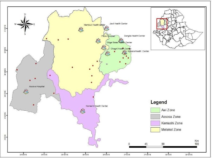
The region has two hospitals (one additional hospital was built in 2018) and 32 health centers. There is no TB culture and DST laboratories in the region during the study. Hence, the laboratory confirmation of TB in the area solely depends on sputum smear microscopy. In the current study, the two general hospitals, Assosa and Pawe, and four health centers, Felegeselam, Mambuk, Dangur, and Gilgelbeles, were included based on the availability of sputum smear microscopy and TB clinic as part of their routine TB diagnosis and treatment service. Additional three health centers were included from the Awi zone of the Amhara region, geographically proximal to the study region (Fig 1).

Fig 1. Map of the study area, Benishangul Gumuz region and its surroundings.

Fig 1

Health institutions included in the current study are indicated by a house symbol H, and the hospitals encircled red. Dots represent towns in the region.

Study design

A cross-sectional study was conducted from October 2013—June 2014. The study participants were newly diagnosed sputum smear-positive pulmonary TB (PTB) cases. All PTB suspects sent for sputum smear examination were asked for informed consent, and only patients with Acid-fast-bacilli (AFB) smear-positive sputum were enrolled. Before data collection, two days of theoretical and practical training was given to the personnel collecting data mainly focusing on research ethics, documentation, safety, sample collection, storage, and patient handling.

Data collection

Trained nurses working at TB clinics collected sociodemographic and clinical data of participants. Trained laboratory professionals collected sputum samples. The HIV status of the participant was obtained from the patient’s clinical record.

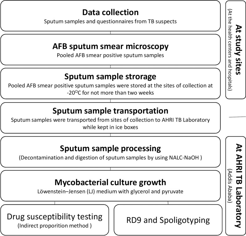
Participants were requested to give three sputum samples (Spot-Morning-Spot) as part of their routine diagnostic procedure and checked for AFB at the site of collection. AFB smear-positive sputum samples of the same patient were pooled and temporarily stored at -20°C until transported to AHRI TB laboratory at Addis Ababa, within no more than two weeks [18]. Upon arrival at AHRI, sputum samples were processed immediately by using the N-acetyl L- cysteine- sodium hydroxide (NALC-NaOH) method for sputum digestion and decontamination [19]. Fig 2 depicts an overview of the workflow for data collection and the laboratory methods applied.

Fig 2. Overview of the workflow for data collection and the laboratory methods.

Fig 2

Mycobacterial culture

Egg-based Löwenstein-Jensen (LJ) culture medium was used following the WHO standard procedure [20]. Briefly, an equal volume of NALC-NaOH and sputum samples was left standing for 15 minutes for digestion and decontamination. Sedimentation was achieved by centrifugation at 3000 rpm for 15 minutes at +4°C. The supernatant was discarded, and the sediment was inoculated onto two freshly prepared LJ medium slants, one containing 0.6% glycerol and the other with 0.6% sodium pyruvate to facilitate the growth of M. tuberculosis and M. bovis, respectively.

The Inoculated samples were incubated at 37°C and followed-up weekly for eight weeks. Mycobacterial growth was provisionally confirmed by typical colony characteristics (S1 Fig) and AFB sputum smear microscopy.

Drug susceptibility testing

DST was performed based on the indirect proportion method following standard procedures [21]. Briefly, a 24-well agar plate was prepared with 2.5 ml of Middlebrook 7H10 medium supplemented with 10% OADC and 5% glycerol per each well. The bacterial suspension was prepared in 0.2μm filter-sterilized MQ-H2O and the turbidity was adjusted to McFarland standard 1.0. Four drugs, namely Isoniazid, Rifampicin, Streptomycin, and Ethambutol, were used with critical concentration breakpoints of 0.2μg/ml, 1μg/ml, 2μg/ml, and 5μg/ml, respectively. Two drug-free wells, one with 1: 100 dilution and another containing undiluted bacteria suspension, were included as controls.

A strain was considered as “Susceptible” if no growth or considerably less than 1% growth was observed on the well containing the critical concentration of the corresponding drug compared to the control with 1% inoculum. A strain was considered as “Resistant” when growth on the well with the drug was higher than the control with 1% inoculum [22]. Any contaminated or borderline results were repeated.

Molecular typing using RD9 and spoligotyping

Mycobacterial species and M. tuberculosis strain identification were carried out at AHRI TB molecular laboratory.

For molecular typing, DNA was obtained by taking a loop full colony from LJ growth and suspended in 50μl of 0.2μm filter-sterilized 1% TE buffer in 2ml tubes. The suspension was heated at 80°C in the water bath for 45 minutes followed by sonication for an additional 15 minutes. The heat-killed product was centrifuged to 13000 rpm for 10 minutes at +4°C, and the supernatant was collected in a new tube.

M. tuberculosis was differentiated from other species of mycobacteria based on PCR targeting RD9, as previously described by Parsons et al. [23]. A multiplex PCR that was designed to amplify and detect RD9 using the following primers was employed; Forward RD9_FlankFW: 5’-AACACGGTCACGTTGTCGTG-3’, Reverse RD9_FlankRev: 5’-CAAACCAGCAGCTGTCGTTG-3’, and Internal RD9_InternalR: 5’-TTGCTTCCCCGGTTCGTCTG-3’. A PCR product of 396 bp indicates the presence of RD9 which is unique to M. tuberculosis and is absent from all other MTB complex members. Thus, selective amplification of the region targeted by the internal primer demonstrates M. tuberculosis. The strains were further typed by spoligotyping based on polymorphism in the direct repeat (DR) locus using a method described previously [10, 24]. Briefly, the DR region was amplified using primers, DRa (biotin-labeled): 5’-GGTTTTGGGTCTGACGAC-3’ and DRb: 5’-CCGAGAGGGGACGGAAAC-3’. After PCR amplification, the PCR products were hybridized to 43 spacer oligonucleotides of the corresponding DR region. The hybridization patterns obtained from the reaction were converted into binary and octal formats. The mycobacterial isolates identified were assigned into Spoligotype International Types (SIT), family, and their lineages using databases [12].

Quality control

The control strains M. tuberculosis H37Rv (ATCC 27294) and M. bovis (AF 61/2122/97) with known genotype and drug resistance patterns were run together during all laboratory assays, including growth, detection, DST, RD9 typing, and spoligotyping.

Data analysis

All sociodemographic and clinical data of the study participants and the laboratory test results were entered and analyzed using SPSS v.24 (IBM Corp., Armonk, N.Y., USA). Association between drug resistance, strain type, and sociodemographic and clinical data was assessed. Statistical significance was considered at a P-value of less than 0.05.

Ethical statement

The research ethics review committee of Addis Ababa University and the AHRI/ALERT ethics review committee evaluated and approved the proposal (Reg. No. PO12/13). Institutional support letters were obtained from regional, zonal, and local health administrations. Study participants were provided with adequate information, and the study participants were recruited only upon signing the informed consent. The results from DST were communicated to the relevant attending physician as soon as available, for better and timely management of patients. MDR-TB cases identified were referred either to the MDR-TB treatment center at St. Peter’s TB Specialized Hospital, Addis Ababa, or to Gondar University Hospital MDR-TB facility.

Results

Sociodemographic and clinical data of culture positive TB patients

From 107 smear-positive sputum samples collected, 81.3% (87/107) were culture positive and available for further analysis. Spoligotyping was not carried out for 12 isolates due to a technical problem, and therefore, only 75 were available for strain typing and cluster analysis.

The mean age of participants was 29 ± 9.2 SD years and ranged from 15 to 65. Participants between 18 and 47 years old accounted for 88.5% (77/87). Data on HIV status was available for 71.3% (62/87), among which 16.1% (10/62) were seropositive. There were more male patients than females with a 3:1 male to female ratio, and 55.2% of participants were married (Table 1).

Table 1. Sociodemographic and clinical characteristics of culture positive TB patients (n = 87) from the Benishangul Gumuz region and its surroundings, northwest Ethiopia from October 2013-June 2014.

Characteristics  Categories Variables Frequency
N (%)
Sociodemographic characteristics Gender Male 65 (74.7)
Female 22 (25.3)
Residence Urban 22 (25.3)
Rural 65 (74.7)
Occupation Farmer 40 (46)
Merchant 6 (6.9)
Gov’t employee 11 (12.6)
Student 8 (9.2)
Daily laborer 21 (24.1)
Prisoner 1 (1.1)
Educational status Illiterate 34 (39.1)
Grade1-4 10 (11.5)
Grade 5–8 18 (20.7)
Grade 9–12 16 (18.4)
Diploma and+ 9 (6.9)
Marital status Married 48 (55.2)
Single 36 (41.4)
Divorced 1 (1.1)
Widowed 2 (2.3)
Age groups
<18 5 (5.7)
18–27 38 (43.7)
28–37 31 (35.6)
38–47 8 (9.2)
48–57 4 (4.6)
>57 1 (1.1)
Clinical characteristics HIV Status Positive 10 (11.5)
Negative 52 (59.8)
Unknown 25 (28.7)
TB patient contact history Yes 20 (23)
No 51 (58.6)
Unknown 16 (18.4)
Previous anti-TB treatment Yes 12 (13.8)
No 73 (83.9)
Unknown 2 (2.3)

Anti-TB drug resistance pattern

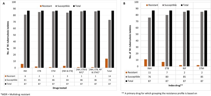
Among the 87 M. tuberculosis isolates that were tested against Isoniazid, Rifampicin, Ethambutol, and Streptomycin (Table 2A and Fig 3A), resistance to any of the drugs tested was observed in 16.1% (14/87) of the strains.

Table 2. A. Drug resistance pattern of first-line anti-TB drugs among culture positive TB patients (n = 87) in the Benishangul Gumuz region and its surroundings, Northwest Ethiopia, 2013/14.

A
Resistance profile Resistant
N (%)
Susceptible
N (%)
Remark
INH 6 (6.90) 81 (93.10) Monodrug resistant
ETB 2 (2.30) 85 (97.70)
STM 1 (1.15) 86 (98.85)
INH and ETB 3 (3.45) 84 (96.55) Poly drug-resistant
INH, ETB, and RIF 1 (1.15) 86 (98.85) MDR*/Poly drug-resistant
INH, ETB, RIF, and STM 1 (1.15) 86 (98.85)
Total 14 (16.10) 73 (83.90)
B
Index drug* Categories Resistant
N (%)
Susceptible
N (%)
INH INH 6 (6.90) 81 (93.10)
INH and ETB 3 (3.40) 84 (96.60)
INH, ETB, and RIF 1 (1.15) 86 (98.85)
INH, ETB, RIF, and STM 1 (1.15) 86 (98.85)
Total 11 (12.60) 76 (87.40)
ETB ETB 2 (2.30) 85 (97.70)
INH and ETB 3 (3.40) 84 (96.60)
INH, ETB, and RIF 1 (1.15) 86 (98.85)
INH, ETB, RIF, and STM 1 (1.15) 86 (98.85)
Total 7 (8.0) 80 (92.0)
RIF INH, ETB, and RIF 1 (1.15) 86 (98.85)
INH, ETB, RIF, and STM 1 (1.15) 86 (98.85)
Total 2 (2.30) 85 (97.70)
STM STM 1 (1.15) 86 (98.85)
INH, ETB, RIF, and STM 1 (1.15) 86 (98.85)
Total 2 (2.30) 85 (97.70)

INH: Isoniazid, ETB: Ethambutol, RIF: Rifampicin, STM: Streptomycin; *MDR = Multidrug resistant

* “Index drug” stands for the primary drug for which grouping the resistance profile is based.

Fig 3.

Fig 3

A and B. The magnitude of first-line anti-TB drug resistance profile of the M. tuberculosis isolates from culture positive TB patients (n = 87) in the Benishangul Gumuz region and its surroundings, North West Ethiopia, 2013/14.

Multidrug-resistant TB was detected in 2.3% (2/87), one of the strains was resistant to all four of the drugs tested (Table 2A and Fig 3A). The proportion of monoresistance was 6.9%, 2.3%, and 1.15% for Isoniazid, Ethambutol, and Streptomycin, respectively. There was no monoresistance to Rifampicin. Resistance to two drugs (Isoniazid and Ethambutol) in 3.45% and Polydrug resistance (resistance to more than two drugs) was observed in 2.3% of isolates. In contrast, one triple-drug resistance was identified against the Isoniazid, Ethambutol, and Rifampicin combination.

Stratifying the resistance profile into subgroups containing each drug tested showed any drug resistance of 12.6%, 8.0%, 2.30%, and 2.30% related to Isoniazid, Ethambutol, Rifampicin, and Streptomycin respectively (Table 2B and Fig 3B).

Molecular typing and genetic diversity

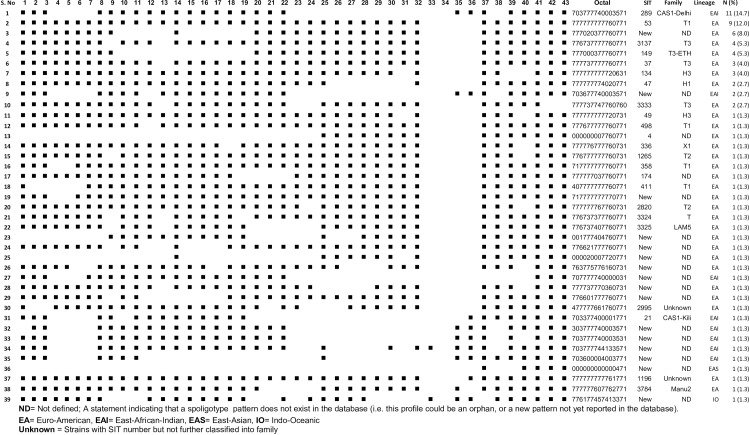
PCR based RD9 typing revealed that all the 75 isolates spoligotyped were M. tuberculosis (S2 Fig). The spoligotyping patterns were compared to the existing strains from databases [11, 12], and assigned into octal, SIT, family, and lineages (Fig 4).

Fig 4. Spoligotyping pattern of M. tuberculosis isolates from culture positive TB patients (n = 75) in the Benishangul Gumuz region and its surroundings, North West Ethiopia, 2013–14.

Fig 4

Thirty-nine distinct spoligotype patterns were identified, with 61.3% (46/ 75) grouped in 10 similar spoligotyping patterns containing two to 11 isolates while 38.7% (29/75) represented as unique patterns. Lineage classification revealed 72.0% (54/75), 25.3% (19/75), 1.3% (1/75) and 1.3% (1/75) isolates into Euro American, EA (Lineage 4), East African Indian, EAL (Lineage 3), East Asian, EAS (Lineage 2) and Indo-Oceanic, IO (Lineage 1), respectively (S1A Table and Fig 4). Three families, namely T, CAS, and H, were predominant, accounting for 61.3% (46/75) of the isolates (S1B Table and Fig 4). Among 61.3% (46/75) of the isolates containing a cluster of two to 11 similar spoligotype patterns, 71.7% (33/46) belonged to EA and 28.3% (13/46) to EAI lineages, while 17% (8/46) were without shared international spoligotype (SIT) so far and thus labeled ‘'New''. Thirty-eight percent (29/75) exhibited a single and unique spoligotyping pattern, with fifteen patterns having defined SIT and the remaining fourteen without SIT assigned as ‘‘New”.

Notably, SIT289 (11 isolates, CAS) and SIT53 (nine isolates, T1) were the most frequent strains accounting for 14.7% (11/75) and 12% (9/75), respectively. The findings were checked for the statistical associations. None of the sociodemographic and clinical variables studied were found to be significantly associated with either drug resistance patterns or circulating strains isolated (Table 3).

Table 3. Bivariate logistic analysis of factors associated with resistance to any one of anti-TB drug.

Variable Any drug resistance COR (95%CI) p-value
Yes N(%*) No N(%*)
Gender M 8 (12.3) 57 (87.7) 1
F 6 (28.3) 16 (72.7) 0.37 (0.11,1.24) 0.107
Residence Urban 1 (4.50) 21 (95.5) 1
Rural 13 (20.0) 52 (80.0) 0.19 (0.02, 1.55) 0.121
HIV status Positive 1 (15.4) 9 (84.6) 1
Negative 8 (10.0) 44 (90.0) 0.73 (0.21,2.58) 0.614
Unknown 5 (20.0) 20 (80.0) 0.44 (0.45, 4.37) 0.487
Previous treatment history Yes 5 (31.3) 11 (68.8) 1
No 9 (12.7) 62 (87.3) 3.13 (0.88,11.12) 0.078
Previous contact with TB patient Yes 6 (22.2) 21 (77.8) 1
No 8 (13.3) 52 (86.7) 1.86 (0.58,6.0) 0.301
Education status Illiterate 7 (20.6) 27(79.4) 1
Literate 7 (13.2) 46 (88.8) 1.70 (0.54,5.38) 0.078
Lineages Lineage 1 (IO) 0 (0.0) 1(100.0) NA NA
Lineage 2 (EAS) 0(0.0) 1(100.0) NA NA
Lineage 3 (EAI) 4(26.7) 11(73.3) NA NA
Lineage 4 (EA) 9(16.1) 47(83.9) 1.40 (0.043, 44.9) 0.85
Strains (SIT) New 5 (15.2) 28 (84.8) 1
Previously defined 8 (19.0) 34 (91.0) 0.76 (0.22,2.58) 0.66

*% calculated by row

Discussion

The main aim of this study was to understand the molecular epidemiology of M. tuberculosis strains circulating in a remote and hard-to-reach region in order to infer lessons for interventions in these and similar areas for which no such information exists. Benishangul Gumuz region and its surroundings are among the areas in Ethiopia where resources and infrastructures are severely limited. Sociodemographic and clinical information of the study participants and the type of MTB strains isolated and their drug susceptibility patterns were analysed. Here, we showed results that either substantiated previously estimated extrapolations from other regions through new evidence to support interventions or provided baseline data for further rigorous studies in the area.

All isolates identified were confirmed as M. tuberculosis by PCR based RD9 typing. This finding concurs with previous reports that have shown M. tuberculosis as responsible for more than 90 percent of pulmonary tuberculosis infections in Ethiopia [25].

Spoligotyping revealed that 68% (51/75) of the strains belonged to groups with assigned SIT in the latest database, SITIVIT2 [12]. In comparison to the studies conducted by other groups [2628], the remaining 32% (24/75) accounted for a slightly higher proportion of ‘'New'' strains from the area, indicating the absence of data from prior studies in the region. Our findings support the expected variation in strain distribution across different ecological niches, and the previous preliminarily results from the region [29, 30].

The identified thirty-nine distinct spoligotype patterns constituting 61.3% (46/75) of isolates clustering in two to 11 similar patterns indicate a high level of M. tuberculosis strain diversity in the area. The predominance of clusters within SIT289 (11 isolates) and SIT53 (nine isolates) strongly suggests an ongoing active transmission of tuberculosis in the area. Remarkably, this is despite the dispersed settlement of the population in the region.

The SIT289 strain was previously reported as predominant in Gambella and Bahir Dar. Both regions share long borders with the current study area in the southwest and northwest parts of the country, respectively [31,32]. SIT53 is frequent in central (Ambo), north (Bahir Dar, Dessie), and south Ethiopia (Dilla) [32,33]. This study site has, despite its relative isolation, an increasingly mixed population, including people originating from different parts of the country resettled in the 1980s [34], which could contribute to the diversity in the pool of circulating mycobacterial strains.

The identified ‘‘New” strain of lineage 2(EAS) deserves particular attention and needs to be characterized further. Lineage 2 is, in general, still very rare in Ethiopia [35]. The Beijing family has previously been reported to be prone to drug resistance. Strains within this lineage are reported to rapidly adapt to environments with variable stress conditions, including antibiotics [36]. With increased trade and travel between East Asia and Ethiopia, the risk of multiple introductions of Lineage 2 into the country remains high. It is essential to pay special attention to this risk, monitor detected strains, and conduct further studies with broader context to better design targeted control efforts.

Strains without an assigned spoligotype, so-called ‘'New'', made up 29.3% (22/75) of all isolates, and eight strains existed in clusters of two and six isolates each. The epidemiological significance of these strains needs further investigation using clinical and molecular tools with better discriminatory power.

Lineage 3(CAS-Delhi, the principal family, or CAS) with 11 strains in a cluster, which has also been reported as the dominant mycobacterial strain circulating in Sudan [37], could hint cross-border transmission since people from the current study area share market places and enjoy free cross-border movement into Sudan [38].

The dominant prevalence of lineage 4 (EA) and lineage 3 (EAI) has already been shown in previous studies from Ethiopia [1315, 25]. Lineage 1(IO, ancestral) seems to be more prevalent in southern regions of Ethiopia and may be shared with the Eastern African coast and Kenya [3941]. From the same setting, in a preliminarily study from spoligotyping of 33 isolates, the only two lineages reported were lineage 4 (EA) and lineage 1 (IO, ancestral). Moreover, there were 63.6% (22/33) of strains with ‘‘New” spoligotype patterns that have not been previously reported [30]. This finding highlights the need for further investigation of the molecular epidemiology of M. tuberculosis in the vast border regions of the country to understand trends better and respond to emerging challenges.

The observed magnitude of MDR-TB and resistance to any one first-line anti-TB drug is comparable, while Isoniazid monoresistance is considerably higher than neighbouring sites. Importantly Rifampicin, Streptomycin, and Ethambutol monoresistance were lower than in most previous studies elsewhere in the country [4044]. The reasons need to be investigated but could include factors related to the study design, study period, sample size or study population (Urban/rural, access to drugs, health-seeking behavior, mobility of population), among others.

This study has limitations such as the sample size was relatively small, and the yield of culture was further reduced because of logistic challenges. Furthermore, there are no data on drug resistance mutations among the identified Mycobacterium strains. We relied on spoligotyping alone to detect transmission trends, a method that tends to overestimate rates and indicates older events than whole-genome sequencing, which is increasingly accepted as a new gold standard. Nevertheless, the number and diversity of isolates detected provided adequate data with interesting findings and meaningful conclusions. Although the indications from spoligotyping may lack precision in the magnitude of transmission, the interpretation we made from the findings that there is active ongoing transmission in the area is valid [38].

In summary, the results provided insight into the molecular diversity and drug resistance pattern of TB strains circulating in a peripheral predominantly rural region of Ethiopia where the population density is low, but where scattered urban centers have highly mobile populations and serve as hubs of cross border trade and travel. The findings show that there is much diversity but also clustering in circulating M. tuberculosis strains in this setting where cross border movements are frequent and where health care services are generally scanty and inadequate. The study has also revealed the presence of several yet unknown strain types whose significance in driving tuberculosis is unexplored. In addition, there are indications that multidrug resistance is emerging and a strain that belongs to lineage 2 has been detected. It is, therefore, important to build local DST facilities in the area to detect and treat MDR TB early. It is critical to monitor the molecular epidemiology of tuberculosis in the Benishangul Gumuz region and similar regions of the country closely to predict trends in transboundary transmission and respond with appropriate interventions.

Supporting information

S1 Fig. Colony characteristic used for identification of the growth of Mycobacterium tuberculosis.

(PDF)

S2 Fig. PCR for RD9 deletion typing.

(PDF)

S1 Table

A and B. Frequency of M. tuberculosis family and Lineages.

(PDF)

Acknowledgments

The authors would like to thank all the study participants. We would also like to extend our gratitude to Mr. Shiferaw Bekele for his assistance and guidance during laboratory work, and Mr. Legesse Negash during data entry and statistical analysis. We are thankful to medical laboratory professionals and nurses working at TB clinics of the study area for their commitment and dedicated assistance during data collection. The authors would like to mention and appreciate the Benishangul Gumuz regional health bureau and Pawe woreda TB program coordinating offices for facilitating logistics for the transportation of the identified MDR-TB patients to the treatment initiation centers (TICs). Lastly, we acknowledge Mari Kaarbo for her extensive proofreading of the manuscript.

Data Availability

All relevant data are within the manuscript and its Supporting Information files.

Funding Statement

This work is fully supported by AHRI core budget. AHRI receives core financial support from Swedish International Development Cooperation Agency (SIDA), Norwegian Agency for Development Cooperation (NORAD), and from the Government of Ethiopia through Ministry of Health. AA holds a grant from Human Hereditary and Health in Africa (H3Africa) [H3A/18/003] implemented by the African Academy of Sciences (AAS) and the NEPAD Agency's Alliance for Accelerating Excellence in Science in Africa (AESA) in partnership with Wellcome and a grant from the National Institutes of Health (NIH) Fogarty International Center Global Infectious Diseases entitled “Ethiopia-Emory TB Research Training Program” (D43TW009127).

References

  • 1.World Health Organization. Global Tuberculosis Report 2018. Available: https://www.who.int/tb/publications/global_report/en/. Accessed 19 March 2019
  • 2.Nahid P, Pai M, Hopewell PC. Advances in the diagnosis and treatment of tuberculosis. Proc Am Thorac Soc. 2006;3(1):103–10. 10.1513/pats.200511-119JH [DOI] [PMC free article] [PubMed] [Google Scholar]
  • 3.The Federal Ministry of Health Ethiopia. First Ethiopian national population-based Tuberculosis Prevalence Survey.2011. Available: https://www.ephi.gov.et/. Accessed 13 September 2018.
  • 4.World Health Organization. Global tuberculosis report. 2015. Available: http://apps.who.int/iris/bitstream/10665/191102/1/9789241565059_eng.pdf?ua=1. Accessed 13 September 2018.
  • 5.World Health Organization. Global tuberculosis report. 2016. Available: https://apps.who.int/medicinedocs/documents/s23098en/s23098en.pdf Accessed13 September 2018.
  • 6.Gebreegziabher SB, Bjune GA, Yimer SA. Total Delay Is Associated with unfavorable Treatment Outcome among Pulmonary Tuberculosis Patients in West Gojjam Zone, Northwest Ethiopia: A Prospective Cohort Study. PLOS One. 2016; 11(7). [DOI] [PMC free article] [PubMed] [Google Scholar]
  • 7.Ramage HR, Connolly LE, Cox JS. Comprehensive Functional Analysis of Mycobacterium tuberculosis Toxin-Antitoxin Systems: Implications for Pathogenesis, Stress Responses, and Evolution. PLOS Genetics. 2009; 5(12). [DOI] [PMC free article] [PubMed] [Google Scholar]
  • 8.Stucki D, Brites D, Jeljeli L, Coscolla M, Liu Q, Trauner A, et al. Mycobacterium tuberculosis lineage 4 comprises globally distributed and geographically restricted sublineages. Nat Genet. 2016;48(12):1535–43. 10.1038/ng.3704 [DOI] [PMC free article] [PubMed] [Google Scholar]
  • 9.Gagneux S, Small PM. Global phylogeography of Mycobacterium tuberculosis and implications for tuberculosis product development. Lancet Infect Dis. 2007;7(5):328–37. 10.1016/S1473-3099(07)70108-1 [DOI] [PubMed] [Google Scholar]
  • 10.Kamerbeek J, Schouls L, Kolk A, van Agterveld M, van Soolingen D, Kuijper S, et al. Simultaneous detection and strain differentiation of Mycobacterium tuberculosis for diagnosis and epidemiology. J Clin Microbiol. 1997; 35(4):907–14. [DOI] [PMC free article] [PubMed] [Google Scholar]
  • 11.Shabbeer A, Cowan LS, Ozcaglar C, Rastogi N, Vandenberg SL, Yener B, et al. TB-Lineage: an online tool for classification and analysis of strains of Mycobacterium tuberculosis complex. Infect Genet Evol. 2012;12(4):789–97. 10.1016/j.meegid.2012.02.010 [DOI] [PubMed] [Google Scholar]
  • 12.Couvin D, David A, Zozio T, Rastogi N. Macro-geographical specificities of the prevailing tuberculosis epidemic as seen through SITVIT2, an updated version of the Mycobacterium tuberculosis genotyping database. Infection Genetics and Evolution. 2019; 72:31–43. [DOI] [PubMed] [Google Scholar]
  • 13.Tulu B, Ameni G. Spoligotyping based genetic diversity of Mycobacterium tuberculosis in Ethiopia: a systematic review. BMC Infect Dis. 2018;18(1):140 10.1186/s12879-018-3046-4 [DOI] [PMC free article] [PubMed] [Google Scholar]
  • 14.Agonafir M, Lemma E, Wolde-Meskel D, Goshu S, Santhanam A, Girmachew F, et al. Phenotypic and genotypic analysis of multidrug-resistant tuberculosis in Ethiopia. Int J Tuberc Lung Dis. 2010; 14(10):1259–65. [PubMed] [Google Scholar]
  • 15.Mekonnen D, Derbie A, Chanie A, Shumet A, Biadglegne F, Kassahun Y, et al. Molecular epidemiology of M. tuberculosis in Ethiopia: A systematic review and meta-analysis. Tuberculosis. 2019;118. [DOI] [PMC free article] [PubMed] [Google Scholar]
  • 16.Central statistical agency (CSA). Population and Housing Census of Ethiopia: Results for Benishangul-Gumuz Region, Vol. 1; 1994. Available: www.csa.gov.et/census-report/complete-report/census-1994. [Google Scholar]
  • 17.Unicef-Ethiopian Country office. Benishangul-Gumuz Briefing Note. 2016. Available: https://www.unicef.org/ethiopia/ECO/Benishangul/Gumuz/Brieifing/Note.2016. Accessed 13.09.2018.
  • 18.World Health Organization. Guidelines for surveillance of drug resistance in tuberculosis. 2009; 4th ed. Available: https://www.who.int/tb/publications/surveillance_guidelines/en/. Accessed 13.09.2018.
  • 19.Kubica GP, Dye WE, Cohn ML, Middlebrook G. Sputum digestion and decontamination with N-acetyl-L-cysteine-sodium hydroxide for culture of mycobacteria. Am Rev Respir Dis. 1963;87:775–9. 10.1164/arrd.1963.87.5.775 [DOI] [PubMed] [Google Scholar]
  • 20.Global Laboratory initiative. Mycobacteriology laboratory manual. 2014:47–51. [Google Scholar]
  • 21.van Klingeren B, Dessens-Kroon M, van der Laan T, Kremer K, van Soolingen D. Drug susceptibility testing of Mycobacterium tuberculosis complex by use of a high-throughput, reproducible, absolute concentration method. J Clin Microbiol. 2007; 45(8):2662–8. 10.1128/JCM.00244-07 [DOI] [PMC free article] [PubMed] [Google Scholar]
  • 22.Wedajo W, Schon T, Bedru A, Kiros T, Hailu E, Mebrahtu T, et al. A 24-well plate assay for simultaneous testing of first and second line drugs against Mycobacterium tuberculosis in a high endemic setting. BMC Res Notes. 2014; 7:512 10.1186/1756-0500-7-512 [DOI] [PMC free article] [PubMed] [Google Scholar]
  • 23.Parsons LM, Brosch R, Cole ST, Somoskovi A, Loder A, Bretzel G, et al. Rapid and simple approach for identification of Mycobacterium tuberculosis complex isolates by PCR-based genomic deletion analysis. J Clin Microbiol. 2002; 40(7):2339–45. 10.1128/JCM.40.7.2339-2345.2002 [DOI] [PMC free article] [PubMed] [Google Scholar]
  • 24.van Embden JD, van Gorkom T, Kremer K, Jansen R, van Der Zeijst BA, Schouls LM. Genetic variation and evolutionary origin of the direct repeat locus of Mycobacterium tuberculosis complex bacteria. J Bacteriol. 2000;182(9):2393–401. 10.1128/jb.182.9.2393-2401.2000 [DOI] [PMC free article] [PubMed] [Google Scholar]
  • 25.Mihret A, Bekele Y, Aytenew M, Assefa Y, Abebe M, Wassie L, et al. Modern lineages of Mycobacterium tuberculosis in Addis Ababa, Ethiopia: implications for the tuberculosis control programe. African Health Sciences. 2012;12(3):339–44. 10.4314/ahs.v12i3.15 [DOI] [PMC free article] [PubMed] [Google Scholar]
  • 26.Esmael A, Wubie M, Desta K, Ali I, Endris M, et al. Genotyping and Drug Resistance Patterns of M. tuberculosis in Eastern Amhara region, Ethiopia. J Clin Diagn Res. 2014; 2:102. [Google Scholar]
  • 27.Garedew L, Mihret A, Mamo G, Abebe T, Firdessa R, Bekele Y, et al. Strain diversity of mycobacteria isolated from pulmonary tuberculosis patients at Debre Birhan Hospital, Ethiopia. Int J Tuberc Lung D. 2013; 17(8):1076–81. [DOI] [PubMed] [Google Scholar]
  • 28.Diriba B, Berkessa T, Mamo G, Tedla Y, Ameni G. Spoligotyping of multidrug-resistant Mycobacterium tuberculosis isolates in Ethiopia. Int J Tuberc Lung D. 2013; 17(2):246–50. [DOI] [PubMed] [Google Scholar]
  • 29.Ford CB, Shah RR, Maeda MK, Gagneux S, Murray MB, Cohen T, et al. Mycobacterium tuberculosis mutation rate estimates from different lineages predict substantial differences in the emergence of drug-resistant tuberculosis. Nature Genetics. 2013;45(7):784–90. 10.1038/ng.2656 [DOI] [PMC free article] [PubMed] [Google Scholar]
  • 30.Disassa H, Tafess K, Worku A, Ameni G. A preliminary study on molecular characterization of Mycobacterium tuberculosis in Benishangul Gumuz region, Western Ethiopia. British Microbiol Res J. 2015; 10(6):1–10. [Google Scholar]
  • 31.Alemu J, Mamo G, Ameni G, Pal M. Molecular Epidemiology of Bovine Tuberculosis in Cattle and its Public Health Implications in Gambella Town and its Surroundings, Gambella Regional State, Ethiopia. Molecular Microbiology Research. 2016; 6(1): 1–15. [Google Scholar]
  • 32.Nuru A, Mamo G, Worku A, Admasu A, Medhin G, Pieper R, et al. Genetic Diversity of Mycobacterium tuberculosis Complex Isolated from Tuberculosis Patients in Bahir Dar City and Its Surroundings, Northwest Ethiopia. Biomed Res Int. 2015;2015:174732 10.1155/2015/174732 [DOI] [PMC free article] [PubMed] [Google Scholar]
  • 33.Maru M, Mariam SH, Airgecho T, Gadissa E, Aseffa A. Prevalence of Tuberculosis, Drug Susceptibility Testing, and Genotyping of Mycobacterial Isolates from Pulmonary Tuberculosis Patients in Dessie, Ethiopia. Tuberc Res Treat. 2015;2015:215015 10.1155/2015/215015 [DOI] [PMC free article] [PubMed] [Google Scholar]
  • 34.Central statistical agency of Ethiopia. Population and Housing Census of Ethiopia: Results for Benishangul-Gumuz Region, Vol. 1; 1994. Accessed from www.csa.gov.et/census-report/complete-report/census-1994. [Google Scholar]
  • 35.Firdessa R, Berg S, Hailu E, Schelling E, Gumi B, Erenso G, et al. Mycobacterial lineages causing pulmonary and extrapulmonary tuberculosis, Ethiopia. Emerg Infect Dis. 2013; 19(3):460–3. 10.3201/eid1903.120256 [DOI] [PMC free article] [PubMed] [Google Scholar]
  • 36.Ebrahimi-Rad M, Bifani P, Martin C, Kremer K, Samper S, Rauzier J, et al. Mutations in putative mutator genes of Mycobacterium tuberculosis strains of the W-Beijing family. Emerg Infect Dis. 2003;9(7):838–45. 10.3201/eid0907.020803 [DOI] [PMC free article] [PubMed] [Google Scholar]
  • 37.Sharaf Eldin GS, Fadl-Elmula I, Ali MS, Ali AB, Salih AL, Mallard K, et al. Tuberculosis in Sudan: a study of Mycobacterium tuberculosis strain genotype and susceptibility to anti-tuberculosis drugs. BMC Infect Dis. 2011;11:219 10.1186/1471-2334-11-219 [DOI] [PMC free article] [PubMed] [Google Scholar]
  • 38.Comas I, Hailu E, Kiros T, Bekele S, Mekonnen W, Gumi B, et al. Population Genomics of Mycobacterium tuberculosis in Ethiopia Contradicts the Virgin Soil Hypothesis for Human Tuberculosis in Sub-Saharan Africa. Curr Biol. 2015; 25(24):3260–6. 10.1016/j.cub.2015.10.061 [DOI] [PMC free article] [PubMed] [Google Scholar]
  • 39.Mbugi EV, Katale BZ, Streicher EM, Keyyu JD, Kendall SL, Dockrell HM, et al. Mapping of Mycobacterium tuberculosis Complex Genetic Diversity Profiles in Tanzania and Other African Countries. PLOS One. 2016; 11(5):e0154571 10.1371/journal.pone.0154571 [DOI] [PMC free article] [PubMed] [Google Scholar]
  • 40.Yimer SA, Agonafir M, Derese Y, Sani Y, Bjune GA, Holm-Hansen C. Primary drug resistance to anti-tuberculosis drugs in major towns of Amhara region, Ethiopia. APMIS. 2012; 120(6):503–9. 10.1111/j.1600-0463.2011.02861.x [DOI] [PubMed] [Google Scholar]
  • 41.Bruchfeld J, Aderaye G, Palme IB, Bjorvatn B, Ghebremichael S, Hoffner S, et al. Molecular epidemiology and drug resistance of Mycobacterium tuberculosis isolates from Ethiopian pulmonary tuberculosis patients with and without human immunodeficiency virus infection. J Clin Microbiol. 2002; 40(5):1636–43. 10.1128/JCM.40.5.1636-1643.2002 [DOI] [PMC free article] [PubMed] [Google Scholar]
  • 42.Demissie M, Gebeyehu M, Berhane Y. Primary resistance to anti-tuberculosis drugs in Addis Ababa, Ethiopia. Int J Tuberc Lung Dis. 1997; 1(1):64–7. [PubMed] [Google Scholar]
  • 43.Tessema B, Beer J, Emmrich F, Sack U, Rodloff AC. First- and second-line anti-tuberculosis drug resistance in Northwest Ethiopia. Int J Tuberc Lung Dis. 2012; 16(6):805–11. 10.5588/ijtld.11.0522 [DOI] [PubMed] [Google Scholar]
  • 44.Kassu D, Daniel A, Eshetu L, Mekdes G, Beniam F. Drug susceptibility of Mycobacterium tuberculosis isolate from smear-negative pulmonary tuberculosis patients, Addis Ababa, Ethiopia. Ethiop J Health Dev. 200822(2):212–215. [Google Scholar]

Decision Letter 0

Igor Mokrousov

10 Jan 2020

PONE-D-19-25370

Genetic Diversity and Drug Resistance Pattern of Mycobacterium tuberculosis Strains from Pulmonary Tuberculosis Patients in Benishangul Gumuz Region and Its Surroundings, Northwest Ethiopia

PLOS ONE

Dear Mr. Lobie,

Thank you for submitting your manuscript to PLOS ONE. After careful consideration, we feel that it has merit but does not fully meet PLOS ONE’s publication criteria as it currently stands. Therefore, we invite you to submit a revised version of the manuscript that addresses ALL points raised during the review process, in particular, an interest of this study should be better demonstrated and it should be better placed in the context of the published papers on Ethiopia.

English language should be edited by a native speaker (and expert in the field).

We would appreciate receiving your revised manuscript by Feb 24 2020 11:59PM. When you are ready to submit your revision, log on to https://www.editorialmanager.com/pone/ and select the 'Submissions Needing Revision' folder to locate your manuscript file.

If you would like to make changes to your financial disclosure, please include your updated statement in your cover letter.

To enhance the reproducibility of your results, we recommend that if applicable you deposit your laboratory protocols in protocols.io, where a protocol can be assigned its own identifier (DOI) such that it can be cited independently in the future. For instructions see: http://journals.plos.org/plosone/s/submission-guidelines#loc-laboratory-protocols

Please include the following items when submitting your revised manuscript:

  • A rebuttal letter that responds to each point raised by the academic editor and reviewer(s). This letter should be uploaded as separate file and labeled 'Response to Reviewers'.

  • A marked-up copy of your manuscript that highlights changes made to the original version. This file should be uploaded as separate file and labeled 'Revised Manuscript with Track Changes'.

  • An unmarked version of your revised paper without tracked changes. This file should be uploaded as separate file and labeled 'Manuscript'.

Please note while forming your response, if your article is accepted, you may have the opportunity to make the peer review history publicly available. The record will include editor decision letters (with reviews) and your responses to reviewer comments. If eligible, we will contact you to opt in or out.

We look forward to receiving your revised manuscript.

Kind regards,

Igor Mokrousov, Ph.D., D.Sc.

Academic Editor

PLOS ONE

Journal Requirements:

When submitting your revision, we need you to address these additional requirements.

1. Please ensure that your manuscript meets PLOS ONE's style requirements, including those for file naming. The PLOS ONE style templates can be found at

http://www.journals.plos.org/plosone/s/file?id=wjVg/PLOSOne_formatting_sample_main_body.pdf and http://www.journals.plos.org/plosone/s/file?id=ba62/PLOSOne_formatting_sample_title_authors_affiliations.pdf

2. Thank you for stating the following in the Acknowledgments Section of your manuscript:

'...we would like to mention and appreciate that financial and logistic support for the transportation of the identified MDR-TB patients was facilitated by Benishangul Gumuz regional health bureau and Pawe woreda TB program coordinating office.'

We note that you have provided funding information that is not currently declared in your Funding Statement. However, funding information should not appear in the Acknowledgments section or other areas of your manuscript. We will only publish funding information present in the Funding Statement section of the online submission form.

Please remove any funding-related text from the manuscript and let us know how you would like to update your Funding Statement. Currently, your Funding Statement reads as follows:

'The funders had no role in study design, data collection and analysis, decision to publish, or preparation of the manuscript.'

  1. Please provide an amended Funding Statement that declares *all* the funding or sources of support received during this specific study (whether external or internal to your organization) as detailed online in our guide for authors at http://journals.plos.org/plosone/s/submit-now

  1. Please state what role the funders took in the study.  If any authors received a salary from any of your funders, please state which authors and which funder. If the funders had no role, please state: "The funders had no role in study design, data collection and analysis, decision to publish, or preparation of the manuscript."

c. Please include your amended statements within your cover letter; we will change the online submission form on your behalf.

3. Please upload a new copy of Figure 1 as the detail is not clear. Please follow the link for more information: http://blogs.PLOS.org/everyone/2011/05/10/how-to-check-your-manuscript-image-quality-in-editorial-manager/

4. Please include a caption for figure 3.

5. Your ethics statement must appear in the Methods section of your manuscript. If your ethics statement is written in any section besides the Methods, please move it to the Methods section and delete it from any other section. Please also ensure that your ethics statement is included in your manuscript, as the ethics section of your online submission will not be published alongside your manuscript.

[Note: HTML markup is below. Please do not edit.]

Reviewers' comments:

Reviewer's Responses to Questions

Comments to the Author

1. Is the manuscript technically sound, and do the data support the conclusions?

The manuscript must describe a technically sound piece of scientific research with data that supports the conclusions. Experiments must have been conducted rigorously, with appropriate controls, replication, and sample sizes. The conclusions must be drawn appropriately based on the data presented.

Reviewer #1: Yes

Reviewer #2: Partly

**********

2. Has the statistical analysis been performed appropriately and rigorously?

Reviewer #1: N/A

Reviewer #2: Yes

**********

3. Have the authors made all data underlying the findings in their manuscript fully available?

The PLOS Data policy requires authors to make all data underlying the findings described in their manuscript fully available without restriction, with rare exception (please refer to the Data Availability Statement in the manuscript PDF file). The data should be provided as part of the manuscript or its supporting information, or deposited to a public repository. For example, in addition to summary statistics, the data points behind means, medians and variance measures should be available. If there are restrictions on publicly sharing data—e.g. participant privacy or use of data from a third party—those must be specified.

Reviewer #1: Yes

Reviewer #2: Yes

**********

4. Is the manuscript presented in an intelligible fashion and written in standard English?

PLOS ONE does not copyedit accepted manuscripts, so the language in submitted articles must be clear, correct, and unambiguous. Any typographical or grammatical errors should be corrected at revision, so please note any specific errors here.

Reviewer #1: Yes

Reviewer #2: No

**********

5. Review Comments to the Author

Please use the space provided to explain your answers to the questions above. You may also include additional comments for the author, including concerns about dual publication, research ethics, or publication ethics. (Please upload your review as an attachment if it exceeds 20,000 characters)

Reviewer #1: I found several works on the genotyping of MTB in Benishangul Gumuz Region and in Ethiopia.

(Disassa H., A Preliminary Study on Molecular Characterization of Mycobacterium tuberculosis in Benishangul Gumuz Region, Western Ethiopia /British Microbiology Research Journal 10(6): 1-10, 201). Perhaps the author should compare their results with the data in this publication.

Tulu B. et al. Spoligotyping based genetic diversity of Mycobacterium tuberculosis in Ethiopia: asystematic review BMC Infectious Diseases (2018) 18:140

Mekonnen D et al. Molecular epidemiology of M. tuberculosis in Ethiopia: A systematic review and meta-analysis/ Tuberculosis Volume 118, September 2019, 101858.

Perhaps you should compare your results with the data in this publication.

The following errors were identified in the article:

129 line is not clear what is AHRI, where is this organization located?

135-136 lines are not the correct interpretation, because decontamination does not occur before, but after mixing within 15 minutes, and centrifugation - not 3000 rpm, but 3000 g should be, and temperature? It must be 4 C.

148 - 149 the term minimum inhibitory concentration of a breakpoint is better replaced by critical concentrations

183 error in numbers 81.3% (75/87)

188 - 189 woman and man were mixed up

338 - 339 to talk about «on-going active transmission of tuberculosis» is not right if spoligotyping is used

Reviewer #2: The authors studied a panel of 87 MTB isolates from TB patients from NorthWest Ethiopia (Benishangul Gumuz and its surroundings). The main goal was to investigate drug susceptibility patterns and genetic diversity of MTB Strains in this setting.

The subject is scientifically important but the novelty for science provided by this paper is meaningless; the study is somehow old fashioned. Indeed, Disassa et al. (2015) conducted a preliminary study for molecular characterization of MTB strains in the same area (n=33) from November 2012 to April 2013 . Moreover, Tulu and Ameni. (2018) made a review of articles published on M. tuberculosis strains and lineages in Ethiopia (up to 21 articles with spoligotyping results). SITs from many regions of Ethiopia have been extensively described and well documented. The authors report the results of routine drug susceptibility tests to first line anti TB drugs. Although cultures and DSTs and Gold standard tests, the authors did not use any molecular method to assess drug resistant TB. For genetic diversity, authors used spoligotyping which tend to overestimate links between isolates which is inconclusive for transmission routes of TB.

Other aspects that could be addressed by authors to strength the paper are:

1) To perform 15 or 24 loci MIRU-VNTR genotyping to strengthen data on MTB diversity.

2) To determine the mutations related to resistance to main anti TB drugs i.e. INH and RIF and to compare these genotyping results with those of conventional tests (drug susceptibility testing).

3) To add data regarding TB prevalence in northwest Ethiopia and give more details for the choice of this setting.

4) To review methodology section as they are too much details of well-known procedures ( for sputa collection and media preparation)...Also, there was no brief description for PCR targeting the RD9 and for spoligotyping method.

5) The autors have to check the % of the resistance to each drug, i have noticed many discordances between tha main text and table II (i.e. Rif).

6) The paper requires extensive English editing.

**********

6. PLOS authors have the option to publish the peer review history of their article (what does this mean?). If published, this will include your full peer review and any attached files.

If you choose “no”, your identity will remain anonymous but your review may still be made public.

Do you want your identity to be public for this peer review? For information about this choice, including consent withdrawal, please see our Privacy Policy.

Reviewer #1: No

Reviewer #2: Yes: Imane CHAOUI

[NOTE: If reviewer comments were submitted as an attachment file, they will be attached to this email and accessible via the submission site. Please log into your account, locate the manuscript record, and check for the action link "View Attachments". If this link does not appear, there are no attachment files to be viewed.]

While revising your submission, please upload your figure files to the Preflight Analysis and Conversion Engine (PACE) digital diagnostic tool, https://pacev2.apexcovantage.com/. PACE helps ensure that figures meet PLOS requirements. To use PACE, you must first register as a user. Registration is free. Then, login and navigate to the UPLOAD tab, where you will find detailed instructions on how to use the tool. If you encounter any issues or have any questions when using PACE, please email us at figures@plos.org. Please note that Supporting Information files do not need this step.

PLoS One. 2020 Apr 8;15(4):e0231320. doi: 10.1371/journal.pone.0231320.r003

Author response to Decision Letter 0


10 Feb 2020

Response to the journal requirements (Editor) and Reviewers

Journal Requirements (response to the editor):

When submitting your revision, we need you to address these additional requirements.

1. Please ensure that your manuscript meets PLOS ONE's style requirements, including those for file naming. The PLOS ONE style templates can be found at

http://www.journals.plos.org/plosone/s/file?id=wjVg/PLOSOne_formatting_sample_main_body.pdf and http://www.journals.plos.org/plosone/s/file?id=ba62/PLOSOne_formatting_sample_title_authors_affiliations.pdf

� As the requirement and requested by academic editor of the journal the authors adhered to templates provided and found it useful in shaping the manuscript.

� To mention title, current address, corresponding author’s initial, fonts of title and subtitles, references, figure and tables and supplementary information among other points were addressed.

2. Thank you for stating the following in the Acknowledgments Section of your manuscript:

'...we would like to mention and appreciate that financial and logistic support for the transportation of the identified MDR-TB patients was facilitated by Benishangul Gumuz regional health bureau and Pawe woreda TB program coordinating office.'

We note that you have provided funding information that is not currently declared in your Funding Statement. However, funding information should not appear in the Acknowledgments section or other areas of your manuscript. We will only publish funding information present in the Funding Statement section of the online submission form.

Please remove any funding-related text from the manuscript and let us know how you would like to update your Funding Statement. Currently, your Funding Statement reads as follows:

'The funders had no role in study design, data collection and analysis, decision to publish, or preparation of the manuscript.'

Please provide an amended Funding Statement that declares *all* the funding or sources of support received during this specific study (whether external or internal to your organization) as detailed online in our guide for authors at http://journals.plos.org/plosone/s/submit-now

Please state what role the funders took in the study. If any authors received a salary from any of your funders, please state which authors and which funder. If the funders had no role, please state: "The funders had no role in study design, data collection and analysis, decision to publish, or preparation of the manuscript."

c. Please include your amended statements within your cover letter; we will change the online submission form on your behalf.

� Funding related text from the acknowledgement section of the manuscript is removed and re-written as follow on line no. 366-368 :-

‘’… Lastly, we would like to mention and appreciate the Benishangul Gumuz regional health bureau and Pawe woreda TB program coordinating offices for facilitating logistics for the transportation of the identified MDR-TB patients to the treatment initiation centers (TICs). ‘’

Funding statement, (this will be move to the cover letter as mentioned by the academic editor)

Amendment to the funding statement declaration is included in the cover letter and stated as follow.

� ‘’This work is fully supported by AHRI core budget. AHRI receives core financial support from Swedish International Development Cooperation Agency (SIDA), Norwegian Agency for Development Cooperation (NORAD), and from the Government of Ethiopia through Ministry of Health.

� ‘’AA holds a grant from Human Hereditary and Health in Africa (H3Africa) [H3A/18/003] implemented by the African Academy of Sciences (AAS) and the NEPAD Agency's Alliance for Accelerating Excellence in Science in Africa (AESA) in partnership with Wellcome and a grant from the National Institutes of Health (NIH) Fogarty International Center Global Infectious Diseases entitled “Ethiopia-Emory TB Research Training Program” (D43TW009127).’’

� Any statements related to funding is removed from the manuscript.

3. Please upload a new copy of Figure 1 as the detail is not clear. Please follow the link for more information: http://blogs.PLOS.org/everyone/2011/05/10/how-to-check-your-manuscript-image-quality-in-editorial-manager/

� Figure 1 is checked for its quality using PACE as recommended and clear and the corrected version is uploaded.

4. Please include a caption for figure 3.

� Figure 3 is now renamed as ‘Figure 4 ‘and caption is added as ‘’ Figure 4. Spoligotyping pattern of M. tuberculosis isolates from pulmonary TB patients from the Benishangul Gumuz region and its surroundings in North West Ethiopia, 2013/14.’’.

5. Your ethics statement must appear in the Methods section of your manuscript. If your ethics statement is written in any section besides the Methods, please move it to the Methods section and delete it from any other section. Please also ensure that your ethics statement is included in your manuscript, as the ethics section of your online submission will not be published alongside your manuscript.

� Ethics statement is REVISED and moved to the method section (line no. 198-206).

‘’Ethical statement

The research review committee from Addis Ababa University and AHRI/ALERT health research and ethics review committee evaluated and approved the proposal with the project Reg. No. PO12/13 (S1 File). Institutional support letters obtained from regional, zonal, and local health administrations. Study participants provided with sufficient information, and only volunteers recruited after signing informed consent. Results from DST was communicated to the relevant caregiver as soon as available, for better and timely management of patients. MDR-TB cases identified were referred to either the MDR-TB treatment center at St. Peter TB specialized hospital, Addis Ababa, or to Gondar University Hospital MDR-TB facility, Gondar.’’

Response to the reviewers

Thank you for the valuable and valid comments that helped us to improve our work.

By appreciating, the effort made and time invested to bring these important points to our attention, the authors addressed all the comments from the journal editor as well as reviewers as curiously as possible. Some points, which are relevant but failed to be addressed by the virtue of limited capacity of the authors, were either strongly recommended to be taken into account in the future work plan or mentioned as limitations.

Reviewer 1.

1. Comment to compare our results with data from the following published articles of

Disassa H. et al., 2015; Tulu B. et al., 2018; and Mekonnen D. et al, 2019.

� Our results were compared and further discussed using the recommended papers.

Specifically, Disassa H. et al., 2015 is cited as Ref. 30, and its findings are compared and discussed in line with our findings as shown on lines no. 326-331,

- Findings from the systematic review papers by Tulu B. et al., 2018 and Mekonnen D. et al., 2019 were also cited as Ref. no 13 and 15, respectively; and their findings were compared, and discussed in the document lines number 92-95 and 324-325.

- In both review papers, the results presented regarding the Benishangul Gumuz region (the current study area) were from a single preliminary study by Disassa H. et al., 2015, and the conclusions and recommendations ask for more research in settings like Benishangul Gumuz region, refer to the conclusion from Tulu B. et al, 2018 on page 8 of 10.

2. 129 line is not clear what is AHRI, where is this organization located?

- AHRI is defined as Armauer Hansen Research Institute and located at Addis Ababa, Ethiopia. Additional explanation on the funding sources of the institute is also presented and submitted on a separate letter of the funding statement to the academic editor as follow:-

’’This work is fully supported by AHRI core budget. AHRI receives core financial support from Swedish International Development Cooperation Agency (SIDA), Norwegian Agency for Development Cooperation (NORAD), and from the Government of Ethiopia through Ministry of Health’’

3. 135-136 lines are not the correct interpretation, because decontamination does not occur before, but after mixing within 15 minutes, and centrifugation - not 3000 rpm, but 3000 g should be, and temperature? It must be 4 C.

� This statement is corrected and presented on line 144-146 as follow:-

‘’…… Briefly, an equal volume of NALC-NaOH and sputum samples was left standing for 15 minutes for digestion and decontamination. Sedimentation was achieved by centrifugation at 3000 rpm for 15 minutes at +4OC. ‘’

4. 148 - 149 the term minimum inhibitory concentration of a breakpoint is better replaced by critical concentrations

� The phrase ‘’minimum inhibitory concentrations’’ is replaced by ‘’critical concentrations’’, and indicated on line 156-158 as follow

‘’…. Four drugs, namely Isoniazid, Rifampicin, Streptomycin, and Ethambutol, were used with critical concentration breakpoints of 0.2μg/ml, 1μg/ml, 2μg/ml, and 5μg/ml, respectively.’’

� It is also corrected on line 161

5. 183 error in numbers 81.3% (75/87)

� This is now corrected and stated on line 214 as ‘’…… 81.3% (87/107) were culture positive and available for further analysis’’. Calculations for % and number were also checked across the whole manuscript and corrected when find.

6. 188 - 189 woman and man were mixed up

� The statement mentioned as ‘’There were more female patients than male with 3:1 male to female ratio’’ was wrong and replaced by the statement ‘’…. There were more male patients than females with a 3:1 male to female ratio’’ on line no. 219-220.

7. 338 - 339 to talk about «on-going active transmission of tuberculosis» is not right if spoligotyping is used.

� For the discussions made based on the finding from spoligotyping, revision has been made and conclusive presentations were corrected, as results from the spoligotyping are valid and suggestive but not conclusive.

� Line 300-302 ‘’…The predominance of clusters within SIT289 (11 isolates) and SIT53 (nine isolates) could strongly suggest an on-going active transmission of tuberculosis in the area’’, and

� line 52-53 ‘’….Clustered spoligotype patterns suggest on-going active tuberculosis transmission in the area.’’

Reviewer 2.

The authors report the results of routine drug susceptibility tests to first line anti TB drugs. Although cultures and DSTs and Gold standard tests, the authors did not use any molecular method to assess drug resistant TB. For genetic diversity, authors used spoligotyping, which tend to overestimate links between isolates that is inconclusive for transmission routes of TB.

Other aspects that could be addressed by authors to strength the paper are:

1) To perform 15 or 24 loci MIRU-VNTR genotyping to strengthen data on MTB diversity.

2) To determine the mutations related to resistance to main anti TB drugs i.e. INH and RIF and to compare these genotyping results with those of conventional tests (drug susceptibility testing).

The authors strongly agree the need and importance of 15 or 24 loci MIRU-VNTR for genetic typing, and determining genetic mutation to the main drugs tested in study. Nevertheless, we could not able to address this only because technical, logistic and financial issues during the study period.

Some of the reasons and pertinent justifications of our findings are stated below.

a. There was no MIRU-VNTR in Ethiopia during the study period and still there no this facility available. Obviously, this demand to establish collaboration from abroad that may incur us many things and further delay, if it ends successfully.

b. Though this study at the beginning was designed to address only the genetic diversity of M. tuberculosis from the Benishangul Gumuz region (where there was no data available then), we decided to add the drug susceptibility profile study of the isolates using indirect proportion method, which is recommended by WHO as a gold standard method. Unlike most studies focusing on either molecular epidemiology or drug resistance alone, our work addressed both issues that strengthened the findings, especially to the current study setting. Despite its importance to complement the observed DST results, our results on drug resistance lack molecular methods to determine which resistance gene the bacteria carry, and this is explained as a limitation ….[line 345-347]

c. Among limitations mentioned and recommendations made to be addressed in the future work for the current settings, molecular epidemiology using molecular tools with high discriminatory power like MIRU-VNTR and WGS were given emphasis.

d. Furthermore, two systematic reviews by Tulu B. et al., 2018 (ref. 13), and Mekonnen D. et al, 2019 (Ref. 15) which were recently published and extensively addressing the big picture of genetic diversity of M. tuberculosis in the country, were mainly based on spoligotyping. Both the reviews used overlapping studies with only few (7/31) studies included by Mekonnen D. et al. addressed additional results from MIRU-VNTR/SNP and both failed to compare results from Benishangul Gumuz region because of limited results presented from a single preliminary study by Disassa H. et al., 2015. The authors of these studies were opted to conclude by recommending the need for further study from the region (the current study area). See conclusion by Tulu B. et al., 2018 on page 8 of 10.

We therefore, strongly believe making data avail from spoligotyping with interesting findings and suggestive conclusion is valid (Ref. 10 and 38). Moreover, the drug resistance profile of the isolates will influence the need for further interventions and research in the area especially when and where there is no enough information.

3) To add data regarding TB prevalence in northwest Ethiopia and give more details for the choice of this setting.

Prevalence of Tuberculosis in Northwest Ethiopia

� Except the national TB prevalence survey from 2011 (Ref. 3), there no regional TB prevalence for each region in Ethiopia. However, we managed to extrapolated results from individual studies done the surrounding areas to the current study region like Bahir Dar (Ref. 32), Gondar, Bahir Dar and Debremarkos (Ref. 43) West Gojjam zone (Ref.6), Dessie (Ref.34), in East Amhara (Ref. 26) and major towns in Amhara (Ref. 40) were extensively discussed in the paper.

� Findings from the review papers mentioned above and addressing molecular epidemiology of M. tuberculosis at the national level by Tulu B. et al., 2018 (ref. 13); and Mekonnen D. et al, 2019 (Ref. 15) were discussed.

� Moreover, findings from a study by Disassa H., et al., 2015 (Ref. 30) which was based on Spoligotyping of 33 mycobacterial isolates from the similar setting, were compared and discussed as stated on line no. 326-331.

Details for choosing this setting

� More details explaining the demographic and geographic parameters of the study setting were presented under ‘’study setting’’ section line no. 100-118. Briefly, the main reason to choose the current setting, as presented in lines 26-28, is ‘’Because of the supposedly high initial cost and limitation of resources, there is limited information about drug resistance patterns and circulating strains in peripheral and emerging regions of Ethiopia.’’ As mentioned in the study setting section, Benishangul Gumuz is one of the emerging regions in the country with severely limited resources in terms of infrastructure including health facilities. In addition, the recent reviews from the country are lacking information on genetic diversity of M. tuberculosis from the area and recommended the need for further studies in the emerging regions like Benishangul Gumuz region (Tulu B. et al., 2018 (ref. 13, conclusion remark on page 8 of 10); and Mekonnen D. et al., 2019 (Ref. 15)), and therefore, we believe this study helps to call further studies needed to narrow the wider gaps existing in peripheral settings including Benishangul Gumuz region.

� Moreover, there no single study so far presented drug resistance profile of M. tuberculosis complemented with strain diversity in Benishangul Gumuz region, EXCEPT single preliminary study on molecular epidemiology based on spoligotyping of 33 isolates (by Disassa H. et al., 2015).

4) To review methodology section, as they are too many details of well-known procedures (for sputa collection and media preparation)...Also, there was no brief description for PCR targeting the RD9 and for spoligotyping method.

� Details from the method section are now removed, and a brief description for PCR targeting the RD9 and Spoligotying method is added. The authors briefly provided basic details of the method for the purpose of simple understanding and clarification.

Descriptions for PCR targeting RD9 and Spoligotyping

� Description for PCR targeting Spoligotyping and RD9 were explained on line 172-184 as:-

‘’ M. tuberculosis was differentiated from other species of mycobacteria based on PCR targeting the RD9, as previously described by Parsons et al. (Ref.23). A multiplex PCR was designed to amplify and detect the RD9 using the following primers; Forward RD9_FlankFW: 5’-AACACGGTCACGTTGTCGTG-3’, Reverse RD9_FlankRev: 5’-CAAACCAGCAGCTGTCGTTG-3’, and Internal RD9_InternalR: 5’-TTGCTTCCCCGGTTCGTCTG-3’). The PCR product of 396bp was interpreted as RD9 is present indicating M. tuberculosis.

The strains were further typed by spoligotyping based on polymorphism in the direct repeat (DR) locus using a method described previously [Ref.10 and 24]. Briefly, the DR region was amplified using primers, DRa: 5’-GGTTTTGGGTCTGACGAC-3’ and DRb: 5’-CCGAGAGGGGACGGAAAC-3’. After PCR amplification, the PCR products were hybridized to 43 spacer oligonucleotides of the corresponding DR region. The hybridization patterns obtained from the reaction were converted into binary and octal formats. A database with a broad set of strains checked for SIT (Spoligotype International Type), their corresponding numbers, family, and lineages, and assigned to the isolates (Ref. 12).’’

5) The authors have to check the % of the resistance to each drug; I have noticed many discordances between the main text and table II (i.e. Rif).

� To make the presentation clear and simple for the readers, we made new table (Table 2A and B) and figure (Figure 3A and 3B). The % of drug resistance to each drug and combination of drugs is now calculated and presented in elaborated fashion.

6) The paper requires extensive English editing.

� Comments on English writing were checked and addressed after having reviewed by peers and experts in the area.

Attachment

Submitted filename: Response for the comments from reviewers and editor.docx

Decision Letter 1

Igor Mokrousov

28 Feb 2020

PONE-D-19-25370R1

Genetic diversity and drug resistance pattern of Mycobacterium tuberculosis strains from pulmonary tuberculosis patients in Benishangul Gumuz region and its surroundings, Northwest Ethiopia

PLOS ONE

Dear Mr. Lobie,

Thank you for submitting your manuscript to PLOS ONE. After careful consideration, we feel that it has merit but does not fully meet PLOS ONE’s publication criteria as it currently stands. Therefore, we invite you to submit a revised version of the manuscript that addresses the points raised during the review process.

Please address additional minor comments made by the reviewer.

We would appreciate receiving your revised manuscript by Apr 13 2020 11:59PM. When you are ready to submit your revision, log on to https://www.editorialmanager.com/pone/ and select the 'Submissions Needing Revision' folder to locate your manuscript file.

If you would like to make changes to your financial disclosure, please include your updated statement in your cover letter.

To enhance the reproducibility of your results, we recommend that if applicable you deposit your laboratory protocols in protocols.io, where a protocol can be assigned its own identifier (DOI) such that it can be cited independently in the future. For instructions see: http://journals.plos.org/plosone/s/submission-guidelines#loc-laboratory-protocols

Please include the following items when submitting your revised manuscript:

  • A rebuttal letter that responds to each point raised by the academic editor and reviewer(s). This letter should be uploaded as separate file and labeled 'Response to Reviewers'.

  • A marked-up copy of your manuscript that highlights changes made to the original version. This file should be uploaded as separate file and labeled 'Revised Manuscript with Track Changes'.

  • An unmarked version of your revised paper without tracked changes. This file should be uploaded as separate file and labeled 'Manuscript'.

Please note while forming your response, if your article is accepted, you may have the opportunity to make the peer review history publicly available. The record will include editor decision letters (with reviews) and your responses to reviewer comments. If eligible, we will contact you to opt in or out.

We look forward to receiving your revised manuscript.

Kind regards,

Igor Mokrousov, Ph.D., D.Sc.

Academic Editor

PLOS ONE

[Note: HTML markup is below. Please do not edit.]

Reviewers' comments:

Reviewer's Responses to Questions

Comments to the Author

1. If the authors have adequately addressed your comments raised in a previous round of review and you feel that this manuscript is now acceptable for publication, you may indicate that here to bypass the “Comments to the Author” section, enter your conflict of interest statement in the “Confidential to Editor” section, and submit your "Accept" recommendation.

Reviewer #2: All comments have been addressed

**********

2. Is the manuscript technically sound, and do the data support the conclusions?

The manuscript must describe a technically sound piece of scientific research with data that supports the conclusions. Experiments must have been conducted rigorously, with appropriate controls, replication, and sample sizes. The conclusions must be drawn appropriately based on the data presented.

Reviewer #2: Yes

**********

3. Has the statistical analysis been performed appropriately and rigorously?

Reviewer #2: Yes

**********

4. Have the authors made all data underlying the findings in their manuscript fully available?

The PLOS Data policy requires authors to make all data underlying the findings described in their manuscript fully available without restriction, with rare exception (please refer to the Data Availability Statement in the manuscript PDF file). The data should be provided as part of the manuscript or its supporting information, or deposited to a public repository. For example, in addition to summary statistics, the data points behind means, medians and variance measures should be available. If there are restrictions on publicly sharing data—e.g. participant privacy or use of data from a third party—those must be specified.

Reviewer #2: Yes

**********

5. Is the manuscript presented in an intelligible fashion and written in standard English?

PLOS ONE does not copyedit accepted manuscripts, so the language in submitted articles must be clear, correct, and unambiguous. Any typographical or grammatical errors should be corrected at revision, so please note any specific errors here.

Reviewer #2: No

**********

6. Review Comments to the Author

Please use the space provided to explain your answers to the questions above. You may also include additional comments for the author, including concerns about dual publication, research ethics, or publication ethics. (Please upload your review as an attachment if it exceeds 20,000 characters)

Reviewer #2: The authors agreed on the importance of MIRU VNTR typing, they stated that this method is not established in Ethiopia because of several constraints.

The autors should keep im nind that "home" MIRU- VNTR is not as cumbersome and it does not require expansive reagents to be done.

In methodology section, the authors have to specify that the primer DRa is biotin labeled.

For PCR targeting RD9, the authors have to sepcify that the PCR product of 396bp is related to internal primers.

The authors have to reformulate the following sentence:" A database with a broad set of strains checked for SIT (Spoligotype International Type), their corresponding numbers, family, and lineages, and assigned to the isolates"

**********

7. PLOS authors have the option to publish the peer review history of their article (what does this mean?). If published, this will include your full peer review and any attached files.

If you choose “no”, your identity will remain anonymous but your review may still be made public.

Do you want your identity to be public for this peer review? For information about this choice, including consent withdrawal, please see our Privacy Policy.

Reviewer #2: Yes: Imane CHAOUI

[NOTE: If reviewer comments were submitted as an attachment file, they will be attached to this email and accessible via the submission site. Please log into your account, locate the manuscript record, and check for the action link "View Attachments". If this link does not appear, there are no attachment files to be viewed.]

While revising your submission, please upload your figure files to the Preflight Analysis and Conversion Engine (PACE) digital diagnostic tool, https://pacev2.apexcovantage.com/. PACE helps ensure that figures meet PLOS requirements. To use PACE, you must first register as a user. Registration is free. Then, login and navigate to the UPLOAD tab, where you will find detailed instructions on how to use the tool. If you encounter any issues or have any questions when using PACE, please email us at figures@plos.org. Please note that Supporting Information files do not need this step.

PLoS One. 2020 Apr 8;15(4):e0231320. doi: 10.1371/journal.pone.0231320.r005

Author response to Decision Letter 1


11 Mar 2020

Response to the reviewers (a minor revision)

Thank you again for your valuable and valid comments that helped us to improve our work.

The authors find all the comments to be scientifically pertinent and, therefore, addressed as presented below. Comments are indicated by numbering from 1 to 4 and bolded.

Reviewer 2.

1. In methodology section, the authors have to specify that the primer DRa is biotin labeled.

Response: In the manuscript line no. 180 – 181: the primers used for spoligotyping are presented as:

(….DRa (biotin-labeled):5’-GGTTTTGGGTCTGACGAC-3’and DRb: 5’-CCGAGAGGGGACGGAAAC-3’)

2. For PCR targeting RD9, the authors have to sepcify that the PCR product of 396bp is related to internal primers.

Response: On line 176-178 stated as-

‘’A PCR product of 396 bp indicates the presence of RD9 which is unique to M. tuberculosis and is absent from all other MTB complex members. Thus, selective amplification of the region targeted by the internal primer demonstrates M. tuberculosis.’’

3. The authors have to reformulate the following sentence:" A database with a broad set of strains checked for SIT (Spoligotype International Type), their corresponding numbers, family, and lineages, and assigned to the isolates"

Response: This statement is reformulated and presented on line no. 183-185 as:

‘’The mycobacterial isolates identified were assigned into Spoligotype International Types (SIT), family, and lineages using databases.’’

4. Is the manuscript presented in an intelligible fashion and written in standard English? No

Response: To improve the intelligibility and comprehensiveness of the manuscript, senior researchers with ample expertise in TB research were invited and extensively revised the manuscript; and the revised manuscript went through additional proofreading process by a native English speaker and senior scientist. This person is acknowledged as indicated on line no. 374-375.

Attachment

Submitted filename: Response for the comments from editor and reviewers.docx

Decision Letter 2

Igor Mokrousov

23 Mar 2020

Genetic diversity and drug resistance pattern of Mycobacterium tuberculosis strains isolated from pulmonary tuberculosis patients in the Benishangul Gumuz region and its surroundings, Northwest Ethiopia

PONE-D-19-25370R2

Dear Dr. Lobie,

We are pleased to inform you that your manuscript has been judged scientifically suitable for publication and will be formally accepted for publication once it complies with all outstanding technical requirements.

Within one week, you will receive an e-mail containing information on the amendments required prior to publication. When all required modifications have been addressed, you will receive a formal acceptance letter and your manuscript will proceed to our production department and be scheduled for publication.

Shortly after the formal acceptance letter is sent, an invoice for payment will follow. To ensure an efficient production and billing process, please log into Editorial Manager at https://www.editorialmanager.com/pone/, click the "Update My Information" link at the top of the page, and update your user information. If you have any billing related questions, please contact our Author Billing department directly at authorbilling@plos.org.

If your institution or institutions have a press office, please notify them about your upcoming paper to enable them to help maximize its impact. If they will be preparing press materials for this manuscript, you must inform our press team as soon as possible and no later than 48 hours after receiving the formal acceptance. Your manuscript will remain under strict press embargo until 2 pm Eastern Time on the date of publication. For more information, please contact onepress@plos.org.

With kind regards,

Igor Mokrousov, Ph.D., D.Sc.

Academic Editor

PLOS ONE

Additional Editor Comments (optional):

Reviewers' comments:

Acceptance letter

Igor Mokrousov

25 Mar 2020

PONE-D-19-25370R2

Genetic diversity and drug resistance pattern of Mycobacterium tuberculosis strains isolated from pulmonary tuberculosis patients in the Benishangul Gumuz region and its surroundings, Northwest Ethiopia

Dear Dr. Lobie:

I am pleased to inform you that your manuscript has been deemed suitable for publication in PLOS ONE. Congratulations! Your manuscript is now with our production department.

If your institution or institutions have a press office, please notify them about your upcoming paper at this point, to enable them to help maximize its impact. If they will be preparing press materials for this manuscript, please inform our press team within the next 48 hours. Your manuscript will remain under strict press embargo until 2 pm Eastern Time on the date of publication. For more information please contact onepress@plos.org.

For any other questions or concerns, please email plosone@plos.org.

Thank you for submitting your work to PLOS ONE.

With kind regards,

PLOS ONE Editorial Office Staff

on behalf of

Dr Igor Mokrousov

Academic Editor

PLOS ONE

Associated Data

    This section collects any data citations, data availability statements, or supplementary materials included in this article.

    Supplementary Materials

    S1 Fig. Colony characteristic used for identification of the growth of Mycobacterium tuberculosis.

    (PDF)

    S2 Fig. PCR for RD9 deletion typing.

    (PDF)

    S1 Table

    A and B. Frequency of M. tuberculosis family and Lineages.

    (PDF)

    Attachment

    Submitted filename: Main manuscript_With track changes.docx

    Attachment

    Submitted filename: Response for the comments from reviewers and editor.docx

    Attachment

    Submitted filename: Response for the comments from editor and reviewers.docx

    Data Availability Statement

    All relevant data are within the manuscript and its Supporting Information files.


    Articles from PLoS ONE are provided here courtesy of PLOS

    RESOURCES