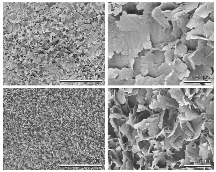
Figure 3.
Olive (Olea europaea) fruit surfaces of the cultivars Manzanilla (a,b) and Picholine (c,d) in the cryo-scanning electron microscope (SEM). In (b) and (d), details of the epicuticular wax (EW) coverage composed of flat projections (platelets) with irregular sinuate margins, are shown. The platelets are attached to the fruit surface by their narrow side protruding from the surface at different angles (compare (b) with (c)).

