Abstract
Simple Summary
Reproductive traits are economically important in the livestock industry, and this is of greater relevance when it comes to indigenous animals, since their study allows improving their use and management. Through a genome-wide association study (GWAS), the reproductive trait of the litter size (prolificity) was analyzed in the indigenous Pelibuey sheep. Several single-nucleotide polymorphisms (SNPs) and candidate genes potentially associated with litter size trait were found in the multiparous ewe’s group. These findings help to understand the genetic basis of reproductive traits of hairy Pelibuey sheep.
Abstract
The Pelibuey sheep has adaptability to climatic variations, resistance to parasites, and good maternal ability, whereas some ewes present multiple births, which increases the litter size in farm sheep. The litter size in some wool sheep breeds is associated with the presence of mutations, mainly in the family of the transforming growth factor β (TGF-β) genes. To explore genetic mechanisms underlying the variation in litter size, we conducted a genome-wide association study in two groups of Pelibuey sheep (multiparous sheep with two lambs per birth vs. uniparous sheep with a single lamb at birth) using the OvineSNP50 BeadChip. We identified a total of 57 putative SNPs markers (p < 3.0 × 10⁻³, Bonferroni correction). The candidate genes that may be associated with litter size in Pelibuey sheep are CLSTN2, MTMR2, DLG1, CGA, ABCG5, TRPM6, and HTR1E. Genomic regions were also identified that contain three quantitative trait loci (QTLs) for aseasonal reproduction (ASREP), milk yield (MY), and body weight (BW). These results allowed us to identify SNPs associated with genes that could be involved in the reproductive process related to prolificacy.
Keywords: prolificacy, Pelibuey sheep, genome-wide association study
1. Introduction
The sheep meat demand in Mexico is not covered by national production, partly due to the country’s low productive efficiency [1], creating opportunities to intensify lamb production in each region of the country [2]. Hair sheep that guarantee a relatively constant production of lamb throughout the year and the availability of meat [3], such as Pelibuey ewes, predominate throughout the Mexican territory under different types of climates. They are characterized by their rusticity, high adaptability to different environments, excellent maternal ability, prolificacy, and active reproduction most of the year [4,5]. The prolificacy in the Pelibuey sheep is ~1.5 lamb per calving, which could be of great interest to breeders. In different breeds of sheep, it was found that prolificacy is associated with mutations in different genes, which were identified mainly in wool breeds [6,7,8], but few studies were done on hair breeds such as Pelibuey sheep. In this way, several studies showed that genes related to the transforming factor group β (TGF-β), bone morphogenetic protein 15 (BMP15), growth differentiation factor 9 (GDF9), and bone morphogenetic protein of the receptor-IB (BMPR-IB) are essential for normal follicular development in the primary follicle stage in sheep [9,10,11]. TGF-β plays a role in the regulation of oocyte maturation and follicular development, and it is probably involved in cumulus expansion in mice [12], and granulosa cell proliferation (GC) [13]. The single-nucleotide polymorphisms (SNPs) identified in these genes were shown to be associated with an increase of ovulation rate (OR) and litter size [14]. Nine SNPs were evidenced in the gene BMP15 such as FecXI, FecXH [6], FecXB, FecXG [10], FecXL [14], FecX2W [15], FecXO, FecXGr [16], FecXBar [17], and deletion of 17 base pairs in FecXR [9]. In the GDF9 gene, eleven point mutations were reported: G1, G2, G3, G4, G5, G6, G7, G8 [10], FecTT [18], FecGE [19], and FecGWNS [20]. These mutations are strongly associated with litter size in sheep and are potential molecular markers used in breeding programs to increase productivity and efficiency of lamb meat production [21]. The use of genetic markers can reduce generational time in the genetic selection process through introgression of wild alleles in the population and ensuring a high production on farms. Genome-wide association studies (GWAS) are used for scanning markers across the complete genome to identify genetic variations associated with a particular trait [22]. GWAS are widely used for the detection of single-nucleotide polymorphisms (SNPs) associated with economically important traits, revolutionizing the way to locate regions of quantitative trait loci (QTL). In sheep, they were used to identify markers associated with resistance to parasites [23], selection in locally adapted livestock [24], fat deposition in the tails [25], body weight [26], and prolificacy [9,25,27]. In Ile de France sheep, using Illumina Ovine 50K, four SNPs (s17197, s48166, s25202, and OAR5_47774570) associated with prolificacy were reported in the GDF9 gene [27]. In four breeds, statistically significant SNPs were reported (rs416717560 and rs421635584 in Wadi, rs429755189 in Hu, rs412280524 and rs401960737 in Finnsheep, and rs423810437 in Romanov sheep) for litter size [25]. However, these studies were conducted on wool sheep, while there is no research on hair breeds such as the Pelibuey sheep. Therefore, the objective of this study is to identify SNPs associated with litter size in multiparous Pelibuey sheep via micro-array analysis using the OvineSNP50 BeadChip, as well as to integrate the analysis of metabolic pathways associated with SNPs.
2. Materials and Methods
2.1. Animals Used and Obtaining Samples
For our study, we used 48 Pelibuey ewes, with record of three consecutive births. Those with two or more lambs per birth were considered prolific sheep (case group, n = 24) and those with a single lamb at birth were considered non-prolific (control group, n = 24). DNA was extracted from blood samples: eight samples of prolific and six samples of non-prolific sheep were obtained from the sheep farm “El Rodeo” located in Tabasco, Mexico (17°50′39″ north (N) and 92°49′01″ west (W)); seven samples of prolific sheep and 13 samples of non-prolific sheep were obtained from the farm “Las Potrancas” in Tabasco (18°03′04″ N and 92°49′23″ W); five prolific sheep and five non-prolific sheep were obtained from “San Alberto” located in Yucatán, Mexico (20°49′42.09″ N and 89°48′42.29″ W); four prolific sheep were obtained from the farm “El Cortijo” located in Campeche, Mexico (19°43′48.92″ N and 90°05′11.58″ W). Subsequently, the blood samples were stored at −4 °C until DNA extraction.
2.2. Extraction of DNA and Genotyping
DNA was extracted from 200 μL of blood, using the Quick-DNA™ Miniprep kit (Zymo Research, Irvine, CA, USA), according to the manufacturer’s protocol. The DNA obtained was purified using the DNA Clean & Concentrator™ -5 kit (Zymo Research). The DNA concentration was measured with a Nanodrop Lite (Thermo Scientific®, Wilmington, DE, USA), and integrity was visualized on a 1.5% agarose gel. The genotyping of the two experimental groups was performed with a medium-density array Illumina OvineSNP50 Beadchip (54, 241 SNPs) in the Illumina HiScan System according to the manufacturer’s instructions, at GeneSeek (Lincoln, NE, USA).
2.3. Genotyping Analysis and Data Quality Control
A total of 54,241 SNPs were obtained from the genotyping data. The PLINK v1.07 software [28] was used for quality control (QC). The SNPs that met the following criteria were selected: SNPs that were positioned on chromosome (1 to 27), call rate < 0.95 (--geno: 0.05 and --mind: 0.05), and minor allele frequency (MAF) < 0.02 [28]. SNPs that failed the Hardy–Weinberg equilibrium (HWE) (p-value < 0.001) were excluded [29]. Relatedness of the genetic distances between populations was calculated based on a pairwise state identity data (IBS) distance matrix of all samples, for which the first three multidimensional scale (MDS) dimensions were extracted (–genome, –cluster, –mds-plot 4) and visualized with R v. 3.5.2 [30], and all pairs of individuals with IBS > 0.4 were excluded from further analysis.
2.4. Genome-Wide Association Analysis
The GWAS was conducted using PLINK v. 1.09, consisting of a comparison of allele frequencies between cases and controls, with asymptotic and empirical p-values available. To control the family-wise error rate (FWER), a Bonferroni correction was used as described by Brinster et al. [31]. The suggestive association significance threshold was p < 0.05 ((p < 2.67 × 10⁻⁵) = (p < 0.05/(50,602/27)) at the chromosome-wide level [31,32,33]. The significant association indicates that the chromosome-wide level association considered corresponds to a p-value less than 10−3 [34]. The quantile–quantile (Q–Q) plots were visualized by plotting the distribution of obtained vs. expected log10 (p-value) with inflation factors (λ). The association map and the significant SNPs were visualized in the Manhattan plot with a threshold line. The Manhattan and quantile–quantile graphics were plotted with R v. 3.5.2 [30].
2.5. Candidate Gene and QTLs Associated
The genes identified as associated with significant SNP loci were aligned to confirm their chromosome and physical location, using ovine reference genome OAR_v4.0 with the Genome Data Viewer, available online from the NCBI’s Genome (https://www.ncbi.nlm.nih.gov/genome/gdv/?org=ovis-aries). An SNP was considered to be from a particular gene if it mapped within it. The QTLs were located in the database Jbrowse [35], using online page (https://www.animalgenome.org/) which contains QTLs previously reported in sheep [36,37,38,39].
2.6. Analysis of Gene Ontology and Metabolic Pathways
For the gene ontology (GO) enrichment analysis, the genes identified in GWAS were analyzed using the Database for Annotation, Visualization, and Integrated Discovery (DAVID) platform [40], with the sheep genome OAR_v4. In addition, we performed a metabolic pathway analysis using the Kyoto Encyclopedia of Gene and Genomes (https://www.genome.jp/kegg/genes.html) [41]. To graph GO annotations, the WEGO v2.0 program was used [42].
3. Results
3.1. Genotyping Analysis and Data Quality Control
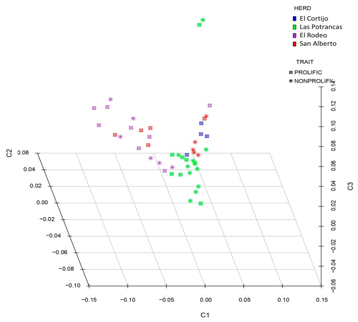
After QC analysis, 50,661 SNPs were genotyped. Subsequently, 59 SNPs were excluded from our dataset as they did not pass the HWE and MAF tests. One non-prolific ewe of 24 ewes was discarded for presenting a low call rate (<95%). Finally, 50,602 autosomal SNPs from 47 Pelibuey sheep were used to carry out genotype association tests. The stratification of alleles in the population based on state identity data (IBS) was plotted on the multidimensional scale (MDS) among the same groups of related sheep. The treatments did not show a clear formation of clusters within the populations, as shown in Figure 1. The distribution of the alleles indicates that there is no genetic difference between the two groups, since they belong to the same breed.
Figure 1.
The multidimensional scale (MDS) analysis of genotypes included in this study. The analysis was performed for the first three components (C1, C2, and C3). The color indications for the herd are as follows: El Cortijo, blue; Las Potrancas, green; El Rodeo, purple; San Alberto, red.
3.2. Genome-Wide Association Analysis
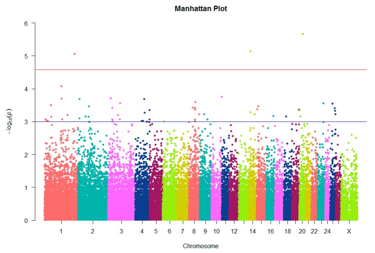
The association analysis was performed on 50,602 SNPs distributed in 27 chromosomes. Of these, only three SNPs which were defined as associated with litter size passed the Bonferroni test correction at the chromosome genome-wide level and 54 SNPs were only suggestive, as shown in Figure 2 and Table S1.
Figure 2.
Manhattan plot showing single-nucleotide polymorphisms (SNPs) associated with litter size on the ovine chromosome. The red line corresponds to the 5% chromosome-wide significance threshold using a Bonferroni correction (2.6 × 10⁻⁵). The blue line corresponds to a suggestive chromosome-wide threshold of 10−3.
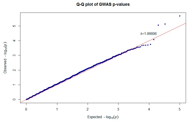
The quantile–quantile plot shows the total distribution of the observed p-values (−log10 p-values) of 50,602 SNPs versus the expected values, showing that some deviated from of the expected with an inflation factor (λ) of 1.06606, as shown in Figure 3.
Figure 3.
Quantile–quantile plot of genome-wide association study (GWAS) shown in the Manhattan plot.
3.3. Gene Ontology and KEGG Analysis
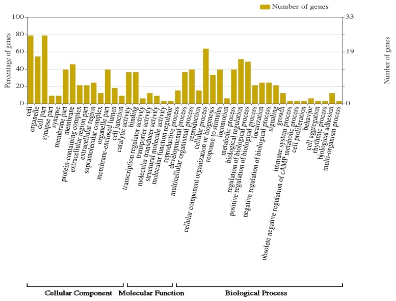
In order to determine the molecular and metabolic functions in which the 57 selected genes participate, the identification of their ontological categories and associated metabolic pathways was carried out. Identified genes were categorized into 46 functional groups of the Gene Ontology (GO) classification, distributed as follows: 25 functional groups for biological process (BP); 14 groups for cellular component (CC); 7 groups for molecular function (MF). The highest abundance of genes was represented in the CC category (Figure 4).
Figure 4.
Gene ontology categories identified in genes with significant SNPs.
Among the ontological categories identified in the 57 genes, those related to reproduction were as follows: GO:0001701: in uterus embryonic development (ANKRD11); GO:0008585: female gonad development (ARID5B); GO:0070373: negative regulation of ERK1 and ERK2 cascade (DLG1); GO:0032870: cellular response to hormone stimulus (ROBO2); GO:0061364: apoptotic process involved in luteolysis (ROBO2); GO:0032275: luteinizing hormone secretion (CGA); GO:0032870: cellular response to hormone stimulus (CGA); GO:0046621: negative regulation of organ growth (CGA); GO:0046884: follicle-stimulating hormone secretion (CGA).
The SNPs were used to identify QTLs previously localized in other studies performed in other sheep populations [36,37,38,39], and all QTLs are shown in Table S2. The analysis of the metabolic pathways in which the significant genes participate, showed that 12 genes are associated with some metabolic pathway (Table 1). Analysis of the DLG1 gene revealed its participation in the Hippo signaling pathway. The CGA gene participates in six important metabolic pathways, including the cAMP signaling pathway, GnRH signaling pathway, ovarian steroidogenesis, prolactin signaling pathway, thyroid hormone synthesis, and regulation of lipolysis in adipocytes, which are associated with reproductive processes.
Table 1.
SNPs identified by chromosome genome-wide association, traits, and biological pathway association with prolificacy.
| SNP ID | Chr | Position (bp) | Gene Name | Gene Description | Traits | Signal Pathway |
|---|---|---|---|---|---|---|
| s71757.1 | 1 | 51,963,826 | ST6GALNAC3 | ST6 N-acetylgalactosaminide alpha-2 6-sialyltransferase 3 | MUSWT, LMYP, BONE_WT, BONEP, FATP [36] | Glycosphingolipid biosynthesis |
| OAR1_155672687.1 | 1 | 189,855,910 | ROBO2 | Roundabout guidance receptor 2 | MUSWT, LMYP, BONE_WT, BONEP, FATP [36] | Axon guidance |
| OAR1_204970872.1 | 1 | 189,855,910 | DLG1 | Discs large MAGUK scaffold protein 1 | ASREP [37], SAOS [43], LMYP, FATP, BONE_WT, MUSWT [36] | Hippo signaling pathway; tight junction; T-cell receptor signaling pathway |
| s09883.1 | 1 | 246,913,454 | CLSTN2 | Calsyntenin 2 | ASREP [37], TFEC_1 [44], FATP [36], FCURV [45] | |
| OAR2_65914681.1 | 2 | 61,498,071 | TRPM6 | Transient receptor potential cation channel subfamily M member 6 | HCWT [46], MF [39], MFY, PP, MY [38], BW [36] | Mineral absorption |
| OAR2_95966123.1 | 2 | 89,499,669 | COL11A1 | Collagen type XI alpha 1 chain | SCS [46], PP [38], LATRICH2 [47], HCWT [36], MF [38] | Protein digestion and absorption |
| OAR3_85112203.1 | 3 | 80,398,784 | ABCG5 | ATP-binding cassette subfamily G member 5 | INTFAT [36], MCLA [48], SL [49] | ABC transporters; fat digestion and absorption; bile secretion; cholesterol metabolism |
| OAR3_104545117_X.1 | 3 | 98,126,615 | HTRA2 | HtrA serine peptidase 2 | FECZ [50], MCLA [48], SL [49], INTFAT [36] | Apoptosis |
| s62827.1 | 8 | 49,878,423 | CGA | Glycoprotein hormones alpha polypeptide | LATRICH2 [47], INTFAT [36], FECGEN [51] | cAMP signaling pathway; GnRH signaling pathway; ovarian steroidogenesis; prolactin signaling pathway; thyroid hormone synthesis; regulation of lipolysis in adipocytes |
| OAR8_53593379.1 | 8 | 49,981,252 | HTR1E | 5-hydroxytryptamine receptor 1E | LATRICH2 [47], INTFAT [36], FECGEN [51] | cAMP signaling pathway; neuroactive ligand–receptor interaction; serotonergic synapse; taste transduction |
| OAR9_36598045.1 | 9 | 34,604,487 | ATP6V1H | ATPase H+ transporting V1 subunit H | HCWT, LMA [36] | Oxidative phosphorylation; metabolic pathways; phagosome; mTOR signaling pathway; synaptic vesicle cycle |
| OAR15_13905772.1 | 15 | 13,872,637 | MTMR2 | Myotubularin related protein 2 | Inositol phosphate metabolism; metabolic pathways; phosphatidylinositol signaling system | |
| s07255.1 | 23 | 47,438,785 | ST8SIA5 | ST8 alpha-N-acetyl-neuraminide alpha-2 8-sialyltransferase 5 | IGA [51], FATP, LMYP, HCWT, BW, FATWT [36], MFY, MY [38] | Glycosphingolipid biosynthesis; metabolic pathways |
| s08197.1 | 25 | 40,382,673 | GRID1 | Glutamate ionotropic receptor delta type subunit 1 | SL, MFDIAM, CVFD_PRI [49] | Neuroactive ligand–receptor interaction |
Chr, chromosome; ID, identifier; bp, base pairs; MUSWT, muscle weight in carcass; LMYP, lean meat yield percentage; BONE_WT, bone weight in carcass; BONEP, carcass bone percentage; FATP, carcass fat percentage; ASREP, aseasonal reproduction; SAOS, Salmonella abortus ovis susceptibility; TFEC_1, Trichostrongylus colubriformis FEC; FCURV, fiber curvature; HCWT, hot carcass weight; MF, milk fat percentage; MFY, milk fat yield; PP, milk protein percentage; MY, milk yield; BW, body weight; SCS, somatic cell score; LATRICH, Trichostrongylus adult and larva count; INTFAT, internal fat amount; MCLA, meat-conjugated linoleic acid content; SL, staple length; FECGEN, fecal egg count; LMA, longissimus muscle area; IGA, immunoglobulin A level; FATWT, fat weight in carcass; MFDIAM, mean fiber diameter; CVFD_PRI, primary fiber diameter coefficient of variance.
3.4. Identification of the Candidate Genes
Functional analysis based on the results of GWAS revealed that the top 10 candidate genes that are involved in the ovary development process in ewes with two lambs at birth were CLSTN2, MTMR2, CCDC174, NOM1, ANKRD11, DLG1, ALPK3, ROBO2, CGA, and KDM4A. The relevant genes identified in the present analysis that were reported in other studies to be potentially associated with reproduction processes were ANKRD11, ARID5B, DLG1, ROBO2, and CGA, as shown in Table 2.
Table 2.
The SNPs identified by GWAS for prolificacy in Pelibuey sheep.
| Chr | SNP | Position (bp) | A1 | A2 | F_A | F_U | MAF | p-Unadjusted | Chis-q | Gene Annotated |
|---|---|---|---|---|---|---|---|---|---|---|
| 1 | s09883.1 | 246,913,454 | A | G | 0.1458 | 0.587 | 0.3617 | 8.61 × 10⁻⁶ | 19.8 | CLSTN2 |
| 15 | OAR15_13905772.1 | 13,872,637 | A | G | 0.04167 | 0.3261 | 0.1809 | 0.0003417 | 12.83 | MTMR2 |
| 19 | s15631.1 | 57,489,437 | A | C | 0.5208 | 0.1739 | 0.3511 | 0.0004272 | 12.41 | CCDC174 |
| 4 | s37914.1 | 117,719,020 | G | A | 0.6667 | 0.3043 | 0.4894 | 0.0004434 | 12.34 | NOM1 |
| 14 | s57545.1 | 13,903,063 | T | C | 0.5417 | 0.1957 | 0.3723 | 0.0005225 | 12.03 | ANKRD11 |
| 1 | OAR1_204970872.1 | 189,855,910 | T | C | 0.3542 | 0.06522 | 0.2128 | 0.0006221 | 11.71 | DLG1 |
| 18 | OAR18_22964031.1 | 22,346,018 | C | T | 0.1875 | 0.5217 | 0.3511 | 0.0006891 | 11.52 | ALPK3 |
| 1 | OAR1_155672687.1 | 144,029,243 | C | T | 0.625 | 0.2826 | 0.4574 | 0.0008655 | 11.1 | ROBO2 |
| 8 | s62827.1 | 49,878,423 | A | G | 0.08333 | 0.3696 | 0.2234 | 0.0008669 | 11.09 | CGA |
| 1 | OAR1_18691972.1 | 18,481,816 | T | C | 0.25 | 0.587 | 0.4149 | 0.0009179 | 10.99 | KDM4A |
Chr, chromosome; bp, base pairs; A1, minor allele 1; A2, major allele 2; F_A, allele 1 frequency among cases; F_U, allele 1 frequency among controls; MAF, minor allele frequency.
4. Discussion
In the present study, we performed a GWAS using the medium-density Illumina OvineSNP50 Genotyping BeadChip. A total of 50,602 SNPs that passed quality control belonging to 47 Pelibuey ewes were used to identify regions associated with litter size. GWAS was able to identify three SNPs significant at 5% at the chromosome-wide level and 54 suggestive SNPs associated with the prolificacy trait. The power of GWAS to detect the true association is determined by many factors such as phenotypic variation, the number of individuals, and allele frequency [52]. The indirect associations are the result of disequilibrium between multiple factors affecting a trait, whereas lack of statistical power can produce spurious associations that are only distantly linked to causal polymorphisms [53]. A low heritability for reproductive traits is associated with a low genetic variability for litter size [54]. A greater heritability of broad sense results in greater −log10 p-values, while the number of loci that affect the trait increases along with environmental interactions with an expected decrease in heritability [55]. We used a low number of samples, which could have had an effect on the number of significant SNPs; however, the inflation factor was 1.06 which indicates a low possibility of false positives. When the inflation factor is small (<1.1) it indicates that stratification cannot be excluded as a possibility in real scenarios, to reduce the possibility of confusion due to the population mix [56].
4.1. Markers Associated with Prolificacy Traits
Four SNPs identified here may be associated with prolificacy; these are OAR1_204970872.1 and s09883.1 for aseasonal reproduction (ASREP), as well as SNPs OAR2_65914681.1 and s07255.1 for milk yield (MY), and body weight (BW), respectively, as shown in Table 1. In addition, we also report other productive traits identified in other breeds such as muscle weight in carcass (MUSWT), carcass fat percentage (FATP), lean meat yield percentage (LMYP), hot carcass weight (HCWT), internal fat amount (INTFAT), fat weight in carcass (FATWT), and milk lactose yield (MLACT).
An important trait is ASREP, with markers located in two regions (OAR1_204970872.1 (Chr 1: 189,855,910 bp), and s09883.1 (Chr 1: 246,913,454 bp) for seasonal reproduction). In Dorset × East Frisian sheep, seven chromosomes (1, 3, 12, 17, 19, 20, and 24) were identified to harbor putative QTLs for traits associated with ASREP; in chromosome 1, a QTL is associated with the associated with the maximum progesterone level during the pre-breeding season [37]. The increased follicular progesterone secretion comes from the ovary containing the active follicles [57]. In Pelibuey sheep, in the postpartum period, progesterone concentrations rise to luteal phase levels but not to the magnitude of luteinizing hormone (LH) [58]. The markers associated with BW trait are s05724.1 (Chr 1: 56,360,553 bp), OAR1_149400642.1 (Chr 1: 138,085,163 bp), OAR1_150722624.1 (Chr 1: 139,388,579 bp), OAR2_65914681.1 (Chr 2: 61,498,071 bp), OAR3_24559969.1 (Chr 3: 22,729,462 bp), s66102.1 (Chr 3: 33,073,804 bp), OAR3_36221107.1 (Chr3: 33,738,283 bp), s72352.1 (Chr 3: 36,572,814 bp), OAR6_14287930.1 (Chr 6: 11,709,842 bp), s50134.1 (Chr 14: 46,046,471 bp), OAR20_31210307.1 (Chr 20: 28,418,356 bp), and s07255.1 (Chr 23: 47,438,785 bp). In Merino sheep, it was reported that BW is associated with the traits of MUSWT, LMYP, bone weight in carcass (BONE_WT), carcass bone percentage (BONEP), and FATP [36]. In Black Bengal goats, there is a positive relationship between withers height (WH), distance between tuber coxae bones (DTC), BW, and litter size, while physical strength was implicated in an increased likelihood of multiple births in goats bearing multiple fetuses from those bearing a single fetus [59].
The SNPs associated with the MY trait are located in OAR2_65914681.1 (Chr 2: 61,498,071 bp), OAR18_22964031.1 (Chr 18: 22,346,018 bp), OAR20_5129052.1 (Chr 20: 5,085,823 bp), OAR20_11623776.1 (Chr 20: 11,249,238 bp), and OAR20_31210307.1 (Chr 20: 28,418,356 bp). In Churra sheep and Lacaune breeds, ewes presenting twin parturitions produced more milk than ewes with single parturitions, and the presence of female lambs had a positive effect on milk yield [60]. In sheep, litter size has a positive correlation with the incidence of breastfeeding and milk production [61]. In Damascus goats, milk production after weaning was hardly affected by post-kidding body weight, and body weight at mating time was linearly related with litter size and weight at subsequent kidding [62], which indicates that it may be associated with litter size in this breed, representing the potential to exploit these genetic traits.
Finally, our study revealed four SNPs s37914.1 (Chr4: 117,719,020 bp), s02969.1 (Chr5: 184,537 bp), OAR15_13905772.1 (Chr15: 13,872,637 bp), and s15631.1 (Chr15: 57,489,437 bp) not previously reported as QTLs on the Jbrowse online page. These four SNPs may have a relationship with the litter size in this breed; however, it is important to carry out analyses of the regions in the corresponding genes (NOM1, LOC101116985, MTMR2, and CCDC174, respectively).
4.2. Gene Ontology and Metabolic Pathways
In different studies, identifications of molecular functions and metabolic pathways for genetic loci associated with diseases or traits obtained from GWAS results were carried out [25,63]. We investigated the molecular functions and metabolic processes of the 57 genes identified as a result of the GWAS analysis. Of the 57 genes analyzed, only 13 genes with metabolic pathways and biological functions of reproduction were identified (Table S3). For example, the DLG1 gene was enriched in six signal pathways; of these, only the Hippo signaling pathway plays a key role in mechanotransduction, providing an understanding of the molecular mechanisms via which cells sense and respond to mechanical signals to regulate cell proliferation and apoptosis for maintaining optimal organ sizes [64]. The Hippo signaling pathway regulates the activation of primordial follicles and increases birth rate, accompanied by increasing levels of 17β-estradiol (E2) and follicle-stimulating hormone (FSH) in mouse [65]. In bovine ovaries, the Hippo pathway transcription co-activators play an important role in GC proliferation and estrogen production, thereby determining normal follicle development [66]. In addition to its role in regulating tissue growth, the pathway was implicated in the control of other biological processes, such as cell-fate determination, mitosis, and pluripotency [67]. The TRPM6 gene was enriched in the mineral absorption pathway; TRPM6 is a member of the melastatin-related transient receptor potential (TRPM) subfamily of ion channels, and it is a polypeptide containing both an ion channel pore and a serine/threonine kinase [68]. Studies in mice showed these channels to have a role in whole-body magnesium homeostasis, as well as additional critical functions during early embryogenesis [69]. The ABCG5 gene was enriched in four pathways, where only the cholesterol metabolism pathway is important, which is utilized for steroid synthesis by ovarian tissue potentially derived from de novo synthesis or cellular uptake of lipoprotein cholesterol [70]. Six important pathways were enriched in the CGA gene, which can be the key to understanding the molecular processes that are important for prolificacy. Three of these pathways were the GnRH signaling pathway, ovarian steroidogenesis, and prolactin signaling pathway. The GnRH signaling pathways are activated by signal transduction cascades, which consequently cause the release of gonadotropins, LH, and FSH for gonadal maturation, the onset of puberty, and ovulation [71]. In mice, using GnRH-Ag caused stimulatory effects on ovarian steroidogenesis and follicular development [72]. Ovarian steroidogenesis in sheep is regulated by multiple signals of transduction such as SHH, WNT, and RHO GTPase, for early folliculogenesis [13]. Prolactin is a hormone that is essential for normal reproduction, and it signals through two types of receptors. The prolactin signaling pathway is initiated by the binding of prolactin with the prolactin receptor (PRLR), which is expressed in a variety of tissues [73]. The HTR1E gene was enriched in the cAMP signaling pathway, whereby the persistent cAMP signals from internalized LH receptors contribute to transmitting LH effects inside follicle cells and the oocyte [74]. However, these genes can be regulated by extrinsic factors such as nutrition.
4.3. Candidate Gene Identification
After the annotation of the genes, only 10 regions were selected according to their biological functions: s09883.1 (CLSTN2), OAR15_13905772.1 (MTMR2), s15631.1 (CCDC174), s37914.1 (NOM1), s57545.1 (ANKRD11), OAR1_204970872.1 (DLG1), OAR18_22964031.1 (ALPK3), OAR1_155672687.1 (ROBO2), s62827.1 (CGA), and OAR1_18691972.1 (KDM4A).
CLSTN2 (Calsyntenin 2), also known as alcadein-γ, is a neuronal cell surface synaptic protein with an evolutionarily conserved role in learning and memory [75]. In the immature rat uterus, the CSTN2 gene increased expression in the follicular phase when the level of 17β-estradiol estradiol (E2) increased during the estrous cycle [76]. More recently, in cow, the CLSTN2 gene was found to play a role related to lipid metabolism, affecting carcass traits [77]. Pensante-Pacheco et al. [78] reported that sheep after calving lose weight and body condition, and lambs born alone were heavier than those born in multiparous litters. Therefore, this gene can act in the metabolic pathways, mainly for the accumulation of energy reserves, allowing animals to survive food shortages that can be used by sheep in reproduction, pregnancy, and lactation; these include changes in synaptic inputs onto GnRH neurons, and the neuroendocrine system signal regulates seasonal reproduction [79]. MTMR2 (myotubularin-related protein 2), belongs to the family of phosphoinositide phosphatases including several members mutated in neuromuscular diseases or those associated with metabolic syndrome, obesity, and cancer [80]. It was also reported in Schwann cells (SC) [81], lipid metabolic processes [82], and the reproductive process [83]. There is a relationship between E2 and SC, since it promotes SC myelination of regenerated sciatic nerves and can promote SC differentiation through the estrogen receptor beta-extracellular signal-regulated kinase 1 and 2 (ERβ-ERK1/2) signaling pathway in rats [84]. It was reported that the MTMR2 and MTMR5 genes are highly expressed in the testicles, especially in germ cells and in Sertoli, and the deactivation of any of these genes produces spermatogenic defects [83]. CCDC174 (coiled-coil domain-containing 174) is essential for neuronal differentiation; in human, mutations affect psychomotor developmental delay and abducens nerve palsy [85]. The CCDC174 gene is not yet reported in reproductive processes; however, other members of this family are associated with these processes. For example, 10 coiled-coil domain-containing (CCDC) messenger RNAs (mRNAs) were found in the glandular epithelium (GE), related to pregnancy recognition and establishment [86]. In humans, during normal healthy pregnancy, CCDC proteins significantly increase in exosomes present in maternal plasma with gestational age during the first trimester of pregnancy [87].
NOM1 (nucleolar protein with MIF4G domain 1) is a member of the MIF4G/MA3 family identified at the chromosome 7q36 breakpoint involved in processes that impact translation [88]. Solomon-Zemler et al. [89] reported that NOM1 has biological pathways associated with nuclear IGF1R (insulin-like growth factor-1 receptor), and inhibition of nuclear IGF1R translocation by dansylcadaverine reduced NOM1 levels in nuclei of MCF7 cells. According to Gunawardena et al. [90], NOM1 is a PP1 (protein phosphatase I) nucleolar targeting subunit, which is an essential eukaryotic serine/threonine phosphatase required for many cellular processes, including cell division, signaling, and metabolism. ANKRD11 (ankyrin repeat domain 11) is a gene directly involved in embryonic and fetal development, which is also associated with maternal nutrition during pregnancy in sheep [91]. DLG1 (discs large MAGUK scaffold protein 1) is a scaffolding protein that, through interaction with diverse cell partners, participates in the control of key cellular processes such as polarity, proliferation, and migration [92]. Cavatorta et al. [93] reported that the DLG1 gene encodes a member of the MAGUK protein family involved in the polarization of epithelial cells. In mouse, DLG1 is expressed at high levels in oocytes and granulosa cells [94]. ALPK3 (alpha kinase 3) is implicated in a large variety of cellular processes such as protein translation, Mg2+ homeostasis, intracellular transport, cell migration, adhesion, and proliferation [95]. A mutation in the ALPK3 gene in human is associated with cardiomyopathy [96]. The overexpression of ALPK3 enhances differentiation of murine embryonic carcinoma cells into cardiomyocytes [95]. ROBO2 (roundabout guidance receptor 2) is important for axon guidance across the midline during central nervous system (CNS) development [97]. In sheep, the ROBO2 gene is essential during the early stages of follicle formation, as well as during primordial follicle maturation-determining processes in ovary development [98], and expression may be regulated by additional factors in the ovary and steroid hormones in other reproductive tissues [99]. CGA (glycoprotein hormone, alpha polypeptide) is the α subunit of glycoprotein hormones, with an important role in the development and function of thyroid and gonads [100]. Two subunits were reported in the FSH; FSHα and FSHβ play key roles in female reproduction, including boars [101,102], where each subunit regulates a variety of functions of the hormone including folding, heterodimerization, secretion, circulatory survival, and bioactivity [103]. Previous reports indicated that FSHα and FSHβ mRNAs were detected only in the pituitary tissue of boar [101]. KDM4A (lysine demethylase 4A) is a lysine demethylase with specificity toward di- and tri-methylated lysine 9 and lysine 36 of histone H3 (H3K9me2/me3 and H3K36me2/me3) [104]; a previous study reported that KDM4A is a maternal factor that plays a key role in embryo survival, the implantation process in mice, and fertility [104]. All these genes may have an important role in cellular processes such as follicular development associated with the regulation of the litter size identified in the Pelibuey sheep. In addition to the 10 SNPs significant at the chromosome-wide level associated with prolificacy, we also report three new uncharacterized proteins were identified in this study. These were OAR1_149400642.1 (Chr 1: 138,085,163 bp) (p < 0.0000837), s11062.1 (Chr 14: 13,674,017 bp) (p < 0.00000729), and OAR20_31210307.1 (Chr 20: 28,418,356 bp) (p < 0.00000211), located in genes LOC101114740, LOC105616840, and LOC101117202, respectively (Table S1). However, the information for these genes is non-existent; thus, it is necessary to expand the research to obtain thorough knowledge associated with the reproduction of sheep.
5. Conclusions
The GWAS performed was able to identify 57 SNPs associated with litter size in the Pelibuey breeds. The candidate genes associated with litter size in Pelibuey that may be involved in the reproduction process are CLSTN2, MTMR2, CCDC174, NOM1, ANKRD11, DLG1, ALPK3, ROBO2, CGA, and KDM4A. In addition, we also report four SNPs not previously documented whose QTLs were previously reported in sheep: s37914.1 (Chr 4: 117,719,020 bp), s02969.1 (Chr 5: 184,537 bp), OAR15_13905772.1 (Chr 15: 13,872,637 bp), and s15631.1 (Chr 19: 57,489,437 bp). The presence of SNPs commonly present in prolific wool sheep breeds such as for the transforming factor group β (TGF β) was not reported in this hair sheep, which may confirm that prolificacy in Pelibuey sheep can be controlled via different underlying genetic mechanisms, such as candidate genes related to reproductive seasonality, milk yield, and body weight. Further validation studies can elucidate the gene network, enabling its incorporation in sheep breeding programs in order to obtain ewes with higher reproductive efficiency.
Acknowledgments
W.H.-M. acknowledges the Conacyt Fellowship No. 274026 awarded to carry out his Doctorate in Science studies within the Doctoral Program in Science in Sustainable Tropical Agriculture, of the Technological Institute of Conkal; the authors also acknowledge C. Walter R. Lanz-Villegas for his contribution in obtaining the samples.
Supplementary Materials
The following are available online at https://www.mdpi.com/2076-2615/10/3/434/s1: Table S1: 57 SNPs considered as candidate markers and genes mapped; Table S2: Regions of chromosome in SNPs identified in the present study and QTLs previously reported for traits different in other sheep breeds; Table S3: KEGG pathway analysis in genes with chromosome-wide level association.
Author Contributions
Conceptualization, R.Z.-B., S.I.R.-P., M.A.M.-N., and W.H.-M.; methodology, R.Z.-B., S.I.R.-P., J.P.R.-U., and W.H.-M.; software, S.I.R.-P., M.A.M.-N., and W.H.-M.; validation, S.I.R.-P., R.C.-C., and W.H.-M.; formal analysis, S.I.R.-P. and W.H.-M.; investigation, W.H.-M.; resources, R.Z.-B.; data curation, R.C.-C., S.I.R.-P., and W.H.-M; writing—original draft preparation, R.Z.-B., M.A.M.-N., J.P.R.-U., and W.H.-M.; writing—review and editing, R.Z.-B., S.I.R.-P., J.P.R.-U., and W.H.-M.; visualization, R.C.-C.; supervision, S.I.R.-P. and W.H.-M.; project administration, R.Z.-B. and W.H.-M.; funding acquisition, R.Z.-B. All authors have read and agreed to the published version of the manuscript.
Funding
This work was financially supported by TecNM (Grant No 5554.19-P) and partially supported by CONACYT (Grant No. 176533).
Conflicts of Interest
The authors declare no conflict of interest.
References
- 1.Bobadilla-Soto E.E., Salas-Razo G., Padillas-Flores J.P., Perea-Peña M. Unit displacement of sheep production in Mexico by effect of imports. Int. J. Dev. Res. 2018;5:3607–3612. [Google Scholar]
- 2.Macías-Cruz U., Álvarez-Valenzuela F.D., Correa-Calderon A., Molina-Ramirez L., González-Reyna A., Soto-Navarro S., Avendaño-Reyes L. Pelibuey ewe productivity and subsequent pre-weaning lamb performance using hair-sheep breeds under a confinement system. J. Appl. Anim. Res. 2009;36:255–260. doi: 10.1080/09712119.2009.9707071. [DOI] [Google Scholar]
- 3.Macías-Cruz U., Álvarez-Valenzuela F., Olguín-Arredondo H., Molina-Ramirez L., Avendaño-Reyes L. Ovejas Pelibuey sincronizadas con progestágenos y apareadas con machos de razas Dorper y Katahdin bajo condiciones estabuladas: Producción de la oveja y crecimiento de los corderos durante el período predestete. Arch. Med. Vet. 2012;44:29–37. doi: 10.4067/S0301-732X2012000100005. [DOI] [Google Scholar]
- 4.Dickson L., Torres G., Aubeterre R.D., Garcia O. Factores que influyen en el intervalo entre partos y la prolificidad de un hato de carneros Pelibuey en Venezuela. Rev. Cuba. Cienc. Agric. 2004;38:13–17. [Google Scholar]
- 5.Aké-López R., Segura-Correa J.C., Quintal-Franco J. Effect of flunixin meglumine on the corpus luteum and possible prevention of embryonic loss in Pelibuey ewes. Small Rumin. Res. 2005;59:83–87. doi: 10.1016/j.smallrumres.2004.11.008. [DOI] [Google Scholar]
- 6.Davis G.H., Balakrishnan L., Ross I.K., Wilson T., Galloway S.M., Lumsden B.M., Hanrahan J.P., Mullen M., Mao X.Z., Wang G.L., et al. Investigation of the Booroola (FecB) and Inverdale (FecX I) mutations in 21 prolific breeds and strains of sheep sampled in 13 countries. Anim. Reprod. Sci. 2006;92:87–96. doi: 10.1016/j.anireprosci.2005.06.001. [DOI] [PubMed] [Google Scholar]
- 7.Monteagudo L.V., Ponz R., Tejedor M.T., Laviña A., Sierra I. A 17 bp deletion in the Bone Morphogenetic Protein 15 (BMP15) gene is associated to increased prolificacy in the Rasa Aragonesa sheep breed. Anim. Reprod. Sci. 2009;110:139–146. doi: 10.1016/j.anireprosci.2008.01.005. [DOI] [PubMed] [Google Scholar]
- 8.Souza C.J.H., MacDougall C., Campbell B.K., McNeilly A.S., Baird D.T. The Booroola (FecB) phenotype is associated with a mutation in the bone morphogenetic receptor type 1 B (BMPR1B) gene. Rapid Comun. 2001;169:R1–R6. doi: 10.1677/joe.0.169r001. [DOI] [PubMed] [Google Scholar]
- 9.Martinez-Royo A., Jurado J.J., Smulders J.P., Marti J.I., Alabart J.L., Roche A., Fantova E., Bodin L., Mulsant P., Serrano M., et al. A deletion in the bone morphogenetic protein 15 gene causes sterility and increased prolificacy in Rasa Aragonesa sheep. Anim. Genet. 2008;39:294–297. doi: 10.1111/j.1365-2052.2008.01707.x. [DOI] [PubMed] [Google Scholar]
- 10.Hanrahan J.P., Gregan S.M., Mulsant P., Mullen M., Davis H.G., Powell R., Galloway M.S. Mutations in the Genes for Oocyte-Derived Growth Factors GDF9 and BMP15 Are Associated with Both Increased Ovulation Rate and Sterility in Cambridge and Belclare Sheep (Ovis aries) Biol. Reprod. 2004;70:900–909. doi: 10.1095/biolreprod.103.023093. [DOI] [PubMed] [Google Scholar]
- 11.Galloway S.M., McNatty K.P., Cambridge L.M., Laitinen M.P., Juengel J.L., Jokiranta T.S., McLaren R.J., Luiro K., Dodds K.G., Montgomery G.W., et al. Mutations in an oocyte-derived growth factor gene (BMP15) cause increased ovulation rate and infertility in a dosage-sensitive manner. Nat. Genet. 2000;25:279–283. doi: 10.1038/77033. [DOI] [PubMed] [Google Scholar]
- 12.Guéripel X., Véronique B., Gougeon A. Oocyte Bone Morphogenetic Protein 15, but not Growth Differentiation Factor 9, Is Increased During Gonadotropin-Induced Follicular Development in the Immature Mouse and Is Associated with Cumulus Oophorus Expansion. Biol. Reprod. 2006;75:836–843. doi: 10.1095/biolreprod.106.055574. [DOI] [PubMed] [Google Scholar]
- 13.Bonnet A., Cabau C., Bouchez O., Sarry J., Marsaud N., Foissac S., Woloszyn F., Mulsant P., Mandon-pepin B. An overview of gene expression dynamics during early ovarian folliculogenesis: Specificity of follicular compartments and bi-directional dialog. BMC Genom. 2013;14:1–19. doi: 10.1186/1471-2164-14-904. [DOI] [PMC free article] [PubMed] [Google Scholar]
- 14.Bodin L., Di Pasquale E., Febre S., Bontoux M., Monget P., Persani L., Mulsant P. A Novel Mutation in the Bone Morphogenetic Protein 15 Gene Causing Defective Protein Secretion Is Associated Lacaune Sheep. Endocrinology. 2007;148:393–400. doi: 10.1210/en.2006-0764. [DOI] [PubMed] [Google Scholar]
- 15.Feary E.S., Juengel J.L., Smith P., French M.C., Connell A.R.O., Lawrence S.B., Galloway S.M., Davis G.H., Mcnatty K.P. Patterns of Expression of Messenger RNAs Encoding GDF9, BMP15, TGFBR1, BMPR1B, and BMPR2 During Follicular Development and Characterization of Ovarian Follicular Populations in Ewes Carrying the Woodlands FecX2 W Mutation 1. Biol. Reprod. 2007;77:990–998. doi: 10.1095/biolreprod.107.062752. [DOI] [PubMed] [Google Scholar]
- 16.Demars J., Fabre S., Sarry J., Rossetti R., Gilbert H., Persani L., Tosser-Klopp G., Mulsant P., Nowak Z., Drobik W., et al. Genome-Wide Association Studies Identify Two Novel BMP15 Mutations Responsible for an Atypical Hyperprolificacy Phenotype in Sheep. PLoS Genet. 2013;9:1–13. doi: 10.1371/journal.pgen.1003482. [DOI] [PMC free article] [PubMed] [Google Scholar]
- 17.Lassoued N., Benkhlil Z., Woloszyn F., Rejeb A., Aouina M., Rekik M., Fabre S., Bedhiaf-Romdhani S. FecXBar a Novel BMP15 mutation responsible for prolificacy and female sterility in Tunisian Barbarine Sheep. BMC Genet. 2017;18:1–10. doi: 10.1186/s12863-017-0510-x. [DOI] [PMC free article] [PubMed] [Google Scholar]
- 18.Nicol L., Bishop S.C., Pong-wong R., Bendixen C., Holm L., Rhind S.M., Mcneilly A.S. Homozygosity for a single base-pair mutation in the oocyte-specific GDF9 gene results in sterility in Thaka sheep. Reprod. Res. 2009;138:921–933. doi: 10.1530/REP-09-0193. [DOI] [PubMed] [Google Scholar]
- 19.Silva B.D.M., Castro E.A., Souza C.J.H., Paiva S.R., Sartori R., Franco M.M., Azevedo H.C., Silva T.A.S.N., Vieira A.M.C., Neves J.P., et al. A new polymorphism in the Growth and Differentiation Factor 9 (GDF9) gene is associated with increased ovulation rate and prolificacy in homozygous sheep. Anim. Genet. 2010;42:89–92. doi: 10.1111/j.1365-2052.2010.02078.x. [DOI] [PubMed] [Google Scholar]
- 20.Våge D.I., Husdal M., Kent M.P., Klemetsdal G., Boman I.A. A missense mutation in growth differentiation factor 9 (GDF9) is strongly associated with litter size in sheep. BMC Genet. 2013;14:1–8. doi: 10.1186/1471-2156-14-1. [DOI] [PMC free article] [PubMed] [Google Scholar]
- 21.Sagar N.G., Kumar S., Baranwal A., Prasad A.R. Introgression of Fecundity Gene (FecB) in Non-Prolific SHEEP Breeds: A Boon for Farmers. Int. J. Sci. Environ. Technol. 2017;6:375–380. [Google Scholar]
- 22.Duncan E.L., Brown M.A. Genome-wide Association Studies. In: Rajesh V., Thakker Michael P., Whyte John A., Eisman T.I., editors. Genetics of Bone Biology and Skeletal Disease. Academic Press; San Diego, CA, USA: 2013. pp. 93–100. [Google Scholar]
- 23.Sallé G., Jacquiet P., Gruner L., Cortet J., Sauvé C., Prévot F., Grisez C., Bergeaud J.P., Schibler L., Tircazes A., et al. A genome scan for QTL affecting resistance to Haemonchus contortus in sheep. J. Anim. Sci. 2012:4690–4705. doi: 10.2527/jas.2012-5121. [DOI] [PubMed] [Google Scholar]
- 24.José J., Gouveia D.S., Rezende S., Mcmanus C.M., Caetano R., Kijas J.W., Facó O., Costa H., De Araujo M., José C., et al. Genome-wide search for signatures of selection in three major Brazilian locally adapted sheep breeds. Livest. Sci. 2017;197:36–45. [Google Scholar]
- 25.Xu S.-S., Gao L., Xie X.-L., Ren Y.-L., Shen Z.-Q., Wang F., Shen M., Eyϸórsdóttir E., Hallsson J.H., Kiseleva T., et al. Genome-Wide Association Analyses Highlight the Potential for Different Genetic Mechanisms for Litter Size Among Sheep Breeds. Front. Genet. 2018;9:1–14. doi: 10.3389/fgene.2018.00118. [DOI] [PMC free article] [PubMed] [Google Scholar]
- 26.Al-mamun H.A., Kwan P., Clark S.A., Ferdosi M.H., Tellam R., Gondro C. Genome-wide association study of body weight in Australian Merino sheep reveals an orthologous region on OAR6 to human and bovine genomic regions affecting height and weight. Genet. Sel. Evol. 2015;47:1–11. doi: 10.1186/s12711-015-0142-4. [DOI] [PMC free article] [PubMed] [Google Scholar]
- 27.Benavides M.V., Souza C.J.H., Moraes J.C.F. How efficiently Genome-Wide Association Studies (GWAS) identify prolificity-determining genes in sheep. Genet. Mol. Res. 2018;17:9–14. doi: 10.4238/gmr16039909. [DOI] [Google Scholar]
- 28.Purcell S., Neale B., Todd-Brown K., Thomas L., Ferreira M.A.R., Bender D., Maller J., Sklar P., de Bakker P.I.W., Daly M.J., et al. PLINK: A Tool Set for Whole-Genome Association and Population-Based Linkage Analyses. Am. J. Hum. Genet. 2007;81:559–575. doi: 10.1086/519795. [DOI] [PMC free article] [PubMed] [Google Scholar]
- 29.Wigginton J.E., Cutler D.J., Abecasis G.R. A note on exact tests of Hardy-Weinberg equilibrium. Am. J. Hum. Genet. 2005;76:887–893. doi: 10.1086/429864. [DOI] [PMC free article] [PubMed] [Google Scholar]
- 30.Turner S.D. qqman: An R package for visualizing GWAS results using Q-Q and manhattan plots. bioRxiv. 2014 doi: 10.1101/005165. [DOI] [Google Scholar]
- 31.Brinster R., Köttgen A., Tayo B.O., Schumacher M., Sekula P. Control procedures and estimators of the false discovery rate and their application in low-dimensional settings: An empirical investigation. BMC Bioinform. 2018;19:1–10. doi: 10.1186/s12859-018-2081-x. [DOI] [PMC free article] [PubMed] [Google Scholar]
- 32.Nakagawa S. A farewell to Bonferroni: The problems of low statistical power and publication bias. Behav. Ecol. 2004;15:1044–1045. doi: 10.1093/beheco/arh107. [DOI] [Google Scholar]
- 33.Balding D.J. A tutorial on statistical methods for population association studies. Nat. Rev. Genet. 2006;7:781–791. doi: 10.1038/nrg1916. [DOI] [PubMed] [Google Scholar]
- 34.Sahana G., Guldbrandtsen B., Bendixen C., Lund M.S. Genome-wide association mapping for female fertility traits in Danish and Swedish Holstein cattle. Anim. Genet. 2010;41:579–588. doi: 10.1111/j.1365-2052.2010.02064.x. [DOI] [PubMed] [Google Scholar]
- 35.Buels R., Yao E., Diesh C.M., Hayes R.D., Munoz-Torres M., Helt G., Goodstein D.M., Elsik C.G., Lewis S.E., Stein L., et al. JBrowse: A dynamic web platform for genome visualization and analysis. Genome Biol. 2016;17:1–12. doi: 10.1186/s13059-016-0924-1. [DOI] [PMC free article] [PubMed] [Google Scholar]
- 36.Cavanagh C.R., Jonas E., Hobbs M., Thomson P.C., Tammen I., Raadsma H.W. Mapping Quantitative Trait Loci (QTL) in sheep. III. QTL for carcass composition traits derived from CT scans and aligned with a meta-assembly for sheep and cattle carcass QTL. Genet. Sel. Evol. 2010;42:1–14. doi: 10.1186/1297-9686-42-36. [DOI] [PMC free article] [PubMed] [Google Scholar]
- 37.Mateescu R.G., Thonney M.L. Genetic mapping of quantitative trait loci for aseasonal reproduction in sheep. Anim. Genet. 2010;41:454–459. doi: 10.1111/j.1365-2052.2010.02023.x. [DOI] [PubMed] [Google Scholar]
- 38.Garcia-Gámez E., Gutiérrez-Gil B., Suarez-Vega A., de la Fuente L.F., Arranz J.J. Identification of quantitative trait loci underlying milk traits in Spanish dairy sheep using linkage plus combined linkage disequilibrium and linkage analysis approaches. J. Dairy Sci. 2013;96:6059–6069. doi: 10.3168/jds.2013-6824. [DOI] [PubMed] [Google Scholar]
- 39.Gutiérrez-Gil B., El-Zarei M.F., Alvarez L., Bayón Y., De La Fuente L.F., San Primitivo F., Arranz J.J. Quantitative trait loci underlying milk production traits in sheep. Anim. Genet. 2009;40:423–434. doi: 10.1111/j.1365-2052.2009.01856.x. [DOI] [PubMed] [Google Scholar]
- 40.Huang D.W., Sherman B.T., Lempicki R.A. Bioinformatics enrichment tools: Paths toward the comprehensive functional analysis of large gene lists. Nucleic Acids Res. 2009;37:1–13. doi: 10.1093/nar/gkn923. [DOI] [PMC free article] [PubMed] [Google Scholar]
- 41.Aoki-kinoshita K.F., Kanehisa M. Gene Annotation and Pathway Mapping in KEGG. Methods Mol Biol. 2007;396:71–91. doi: 10.1007/978-1-59745-515-2_6. [DOI] [PubMed] [Google Scholar]
- 42.Ye J., Fang L., Zheng H., Zhang Y., Chen J., Zhang Z., Wang J., Li S., Li R., Bolund L., et al. WEGO: A web tool for plotting GO annotations. Nucleic Acids Res. 2006;34:w293–w297. doi: 10.1093/nar/gkl031. [DOI] [PMC free article] [PubMed] [Google Scholar]
- 43.Lantier I., Moreno C.R., Berthon P., Sallé G., Pitel F., Schibler L., Gautier-Bouchardon A.V., Boivin R., Weisbecker J.L., François D., et al. Quantitative trait loci for resistance to infection in sheep using a live Salmonella Abortusovis vaccine. Anim. Genet. 2012;43:1–4. doi: 10.1111/j.1365-2052.2011.02291.x. [DOI] [PubMed] [Google Scholar]
- 44.Beh K.J., Hulme D.J., Callaghan M.J., Leish Z., Lenane I., Windon R.G., Maddox J.F. A genome scan for quantitative trait loci affecting resistance to Trichostrongylus colubriformis in sheep. Anim. Genet. 2002;33:97–106. doi: 10.1046/j.1365-2052.2002.00829.x. [DOI] [PubMed] [Google Scholar]
- 45.Roldan D.L., Dodero A.M., Bidinost F., Taddeo H.R., Allain D., Poli M.A., Elsen J.M. Merino sheep: A further look at quantitative trait loci for wool production. Animal. 2010;4:1330–1340. doi: 10.1017/S1751731110000315. [DOI] [PubMed] [Google Scholar]
- 46.Jonas E., Thomson P.C., Hall E.J.S., McGill D., Lam M.K., Raadsma H.W. Mapping quantitative trait loci (QTL) in sheep. IV. Analysis of lactation persistency and extended lactation traits in sheep. Genet. Sel. Evol. 2011;43:1–10. doi: 10.1186/1297-9686-43-22. [DOI] [PMC free article] [PubMed] [Google Scholar]
- 47.Crawford A.M., Paterson K.A., Dodds K.G., Tascon C.D., Williamson P.A., Thomson M.R., Bisset S.A., Beattie A.E., Greer G.J., Green R.S., et al. Discovery of quantitative trait loci for resistance to parasitic nematode infection in sheep: I. Analysis of outcross pedigrees. BMC Genom. 2006;7:1–10. doi: 10.1186/1471-2164-7-178. [DOI] [PMC free article] [PubMed] [Google Scholar]
- 48.Karamichou E., Richardson R.I., Nute G.R., Gibson K.P., Bishop S.C. Genetic analyses and quantitative trait loci detection, using a partial genome scan, for intramuscular fatty acid composition in Scottish Blackface sheep. J. Anim. Sci. 2006;84:3228–3238. doi: 10.2527/jas.2006-204. [DOI] [PubMed] [Google Scholar]
- 49.Ponz R., Moreno C., Allain D., Elsen J.M., Lantier F., Lantier I., Brunel J.C., Pérez-Enciso M. Assessment of genetic variation explained by markers for wool traits in sheep via a segment mapping approach. Mamm. Genome. 2001;12:569–572. doi: 10.1007/s003350030007. [DOI] [PubMed] [Google Scholar]
- 50.Phua S.H., Dodds K.G., Morris C.A., Henry H.M., Beattie A.E., Garmonsway H.G., Towers N.R., Crawford A.M. A genome-screen experiment to detect quantitative trait loci affecting resistance to facial eczema disease in sheep. Anim. Genet. 2008;40:73–79. doi: 10.1111/j.1365-2052.2008.01803.x. [DOI] [PubMed] [Google Scholar]
- 51.Atlija M., Arranz J.J., Martinez-Valladares M., Gutiérrez-Gil B. Detection and replication of QTL underlying resistance to gastrointestinal nematodes in adult sheep using the ovine 50K SNP array. Genet. Sel. Evol. 2016;48:1–16. doi: 10.1186/s12711-016-0182-4. [DOI] [PMC free article] [PubMed] [Google Scholar]
- 52.Alqudah A.M., Sallam A., Stephen Baenziger P., Börner A. GWAS: Fast-Forwarding Gene Identification in Temperate Cereals: Barley as a Case Study—A review. J. Adv. Res. 2019;22:119–135. doi: 10.1016/j.jare.2019.10.013. [DOI] [PMC free article] [PubMed] [Google Scholar]
- 53.Platt A., Vilhjálmsson B.J., Nordborg M. Conditions under which genome-wide association studies will be positively misleading. Genetics. 2010;186:1045–1052. doi: 10.1534/genetics.110.121665. [DOI] [PMC free article] [PubMed] [Google Scholar]
- 54.Lôbo A.M.B.O., Lôbo R.N.B., Paiva S.R., De Oliveira S.M.P., Facó O. Genetic parameters for growth, reproductive and maternal traits in a multibreed meat sheep population. Genet. Mol. Biol. 2009;32:761–770. doi: 10.1590/S1415-47572009005000080. [DOI] [PMC free article] [PubMed] [Google Scholar]
- 55.Kaler A.S., Purcell L.C. Estimation of a significance threshold for genome-wide association studies. BMC Genom. 2019;20:1–8. doi: 10.1186/s12864-019-5992-7. [DOI] [PMC free article] [PubMed] [Google Scholar]
- 56.Georgiopoulos G., Evangelou E. Power considerations for λ inflation factor in meta-analyses of genome-wide association studies. Genet. Res. 2016;98:1–14. doi: 10.1017/S0016672316000069. [DOI] [PMC free article] [PubMed] [Google Scholar]
- 57.England B.G., Webb R., Dahmer M.K. Follicular Steroidogenesis and Gonadotropin Binding to Ovine Follicles during the Estrous Cycle. Endocrinology. 1981;109:881–887. doi: 10.1210/endo-109-3-881. [DOI] [PubMed] [Google Scholar]
- 58.Gonzalez A., Murphy B.D., de Alba M.J., Manns J.G. Endocrinology of the postpartum period in the pelibuey ewe. J. Anim. Sci. 1987;64:1717–1724. doi: 10.2527/jas1987.6461717x. [DOI] [PubMed] [Google Scholar]
- 59.Haldar A., Pal P., Datta M., Paul R., Pal S.K., Majumdar D. Prolificacy and Its Relationship with Age, Body Weight, Parity, Previous Litter Size and Body Linear Type Traits in Meat-type Goats. Asian-Australas. J. Anim. Sci. 2014;27:628–634. doi: 10.5713/ajas.2013.13658. [DOI] [PMC free article] [PubMed] [Google Scholar]
- 60.Abecia J.A., Palacios C. Ewes giving birth to female lambs produce more milk than ewes giving birth to male lambs. Ital. J. Anim. Sci. 2018;17:736–739. doi: 10.1080/1828051X.2017.1415705. [DOI] [Google Scholar]
- 61.Hinch G.N. The Sucking Behaviour of Triplet, Twin and Single Lambs at Pasture. Appl. Anim. Behav. Sci. 1989;22:39–48. doi: 10.1016/0168-1591(89)90078-6. [DOI] [Google Scholar]
- 62.Constantinou A. Genetic and Environmental Relationships of Body Weight, Milk Yield and Litter Size in Damascus Goats. Small Rumin. Res. 1989;2:163–174. doi: 10.1016/0921-4488(89)90041-2. [DOI] [Google Scholar]
- 63.Martinez-royo A., Alabart L., Sarto P., Serrano M., Lahoz B., Folch J., Hugo J. Theriogenology Genome-wide association studies for reproductive seasonality traits in Rasa Aragonesa sheep breed. Theriogenology. 2017;99:21–29. doi: 10.1016/j.theriogenology.2017.05.011. [DOI] [PubMed] [Google Scholar]
- 64.Kawashima I., Kawamura K. Systems Biology in Reproductive Medicine Regulation of follicle growth through hormonal factors and mechanical cues mediated by Hippo signaling pathway. Syst. Biol. Reprod. Med. 2018;64:3–11. doi: 10.1080/19396368.2017.1411990. [DOI] [PubMed] [Google Scholar]
- 65.Ye H., Li X., Zheng T., Hu C., Pan Z., Huang J., Li J., Li W., Zheng Y. The Hippo Signaling Pathway Regulates Ovarian Function via the Proliferation of Ovarian Germline Stem Cells. Cell. Physiol. Biochem. 2017;41:1051–1062. doi: 10.1159/000464113. [DOI] [PubMed] [Google Scholar]
- 66.Plewes M.R., Hou X., Zhang P., Liang A., Hua G., Wood J.R., Cupp A.S., Lv X., Wang C., Davis J.S. Yes-associated protein 1 is required for proliferation and function of bovine granulosa cells in vitro. Biol. Reprod. 2019;101:1001–1017. doi: 10.1093/biolre/ioz139. [DOI] [PMC free article] [PubMed] [Google Scholar]
- 67.Harvey K.F., Hariharan I.K. The Hippo Pathway. Cold Spring Harb. Perspect. Biol. 2012;4:1–4. doi: 10.1101/cshperspect.a011288. [DOI] [PMC free article] [PubMed] [Google Scholar]
- 68.Krapivinsky G., Krapivinsky L., Renthal N.E., Santa-cruz A., Manasian Y., Clapham D.E. Histone phosphorylation by TRPM6’s cleaved kinase attenuates adjacent arginine methylation to regulate gene expression. Proc. Natl. Acad. Sci. USA. 2017;114:E7092–E7100. doi: 10.1073/pnas.1708427114. [DOI] [PMC free article] [PubMed] [Google Scholar]
- 69.Komiya Y., Runnels L.W. TRPM Channels and Magnesium in Early Embryonic Development. Int. J. Dev. Biol. 2015;59:281–288. doi: 10.1387/ijdb.150196lr. [DOI] [PMC free article] [PubMed] [Google Scholar]
- 70.Grummer R.R., Carroll D.J. A review of lipoprotein cholesterol metabolism: Importance to ovarian function. J. Anita. Sci. 2018;66:3160–3173. doi: 10.2527/jas1988.66123160x. [DOI] [PubMed] [Google Scholar]
- 71.Nelson S.B., Eraly S.A., Mellon P.L. The GnRH promoter: Target of transcription factors, hormones, and signaling pathways. Mol. Cell. Endocrinol. 1998;140:151–155. doi: 10.1016/S0303-7207(98)00043-4. [DOI] [PubMed] [Google Scholar]
- 72.Singh P., Krishna A. Effects of GnRH agonist treatment on steroidogenesis and folliculogenesis in the ovary of cyclic mice. J. Ovarian Res. 2010;3:1–13. doi: 10.1186/1757-2215-3-26. [DOI] [PMC free article] [PubMed] [Google Scholar]
- 73.Radhakrishnan A., Raju R., Tuladhar N., Subbannayya T., Thomas J.K., Goel R., Telikicherla D., Palapetta S.M., Rahiman B.A., Venkatesh D.D., et al. A pathway map of prolactin signaling. J. Cell Commun. Signal. 2012;6:169–173. doi: 10.1007/s12079-012-0168-0. [DOI] [PMC free article] [PubMed] [Google Scholar]
- 74.Lyga S., Volpe S., Werthmann R.C., Götz K., Sungkaworn T., Lohse M.J., Calebiro D. Persistent cAMP Signaling by Internalized LH Receptors in Ovarian Follicles. Endocrinology. 2016;157:1613–1621. doi: 10.1210/en.2015-1945. [DOI] [PubMed] [Google Scholar]
- 75.Lipina T.V., Prasad T., Yokomaku D., Luo L., Connor S.A., Kawabe H., Wang Y.T., Brose N., Roder J.C., Craig A.M. Cognitive Deficits in Calsyntenin-2-deficient Mice Associated with Reduced GABAergic Transmission. Neuropsychopharmacology. 2016;41:802–810. doi: 10.1038/npp.2015.206. [DOI] [PMC free article] [PubMed] [Google Scholar]
- 76.Hong E.J., Park S.H., Choi K.C., Leung P.C.K., Jeung E.B. Identification of estrogen-regulated genes by microarray analysis of the uterus of immature rats exposed to endocrine disrupting chemicals. Reprod. Biol. Endocrinol. 2006;4:1–12. doi: 10.1186/1477-7827-4-49. [DOI] [PMC free article] [PubMed] [Google Scholar]
- 77.Santana M.H.A., Ventura R.V., Utsunomiya Y.T., Neves H.H.R., Alexandre P.A., Oliveira Junior G.A., Gomes R.C., Bonin M.N., Coutinho L.L., Garcia J.F., et al. A genomewide association mapping study using ultrasound-scanned information identifies potential genomic regions and candidate genes affecting carcass traits in Nellore cattle. J. Anim. Breed. Genet. 2015;132:420–427. doi: 10.1111/jbg.12167. [DOI] [PubMed] [Google Scholar]
- 78.Pesántez-Pacheco J.L., Heras-Molina A., Torres-Rovira L., Sanz-Fernández M.V., García-Contreras C., Vázquez-Gómez M., Feyjoo P., Cáceres E., Frías-Mateo M., Hernández F., et al. Influence of maternal factors (Weight, body condition, parity, and pregnancy rank) on plasma metabolites of dairy ewes and their lambs. Animals. 2019;9:122. doi: 10.3390/ani9040122. [DOI] [PMC free article] [PubMed] [Google Scholar]
- 79.Lehmana M.N., Ladhaa Z., Coolena L.M., Hilemanb S.M., Connorsb J.M., Goodmanb R.L. Neuronal plasticity and seasonal reproduction in sheep. Eur. J. Neurosci. 2010;23:1–28. doi: 10.1111/j.1460-9568.2010.07530.x. [DOI] [PMC free article] [PubMed] [Google Scholar]
- 80.Hnia K., Vaccari I., Bolino A., Laporte J. Myotubularin phosphoinositide phosphatases: Cellular functions and disease pathophysiology. Trends Mol. Med. 2012;18:317–327. doi: 10.1016/j.molmed.2012.04.004. [DOI] [PubMed] [Google Scholar]
- 81.Chojnowski A., Ravisé N., Bachelin C., Depienne C., Ruberg M., Brugg B., Laporte J., Evercooren A.B., Leguern E. Silencing of the Charcot–Marie–Tooth associated MTMR2 gene decreases proliferation and enhances cell death in primary cultures of Schwann cells. Neurobiol. Dis. 2007;26:323–331. doi: 10.1016/j.nbd.2006.12.018. [DOI] [PubMed] [Google Scholar]
- 82.Sosa-Madrid B.S., Hernández P., Blasco A., Haley C.S., Fontanesi L., Santacreu M.A., Pena R.N., Navarro P., Ibáñez-Escriche N. Genomic regions influencing intramuscular fat in divergently selected rabbit lines. Anim. Genet. 2020;51:58–69. doi: 10.1111/age.12873. [DOI] [PMC free article] [PubMed] [Google Scholar]
- 83.Mruk D.D., Cheng C.Y. The myotubularin family of lipid phosphatases in disease and in spermatogenesis. Biochem. J. 2011;176:253–262. doi: 10.1042/BJ20101267. [DOI] [PMC free article] [PubMed] [Google Scholar]
- 84.Gu Y., Wu Y., Su W., Xing L.Y., Shen Y., He X., Li L., Yuan Y., Tang X., Chen G. 17β-estradiol enhances schwann cell differentiation via the ERβ-ERK1/2 signaling pathway and promotes remyelination in injured sciatic nerves. Front. Pharmacol. 2018;9:1–12. doi: 10.3389/fphar.2018.01026. [DOI] [PMC free article] [PubMed] [Google Scholar]
- 85.Volodarsky M., Lichtig H., Leibson T., Sadaka Y., Kadir R., Perez Y., Liani-leibson K., Gradstein L., Shaco-levy R., Shorer Z., et al. CDC174, a novel component of the exon junction complex whose mutation underlies a syndrome of hypotonia and psychomotor developmental delay. Hum. Mol. Genet. 2015;24:6485–6491. doi: 10.1093/hmg/ddv357. [DOI] [PubMed] [Google Scholar]
- 86.Zeng S., Bick J., Ulbrich S.E., Bauersachs S. Cell type-specific analysis of transcriptome changes in the porcine endometrium on Day 12 of pregnancy. BMC Genom. 2018;19:1–19. doi: 10.1186/s12864-018-4855-y. [DOI] [PMC free article] [PubMed] [Google Scholar]
- 87.Sarker S., Scholz-Romero K., Perez A., Illanes S.E., Mitchell M.D., Rice G.E., Salomon C. Placenta-derived exosomes continuously increase in maternal circulation over the first trimester of pregnancy. J. Transl. Med. 2014;12:1–19. doi: 10.1186/1479-5876-12-204. [DOI] [PMC free article] [PubMed] [Google Scholar]
- 88.Simmons H.M., Ruis B.L., Kapoor M., Hudacek A.W., Conklin K.F. Identification of NOM1, a nucleolar, eIF4A binding protein encoded within the chromosome 7q36 breakpoint region targeted in cases of pediatric acute myeloid leukemia. Gene. 2005;347:137–145. doi: 10.1016/j.gene.2004.12.027. [DOI] [PubMed] [Google Scholar]
- 89.Solomon-zemler R., Pozniak Y., Geiger T., Werner H. Identification of nucleolar protein NOM1 as a novel nuclear IGF1R-interacting protein. Mol. Genet. Metab. 2019;126:259–265. doi: 10.1016/j.ymgme.2019.01.002. [DOI] [PubMed] [Google Scholar]
- 90.Gunawardena S.R., Ruis B.L., Meyer J.A., Kapoor M., Conklin K.F. NOM1 Targets Protein Phosphatase I to the Nucleolus. J. Biol. Chem. 2008;283:398–404. doi: 10.1074/jbc.M706708200. [DOI] [PubMed] [Google Scholar]
- 91.Peñagaricano F., Wang X., Rosa G.J.M., Radunz A.E., Khatib H. Maternal nutrition induces gene expression changes in fetal muscle and adipose tissues in sheep. BMC Genet. 2014;15:1–13. doi: 10.1186/1471-2164-15-1034. [DOI] [PMC free article] [PubMed] [Google Scholar]
- 92.Marziali F., Dizanzo M.P., Cavatorta A.L., Gardiol D. Differential expression of DLG1 as a common trait in different human diseases: An encouraging issue in molecular pathology. Biol. Chem. 2018;400:699–710. doi: 10.1515/hsz-2018-0350. [DOI] [PubMed] [Google Scholar]
- 93.Cavatorta A.L., Di Gregorio A., Valdano M.B., Marziali F., Cabral M., Bottai H., Cittadini J., Nocito A.L., Gardiol D. DLG1 polarity protein expression associates with the disease progress of low-grade cervical intraepithelial lesions. Exp. Mol. Pathol. 2017;102:65–69. doi: 10.1016/j.yexmp.2016.12.008. [DOI] [PubMed] [Google Scholar]
- 94.Huang J.H.Y., Rajkovic A., Szafranski P., Ochsner S., Richards J., Goode S. Expression of Drosophila neoplastic tumor suppressor genes discslarge, scribble, and lethal giant larvae in the mammalian ovary. Gene Expr. Patterns. 2003;3:3–11. doi: 10.1016/S1567-133X(02)00096-0. [DOI] [PubMed] [Google Scholar]
- 95.Middelbeek J., Clark K., Leeuwen F.N.V. The alpha-kinase family: An exceptional branch on the protein kinase tree. Cell. Mol. Life Sci. 2010;67:875–890. doi: 10.1007/s00018-009-0215-z. [DOI] [PMC free article] [PubMed] [Google Scholar]
- 96.Jaouadi H., Kraoua L., Chaker L., Atkinson A., Benkhalifa R., Mrad R., Abdelhak S., Zaffran S. Novel ALPK3 mutation in a Tunisian patient with pediatric cardiomyopathy and facio-thoraco-skeletal features. J. Hum. Genet. 2018;63:1077–1082. doi: 10.1038/s10038-018-0492-1. [DOI] [PubMed] [Google Scholar]
- 97.Sundaresan V., Mambetisaeva E., William A., Annan A., Knöll B., Guy T., Lawrence B. Dynamic Expression Patterns of Robo (Robo1 and Robo2) in the Developing Murine Central Nervous System. J. Comp. Neurol. 2004;468:467–481. doi: 10.1002/cne.10984. [DOI] [PubMed] [Google Scholar]
- 98.Dickinson R.E., Hryhorskyj L., Tremewan H., Hogg K., Thomson A.A., McNeilly A.S., Duncan W.C. Europe PMC Funders Group Involvement of the SLIT/ROBO pathway in follicle development in the fetal ovary. Reproduction. 2010;139:395–407. doi: 10.1530/REP-09-0182. [DOI] [PMC free article] [PubMed] [Google Scholar]
- 99.Dickinson R.E., Duncan W.C., Europe PMC Funders Group The SLIT/ROBO pathway: A regulator of cell function with implications for the reproductive system. Reproduction. 2010;139:697–704. doi: 10.1530/REP-10-0017. [DOI] [PMC free article] [PubMed] [Google Scholar]
- 100.Jamalvandi M., Motovali-bashi M., Amirmahani F., Parisa D., Goharrizi J.K. Association of T/A polymorphism in miR-1302 binding site in CGA gene with male infertility in Isfahan population. Mol. Biol. Rep. 2018;45:413–417. doi: 10.1007/s11033-018-4176-x. [DOI] [PubMed] [Google Scholar]
- 101.Li W., Quan Y., Zhang M., Wang K., Zhu M., Chen Y., Li Q. Effects of pituitary-specific overexpression of FSH α/β on reproductive traits in transgenic boars. J. Anim. Sci. Biotechnol. 2017;8:1–8. doi: 10.1186/s40104-017-0208-y. [DOI] [PMC free article] [PubMed] [Google Scholar]
- 102.Mcdonald R., Sadler C., Kumar T.R. Gain–of–Function Genetic Models to Study FSH Action. Front. Endocrinol. 2019;10:1–17. doi: 10.3389/fendo.2019.00028. [DOI] [PMC free article] [PubMed] [Google Scholar]
- 103.Ulloa-aguirre A., Dias J.A., Bousfield G.R. Gonadotropins. In: Simoni M., Huhtaniem I.T., editors. Endocrinology of the Testis and Male Reproduction. Springer; Berlin/Heidelberg, Germany: 2017. pp. 1–52. [Google Scholar]
- 104.Sankar A., Kooistra S.M., Gonzalez J.M., Ohlsson C., Poutanen M., Helin K. Maternal expression of the histone demethylase Kdm4a is crucial for pre-implantation development. Development. 2017;144:3264–3277. doi: 10.1242/dev.155473. [DOI] [PubMed] [Google Scholar]
Associated Data
This section collects any data citations, data availability statements, or supplementary materials included in this article.


