Abstract
The ability to catalyse a reaction acting on different substrates, known as “broad-specificity” or “multi-specificity”, and to catalyse different reactions at the same active site (“promiscuity”) are common features among the enzymes. These properties appear to go against the concept of extreme specificity of the catalytic action of enzymes and have been re-evaluated in terms of evolution and metabolic adaptation. This paper examines the potential usefulness of a differential inhibitory action in the study of the susceptibility to inhibition of multi-specific or promiscuous enzymes acting on different substrates. Aldose reductase is a multi-specific enzyme that catalyses the reduction of both aldoses and hydrophobic cytotoxic aldehydes and is used here as a concrete case to deal with the differential inhibition approach.
Keywords: Differential inhibitors, multi-specific enzymes, promiscuous enzymes, aldose reductase
Introduction
The concept of “one enzyme-one substrate”, along with the “lock and key” theory, is often referred to when presenting enzymes as extremely specific biocatalysts, but is easily disproven in reality. In fact, numerous multispecific enzymes, able to catalyse in the active site the same transformation of different substrate, are known1–3. Furthermore “promiscuous enzymes”4,5 are able to catalyse different reactions in the same active site, in addition to the reaction considered to be native. These reactions may occur through either the same or alternative mechanisms to those for the native reaction. The multipotency/broad-specificity of enzymes also appears in the so-called “moonlighting” behaviour of enzymes. In these cases, a different microenvironment in terms of the active site is recruited on the protein and a completely different function is conducted6,7. The relevance and the usefulness of these “unspecific behaviours” of enzymes (i.e. multi-specificity, promiscuity and moonlighting) have been widely considered in terms of both metabolic control and metabolism evolution and adaptation8–12. Enzyme multi-specificity and promiscuity are suggested to be key factors in evolution and adaptation, which is of great interest, in particular when considered in conjunction with the specific structural features of molecules to be recruited as substrates13,14, and may provide insights into the genesis of the native and physiologically relevant functions of the enzymes.
One aspect of the multi-specificity of enzymes, which to our knowledge has not been specifically addressed, concerns the feasibility, metabolic significance and usefulness of a differential inhibitory action directed at one or more, but not to all, of the reactions catalysed by a multi-specific enzyme. Our search for useful inhibitors of the multi-specific enzyme aldose reductase (AKR1B1) has led to our proposal of a new strategic approach for inhibiting the enzyme15. The aim of aldose reductase differential inhibition was to specifically target deleterious catalytic actions of the enzyme without interfering with its advantageous catalytic functions.
In this paper, we propose that this inhibition strategy can be generalised as a fine approach to controlling enzyme activity, and we provide an overview of the conditions determining or favouring differential inhibition.
“Differential” inhibition
The term “differential inhibition” refers to the inhibition of a multi-specific or a promiscuous enzyme acting on one or more specific substrates, while the transformation of other substrates remains unaffected or affected at a reduced extent. Differential inhibition is defined as the difference between the percent of inhibition of the reaction that is more sensitive to the inhibitor and that of other substrate transformation in the same reaction conditions. A differential inhibition may also occur for multi-specific and promiscuous enzymes in the presence of classical (i.e. not differential) inhibitors. This can depend on the conditions in which the enzyme’s susceptibility to inhibition is tested, and is linked to the kinetic parameters characterising the transformation of the two substrates. An intuitive example is that of a Michaelian enzyme acting on two competing substrates A and B. For simplicity, we can consider that they are transformed with the same kcat, into the products P and Q, respectively. In the presence of a classical competitive inhibitor (I), differential inhibition is predicted to occur in a situation (condition a) in which the KM for the two substrates (i.e. KA and KB) are significantly different. In a typical scenario, the inhibition test is performed separately on each of the two substrates present in the assay mixture. Here, by using the simple Michaelis and Menten steady-state kinetic equation, imposing a 5-fold difference in KM values between the two substrates (i.e. KB/KA = 5), and considering [I] = Ki and [A] = [B] = KB, a differential inhibition of B with respect to A of approximately 19% will result. This value increases to approximately 25% for a KB/KA ratio of 10. Similarly, a differential inhibition may occur in a situation (condition b) in which the KM of the two substrates are similar in magnitude, but their concentrations are significantly different. Here, if [B] is fixed at the KM value while [A] is kept at 5-fold the KM value and [I] = Ki, a differential inhibition of approximately 19% between the B transformation and the A transformation can be predicted. This value increases to 25% if [A] is raised to 10-fold KM, or to approximately 26% if [B] is decreased to a value of KM/2.
When both substrates are simultaneously present in the above conditions, the equation rate must consider, in addition to the inhibitor, the reciprocal influence exerted by the two substrates16. Thus, for one of the cases described above (condition a) with KB/KA = 5, the relative equations describing products formation are:
| (1) |
| (2) |
Thus, essentially due to the different reciprocal influence of the two substrates, an increase in differential inhibition from 19%, which is assumed when each substrate is present alone, to 24% is predicted. Similarly, when the parameters describing the condition b (i.e. KA=KB, [B] = KM and [A] = 5 KM) are inserted into Equations 1 and 2, the differential inhibition of B versus A, predicted to be 19% when each substrate is present alone, increases to 24%. In the same conditions as above, but with [B] = KM/2, an increase in differential inhibition from 26% when each substrate is present alone to 29% when the two substrates are simultaneously present is predicted.
Although the above conditions are simply imposed to provide an immediate result, many other different combinations of kinetic parameters and concentrations of substrates and inhibitor can occur, which may enhance or attenuate the apparent inhibitory differential effect. When the different reaction conditions for the two substrates represent possible in vivo physio-pathological situations the resulting differential inhibition may be a useful modulatory action for controlling enzyme function.
Intra-site differential inhibitors
In all the cases discussed above, we assume that the inhibitor we define as “classical” will intervene in the reaction irrespective of the substrate that the enzyme will transform. However, an inhibitory molecule may intervene differently in the transformation of two substrates when the interactions between the substrates and the active site are not the same. Thus, depending on the structural features of the substrates, different functional groups may be recruited and/or a different steric hindrance may result. This may well be the case for promiscuous enzymes, for which the two substrates undergo different reactions, thus enabling them to interact with different protein groups. In the most general case of multi-specific enzymes, which may or may not share the same pattern of functional groups that allow catalysis, the substrates may lead to a very specific range of interactions, particularly if they are significantly different in their structural features. This can then lead to a different interaction with the inhibitor. These conditions are the basis for a “mechanistic” generation of differential inhibition, in which the inhibitor is the active part of the phenomenon.
Thus, the simplest definition of a “complete” intra-site differential inhibitor (DI) is a molecule that can interfere specifically with the transformation of one or more substrates while leaving the transformation of one or more other substrates free to occur. Differential inhibitors, which allow the enzyme to act on one of the possible substrates, may modulate the functions associated with multi-specific activity17–19. Differential inhibition refers to multi-specific and promiscuous enzymes in which the catalytic action for all the substrates takes place at the same active site. On the other hand, the activity of moonlighting enzymes, which is associated with different active sites, can be modulated by the classical site-specific inhibition approach.
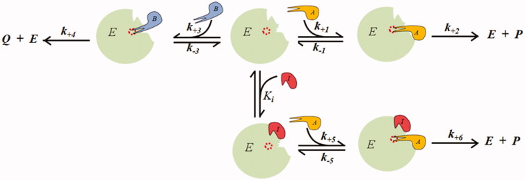
As classical inhibitors, differential inhibitors may intervene in catalysis through different models of action16. A schematic representation of a competitive inhibition model for a DI, the most adequate to express a differential inhibitory action, is given in Figure 1.
Figure 1.
Competitive model of an intra-site differential inhibitor.
Here, only the transformation of the substrate B into the product Q is susceptible to inhibition, while the transformation of the substrate A into the product P is unaffected by the inhibitor. When analysed in steady state conditions, the inhibition model of Figure 1 can be described by two rate equations16. The generation rate of product Q is given by Equation 2, while Equation 3 gives the generation rate of product P; here, an apparent increase rather than a decrease in the reaction rate is observed in the presence of the DI.
| (3) |
The differential action of the DI on the transformation of substrate B will relieve the competitive inhibitory action substrate B exerts on the transformation of substrate A. This gain in the A rate transformation will increase with the DI concentration. The direct inhibitory effect on the B transformation and the indirect enhancing effect on the A transformation exerted by a competitive DI acting in the presence of the two substrates are reported in Figure 2(A,B), respectively. The progressive increase of differential inhibition as a function of DI concentration observed when [A] = [B] = 2KM is reported in Figure 2(C).
Figure 2.
Predicted effect of a competitive DI on the transformation of two substrates catalysed by a multi-specific enzyme. The kinetic behaviour is described by the model reported in Figure 1 with the two substrates displaying the same KM (i.e. KA = KB = 5 concentration units) and the same kcat (100 time−1 units) and is represented by the Hanes–Wolff plot. Panel A: the transformation of substrate B, which is susceptible to inhibition, is analysed. The red open and closed circles refer to substrate B present alone and in the presence of A at a concentration equal to 2KM, respectively. Symbols ▪, ▲, ♦ and ▼refer to red closed circles in the presence of [I] equal to 1, 2, 3, and 5 times the Ki value, respectively. The Ki value is considered as KM/10. Panel B: the transformation of the substrate A, which is insensitive to direct inhibition, is analysed. Blue open and closed circles refer to substrate A alone and in the presence of B at a concentration equal to 2KM, respectively. Symbols ▪, ▲, ♦ and ▼ refer to blue closed circles in the presence of [I] equal to 0.5, 1, 2, and 5 times the Ki value, respectively. The Ki value is considered as KM/10. In Panels A and B, the increase in [I] is emphasised by the red arrows. In Panel C, the B rate transformation (red curve) and A rate transformation (blue curve) are reported as a function of the inhibitor concentration expressed as Ki fold. The substrate concentrations were considered fixed at the 2KM value and the Ki value is considered as KM/10. The dotted line refers to the differential inhibition.
The competitive inhibitor for substrate B may also be able to partially inhibit the transformation of the competing substrate A. The model in Figure 1 can also describe this, in which the ternary complex EIA evolves into a product with a kinetic constant k+6 lower than k+2.
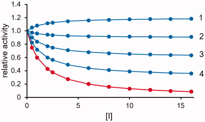
The effect of this situation is that the differential inhibition is progressively reduced along with the increase in difference between k+6 and k+2, as shown in Figure 3. As mentioned above, the competitive type of inhibition is the most effective and theoretically is the only model in which an inhibitor can be a “complete” differential inhibitor. Non-competitive inhibition requires the generation of a ternary complex ESI. This implies a reduction in the catalyst available for catalysis, which will also indirectly affect the reaction that is preserved. A differential inhibitory action, although incomplete, may also occur for non-competitive models of inhibition, depending on the kinetic parameters of the transformation of the two substrates.
Figure 3.
Effect of a competitive DI acting as a partial inhibitor of A transformation. The transformation rate of substrate B (red curve) and substrate A (blue curves) (see Figure 1) as a function of the inhibitor concentration (expressed as Ki fold) is reported. The substrate concentration is considered fixed at the KM value (KA = KB). Curves 1, 2, 3 and 4 refer to k+6 values equal to 0.8, 0.6, 0.4 and 0.2 times the k+2 value, respectively. The Ki value is considered as KM/10.
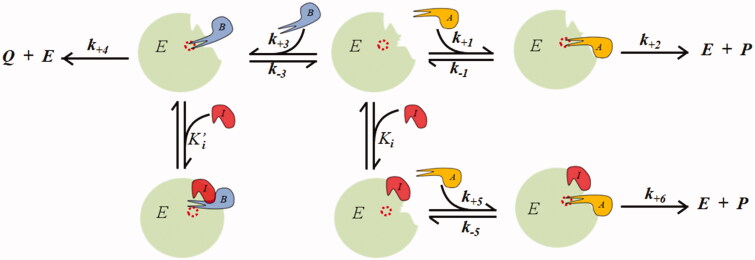
This can be rationalised through the model shown in Figure 4, in which the DI acts on the B transformation as a non-competitive inhibitor. Here, a steady state kinetic equation can be derived (Equation 4), which also describes the influence of the inhibitor on the reaction to be preserved (A transformation), which depends on the inhibitory features of the B transformation.
| (4) |
| (5) |
Figure 4.
Non-competitive model of an intra-site differential inhibitor.
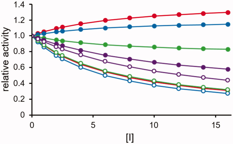
Computer simulations of Equations 4 and 5 are reported in Figure 5. The K′i/Ki ratio, which expresses the relative efficiency of the inhibitor in targeting the complex EB with respect to the free enzyme, clearly modulates the engagement of A in the inhibition process. Thus, the inhibitor progressively loses its character of DI, extending its action on the transformation of the substrate A.
Figure 5.
Effect of a non-competitive DI. The rate transformation of substrate B (open circles, see Equation (5) and substrate A (closed circles, see Equation (4) according to the model of Figure 4 are reported as a function of the inhibitor concentration (expressed as lowest Ki fold). The substrates concentration is considered fixed at the KM value (KA = KB). The following combinations of inhibition constants are considered: red curves: Ki= KM/10 and K′i= KM; blue curves Ki= KM/10 and K′i = KM/5; green curves Ki= KM/5 and K′i= KM/10; violet curves Ki= KM and K′i= KM/10.
The experimental measurements of the inhibition kinetic parameters that were independently conducted on the two substrates (as usually occurs in inhibitor screening) refer to “apparent” inhibitory constants (i.e. appKi, appK′i), whose relative values may lead to apparent incongruities. For example, if the experimental data indicate a mixed type of inhibition for substrate B and an uncompetitive inhibition for substrate A, as in neohesperidin dihydrochalcone (NHDC), which has been assessed as an inhibitor of the reduction of L-idose (substrate B) and HNE (substrate A) catalysed by AKR1B120. The results of the analysis of substrate B shows that the inhibitor may target the free enzyme, and thus this should also result from the analysis of substrate A. However, the data suggest that the inhibitor targets only the EA complex. This apparently contradictory situation can be rationalised although not completely, in the following ways: (i) the measured apparent inhibitory constants can be regarded as true dissociation constants, in which the K′i for substrate A is significantly lower than the Ki measured for substrate B (which is not the case for NHDC); and/or (ii) the measured constants are indeed “apparent” and the ternary complexes EIA, which is generated by adding A to the EI complex, and EAI, which is generated by adding I to the EA complex, are kinetically different and only EIA is able to evolve into products. Thus, once the potential DI has been identified through individual substrate analysis, it will be necessary to proceed analysing the simultaneous presence of the competing substrates for a conclusive evaluation of the differential inhibitory ability of the selected molecule.
DIs must exhibit more restrictive features than classical inhibitors. The strategic approach to searching for classical inhibitors is typically to disclose molecules able to specifically intervene on the target enzyme, and that interact with as many groups as possible at the active site. These are crucial for recognition and/or catalysis. This approach is often supported by molecular modelling and diffraction studies on the complex enzyme/inhibitor and is aimed at the improvement of both selectivity towards the target enzyme and inhibition potency. A very powerful inhibitor binding capacity at the active site is a very poor feature for a DI as it must limit the number of interactions, and for the substrate that is not expected to be affected it must preserve any possible interaction that can help enable its transformation. This consideration suggests that a significant inhibition potency cannot generally be an expected feature for a DI.
Different features in the bindings of the competing substrates may not enable the inhibitor to intervene only on the transformation of one of them. Therefore, it is possible that inhibition may indeed take place, although only partially or with different models of action, on the two different substrates. Thus, although molecules considered as inhibitors in the above examples cannot be defined as “complete” DIs, they still discriminate, at a mechanistic level, the different substrate molecules. This offers another instrument, which can be combined with the set of conditions in which the enzymatic reaction is operating, to elicit differential inhibition of one substrate with respect to the other.
Aldose reductase differential inhibition
The multi-specific enzyme on which differential inhibition was first proposed as a potentially useful inhibition approach is AKR1B1 (EC 1.1.1.21). This enzyme is an aldo-keto reductase able to catalyse the NADPH-dependent reduction of numerous aldehydic compounds. AKR1B1 is usually presented as the first enzyme of the so-called polyol pathway, in which glucose is reduced by the enzyme to sorbitol, which is in turn transformed into fructose through a NAD+-dependent oxidation catalysed by sorbitol dehydrogenase21. Due to the relatively poor affinity the enzyme shows towards glucose22–26, it is debateable whether glucose can be considered as the native substrate for AKR1B1. However, although the polyol pathway is not a highly efficient metabolic route for glucose metabolism, it has been linked to the cellular osmotic control mediated by intracellular sorbitol levels27,28. The situation changes in hyperglycaemic conditions, where the flux of the pathway dramatically increases, thus making it a co-causative factor in the aetiology of secondary diabetic complications. The accumulation of sorbitol and the consequent osmotic unbalance, together with an alteration of the proper redox status of both NAD and NADP cofactors and the accumulation of the potent glycating agent fructose, lead to cell damage29–32. Thus, AKR1B1 has been extensively studied in terms of its inhibition33–38.
In addition to its involvement in the polyol pathway, AKR1B1 has been recognised as fulfilling a detoxifying role, as it can efficiently reduce various toxic hydrophobic aldehydes, such as alkanals and alkenals generated through membrane lipid peroxidation as a result of oxidative stress39–42. Glutathionylated alkenals, generated through the action of glutathione S-transferases43,44, are also substrates for AKR1B115,45. These compounds, devoid of the reactive double bond and with more hydrophilicity than their un-glutathionylated counterparts, represent significant intermediates in the transformation of alkenals.46 Thus, the ability of AKR1B1 to catalyse their reduction appears to accomplish the detoxification. However, the reduction of 3-glutathionyl,4-hydroxynonanal (GSHNE), which is the glutathionyl adduct of HNE and a representative lipid peroxidative product, generates 3-glutathionyl-2,4-dihydroxynonane. This molecule has been reported to activate the NF-kB signalling cascade, thus promoting inflammation47–49. This may well represent the base of the anti-inflammatory action reported for a numbers of aldose reductase inhibitors (ARIs)50.
The multifaceted activities of AKR1B1 have antagonistic effects that depend on the substrate undergoing reduction, and drug development from ARIs has been relatively poor when compared to the in vitro discovery of selective and potent inhibitors of the enzyme. Thus, we propose a new strategy to approaching AKR1B1 inhibition15 and use terms such as “aldose reductase differential inhibitors” (ARDIs) and “intra site differential inhibition” to refer to molecules and conditions, respectively, which are able to determine an inhibition of the enzyme depending on the nature of the substrate the enzyme is working on. Within this frame, useful ARDIs include molecules able to intervene on glucose reduction, leaving unaltered, or less affected, the detoxifying activity of the enzymes towards hydrophobic aldehydes. Molecules able to differentially inhibit the reduction of GSHNE with respect to toxic hydrophobic aldehydes may be valuable, because of the inflammatory potential associated with GSHNE reduction.
Although a “complete” intra-site differential inhibitor as defined above has not as yet been envisaged for AKR1B1, molecules able to differentially inhibit aldoses reduction and/or GSHNE reduction versus HNE reduction have been proposed. The typical basic conditions to illustrate in vitro the differential inhibition of AKR1B1 mimic those occurring in a hyperglycaemic status, with the aldose substrates kept at the mM level, and the toxic aldehydes (i.e., HNE) or their glutathionyl-derivatives (i.e., GSHNE) kept at the µM level. D,L-glyceraldehyde, the most common substrate used in AKR1B1 inhibition studies, was also utilised as a substrate in differential inhibition studies. However, the evidence of incomplete inhibitory action exerted on the enzyme activity by aldose hemiacetals51,52, which cannot take place with a triose, suggested the use of an aldohexose as the substrate. Thus, L-idose, an epimer of D-glucose at C5, with a free aldehyde form approximately 80 times higher than what was observed for glucose, was chosen as elective substrate for inhibition studies53. Finally, the problem of poor solubility of molecules often encountered in inhibition studies of AKR1B1 was overcome by using a proper aqueous cocktail of either methanol or dimethyl sulfoxide, after evaluating the limits of their suitability for the enzyme assay20. In these conditions, supported by kinetic analysis of the inhibitory action towards the different substrates, a number of molecules, coming both from organic synthesis and from natural sources, were identified as having differential inhibitory abilities.
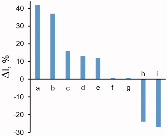
Thus, chemically synthesised compounds such as D-glyceramide or D-gluconamide were found to be inhibitors of the bovine lens enzyme15, and differentially inhibit the glyceraldehyde reduction versus the HNE reduction. D-glyceramide was also differentially active towards GSHNE and, to a lesser extent, towards D-glucose reduction. A differential inhibitory action on the human recombinant AKR1B1 towards L-idose and GSHNE reduction versus HNE reduction was also reported for some pyrazolepyrimidine derivatives54. The differential effects of selected compounds on AKR1B1 activity, derived from either organic synthesis or from natural sources, are summarised in Figure 6. Data relative to classical ARIs are also included as negative controls. Examples of molecules preferentially inhibiting HNE reduction over glyceraldehyde reduction are also reported.
Figure 6.
Differential inhibition of AKR1B1 by synthetic and natural compounds. The bars refer to the differential inhibition between aldoses reduction and HNE reduction. The letters along the bars refer to: a: D-glyceramide15; b: D-gluconamide15; c: pyrazol[1,5-a]pyrimidine derivative (compound 5e52); d: soyasaponin55; e: NHDC20; f: sorbinil52; g: epalrestat52; h: [1,2,4]oxadiazol-5-yl-acetic acid derivative (compound 915); i: Saccharin derivative (compound 1615).
Natural sources represent an almost limitless resource of biomolecules that can potentially affect the activity of enzymes, and this obviously also applies to the present inhibitory approach. Components exhibiting differential inhibitory ability were found in the fractionation of methanolic extracts of some edible vegetables55. The Zolfino bean, a variety of Phaseolus vulgaris, has been revealed to be a very promising source of ARDIs. After the bean was characterised in terms of classical AKR1B1 inhibitory activity56, components identified as soya saponins were isolated and shown to differentially inhibit to some extent L-idose reduction versus HNE reduction57.
Conclusions
The ability of enzymes to convert molecules that are structurally different through the same type of reaction, and the possibility of the same enzyme catalysing different reactions, may require the modulation of the enzyme activity depending on the substrate undergoing transformation. Thus, the “differential inhibitors” analysed in this work are the instruments that can specifically address the catalytic potential of multi-specific or promiscuous enzymes towards one of their possible multiple functions.
Although no “complete” differential inhibitors of the multi-specific enzyme AKR1B1 have as yet been disclosed, knowledge of the interactive features and structural restraints a molecule needs to act as an ARDI is increasing. Studies conducted up to now on ARIs, in which ARDIs may have been disregarded, can be revisited, as an advantageous starting point to further this novel inhibition approach. Positive results for this enzyme target will also more generally confirm the potential of this intriguing inhibition approach for multi-specific or promiscuous enzymes.
Funding Statement
This work was supported by Pisa University.
Disclosure statement
No potential conflict of interest was reported by the author(s).
References
- 1.Skidgel RA, Erdös EG.. The broad substrate specificity of human angiotensin I converting enzyme. Clin Exp Hypertens 1987;9:243–59. [DOI] [PubMed] [Google Scholar]
- 2.Park KH, Kim TJ, Cheong TK, et al. . Structure, specificity and function of cyclomaltodextrinase, a multispecific enzyme of the alpha-amylase family. Biochim Biophys Acta 2000;1478:165–85. [DOI] [PubMed] [Google Scholar]
- 3.Chakraborty S, Asgeirsson B, Rao BJ.. A Measure of the broad substrate specificity of enzymes based on ‘duplicate’ catalytic residues. PLOS One 2012;7:e49313. [DOI] [PMC free article] [PubMed] [Google Scholar]
- 4.Khersonsky O, Roodveldt C, Tawfik DS.. Enzyme promiscuity: evolutionary and mechanistic aspects. Curr Opin Chem Biol 2006;10:498–508. [DOI] [PubMed] [Google Scholar]
- 5.Hult K, Berglund P.. Enzyme promiscuity: mechanism and applications. Trends Biotechnol 2007;25:231–7. [DOI] [PubMed] [Google Scholar]
- 6.Copley SD. Enzymes with extra talents: moonlighting functions and catalytic promiscuity. Curr Opin Chem Biol 2003;7:265–72. [DOI] [PubMed] [Google Scholar]
- 7.Huberts D, van der Klei IJ.. Moonlighting proteins: an intriguing mode of multitasking. Biochim Biophys Acta 2010;1083:520–5. [DOI] [PubMed] [Google Scholar]
- 8.O’Brien PJ, Herschlag D.. Catalytic promiscuity and the evolution of new enzymatic activities. Chem Biol 1999;6:R91–RlO5. [DOI] [PubMed] [Google Scholar]
- 9.Aharoni A, Gaidukov L, Khersonsky O, et al. . The ‘evolvability’ of promiscuous protein functions. Nat Genet 2005;37:73–6. [DOI] [PubMed] [Google Scholar]
- 10.Khersonsky O, Tawfik DS.. Enzyme promiscuity: a mechanistic and evolutionary perspective. Annu Rev Biochem 2010;79:471–505. [DOI] [PubMed] [Google Scholar]
- 11.Erijman A, Aizner Y, Shifman JM.. Multispecific recognition: mechanism, evolution, and design. Biochemistry 2011;50:602–11. [DOI] [PubMed] [Google Scholar]
- 12.Levin M, Amar D, Aharoni A.. Employing directed evolution for the functional analysis of multi-specific proteins. Bioorg Med Chem 2013;21:3511–6. [DOI] [PubMed] [Google Scholar]
- 13.Del-Corso A, Cappiello M, Moschini R, et al. . How the chemical features of molecules may have addressed the settlement of metabolic steps. Metabolomics 2018;14:2. [DOI] [PubMed] [Google Scholar]
- 14.Del-Corso A, Cappiello M, Moschini R, et al. . The furanosidic scaffold of D-ribose: a milestone for cell life. Biochem Soc Trans 2019;47:1931–40. [DOI] [PubMed] [Google Scholar]
- 15.Del-Corso A, Balestri F, Di Bugno E, et al. . A new approach to control the enigmatic activity of aldose reductase. PLOS One 2013;8:e74076. [DOI] [PMC free article] [PubMed] [Google Scholar]
- 16.Cappiello M, Moschini R, Balestri F, et al. . Basic models for differential inhibition of enzymes. Biochem Biophys Res Commun 2014;445:556–60. [DOI] [PubMed] [Google Scholar]
- 17.Berger I, Guttman C, Amar D, et al. . The molecular basis for the broad substrate specificity of human sulfotransferase 1A1. PLOS One 2011;6:e26794. [DOI] [PMC free article] [PubMed] [Google Scholar]
- 18.Gonzalez-Villalobos RA, Shen XZ, Bernstein EA, et al. . Rediscovering ACE: novel insights into the many roles of the angiotensin-converting enzyme. J Mol Med (Berl) 2013;91:1143–54. [DOI] [PMC free article] [PubMed] [Google Scholar]
- 19.Del-Corso A, Cappiello M, Mura U.. From a dull enzyme to something else: facts and perspectives regarding aldose reductase. Curr Med Chem 2008;15:1452–61. [DOI] [PubMed] [Google Scholar]
- 20.Misuri L, Cappiello M, Balestri F, et al. . The use of dimethylsulfoxide as a solvent in enzyme inhibition studies: the case of aldose reductase. J Enz Inhib Med Chem 2017;32:1152–8. [DOI] [PMC free article] [PubMed] [Google Scholar]
- 21.Kinoshita JH. A thirty year journey in the polyol pathway. Exp Eye Res 1990;50:567–73. [DOI] [PubMed] [Google Scholar]
- 22.(a) Cappiello M, Voltarelli M, Giannessi M, et al. . Glutathione dependent modification of bovine lens aldose reductase. Exp Eye Res 1994;58:491–501. [DOI] [PubMed] [Google Scholar]; (b) Hayman S, Kinoshita JH.. Isolation and properties of lens aldose reductase. J Biol Chem 1965;240:877–82. [PubMed] [Google Scholar]
- 23.Petrash JM, Harter TM, Devine CS, et al. . Involvement of cysteine residues in catalysis and inhibition of human aldose reductase. Site-directed mutagenesis of Cys-80, -298, and -303. J Biol Chem 1992;267:24833–40. [PubMed] [Google Scholar]
- 24.Bhatnagar A, Liu SQ, Ueno N, et al. . Human placental aldose reductase: role of Cys-298 in substrate and inhibitor binding. Biochim Biophys Acta 1994;1205:207–14. [DOI] [PubMed] [Google Scholar]
- 25.Gabbay KH, Tze WJ.. Inhibition of glucose-induced release of insulin by aldose reductase inhibitors. Proc Natl Acad Sci USA 1972;69:1435–9. [DOI] [PMC free article] [PubMed] [Google Scholar]
- 26.Vander Jagt DL, Hunsaker LA, Robinson B, et al. . Aldehyde and aldose reductases from human placenta. Heterogeneous expression of multiple enzyme forms. J Biol Chem 1990;265:10912–8. [PubMed] [Google Scholar]
- 27.Yancey P, Clark M, Hand S, et al. . Living with water stress: evolution of osmolyte systems. Science 1982;217:1214–22. [DOI] [PubMed] [Google Scholar]
- 28.Bagnasco S, Balaban R, Fales HM, et al. . Predominant osmotically active organic solutes in rat and rabbit renal medullas. J Biol Chem 1986;261:5872–7. [PubMed] [Google Scholar]
- 29.Yabe-Nishimura C. Aldose reductase in glucose toxicity: a potential target for the prevention of diabetic complications. Pharmacol Rev 1998;50:21–33. [PubMed] [Google Scholar]
- 30.Oyama T, Miyasita Y, Watanabe H, Shirai K.. The role of polyol pathway in high glucose-induced endothelial cell damages. Diabetes Res Clin Pract 2006;73:227–34. [DOI] [PubMed] [Google Scholar]
- 31.Lorenzi M. The polyol pathway as a mechanism for diabetic retinopathy: attractive, elusive, and resilient. Exp Diabetes Res 2007;2007:61038. [DOI] [PMC free article] [PubMed] [Google Scholar]
- 32.Alexiou P, Pegklidou K, Chatzopoulou M, et al. . Aldose reductase enzyme and its implication to major health problems of the 21st century. Curr Med Chem 2009;16:734–52. [DOI] [PubMed] [Google Scholar]
- 33.Maccari R, Ottanà R.. Targeting aldose reductase for the treatment of diabetes complications and inflammatory diseases: new insights and future directions. J Med Chem 2015;58:2047–67. [DOI] [PubMed] [Google Scholar]
- 34.Grewal AS, Bhardwaj S, Pandita D, et al. . Updates on aldose reductase inhibitors for management of diabetic complications and non-diabetic diseases. Mini Rev Med Chem 2016;16:120–62. [DOI] [PubMed] [Google Scholar]
- 35.ElGamal H, Munusamy S.. Aldose reductase as a drug target for treatment of diabetic nephropathy: promises and challenges. Protein Pept Lett 2017;24:71–7. [DOI] [PubMed] [Google Scholar]
- 36.Chatzopoulou M, Pegklidou K, Papastavrou N, Demopoulos VJ.. Development of aldose reductase inhibitors for the treatment of inflammatory disorders. Expert Opin Drug Discov 2013;8:1365–80.24090200 [Google Scholar]
- 37.Colín-Lozano B, Estrada-Soto S, Chávez-Silva F, et al. . Design, synthesis and in combo antidiabetic bioevaluation of multitarget phenylpropanoic acids. Molecules 2018;23. [DOI] [PMC free article] [PubMed] [Google Scholar]
- 38.Maccari R, Del-Corso A, Paoli P, et al. . An investigation on 4-thiazolidinone derivatives as dual inhibitors of aldose reductase and protein tyrosine phosphatase 1B, in the search for potential agents for the treatment of type 2 diabetes mellitus and its complications. Bioorg Med Chem Lett 2018;28:3712–20. [DOI] [PubMed] [Google Scholar]
- 39.Srivastava S, Chandra A, Bhatnagar A, et al. . Lipid peroxidation product, 4-hydroxynonenal and its conjugate with GSH are excellent substrates of bovine lens aldose reductase. Biochem Biophys Res Commun 1995;217:741–6. [DOI] [PubMed] [Google Scholar]
- 40.Vander Jagt DL, Kolb NS, Vander Jagt TJ, et al. . Substrate specificity of human aldose reductase: identification of 4-hydroxynonenal as an endogenous substrate. Biochim Biophys Acta 1995;1249:117–26. [DOI] [PubMed] [Google Scholar]
- 41.Srivastava S, Watowich SJ, Petrash JM, et al. . Structural and kinetic determinants of aldehyde reduction by aldose reductase. Biochemistry 1999;38:42–54. [DOI] [PubMed] [Google Scholar]
- 42.Grimshaw CE. Aldose reductase: model for a new paradigm of enzymic perfection in detoxification catalysts. Biochemistry 1992;31:10139–45. [DOI] [PubMed] [Google Scholar]
- 43.Hubatsch I, Ridderstrom M, Mannervik B.. Human glutathione transferase A4-4: An alpha class enzyme with high catalytic e_ciency in the conjugation of 4-hydroxynonenal and other genotoxic products of lipid peroxidation. Biochem J 1998;330:175–9. [DOI] [PMC free article] [PubMed] [Google Scholar]
- 44.Hou L, Honaker MT, Shireman LM, et al. . Functional promiscuity correlates with conformational heterogeneity in A-class glutathione S-transferases. J Biol Chem 2007;282:23264–74. [DOI] [PubMed] [Google Scholar]
- 45.Ramana KV, Dixit BL, Srivastava S, et al. . Selective recognition of glutathiolated aldehydes by aldose reductase. Biochemistry 2000;39:12172–80. [DOI] [PubMed] [Google Scholar]
- 46.Esterbauer H, Schaur RJ, Zollner H.. Chemistry and biochemistry of 4-hydroxynonenal, malondialdehyde and related aldehydes. Free Radic Biol Med 1991;11:81–128. [DOI] [PubMed] [Google Scholar]
- 47.Frohnert BI, Bernlohr DA.. Glutathionylated products of lipid peroxidation. A novel mechanism of adipocyte to macrophage signaling. Adipocyte 2014;3:224–9. [DOI] [PMC free article] [PubMed] [Google Scholar]
- 48.Frohnert BI, Long EK, Hahn WS, Bernlohr DA.. Glutathionylated lipid aldehydes are products of adipocyte oxidative stress and activators of macrophage inflammation. Diabetes 2014;63:89–100. [DOI] [PMC free article] [PubMed] [Google Scholar]
- 49.Srivastava S, Ramana KV, Bhatnagar A, Srivastava SK.. Synthesis, quantification, characterization, and signaling properties of glutathionyl conjugates of enals. Meth Enzymol 2010;474:297–313. [DOI] [PMC free article] [PubMed] [Google Scholar]
- 50.Chang KC, Petrash JM.. Aldo-keto reductases: Multifunctional proteins as therapeutic targets in diabetes and inflammatory disease. Adv Exp Med Biol 2018;1032:173–202. [DOI] [PMC free article] [PubMed] [Google Scholar]
- 51.Balestri F, Cappiello M, Moschini R, et al. . Modulation of aldose reductase activity by aldose hemiacetals. Biochim Biophys Acta 2015;1850:2329–39. [DOI] [PubMed] [Google Scholar]
- 52.Cappiello M, Balestri F, Moschini R, et al. . Apparent cooperativity and apparent hyperbolic behavior of enzyme mixtures acting on the same substrate. J Enz Inhib Med Chem 2016;31:1556–9. [DOI] [PubMed] [Google Scholar]
- 53.Balestri F, Cappiello M, Moschini R, et al. . l-Idose: an attractive substrate alternative to d-glucose for measuring aldose reductase activity. Biochem Biophys Res Commun 2015;456:891–5. [DOI] [PubMed] [Google Scholar]
- 54.Balestri F, Quattrini L, Coviello V, et al. . Acid derivatives of pyrazolo[1,5-α] pyrimidine as aldose reductase differential inhibitors. Cell Chem Biol 2018;25:1414–8. [DOI] [PubMed] [Google Scholar]
- 55.Balestri F, Sorce C, Moschini R, et al. . Edible vegetables as a source of aldose reductase differential inhibitors. Chem Biol Interact 2017;276:155–9. [DOI] [PubMed] [Google Scholar]
- 56.Balestri F, Rotondo R, Moschini R, et al. . Zolfino landrace (Phaseolus vulgaris L.) from Pratomagno: general and specific features of a functional food. Food Nutr Res 2016;60:31792. [DOI] [PMC free article] [PubMed] [Google Scholar]
- 57.Balestri F, De Leo M, Sorce C, et al. . Soyasaponins from Zolfino bean as aldose reductase differential inhibitors. J Enz Inhib Med Chem 2019;34:350–60. [DOI] [PMC free article] [PubMed] [Google Scholar]


