

Abstract
Lymphedema is a chronic debilitating disease in which impaired drainage of lymphatic fluid causes accumulation of fluid in the soft tissues resulting in a swollen heavy limb. This ultimately leads to severe fibrosis, recurrent infections, non-healing wounds, and a poorly functioning limb that negatively affects a patient’s quality of life. Primary lymphedema is due to abnormal development of the lymphatic system and patients can present with lymphedema at birth or later in life. Secondary lymphedema is caused by damage to the lymphatic system from infection, surgery to treat malignancies, trauma, and obesity. In the past, the only treatment was controlling the swelling to prevent progression of the disease by lymphatic therapy and various types of compression which is still currently the first line treatment. Advances in supermicrosurgery (connecting vessels less than 0.8 mm) have made way for surgical treatment options for lymphedema, including lymphovenous bypass and vascularized lymph node transplant. These new surgical treatment options combined with lymphatic therapy and compression have led to better results and improved patient’s quality of life. After reading this article, the participant should be familiar with diagnosis, imaging, and surgical treatment of lymphedema.
Introduction
Lymphedema is a chronic, progressive, and incurable disease in which there is impaired drainage of interstitial fluid through the lymphatic system, resulting in accumulation of protein-rich fluid in the interstitial space. This fluid accumulation causes an inflammatory response that induces lipogenesis and fat deposition with connective tissue overgrowth. Eventually patients develop irreversible induration and fibrosis of the affected area.1
Primary lymphedema occurs due to structural and functional abnormalities of the lymphatic channels and can become symptomatic at birth, adolescence, or adulthood. Secondary lymphedema develops after disruption of normal lymphatic channels, leading to obstruction of the lymphatic system. The most common cause of secondary lymphedema worldwide is filariasis secondary to infection with Wuscheria bancrofti. In developed countries, however, secondary lymphedema is primarily caused by iatrogenic injury during the treatment of various malignancies including breast, gynecologic, and skin cancers.2 Other causes include traumatic injuries, obesity, and infections.
Regardless of etiology, patients with lymphedema exhibit both physical and psychological morbidity that ultimately leads to a decreased quality of life.3 The affected limb becomes large, swollen, heavy, and painful preventing patients from enjoying hobbies and active lifestyle, exercising, performing activities of daily living, and working which ultimately leads to severe disability. Patients cannot fit into normal clothes and shoes and is a constant painful reminder that they had cancer. As the disease progresses, patients develop recurrent infections and non-healing wounds from even minor trauma requiring intensive wound care, multiple rounds of antibiotics, repeated hospitalization, surgical debridement, and even amputation. This becomes an economic burden on our patients, their families, and our healthcare system.
For many years the first line treatment of lymphedema has been conservative management with complete decongestive therapy (CDT): a combination of compressive garments, skin hygiene, limb compression, manual lymphatic drainage, and exercise. Decongestive lymphatic therapy is labor intensive and requires access to a specialized therapist.4 CDT requires lifelong patient compliance, as it is not curative and requires continued compression to maintain treatment results.5
Multiple surgical options exist for patients whose lymphedema is not adequately controlled with conservative therapy. Debulking procedures such as liposuction and subcutaneous excision decrease extremity size in order to facilitate hygiene and improve functional status.6 Recent advances in microsurgical techniques have made way for physiologic procedures including lymphovenous bypass and vascularized lymph node transfer which attempt to re-establish normal lymphatic physiology.
Clinical Evaluation
Initial patient evaluation should include a full history and physical exam. Other etiologies of limb edema such as congestive heart failure, renal failure, deep vein thrombosis, and venous insufficiency should be excluded. Venous duplex should be considered to rule out venous incompetence. Physical exam should include a thorough evaluation of the quality of the skin and soft tissue in the affected limb. Particular attention should be paid to the presence of peau d’orange, which indicates soft tissue fibrosis, and Stemmer sign, the inability to grasp the skin overlying the second digit of the foot, as these signs favor a diagnosis of lymphedema over other etiologies of limb swelling.1 Serial limb circumference should be measured and compared to the contralateral side, as this can be helpful in monitoring treatment response.
The International Society of Lymphedema has described a staging system for the clinical assessment of lymphedema that is a helpful guide in determining the appropriate treatment options for patients presenting with lymphedema.
Stage 0: Impaired lymph transport, subtle alterations in tissue and fluid composition. Patients may report symptoms, but swelling is not present.
Stage I: Early accumulation of interstitial fluid that is high in protein. Swelling is present but improves with elevation. Patients may have pitting edema.
Stage II: Swelling is present and does not improve with elevation. Early on, pitting edema will be present. Later in stage II, pitting will not be present as soft tissue fibrosis develops.
Stage III: Pitting is absent, trophic skin changes develop, further deposition of fat and fibrosis (lymphostatic elephantiasis).7
Imaging
Imaging of the lymphatic system is a vital tool in the assessment of lymphedema, as a variety of morphologic changes in the lymphatic system can lead to similar clinical presentations. Indocyanine green lymphography and radionuclide lymphoscintigraphy are essential to the lymphatic surgeon for surgical planning as well as intraoperative visualization of lymphatic channels that are invisible to the naked eye.
Indocyanine Green Lymphography
Indocyanine green (ICG) lymphography is a minimally invasive imaging technique that allows dynamic visualization of lymphatic channels without radiation exposure. ICG has been used for decades across a number of specialties for evaluation of hepatic function, sentinel lymph node detection, cardiac output, and flap viability.8 Its use in evaluation of lymphedema was initially described by Ogata et al. in 2007 and it has since become an irreplaceable tool for real-time assessment of lymphatic channels during lymphatic surgery.9
ICG lymphography is performed by injecting ICG subdermally into the interdigital webspaces of the lymphedematous extremity. Near-infrared light emitted by ICG dye is detected with an infrared camera system. The location, path, and flow of superficial lymphatic channels up to 2cm deep from the skin surface can be detected immediately after injection of ICG.8
Findings can be categorized into linear or dermal backflow patterns. Linear patterns correspond to normally functioning lymphatic channels and can be seen in patients with mild clinical findings of lymphedema. Figure 1. Dermal backflow patterns represent nonlinear, pathologic backflow of lymphatic fluid and are seen in more severe cases of lymphedema.10 Dermal backflow patterns can be further divided into splash, stardust, and diffuse patterns, which correspond to increasing severity of disease and increased fibrosis in the lymphatic channels.4
Figure 1.

Preoperative left upper extremity ICG lymphography demonstrating normal linear pattern in the distal forearm and hand (*) and abnormal dermal backflow in the proximal forearm (**) in a patient with secondary lymphedema after treatment for left breast cancer. The blue lines represent incisions for lymphovenous bypass to shunt lymphatic fluid into veins upstream from the blockage.
Radionuclide Lymphoscintigraphy
Lymphoscintigraphy is considered the gold standard for evaluation of lymphedema and is frequently used to confirm a clinical suspicion of lymphedema. A Technetium-99 (Tc-99) radiotracer is injected subdermally into the first or second interdigital web space of the affected extremity and uptake is assessed using a high resolution gamma camera.11 Lymphoscintigraphy gives providers the ability to measure contrast uptake, visualize number and size of lymphatic vessels and nodes, and assess dermal backflow patterns.12 Lymphatic dysfunction is characterized by delayed, asymmetric, or absent visualization of lymph nodes and lymphatic channels, collateral lymphatic channels, interrupted vascular structures, visualization of the deep lymphatic system, or the presence of dermal back flow.13
Surgical Management
Surgical management of lymphedema can be separated into physiologic procedures, which attempt to re-establish normal lymphatic physiology by augmenting clearance of lymphatic fluid, and ablative procedures, which remove excess subcutaneous tissue to improve patient comfort and facilitate conservative therapies.
Lymphovenous Anastomosis/Bypass
Lymphovenous anastomosis (LVA) is a physiologic microsurgical procedure in which distal lymphatic channels are anastomosed to nearby subdermal veins, allowing bypass of proximal lymphatic obstruction by shunting lymphatic fluid into the venous system. Successful LVA begins with patient selection. Patients with International Society of Lymphedema stage I or II lymphedema who have been compliant with conservative measures but have showed minimal improvement are considered good candidates for LVA.14,15
ICG lymphography is performed preoperatively to identify suitable functioning lymphatic channels and guide placement of incisions. Lymphatic channels that are amenable to LVA demonstrate dynamic lymphatic flow that occurs distally to proximally.14 Local anesthesia is infiltrated into the operative field and isosulfan blue is injected distal to the planned incision. Figure 2. The isosulfan blue stains the lymphatic channels so they may be readily visualized during the dissection. From this point onward, the procedure is performed under microscope magnification. The skin is carefully incised and lymphatic channels are identified within the subcutaneous tissue. Once a satisfactory lymphatic channel has been dissected and transected proximally, a nearby venule with appropriate size match is selected. End-to-end, end-to-side, or side-to-end anastomoses may be performed depending on the size, pressure, and availability of subdermal venules. Lymphatic channels are frequently 0.5–0.8mm in size, making surgeon skill, careful tissue handling, and microsurgical technique paramount to success.16,17 The vessel may be floated open with heparinized saline or stented with a 6–0 prolene suture in order to avoid back-walling and ensure full-thickness bites are taken.14 Patency can be confirmed immediately after the anastomosis is completed with ICG lymphography or isosulfan blue injection; fluorescence or dye should flow from the lymphatic duct into the anastomosed venule. Approximately three to four lymphovenous anastomoses are performed per affected limb. Figure 3.
Figure 2.

End-to-side lymphovenous anastomosis (end of lymphatic vessel into the side of a vein) with isosulfan blue dye confirming patency. Each square on the blue background is 1 x 1 mm.
Figure 3.

End-to-side lymphovenous anastomosis (left). Patency is confirmed with ICG lymphography (right). Fluorescence is noted flowing from the lymphatic vessel into the anastomosed venule.
Postoperatively patients are placed in a compression bandage and typically discharged on the day of surgery or admitted overnight for observation. Patients are encouraged to walk but must abstain from vigorous physical activity. At two weeks the compression bandage is transitioned to a compression garment once the incisions are healed. Lymphatic massage is reinitiated at four weeks postoperatively.
Outcomes after LVA have shown promising results in symptoms and disease severity, particularly in upper extremity lymphedema. In a prospective study of 100 patients undergoing LVA, Chang et al. demonstrated symptomatic improvement in 96 percent of patients with upper extremity lymphedema.15 The same study showed a significant postoperative limb volume reduction at three, six, and 12 months after LVA.15 Table 1. LVA has consistently been shown to significantly decrease episodes of cellulitis in the affected limb.17,18 Disadvantages to LVA include technical difficulty and a paucity of long term outcomes in the literature. Patients should be counseled that LVA is not curative and that fibrosis or failure of the anastomosis could lead to disease relapse. Figure 4.
Table 1.
Change in limb volume after left arm lymphovenous anastomosis. Limb volume of the affected limb had decreased significantly and was comparable to the unaffected limb by 7 weeks postoperatively.

Figure 4.
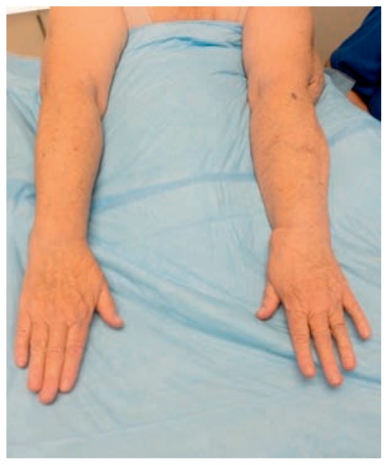
Bilateral upper extremities six months after left upper extremity lymphovenous anastomosis in the same patient. Arm circumference of the affected (left) and unaffected (right) limbs were equivalent.
Vascularized Lymph Node Transfer
In patients whose native lymph node basins are dysfunctional and lymphatic channels are no longer available for LVA, vascularized lymph node transfer (VLNT) may be considered to augment lymphatic drainage of the affected area. The exact mechanism by which VLNT improves lymphedema is still debated, but two leading theories have been described. The first is that transferred lymph nodes act as a sponge that absorbs local lymphatic fluid and redirects it into the vascular system.19 The second theory is that transferred lymph nodes produce vascular endothelial growth factor-C (VEGF-C) which induces local lymphangiogenesis by formation of spontaneous efferent and afferent connections between the transferred nodes and the recipient site.20
VLNT should be considered in patients with significant dermal backflow with no functioning lymphatic vessels on imaging, those with International Society of Lymphedema stage II lymphedema, and those who have not improved after at least 12 months of CDT.4,21 In our practice, we will offer VLNT in patients who do not improve and continue to have pitting edema after LVA or in combination with LVA in patients with more advanced disease. Often these patients have undergone lymph node dissections or radiation for treatment of breast or pelvic cancers. The most common donor site for VLNT is the groin, but harvest of nodes from the lateral thoracic, supraclavicular, submental, and omental systems have been described.19 VLNT using groin and supraclavicular donor sites will be discussed below.
VLNT from the groin is based on the superficial circumflex iliac system or a small medial branch of the femoral artery.22 Dissection should stay superficial to the deep thigh fascia, superior to the groin crease, and lateral to the femoral artery in order to avoid harvesting nodes that are critical to lower extremity lymphatic drainage.23 The groin lymph node flap may be transferred alone or in continuity with abdominal flaps in autologous breast reconstruction, making it an ideal donor for secondary lymphedema of the upper extremity in the setting of breast cancer.23
When transferring supraclavicular lymph nodes, the right side is preferred so as to avoid the thoracic duct on the left. The nodal basin is bound by the sternocleidomastoid muscle medially, the clavicle inferiorly, and the external jugular vein laterally.24 The flap is elevated on the transverse cervical artery. The supraclavicular nodes can be transferred with a skin paddle, which can be useful in patients with radiation skin changes who also require contracture releases at the time of VLNT. Advantages to the supraclavicular VLNT include flap reliability, inconspicuous scar, and a low risk for donor site morbidity and secondary lymphedema.24
There continues to be controversy surrounding whether orthotopic (i.e. groin, axilla) or heterotopic (i.e. elbow, wrist, knee, ankle) recipient site provides the best outcomes. Until now no study has confirmed superior functional outcomes of one method over the other. Advocates of orthotopic placement report advantages including concurrent removal of scar tissue from the recipient site and a better cosmetic outcome.25 Orthotopic placement may make drainage of lymphatic fluid from the hand or forearm more difficult, as the lymph nodes must work against distance and gravity to drain excess lymphatic fluid. In addition, these beds are frequently scarred and irradiated, making dissection technically more challenging. Although heterotopic recipient sites leave a more visible scar, they may be favorable over orthotopic recipient sites in patients who have had prolonged or severe symptoms.22
A dreaded complication of VLNT is the development of secondary lymphatic dysfunction at the donor site. Careful dissection and knowledge of anatomy is essential in avoiding the harvest of lymph nodes that are critical to donor site drainage. Some have advocated reverse lymph node mapping prior to VLNT to identify and protect nodes that are essential to lymph drainage of the donor site.25 However, rates of iatrogenic donor site lymphedema after VLNT appear to be quite low.22
VLNT has been shown to improve overall quality of life and functional status while decreasing skin infection rates and need for CDT.3,22 Significant limb volume reduction of upper and lower extremities after VLNT has been consistently seen in the literature.19,21,22,26 Patients must be counseled on the risk of donor site morbidity and the need for staged revision or debulking procedures in the future.
Ablative Procedures
Ablative procedures remove excess skin and subcutaneous tissue from the affected limb in order to decrease limb volume, improve functional status, and facilitate CDT.6 These procedures may be considered in patients with late stage II and stage III lymphedema who are not candidates for physiologic procedures due to excess adipose deposition and fibrosis in the affected extremity. These techniques are also used after physiologic procedures to address the fat and fibrosis component of the lymphedematous extremity.
Liposuction is minimally invasive, low risk, and is unlikely to further compromise lymphatic drainage of the affected limb.27 Significant limb volume reduction is seen almost immediately in the postoperative period, but liposuction is not curative and patients must continue CDT and lifelong compression to maintain results. Suction-assisted lipectomy may also be used in conjunction with LVA or VLNT to reduce limb size in patients whose lymphatic function has been restored.
The Charles procedure involves radical soft tissue excision to the level of the deep fascia followed by reconstruction with skin grafts. This results in complete disruption of existing distal lymphatic drainage and can lead to exacerbation of distal limb lymphedema.6 Outcomes of the Charles procedure are aesthetically poor. Complications including graft breakdown, chronic ulceration, and hypertrophic scarring may be seen.28
Staged subcutaneous excision is a debulking procedure in which excess subcutaneous tissue is excised from a lymphedematous limb while nerves, blood vessels, and skin are preserved.28 It is less radical than the Charles procedure and does not require skin grafting for wound closure. Although excision procedures carry high morbidity, they may be beneficial in restoring mobility and limb function in patients with devastating advanced disease.
Conclusion
Lymphedema is a chronic and incurable disease that has been shown to decrease function and quality of life. Recent advances in the field of lymphatic surgery have given new hope to patients who suffer from this debilitating disease. Increased patient satisfaction, limb volume reduction, and a decreased need for CDT has been seen with LVA and VLNT. Ablative procedures in combination with CDT continues to be the mainstay of therapy for patients who are not candidates for physiologic procedures. It is critical for all healthcare providers to be comfortable with the diagnosis and treatment of lymphedema so that timely referral to a lymphatic surgeon can be made.
Footnotes
Aurora M. Kareh, MD, is a Resident Physician, Division of Plastic and Reconstructive Surgery, and Kyle Y. Xu, MD, (above), is an Attending Physician, Plastic Surgery, Microsurgery and Hand Surgery, Division of Plastic and Reconstructive Surgery, Department of Surgery, Saint Louis University School of Medicine, St. Louis, Missouri
Contact: kyle.xu@health.slu.edu

Disclosure
None reported.
References
- 1.Warren AG, Brorson H, Borud LJ, Slavin SA. Lymphedema Annals of Plastic Surgery. 2007;59(4):464–472. doi: 10.1097/01.sap.0000257149.42922.7e. [DOI] [PubMed] [Google Scholar]
- 2.Rockson SG, Rivera KK. Estimating the Population Burden of Lymphedema. Annals of the New York Academy of Sciences. 2008;1131(1):147–154. doi: 10.1196/annals.1413.014. [DOI] [PubMed] [Google Scholar]
- 3.De Brucker B, Zeltzer A, Seidenstuecker K, Hendrickx B, Adriaenssens N, Hamdi M. Breast Cancer-Related Lymphedema: Quality of Life after Lymph Node Transfer. Plast Reconstr Surg. 2016;137(6):1673–1680. doi: 10.1097/PRS.0000000000002169. [DOI] [PubMed] [Google Scholar]
- 4.Chang DW, Masia J, Garza R, 3rd, Skoracki R, Neligan PC. Lymphedema: Surgical and Medical Therapy. Plast Reconstr Surg. 2016;138(3 Suppl):209S–218S. doi: 10.1097/PRS.0000000000002683. [DOI] [PubMed] [Google Scholar]
- 5.Lasinski BB, McKillip Thrift K, Squire D, et al. A systematic review of the evidence for complete decongestive therapy in the treatment of lymphedema from 2004 to 2011. PM R. 2012;4(8):580–601. doi: 10.1016/j.pmrj.2012.05.003. [DOI] [PubMed] [Google Scholar]
- 6.Kung TA, Champaneria MC, Maki JH, Neligan PC. Current Concepts in the Surgical Management of Lymphedema. Plast Reconstr Surg. 2017;139(4):1003e–1013e. doi: 10.1097/PRS.0000000000003218. [DOI] [PubMed] [Google Scholar]
- 7.The Diagnosis and Treatment of Peripheral Lymphedema: 2016 Consensus Document of the International Society of Lymphology. Lymphology. 2016;49(4):170–184. [PubMed] [Google Scholar]
- 8.Narushima M, Yamamoto T, Ogata F, Yoshimatsu H, Mihara M, Koshima I. Indocyanine Green Lymphography Findings in Limb Lymphedema. J Reconstr Microsurg. 2016;32(1):72–79. doi: 10.1055/s-0035-1564608. [DOI] [PubMed] [Google Scholar]
- 9.Ogata F, Azuma R, Kikuchi M, Koshima I, Morimoto Y. Novel lymphography using indocyanine green dye for near-infrared fluorescence labeling. Ann Plast Surg. 2007;58(6):652–655. doi: 10.1097/01.sap.0000250896.42800.a2. [DOI] [PubMed] [Google Scholar]
- 10.Yamamoto T, Narushima M, Doi K, et al. Characteristic indocyanine green lymphography findings in lower extremity lymphedema: the generation of a novel lymphedema severity staging system using dermal backflow patterns. Plast Reconstr Surg. 2011;127(5):1979–1986. doi: 10.1097/PRS.0b013e31820cf5df. [DOI] [PubMed] [Google Scholar]
- 11.Yoshida RY, Kariya S, Ha-Kawa S, Tanigawa N. Lymphoscintigraphy for Imaging of the Lymphatic Flow Disorders. Tech Vasc Interv Radiol. 2016;19(4):273–276. doi: 10.1053/j.tvir.2016.10.009. [DOI] [PubMed] [Google Scholar]
- 12.Garza RM, Chang DW. Lymphovenous bypass for the treatment of lymphedema. Journal of Surgical Oncology. 2018;118(5):743–749. doi: 10.1002/jso.25166. [DOI] [PubMed] [Google Scholar]
- 13.Tiwari P, Coriddi M, Salani R, Povoski SP. Breast and gynecologic cancerrelated extremity lymphedema: a review of diagnostic modalities and management options. World journal of surgical oncology. 2013;11:237. doi: 10.1186/1477-7819-11-237. [DOI] [PMC free article] [PubMed] [Google Scholar]
- 14.Baltzer HL, Winocour S, Harless C, Saint-Cyr M. Lymphaticovenous Bypass: Adaptations and Lessons Learned. Plast Reconstr Surg Glob Open. 2017;5(6):e1328. doi: 10.1097/GOX.0000000000001328. [DOI] [PMC free article] [PubMed] [Google Scholar]
- 15.Chang DW, Suami H, Skoracki R. A prospective analysis of 100 consecutive lymphovenous bypass cases for treatment of extremity lymphedema. Plast Reconstr Surg. 2013;132(5):1305–1314. doi: 10.1097/PRS.0b013e3182a4d626. [DOI] [PubMed] [Google Scholar]
- 16.Chang EI, Skoracki RJ, Chang DW. Lymphovenous Anastomosis Bypass Surgery. Semin Plast Surg. 2018;32(1):22–27. doi: 10.1055/s-0038-1636510. [DOI] [PMC free article] [PubMed] [Google Scholar]
- 17.AlJindan FK, Lin C-Y, Cheng MH. Comparison of Outcomes between Side-to-End and End-to-End Lymphovenous Anastomoses for Early-Grade Extremity Lymphedema. Plastic and Reconstructive Surgery. 2019;144(2):486–496. doi: 10.1097/PRS.0000000000005870. [DOI] [PubMed] [Google Scholar]
- 18.Scaglioni MF, Fontein DBY, Arvanitakis M, Giovanoli P. Systematic review of lymphovenous anastomosis (LVA) for the treatment of lymphedema. Microsurgery. 2017;37(8):947–953. doi: 10.1002/micr.30246. [DOI] [PubMed] [Google Scholar]
- 19.Scaglioni MF, Arvanitakis M, Chen YC, Giovanoli P, Chia-Shen Yang J, Chang EI. Comprehensive review of vascularized lymph node transfers for lymphedema: Outcomes and complications. Microsurgery. 2018;38(2):222–229. doi: 10.1002/micr.30079. [DOI] [PubMed] [Google Scholar]
- 20.Pappalardo M, Patel K, Cheng MH. Vascularized lymph node transfer for treatment of extremity lymphedema: An overview of current controversies regarding donor sites, recipient sites and outcomes. J Surg Oncol. 2018;117(7):1420–1431. doi: 10.1002/jso.25034. [DOI] [PubMed] [Google Scholar]
- 21.Schaverien MV, Coroneos CJ. Surgical Treatment of Lymphedema. Plast Reconstr Surg. 2019;144(3):738–758. doi: 10.1097/PRS.0000000000005993. [DOI] [PubMed] [Google Scholar]
- 22.Cheng MH, Chen SC, Henry SL, Tan BK, Lin MC, Huang JJ. Vascularized groin lymph node flap transfer for postmastectomy upper limb lymphedema: flap anatomy, recipient sites, and outcomes. Plast Reconstr Surg. 2013;131(6):1286–1298. doi: 10.1097/PRS.0b013e31828bd3b3. [DOI] [PubMed] [Google Scholar]
- 23.Tourani SS, Taylor GI, Ashton MW. Vascularized Lymph Node Transfer: A Review of the Current Evidence. Plast Reconstr Surg. 2016;137(3):985–993. doi: 10.1097/01.prs.0000475827.94283.56. [DOI] [PubMed] [Google Scholar]
- 24.Maldonado AA, Chen R, Chang DW. The use of supraclavicular free flap with vascularized lymph node transfer for treatment of lymphedema: A prospective study of 100 consecutive cases. J Surg Oncol. 2017;115(1):68–71. doi: 10.1002/jso.24351. [DOI] [PubMed] [Google Scholar]
- 25.Liu HL, Pang SY, Lee CC, Wong MM, Chung HP, Chan YW. Orthotopic transfer of vascularized groin lymph node flap in the treatment of breast cancer-related lymphedema: Clinical results, lymphoscintigraphy findings, and proposed mechanism. J Plast Reconstr Aesthet Surg. 2018;71(7):1033–1040. doi: 10.1016/j.bjps.2018.02.015. [DOI] [PubMed] [Google Scholar]
- 26.Cook KH, Park MC, Lee IJ, Lim SY, Jung YS. Vascularized Free Lymph Node Flap Transfer in Advanced Lymphedema Patient after Axillary Lymph Node Dissection. J Breast Cancer. 2016;19(1):92–95. doi: 10.4048/jbc.2016.19.1.92. [DOI] [PMC free article] [PubMed] [Google Scholar]
- 27.Boyages J, Kastanias K, Koelmeyer LA, et al. Liposuction for Advanced Lymphedema: A Multidisciplinary Approach for Complete Reduction of Arm and Leg Swelling. Ann Surg Oncol. 2015;22(Suppl 3):S1263–1270. doi: 10.1245/s10434-015-4700-3. [DOI] [PMC free article] [PubMed] [Google Scholar]
- 28.Miller TA, Wyatt LE, Rudkin GH. Staged skin and subcutaneous excision for lymphedema: a favorable report of long-term results. Plast Reconstr Surg. 1998;102(5):1486–1498. doi: 10.1097/00006534-199810000-00022. discussion 1499–1501. [DOI] [PubMed] [Google Scholar]