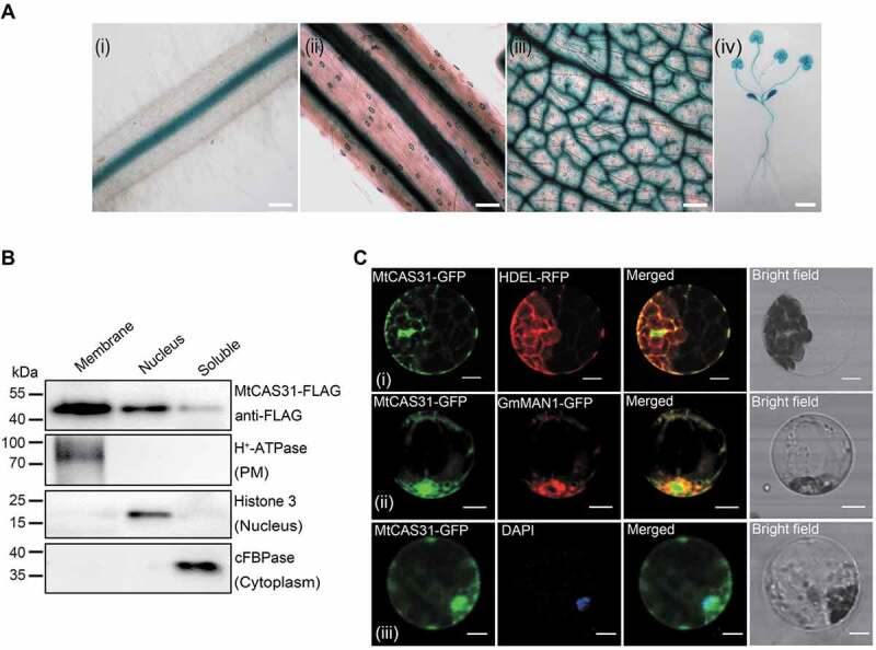
Figure 1.

Expression pattern of MtCAS31. (A) GUS staining of transgenic M. truncatula expressing MtCAS31pro:GUS. (i) Root, bar: 100 μm. (ii) Stem, bar: 100 μm. (iii) Vascular and stomal tissue of a leaf, bar: 100 μm. (iv) Whole plant, bar: 1 cm. (B) Immunoblotting assay of the membrane, nuclear and soluble components separated from N. benthamiana leaf cells expressing CaMV35S:MtCAS31-FLAG. H+-ATPase, Histone 3 and cFBPase were employed as markers of the membrane, nucleus and cytoplasm, respectively. (C) Subcellular localization of MtCAS31-GFP driven by CaMV35S. HDEL-RFP (i) and GmMAN1-RFP (ii) were co-transformed with MtCAS31-GFP. Protoplasts from plants expressing MtCAS31-GFP were stained with DAPI (iii). Fluorescence was detected by confocal laser scanning microscopy with excitation at 488 nm (for the detection of GFP), 546 nm (for the detection of RFP) and 518 nm (for the detection of DAPI). HDEL, endoplasmic reticulum retention signal. GmMAN1, endoplasmic reticulum membrane protein. DAPI, 4ʹ,6-diamidino-2-phenylindole, a nuclear dye. Bar: 10 μm.
