PLOS ONE logoLink to PLOS ONE
. 2020 Apr 9;15(4):e0231220. doi: 10.1371/journal.pone.0231220

A spectral-domain optical coherence tomographic analysis of Rdh5-/- mice retina

Yuting Xie 1, Takayuki Gonome 1, Kodai Yamauchi 1, Natsuki Maeda-Monai 1, Reiko Tanabu 1, Sei-ichi Ishiguro 2, Mitsuru Nakazawa 1,*
Editor: Knut Stieger3
PMCID: PMC7144952  PMID: 32271812

Abstract

Purpose

To investigate the longitudinal findings of spectral-domain optical coherence tomography (SD-OCT) in relation to the morphologic features in Rdh5 knockout (Rdh5-/-) mice.

Materials and methods

The mouse retina was segmented into four layers; the inner retinal (A), outer plexiform and outer nuclear (B), rod/cone (C), and retinal pigment epithelium (RPE)/choroid (D) layers. The thickness of each retinal layer of Rdh5-/- mice was longitudinally and quantitatively measured at six time points from postnatal months (PM) 1 to PM6 using SD-OCT. Age-matched C57BL/6J mice were employed as wild-type controls. The data were statistically compared using Student’s t-test. The fundus appearance was assessed, histologic and ultrastructural examinations were performed in both groups.

Results

Layers A and B were significantly thinner in the Rdh5-/- mice than in the wild-type C57BL/6J mice during the observation periods. Layers C and D became thinner in the Rdh5-/- mice than in the wild-type mice after PM6. Although no abnormalities corresponding to whitish fundus dots were detected by SD-OCT or histologic examinations, the intracellular accumulation of low-density vacuoles was noted in the RPE of the Rdh5-/- mice by electron microscopy. The photoreceptor nuclei appeared less dense in the Rdh5-/- mice than in the wild-type mice.

Discussion

The results from the present study suggest that although it is difficult to detect qualitative abnormalities, SD-OCT can detect quantitative changes in photoreceptors even in the early stage of retinal degeneration induced by the Rdh5 gene mutation in mice.

Introduction

Fundus albipunctatus (FA), as a type of hereditary retinal dystrophy, is a rare eye disorder characterized by an impaired visual ability under dim-light conditions and the presence of numerous white dots that are especially abundant near the mid-periphery and perifovea of the retina [1, 2]. It is inherited in an autosomal recessive pattern. Although mutations of the retinaldehyde binding protein1 (RLBP1) and retinal pigment epithelium (RPE)-specific-65-kDa protein (RPE65) genes have been reported to be associated with FA [3, 4], most cases of FA have been caused by mutations in the 11-cis retinol dehydrogenase 5 (Rdh5) gene [5, 6].

The Rdh5 gene encodes the 11-cis retinol dehydrogenase 5 (RDH5), which is predominantly expressed in the retinal pigment epithelium (RPE), where it converts the molecule 11-cis retinol to 11-cis retinal [710]. 11-cis retinal is the recycling molecule in an integral operation of the visual cycle. It is the chromophore residing in rhodopsin and cone opsins and is needed for the conversion of light to electrical signals [11, 12].

FA as a form of congenital night blindness was originally considered a stationary disease. The scotopic electroretinography (ERG) amplitudes were reportedly significantly reduced when recorded after a standard period of dark adaptation but returned to the normal range after prolonged dark adaptation [1, 6, 13]. Yamamoto et al. suggested that the RPE produces 11-cis retinal at a slower-than-normal rate in genetically confirmed patients with Rdh5 mutations, which may be interpreted by the slow recovery of light sensitivity in ERG after prolonged dark adaptation [5]. Subsequently, Yamamoto et al. reported that mutations in the Rdh5 gene may play a role in the devolvement of macular atrophy with fading of the white dots in FA [14]. Other researchers also noted that the white dots disappeared or became weakened in patients with Rdh5 mutations with increasing age or after uveitis [2, 15].

It was speculated that mutations in the Rdh5 gene may play an important role in the progression of the white dots in the natural course of FA. However, Rdh5 knockout (Rdh5-/-) mice reportedly do not show the typical white dots and retinal degeneration as in humans. Instead, Rdh5-/- mice show normal dark adaptation under standard conditions but display delayed dark adaptation under intense lighting [1618]. In addition, no abnormalities have been detected in the structure or function of the retina of the Rdh5-/- mice, possibly because these mice have other compensatory pathways for regenerating chromophores [19].

Recent studies have shown that FA is not always stationary. Using spectral-domain optical coherence tomography (SD-OCT), researchers have revealed details, such as structural abnormalities in the outer retina and disruption of the junction of the photoreceptor, in some patients with FA [2, 20]. Although SD-OCT is a noninvasive technology that allows us to investigate the morphological characteristics of various retinal diseases [20, 21], it is impossible to directly evaluate the pathological features. In contrast, animal models associated with known gene mutations provide strong evidence that facilitate our understanding of the relationship between the SD-OCT findings and the pathological background [2232]. However, to our knowledge, no study has assessed the relationship between SD-OCT findings and the origin of the structural changes in Rdh5-/- mice.

In this study, we explored the relationship between the SD-OCT findings and the morphologic features caused by mutations in the Rdh5 gene in mice. We believe that the information obtained from this study will help clarify the relationship between the SD-OCT images of FA and the pathologic features in clinical practice.

Materials and methods

Experimental animals

This study was carried out in strict accordance with the regulations of the Association for Research in Vision and Ophthalmology (ARVO) Statement for the Use of Animals in Ophthalmic and Vision Research. The protocol was approved by the Committee on the Ethics of Animal Experiments of the Hirosaki University (Approval Number: M12023). All procedures were performed under general anesthesia as described below, and all efforts were made to minimize suffering.

The Rdh5-/- (B6;129S-Rdh5tm1Drie/J) mice had been created using C57BL/6J (B6) as host mice and 129/Sv (129S) as donor strain by Driessen, et al. [16] and were kindly provided by Dr. Mathew M. LaVail (University of California, San Francisco, CA, USA). C57BL/6J mice were purchased from Clea, Japan (Tokyo, Japan) and were used as wild-type controls. The mice were maintained at the Hirosaki University Graduate School of Medicine Animal Care Service Facility under a cycle of 12 h of light (50 lx illumination) and 12 h of darkness (<10 lx environmental illumination) in an air-conditioned atmosphere. Mice were given free access to food and water.

SD-OCT examination and fundus photography

SD-OCT and fundus photography were performed by the methods as described in detail previously, using a Micron® IV, Image-Guided 830 nm OCT (Phoenix Retinal Imaging System, Phoenix Research Labs, Pleasanton, CA, USA) [22, 23, 25, 27]. In brief, SD-OCT and fundus photography were carried out at 6 points of time from postnatal-month (PM) 1 to PM6 for both Rdh5-/- and C57BL/6J mice. Four to eight mice were investigated at each time point. The mice were anesthetized with an intraperitoneal injection of a mixture of medetomidine hydrochloride (0.315mg/kg), midazolam (2.0mg/kg), and butorphanol tartrate (2.5mg/kg). The pupils were dilated with the instillation of eye drops containing a mixture of 0.5% tropicamide and 0.5% phenylephrine hydrochloride. The mouse ocular fundus was simultaneously monitored by a fundus camera equipped to the Micron® IV, and the position of the retinal SD-OCT image was set circumferentially around the optic disc (360°; diameter, 500 μm; 140 μm away from the optic disc margin, Fig 1) [22]. To analyze the structure of a certain fundus change of interest, the position of the SD-OCT image was set vertically or horizontally, depending on the finding. The corneal surface was protected using a 1.5% hydroxyethylcellulose solution. Fifty images were averaged to eliminate projection artifacts. During all experimental procedures, the physical condition of the mice was monitored by inspection and palpation by the researchers.

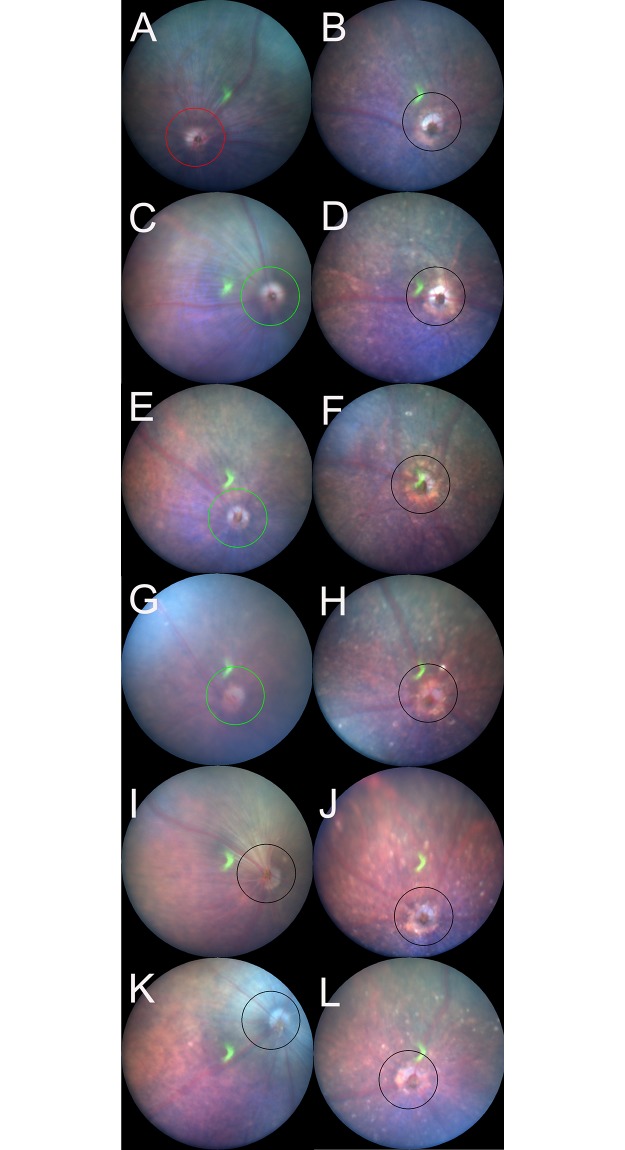
Fig 1. Representative fundus pictures of the C57BL/6J (A, C, E, G, I, and K) and Rdh5-/- (B, D, F, H, J, and L) mice.

Fig 1

Panels A, C, E, G, I, and K correspond to the fundus findings at postnatal-month (PM) 1, PM2, PM3, PM4, PM5, and PM6 of C57BL/6J mice, respectively. Panels B, D, F, H, J, and L correspond to the fundus findings at PM1, PM2, PM3, PM4, PM5, and PM6 of Rdh5-/- mice, respectively. Circles indicate the line at which the OCT images were created.

The analysis of the retinal layer thickness

The retina and choroid were divided into four layers, i.e. the inner retina (A), outer retina (B), photoreceptor segments (C), and RPE/choroid (D) layers. The inner retinal layer A consisted of the retinal nerve fiber layer (NFL), the ganglion cell layer (GCL), the inner plexiform layer (IPL) and the inner nuclear layer (INL); the outer retinal layer B consisted of the outer plexiform layer (OPL) and the outer nuclear layer (ONL); the photoreceptor segments layer C consisted of the photoreceptor inner segment (IS) and outer segment (OS) layers; and combined RPE and the choroid layer (D) (S1 Fig). As previously reported [23, 25, 27], we measured the thickness of layers A, B, C and D using the InSight® software program (Phoenix Research Labs). The borderline between each layer was automatically identified by the software program using the SD-OCT images and was manually modified by the researchers when necessary. The averages were obtained from both eyes of the same animal. The overall average retinal layer thickness was presented as the mean ± standard deviation after the normality of each distribution was confirmed by the Shapiro-Wilk test and Kolmogorov-Smirnov test.

Histological examinations

Histological examinations were performed using eyes enucleated from both Rdh5-/- and C57BL/6J mice in PM3, PM4 and PM6 by the method previously described [22]. Immediately after euthanasia by luxation of the cervical spine, the eyes were excised under a microscope. To prevent possible artificial retinal detachment during further processing, an aliquot of 2% glutaraldehyde and 2% paraformaldehyde solution (pH 7.4) was injected into the anterior chamber through the corneal limbus. After fixation in the same solution for 2 h at room temperature, the eyeballs were re-fixed in 4% paraformaldehyde solution (pH 7.0) for 24 h at 4°C. Paraffin embedding, sectioning, and hematoxylin and eosin (HE) staining were performed as previously described [22]. The HE-stained sections were photographed under a light microscope (DP-74, Olympus, Tokyo, Japan). The histological findings were compared to the corresponding findings of the SD-OCT images.

Electron microscopy

Electron microscopy was performed using eyes enucleated from both Rdh5-/- and C57BL/6J mice in PM4 and PM6 according to the method previously described [22]. Similarly to the preparation for the histological examination, after enucleation, the eyes were immediately fixed with 2.5% glutaraldehyde and 2% paraformaldehyde solution (pH 7.4) for 24 h at 4°C. An aliquot of the same fixation solution was injected into the anterior chamber. The retina and choroid were dissected out, post-fixed in phosphate-buffered 1% osmium tetroxide (pH 7.4) for 3 h at 4°C, dehydrated in an ascending ethanol series (50%-100%), and embedded in epoxy resin. Thin sections (80–90 nm) were stained in uranyl and lead salt solutions. The sections were photographed by a transmission electron microscope (H-7600; Hitachi, Tokyo, Japan) at 100 kV.

Statistical analysis

The statistical analyses of the data obtained in the present study were performed using the SPSS software program (version 25; Statistical Package for the Social Sciences, Chicago, IL, USA). The segmentation data from the two groups were compared using a two-way repeated analysis of variance (ANOVA) after the normality and equality of each distribution were confirmed by the Shapiro-Wilk test and Kolmogorov-Smirnov test, respectively. Student’s t-test was performed to analyze differences in OCT segmentation between age-matched mice groups. P values of < 0.05 were considered as statistically significant.

Results

The fundus findings of C57BL/6J and Rdh5-/- mice

The longitudinal fundus changes of both C57BL/6J and Rdh5-/- mice are shown in Fig 1. In the Rdh5-/- mouse fundi, diffuse whitish spots appeared, especially after PM2 (Fig 1D, 1F, 1H, 1J and 1L), that were not be noted in the C57BL/6J mice (Fig 1C, 1E, 1G, 1I and 1K). However, the presence of such spots has not been previously reported in Rdh5-/- mice [16].

The qualitative analyses of the SD-OCT findings in relation to the retinal structure in Rdh5-/- mice

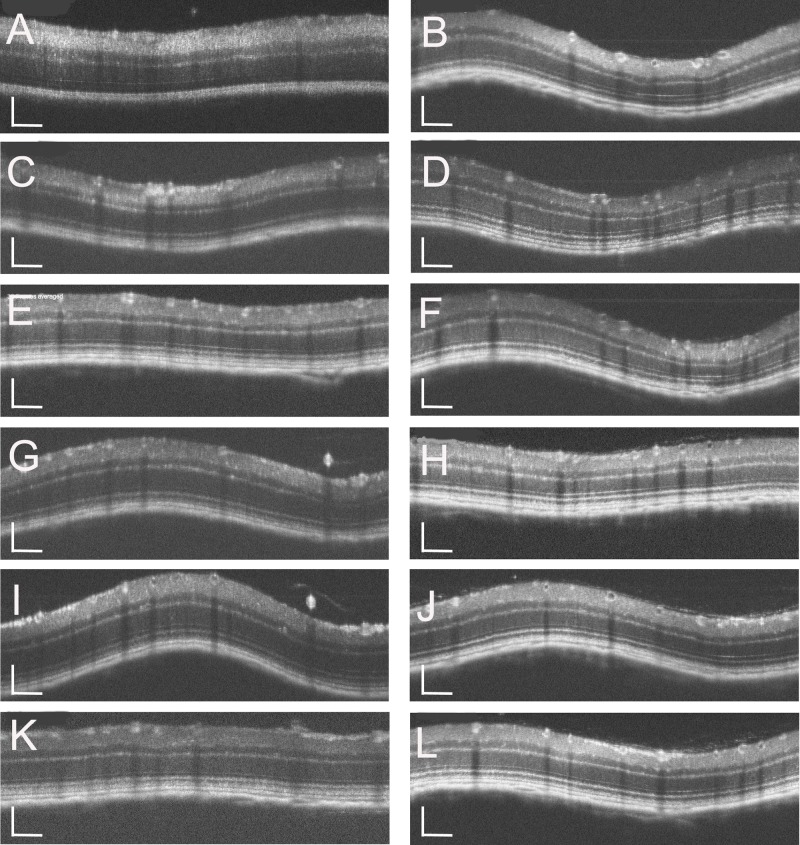
To investigate the characteristics of the retinal development of Rdh5-/- mice, both C57BL/6J and Rdh5-/- mice were subjected to SD-OCT. The typical long-term OCT findings of C57BL/6J and Rdh5-/- mice are presented in Fig 2. In SD-OCT images from both Rdh5-/- and C57BL/6J mice, the retinal layers appeared to be consistent throughout the observation periods. No qualitative differences were observed in the retina between Rdh5-/- and C57BL/6J mice (Fig 2). In addition, this tendency was consistent even in the mid-peripheral areas (Fig 3B and 3D). The wavelike features in some OCT images (Fig 2) were artificially created dependent on the angle between the mouse eye and the eye lens attached to the apparatus. Therefore, this phenomenon was not due to any of phenotypic characteristics but was based on properties of the device.

Fig 2. Representative OCT images of the C57BL/6J (A, C, E, G, I, and K) and Rdh5-/- (B, D, F, H, J, and I) mice.

Fig 2

Panels A, C, E, G, I, and K correspond to PM1, PM2, PM3, PM4, PM5, and PM6 of C57BL/6J mice, respectively. Panels B, D, F, H, J, and L correspond to PM1, PM2, PM3, PM4, PM5, and PM6 of Rdh5-/- mice, respectively. The position of the retinal SD-OCT image was set circumferentially around the optic disc (360°; diameter, 500 μm; 140 μm away from the optic disc margin, indicated by circles in Fig 1). The wavelike features in some images were artificially created depended on the angle of the mouse eye against the eye lens. The SD-OCT images were created using light stimulation at 830 nm. Bar indicates 100μm.

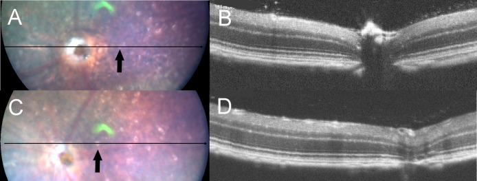
Fig 3. The fundus photographs (panels A and C) and corresponding OCT images (panels B and D) of Rdh5-/- mice.

Fig 3

Panels A and B were taken at PM4, and panels C and D were recorded at PM6. The lines in the left panels A and C indicate the section line of the corresponding OCT images in the right panels B and D, respectively. Note that the orientations of B and D are opposite to those of A and C. Arrows indicate the location of white spots.

Although we tried to identify the location of the whitish spots in the Rdh5-/- mice fundi by SD-OCT, we were unable to detect any corresponding lesions or hyper-reflective elements on SD-OCT images (Fig 3). This point differs from the findings of a previous report which described that discrete hyperreflective spots were reportedly detected in the retina of patients with FA associated with mutations in the Rdh5 gene [33].

The quantitative analyses of the OCT findings in the Rdh5-/- mice

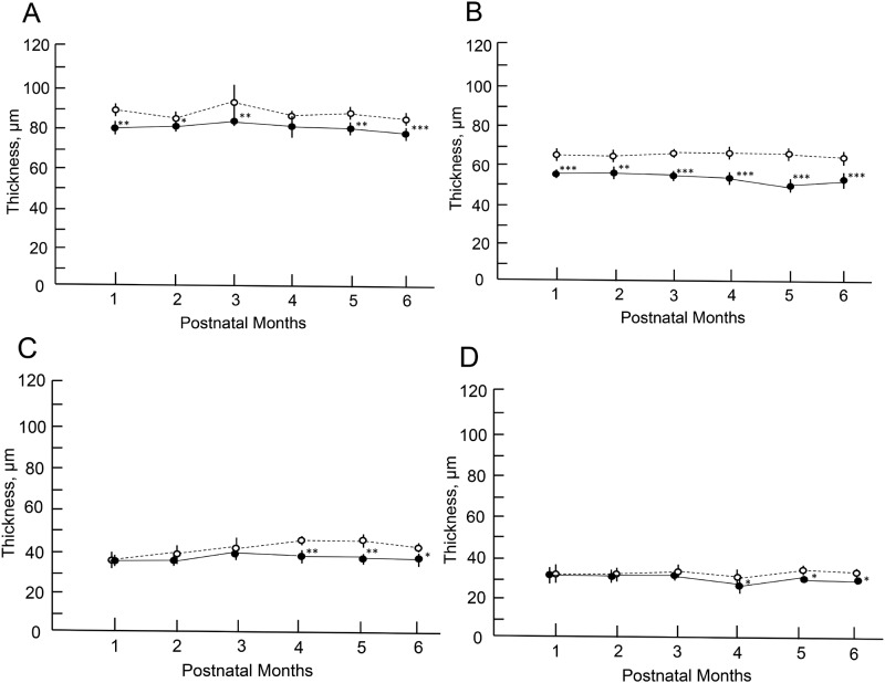
The longitudinal changes of the thickness of the retinal layer are shown in Fig 4, S1 and S2 Tables. Statistically significant differences were found in the thickness of the layers A, B, C and D between two groups at different time points. The thickness of the layer B of the Rdh5-/- mice revealed thinner at all time points in comparison to those of the C57BL/6J mice (Fig 4B). The thickness of layer A showed similar tendency to that of layer B (Fig 4A). In the Rdh5-/- mice, retinal layers C and D became significantly thinner compared to those of the C57BL/6J mice in the late stage (Fig 4C and 4D). These quantitative differences between Rdh5-/- and C57BL/6J mice have not been described previously.

Fig 4. The longitudinal changes in the thicknesses of the retinal layers.

Fig 4

Open circles, C57BL/6J mice; closed circles, Rdh5-/- mice. Panel A, thickness changes in the inner retinal layer (layer A). Panel B, the thickness of the outer retinal layer (layer B). Panel C, the thickness of the photoreceptor inner and outer segments (IS/OS) layer (layer C). Panel D, the thickness of the combined RPE and choroid layers (layer D). Animal numbers: C57BL/6J mice, n = 4 (PM1-4), n = 6 (PM5 and 6); Rdh5-/- mice, n = 6 (PM1-3 and PM6), n = 8 (PM4), n = 4 (PM5). Statistical significance: *, P <0.05; **, P <0.01; ***, P < 0.001 (Student’s t-test).

Retinal morphology by HE staining and electron microscopy

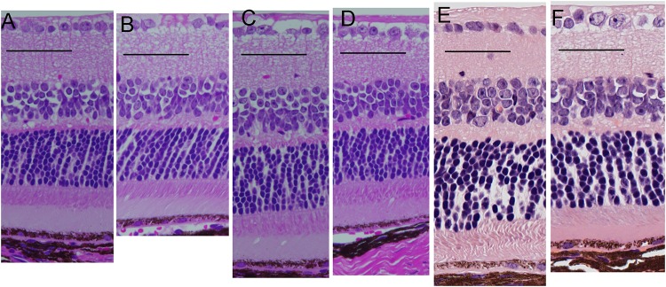
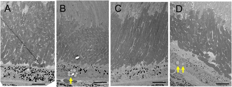
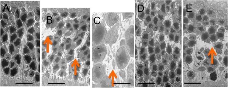
The retinal morphology of the Rdh5-/- and C57BL/6J mice was examined by both light and electron microscopy. Histologically, all cell layers of the retina were comparable between the two groups, with no qualitative differences noted, although the width of the ONL appeared to be thinner in Rdh5-/- mice than in C57BL/6J mice (Fig 5). On electron microscopy, the accumulation of low-density vacuoles was detected in the RPE cells of the Rdh5-/- mice (Fig 6). As was detected by SD-OCT (Fig 4B), the width of the ONL in Rdh5-/- mice was narrower than in C57BL/6J mice (Fig 7). In addition, the interspaces between the nuclei of the photoreceptors in the ONL of Rdh5-/- mice were wider than in C57BL/6J mice (Fig 7). Because the intracellular organelles were observed in the space, these spaces were considered to be cytoplasm of photoreceptor and/or Müller cells. No qualitative differences were found in the retinal layers A and C between C57BL/6J and Rdh5-/- mice.

Fig 5. Light microscopic findings by hematoxylin and eosin staining.

Fig 5

Retinal sections of C57BL/6J (A) and Rdh5-/- (B) mice at PM3, C57BL/6J (C) and Rdh5-/- (D) mice at PM4, and C57BL/6J (E) and Rdh5-/- (F) mice at PM6, respectively.

Fig 6. Electron microscopic findings of the photoreceptor inner and outer segment layer and the retinal pigment epithelial cells (RPE) of C57BL/6J (A) and Rdh5-/- (B) mice at PM4, and C57BL/6J (C) and Rdh5-/- (D) mice at PM6, respectively.

Fig 6

The arrows indicate low electron-density vacuoles in the RPE of Rdh5-/- mice. Bars indicate 10 μm.

Fig 7. Electron microscopic findings of the outer nuclear layer of C57BL/6J (A) and Rdh5-/- (B and C, respectively) mice at PM4, and C57BL/6J (D) and Rdh5-/- (E) mice at PM6, respectively.

Fig 7

Arrows indicate wide interspaces between nuclei of the photoreceptors. Bars indicate 10 μm in panels A, B, D and E, and 2 μm in panel C.

Discussion

In this study, we first described the SD-OCT findings of the retina of Rdh5-/- mice in relation to the morphologic findings. The results obtained may expand our knowledge of the interpretation of the SD-OCT findings in Rdh5-/- mice and may provide some clues for future studies attempting to further our understanding of FA.

Our Rdh5-/- mice displayed whitish spots after PM2 while the C57BL/6J mice showed normal fundus throughout the time course (Fig 1). FA associated with mutations of the Rdh5 gene in human is characterized by the deposition of numerous punctate white spots in the retina [5, 6]. It has been reported that these spots exist even on the day of birth and show little change afterward [33]. Conversely, it has also been reported that the white spots that appeared in the mid- and far-periphery tend to be convergent and shrink, becoming even less apparent with age [2, 12, 15]. The whitish spots on the eyes of our Rdh5-/- mice gradually increased after PM2 and until PM6, these spots showed no tendency to become less obvious or transparent. This may be due to species-related differences between mice and humans. SD-OCT in patients with FA has shown hyper-reflective elements corresponding to white dots in ocular fundus [2, 34]. Querques et al. reported that the disruption of the photoreceptor layer in a patient with FA associated with cone dystrophy at the level of the OS [20]. Shatz et al. and Sergouniotis et al. revealed that hyper-reflective lesions had extended from the RPE to the external limiting membrane [34, 35]. However, we found no such phenomenon in the fundi of the Rdh5-/- mice.

The pathology of FA can be related to the accumulation of cis-retinyl esters [16, 36], and similarly, the accumulation of 13-cis-retinyl ester in RPE can be suspected to be involved in Rdh5-/- mice [16, 17, 37], possibly leading to formation of the small whitish dots seen in the present study. The lack of RDH5 in the Rdh5-/- mice can be countered by high concentrations of 11-cis-retinol [36]. Mice are able to adjust or make use of different metabolic pathways for retinoid metabolism more efficiently than humans, resulting in less noticeable retinal pathology [16, 19]. According to Kim et al., no discernible differences were observed in 2-month-old Rdh5-/- mice retina based on findings of transmission electron microscopy [19]. In mice, although RDH5 is the principle enzyme that produces 11-cis-retinal in the RPE, RDH11 and RDH10 play minor but complementary roles in 11-cis-retinal regeneration [1719, 36, 38, 39]. The results from these previous studies suggest that 11-cis-retinal production may also be catalyzed by other enzymes than RDH5 in the mouse RPE. This may explain why it was difficult to detect any changes in 2-month-old mice retina by electron microscopy in the previous study [19]. In this study, we found the low-density vacuoles accumulated in the RPE of the Rdh5-/- mice at PM4 and PM6 (Fig 6), suggesting that these vacuoles may be derived from the accumulated cis-retinoids and cis-retinyl esters [16].

However, electron microscopy showed 1) the accumulation of low-density vacuoles in the RPE of Rdh5-/- mice and 2) the reduction in the photoreceptor cell density and abundance of the cytosolic space in the ONL. The photoreceptor cell density was relatively scarce in the Rdh5-/- mice compared to C57BL/6J mice (Fig 7). Although the exact mechanism is still unclear, this electron microscopic finding may explain the thinning of the retinal layer B (OPL and ONL) observed by SD-OCT in Rdh5-/- mice (Fig 4) and the thinning of the ONL suggested by histologic analyses (Fig 5). This phenomenon indicates an advantage of SD-OCT over other examinations, as it can detect quantitative changes in the retina more sensitively than a histologic study. In addition, these structural changes detected by SD-OCT may be the earliest abnormality of the Rdh5-/- mice retina, in which overall retinal photoreceptor photoexcitation function is still maintained. Actually, this tendency was observed previous report [16, in their Fig 3F]. However, it is possible that these quantitative changes observed by SD-OCT may be related to the defective photo-recovery function in RDH5-/- mice [16, 19]. Further studies should be carried out in the future to clarify this point. Although SD-OCT was unable to detect any abnormal lesions corresponding to the numerous fundus whitish spots, the abnormal accumulation of low-density vacuoles (Fig 6B) may be related to the pathogenesis of the fundus whitish dots. Based on their configuration and color tones, these dots may have been atrophic changes in the RPE [40] and appear to be difficult to be detected by SD-OCT. While the mechanism underlying why layer A was thinner in Rdh5-/- mice than in C57BL/6J on SD-OCT remains unclear, it is possible that this phenomenon is secondary to the deficiency of RDH5. However, further studies will be needed in order to clarify this point.

Patients with FA may have dysfunction of the cone and rod systems throughout the retina [39, 4143]. Although we did not note any morphological abnormalities of the IS or OS layers in our Rdh5-/- mice, the photoreceptor IS/OS layer became thinner in observational periods after PM4 (Fig 4C). Likewise, the RPE/choroid layer also became thinner in the late stage (Fig 4D). These results suggest that the retinal changes seen in the present study may be those in the early stage of retinal degeneration caused by defect in the Rdh5 gene in mice.

In conclusion, although the SD-OCT technology at present has limited utility in differentiating degeneration and the size variability of the retina, it has the advantages of noninvasively observing longitudinal quantitative changes in the thickness of each retinal layer more sensitively than a histologic study. These results suggest that SD-OCT can detect quantitative abnormalities in photoreceptors induced by Rdh5 gene mutations even in the early stage of retinal degeneration, which may help clarify the molecular pathogenesis of FA.

Supporting information

S1 Fig. Comparison between SD-OCT layers and histological features.

Definition of retinal sublayers A, B, C and D, ELM, IS-EZ and IZ, and comparison between a representative OCT image and histological findings of an Rdh5-/- mouse at PM3. Abbreviations: NFL, nerve fiber layer; GCL, ganglion cell layer; IPL, inner plexiform layer; INL, inner nuclear layer; OPL, outer plexiform layer; ONL, outer nuclear layer; ELM, external limiting membrane; IS-EZ, inner segment ellipsoid zone; IZ, interdigitation zone; RPE, retinal pigment epithelium.

(TIF)

S1 Table. Raw data for C57BL/6J mice.

Raw data for the retinal layer analysis (μm) in C57BL/6J mice.

(PDF)

S2 Table. Raw data for Rdh5-/- mice.

Raw data for the retinal layer analysis (μm) in Rdh5−/− mice.

(PDF)

Acknowledgments

The authors thank Dr. Brian Quinn for editing the English language of this manuscript.

Data Availability

All relevant data are within the manuscript and its Supporting Information files.

Funding Statement

MN Grant-in-Aid for Scientific Research, C-19K09926 and C-16K11313 from Japan Society for the Promotion of Science (Tokyo, Japan) N M-M Grant-in-Aid for Early Career Scientists, B-17K16954 from Japan Society for the Promotion of Science (Tokyo, Japan).

References

  • 1.Skorczyk-Werner A, Pawłowski P, Michalczuk M, et al. Fundus albipunctatus: review of the literature and report of a novel RDH5 gene mutation affecting the invariant tyrosine (p. Tyr175Phe). J Appl Genet, 2015, 56 (3), 317–327. 10.1007/s13353-015-0281-x [DOI] [PMC free article] [PubMed] [Google Scholar]
  • 2.Sergouniotis PI, Sohn EH, Li Z, et al. Phenotypic variability in RDH5 retinopathy (Fundus Albipunctatus). Ophthalmology, 2011, 118 (8), 1661–1670. 10.1016/j.ophtha.2010.12.031 [DOI] [PubMed] [Google Scholar]
  • 3.Naz S, Ali S, Riazuddin S A, et al. Mutations in RLBP1 associated with fundus albipunctatus in consanguineous Pakistani families. Brit J Ophthalmol, 2011, 97 (7), 1019–1024. [DOI] [PMC free article] [PubMed] [Google Scholar]
  • 4.Schatz P, Preising M, Lorenz B, et al. Fundus albipunctatus associated with compound heterozygous mutations in RPE65. Ophthalmology, 2011, 118 (5), 888–894. 10.1016/j.ophtha.2010.09.005 [DOI] [PubMed] [Google Scholar]
  • 5.Yamamoto H, Simon A, Eriksson U, et al. Mutations in the gene encoding 11-cis retinol dehydrogenase cause delayed dark adaptation and fundus albipunctatus. Nat Genet, 1999, 22 (2), 188 10.1038/9707 [DOI] [PubMed] [Google Scholar]
  • 6.Hirose E, Inoue Y, Morimura H, et al. Mutations in the 11-cis retinol dehydrogenase gene in Japanese patients with fundus albipunctatus. Invest Ophthalmol Vis Sci, 2000, 41, (11), 3933–3935. [PubMed] [Google Scholar]
  • 7.Simon A, Hellman U, Wernstedt C, et al. The retinal pigment epithelial-specific 11-cis retinol dehydrogenase belongs to the family of short chain alcohol dehydrogenases. J Biol Chem, 1995, 270 (3), 1107–1112. [PubMed] [Google Scholar]
  • 8.Ruiz A, Winston A, Lim YH, et al. Molecular and biochemical characterization of lecithin retinol acyltransferase. J Biol Chem, 1999, 274 (6), 3834–3841. 10.1074/jbc.274.6.3834 [DOI] [PubMed] [Google Scholar]
  • 9.Simon A, Romert A, Gustafson A L, et al. Intracellular localization and membrane topology of 11-cis retinol dehydrogenase in the retinal pigment epithelium suggest a compartmentalized synthesis of 11-cis retinaldehyde. J Cell Sci, 1999, 112 (4), 549–558. [DOI] [PubMed] [Google Scholar]
  • 10.Lhor M, Salesse C. Retinol dehydrogenases: membrane-bound enzymes for the visual function. Biochem Cell Biol, 2014, 92 (6), 510–523. 10.1139/bcb-2014-0082 [DOI] [PubMed] [Google Scholar]
  • 11.Lidén M, Eriksson U. Understanding retinol metabolism-structure and function of retinol dehydrogenases. J Biol Chem, 2006, 281 (19), 13001–13004. 10.1074/jbc.R500027200 [DOI] [PubMed] [Google Scholar]
  • 12.Parker R O, Crouch R K. Retinol dehydrogenases (RDHs) in the visual cycle. Exp Eye Res, 2010, 91 (6), 788–792. 10.1016/j.exer.2010.08.013 [DOI] [PMC free article] [PubMed] [Google Scholar]
  • 13.Kuehlewein L, Nasser F, Gloeckle N, et al. Fundus albipunctatus associated with cone dysfunction. Retin Cases Brief Rep, 2017, 11, S73–S76. 10.1097/ICB.0000000000000420 [DOI] [PubMed] [Google Scholar]
  • 14.Yamamoto H, Yakushijin K, Kusuhara S, et al. A novel RDH5 gene mutation in a patient with fundus albipunctatus presenting with macular atrophy and fading white dots. Am J Ophthalmol, 2003, 136 (3), 572–574. 10.1016/s0002-9394(03)00332-5 [DOI] [PubMed] [Google Scholar]
  • 15.Imaizumi M, Tatewaki S, Kimoto K, et al. Disappearance of puncta after uveitis in an eye with fundus albipunctatus. Retina, 25 (8), 1096–1098. 10.1097/00006982-200512000-00021 [DOI] [PubMed] [Google Scholar]
  • 16.Driessen CAGG, Winkens H J, Hoffmann K, et al. Disruption of the 11-cis-retinol dehydrogenase gene leads to accumulation of cis-retinols and cis-retinyl esters. Mol Cell Biol, 2000, 20 (12), 4275–4287. 10.1128/mcb.20.12.4275-4287.2000 [DOI] [PMC free article] [PubMed] [Google Scholar]
  • 17.Jang G F, Van Hooser J P, Kuksa V, et al. Characterization of a dehydrogenase activity responsible for oxidation of 11-cis-retinol in the retinal pigment epithelium of mice with a disrupted RDH5 gene: model for the human hereditary disease fundus albipunctatus. J Biol Chem, 2001, 276 (35), 32456–32465. 10.1074/jbc.M104949200 [DOI] [PMC free article] [PubMed] [Google Scholar]
  • 18.Sahu B, Sun W, Perusek L, et al. Conditional ablation of retinol dehydrogenase 10 in the retinal pigmented epithelium causes delayed dark adaption in mice. J Biol Chem, 2015, 290 (45), 27239–27247. 10.1074/jbc.M115.682096 [DOI] [PMC free article] [PubMed] [Google Scholar]
  • 19.Kim TS, Maeda A, Maeda T, et al. Delayed dark adaptation in 11-cis-retinol dehydrogenase deficient mice: a role of RDH11 in visual processes in vivo. J Biol Chem, 2005, 280 (10), 8694–7014. 10.1074/jbc.M413172200 [DOI] [PMC free article] [PubMed] [Google Scholar]
  • 20.Querques G, Carrillo P, Querques L, et al. High-definition optical coherence tomographic visualization of photoreceptor layer and retinal flecks in fundus albipunctatus associated with cone dystrophy. Arch Ophthalmol, 2009, 127 (5), 703–706. [DOI] [PubMed] [Google Scholar]
  • 21.Chen W, Li L, Zhang H, et al. Macular choroidal thickness in highly myopic women during pregnancy and postpartum: a longitudinal study. BMC Pregnancy Childbirth, 2018, 18 (1), 220 10.1186/s12884-018-1865-3 [DOI] [PMC free article] [PubMed] [Google Scholar]
  • 22.Tanabu R, Sato K, Monai N. et al. The findings of optical coherence tomography of retinal degeneration in relation to the morphological and electroretinographic features in RPE65-/- mice. PLoS One, 2019, 14 (1), e0210439 10.1371/journal.pone.0210439 [DOI] [PMC free article] [PubMed] [Google Scholar]
  • 23.Yamauchi K, Tanabu R, Monai N, et al. The spectral-domain optical coherence tomography findings associated with the morphological and electrophysiological changes in a rat model of retinal degeneration, rhodopsin S334ter-4 rats. Biomed Res Int 2018, 2018, ID. 5174986. [DOI] [PMC free article] [PubMed] [Google Scholar]
  • 24.Pinilla I, Fernández-Sánchez L, Segura FJ, et al. Long time remodeling during retinal degeneration evaluated by optical coherence tomography, immunocytochemistry and fundus autofluorescence. Exp Eye Res, 2016, 150, 122–134. 10.1016/j.exer.2015.10.012 [DOI] [PubMed] [Google Scholar]
  • 25.Adachi K, Takahashi S, Yamauchi K, et al. Optical coherence tomography of retinal degeneration in Royal College of Surgeons rats and its correlation with morphology and electroretinography. PLoS One, 2016, 11 (9), e0162835 10.1371/journal.pone.0162835 [DOI] [PMC free article] [PubMed] [Google Scholar]
  • 26.Ryals RC, Andrews MD, Datta S, et al. Long-term characterization of retinal degeneration in Royal College of Surgeons rats using spectral-domain optical coherence tomography. Invest Ophthalmol Vis Sci, 2017, 58 (3), 1378–1386. 10.1167/iovs.16-20363 [DOI] [PMC free article] [PubMed] [Google Scholar]
  • 27.Monai N, Yamauchi K, Tanabu R,et al. Characterization of photoreceptor degeneration in the rhodopsin P23H transgenic rat line 2 using optical coherence tomography. PLoS One, 2018, 13 (3), e0193778 10.1371/journal.pone.0193778 [DOI] [PMC free article] [PubMed] [Google Scholar]
  • 28.Kunchithapautham K, Coughlin B, Crouch RK, Rohrer B. Cone outer segment morphology and cone function in the Rpe65−/− Nrl−/− mouse retina are amenable to retinoid replacement. Invest Ophthalmol Vis Sci, 2009, 50 (10), 4858–4864. 10.1167/iovs.08-3008 [DOI] [PubMed] [Google Scholar]
  • 29.Orhan E, Dalkara D, Neuillé M, et al. Genotype and phenotype characterization of P23H line 1 rat model. PLoS One, 2015, 10 (5), e0127319 10.1371/journal.pone.0127319 [DOI] [PMC free article] [PubMed] [Google Scholar]
  • 30.Cuenca N, Femández-Sánchez L, Sauvé Y, et al. Correlation between SD-OCT, immunocytochemistry and functional findings in an animal model of retinal degeneration. Front Neuroanat, 2014, 8, 151 10.3389/fnana.2014.00151 [DOI] [PMC free article] [PubMed] [Google Scholar]
  • 31.Ohashi-Ikeda H, Sasaoka N, Koike M, et al. Novel VCP modulators mitigate major pathologies of rd10, a mouse model of retinitis pigmentosa. Sci Rep, 2014, 4, 5970 10.1038/srep05970 [DOI] [PMC free article] [PubMed] [Google Scholar]
  • 32.Levine ES, Zam A, Zhang P, et al. Rapid light-induced activation of retinal microglia in mice lacking arrestin-1. Vis Res, 2014, 201, 71–79. [DOI] [PMC free article] [PubMed] [Google Scholar]
  • 33.Sekiya K, Nakazawa M, Ohguro H, et al. Long-term fundus changes due to fundus albipunctatus associated with mutations in the RDH5 gene. Arch Ophthalmol, 2003, 121 (7), 1057–1059. [DOI] [PubMed] [Google Scholar]
  • 34.Schatz P, Preising M, Lorenz B, et al. Lack of autofluorescence in fundus albipunctatus associated with mutations in RDH5. Retina, 2010, 30 (10), 1704–1713. 10.1097/IAE.0b013e3181dc050a [DOI] [PubMed] [Google Scholar]
  • 35.Sergouniotis PI, Sohn EH, Li Z, et al. Phenotypic variability in RDH5 retinopathy (Fundus Albipunctatus). Ophthalmology, 2011, 118 (8), 1661–1670. 10.1016/j.ophtha.2010.12.031 [DOI] [PubMed] [Google Scholar]
  • 36.Farjo KM, Moiseyev G, Takahashi Y, et al. The 11-cis-retinol dehydrogenase activity of RDH10 and its interaction with visual cycle proteins. Invest Ophthalmol Vis Sci, 2009, 50 (11), 5089–5097. 10.1167/iovs.09-3797 [DOI] [PubMed] [Google Scholar]
  • 37.Maeda A, Maeda T, Imanishi Y, et al. Aberrant metabolites in mouse models of congenital blinding diseases: formation and storage of retinyl esters. Biochemistry, 2006, 45 (13), 4210–4219. 10.1021/bi052382x [DOI] [PMC free article] [PubMed] [Google Scholar]
  • 38.Haeseleer F, Jang G F, Imanishi Y, et al. Dual-substrate specificity short-chain retinol dehydrogenases from the vertebrate retina. J Biol Chem, 2002, 277 (47), 45537–45546. 10.1074/jbc.M208882200 [DOI] [PMC free article] [PubMed] [Google Scholar]
  • 39.Maeda A, Maeda T, Palczewski K. Improvement in rod and cone function in mouse model of Fundus albipunctatus after pharmacologic treatment with 9-cis-retinal. Invest Ophthalmol Vis Sci, 2006, 47 (10), 4540–4546. 10.1167/iovs.06-0215 [DOI] [PMC free article] [PubMed] [Google Scholar]
  • 40.Chhablania J, Jonnadula JB, Srinivasa Rao P. et al. Choroidal thickness profile in Retinitis Pigmentosa—Correlation with outer retinal structures. Saudiarabian J Ophthalmol, 2016, 30 (1), 9–13. [DOI] [PMC free article] [PubMed] [Google Scholar]
  • 41.Niwa Y, Kondo M, Ueno S, et al. Cone and rod dysfunction in fundus albipunctatus with RDH5 mutation: an electrophysiological study. Investi Ophthalmol Vis Sci, 2005, 46 (4), 1480–1485. [DOI] [PubMed] [Google Scholar]
  • 42.Cideciyan AV, Haeseleer F, Fariss RN, et al. Rod and cone visual cycle consequences of a null mutation in the 11-cis-retinol dehydrogenase gene in man. Vis Neurosci, 2000, 17 (5), 667–678. 10.1017/s0952523800175029 [DOI] [PMC free article] [PubMed] [Google Scholar]
  • 43.Makiyama Y, Ooto S, Hangai M, et al. Cone abnormalities in fundus albipunctatus associated with RDH5 mutations assessed using adaptive optics scanning laser ophthalmoscopy. Am J Ophthalmol, 2014, 157 (3), 558–570. 10.1016/j.ajo.2013.10.021 [DOI] [PubMed] [Google Scholar]

Decision Letter 0

Knut Stieger

5 Dec 2019

PONE-D-19-30197

A Spectral-Domain Optical Coherence Tomographic Analysis of RDH5-/- Mice Retina

PLOS ONE

Dear Professor Nakazawa,

Thank you for submitting your manuscript to PLOS ONE. After careful consideration, we feel that it has merit but does not fully meet PLOS ONE’s publication criteria as it currently stands. Therefore, we invite you to submit a revised version of the manuscript that addresses the points raised during the review process.

Please pay careful attention to the reviewers comments.

We would appreciate receiving your revised manuscript by Jan 19 2020 11:59PM. When you are ready to submit your revision, log on to https://www.editorialmanager.com/pone/ and select the 'Submissions Needing Revision' folder to locate your manuscript file.

If you would like to make changes to your financial disclosure, please include your updated statement in your cover letter.

To enhance the reproducibility of your results, we recommend that if applicable you deposit your laboratory protocols in protocols.io, where a protocol can be assigned its own identifier (DOI) such that it can be cited independently in the future. For instructions see: http://journals.plos.org/plosone/s/submission-guidelines#loc-laboratory-protocols

Please include the following items when submitting your revised manuscript:

  • A rebuttal letter that responds to each point raised by the academic editor and reviewer(s). This letter should be uploaded as separate file and labeled 'Response to Reviewers'.

  • A marked-up copy of your manuscript that highlights changes made to the original version. This file should be uploaded as separate file and labeled 'Revised Manuscript with Track Changes'.

  • An unmarked version of your revised paper without tracked changes. This file should be uploaded as separate file and labeled 'Manuscript'.

Please note while forming your response, if your article is accepted, you may have the opportunity to make the peer review history publicly available. The record will include editor decision letters (with reviews) and your responses to reviewer comments. If eligible, we will contact you to opt in or out.

We look forward to receiving your revised manuscript.

Kind regards,

Knut Stieger, D.V.M. Ph.D.

Academic Editor

PLOS ONE

Journal Requirements:

When submitting your revision, we need you to address these additional requirements.

1. Please ensure that your manuscript meets PLOS ONE's style requirements, including those for file naming. The PLOS ONE style templates can be found at

http://www.journals.plos.org/plosone/s/file?id=wjVg/PLOSOne_formatting_sample_main_body.pdf and http://www.journals.plos.org/plosone/s/file?id=ba62/PLOSOne_formatting_sample_title_authors_affiliations.pdf

2. We noticed you have some minor occurrence of overlapping text with the following previous work, which needs to be addressed:

https://journals.plos.org/plosone/article?id=10.1371/journal.pone.0210439

In your revision ensure you cite all your sources (including your own works), and quote or rephrase any duplicated text outside the methods section. In the Methods, if you feel it necessary to repeat published protocol details, please rephrase if possible, indicate clearly that you are reproducing published information (e.g. “As described in detail previously [ref],…), and cite the relevant sources. Further consideration is dependent on these concerns being addressed."

3. Thank you for including your ethics statement: All experimental procedures performed in this study conformed to the regulations of the Association for Research in Vision and Ophthalmology (ARVO) Statement for the Use of Animals in Ophthalmic and Vision Research and were reviewed and approved by the Institutional Committee of Ethics for animal experiments (Approval Number: M12023).

Please amend your current ethics statement to include the full name of the ethics committee that approved your specific study.

For additional information about PLOS ONE submissions requirements for ethics oversight of animal work, please refer to http://journals.plos.org/plosone/s/submission-guidelines#loc-animal-research  

Once you have amended this/these statement(s) in the Methods section of the manuscript, please add the same text to the “Ethics Statement” field of the submission form (via “Edit Submission”).

[Note: HTML markup is below. Please do not edit.]

Reviewers' comments:

Reviewer's Responses to Questions

Comments to the Author

1. Is the manuscript technically sound, and do the data support the conclusions?

The manuscript must describe a technically sound piece of scientific research with data that supports the conclusions. Experiments must have been conducted rigorously, with appropriate controls, replication, and sample sizes. The conclusions must be drawn appropriately based on the data presented.

Reviewer #1: Yes

Reviewer #2: Partly

**********

2. Has the statistical analysis been performed appropriately and rigorously?

Reviewer #1: Yes

Reviewer #2: No

**********

3. Have the authors made all data underlying the findings in their manuscript fully available?

The PLOS Data policy requires authors to make all data underlying the findings described in their manuscript fully available without restriction, with rare exception (please refer to the Data Availability Statement in the manuscript PDF file). The data should be provided as part of the manuscript or its supporting information, or deposited to a public repository. For example, in addition to summary statistics, the data points behind means, medians and variance measures should be available. If there are restrictions on publicly sharing data—e.g. participant privacy or use of data from a third party—those must be specified.

Reviewer #1: Yes

Reviewer #2: Yes

**********

4. Is the manuscript presented in an intelligible fashion and written in standard English?

PLOS ONE does not copyedit accepted manuscripts, so the language in submitted articles must be clear, correct, and unambiguous. Any typographical or grammatical errors should be corrected at revision, so please note any specific errors here.

Reviewer #1: Yes

Reviewer #2: Yes

**********

5. Review Comments to the Author

Please use the space provided to explain your answers to the questions above. You may also include additional comments for the author, including concerns about dual publication, research ethics, or publication ethics. (Please upload your review as an attachment if it exceeds 20,000 characters)

Reviewer #1: In this manuscript entitled “A Spectral-Domain Optical Coherence Tomographic Analysis of RDH5-/- Mice Retina” Misuru Nakazawa and colleagues have described a detailed morphological phenotype of a RDH5-/- deficient mouse model for fundus albipunctatus (FA), a human hereditary retinal dystrophy. Based on the first description of the mouse mutant for the 11-cis-retinol dehydrogenase gene (11-cis-RoDH-/- mouse) by Driessen et al., 2000, the authors have investigated the retinal phenotype by using non-invasive diagnostic tools, e.g., OCT, SLO and ERG. Results were confirmed by histological and statistical analyses.

This manuscript offers a broad spectrum of retinal techniques for the investigation of a mild phenotype. The study is related to great diligence of work and is addressed to several interesting aspects in using this model for an improved understanding of FA. The authors have discussed important findings:

1) First description of a detailed morphological data set which reveal a mild phenotype caused by the gene disruption in RDH5.

2) Due to the morphological mild phenotype (decrease in longitudinal in the thickness of the retinal layers), the ERG recordings of mutants are inconspicuously compared to wild-type controls.

3) The whitish spots which are revealed in fundus images can not be depicted in any OCT-scans caused by limitation of the OCT-technique.

Comments and suggested improvements:

Style:

The notation of protein and gene should name according to the guidelines set by the International Committee on Standardized Genetic Nomenclature for Mice (https://www.jax.org/jax-mice-and-services/customer-support/technical-support/genetics-and-nomenclature) both for C57BL6 (e.g. C57BL/6N) and for the mutant.

In the discussion part the sentence “This may be due to spices-related differences...” should correct to “This may be due to species-related differences...”

Results:

Fig 2: The wavelength and the orientation of the OCT-scan (horizontal or vertical orientation) should be completed in the legend and/or in the images. The scans show predominantly a wavelike retinal structure. Is this phenomenon based on properties of the device or may due to any phenotype characteristics?

Fig 3: Please correct the arrow color in C.

Fig 5: Please adapt the position of the scale bars as well the size of images on a consistent standard.

Fig 7: Please adapt and correct the magnification properties in panel A-E. It appears that panel C shows a higher magnification. Panel F is not included in the Fig but described in the legend. Please correct.

Fig 8: It might be helpful for a better understanding of the ERG recordings if the months of investigation and the species (mutant vs control) are included into the figures.

Results and discussion:

The authors have declared no statistical significant differences in the ERG recordings between mutants and controls at PM3 and 5 (Fig 8). But, it might be that the b-wave part in mutations is smaller than in the controls at both time points of investigations by a careful consideration. Therefore, I recommend an overlay of the ERG recordings to proof this suggestion. Furthermore, I recommend to present a separate illustration of the ERG recordings under scotopic (dark adapted), rod-only, as well under scotopic (light adapted), cone-dominated, conditions (please see the publication: Tanimoto et al. 2009, Vision tests in the mouse: Functional phenotyping with electroretinography. Front Biosci (Landmark Ed). Additionally, in 2017, Kuehlewein et al. has described a cone dysfunction in patients with FA.

Reviewer #2: The authors investigated retinal structure and function in RDH5-/- mice. In general, it is a very good work, except for one topic (see below).

There are some remarks the authors should take into the consideration.

M&M:

The authors called the layers of photoreceptor inner segments and outer segments “photoreceptor layer C”, although the photoreceptor cell bodies stretch to the outer rim of the outer plexiform layer, with their nuclei constituting the outer nuclear layer. Maybe the authors could call their layer C “photoreceptor segments layer” or so?

Results:

The authors write: “No qualitative differences were observed in the retina between RDH5-/- and C57BL6 mice. In addition, this tendency was consistent even in the midperipheral areas (Fig 3).” Do they mean Fig. 2 instead of Fig. 3?

The reviewer has the impression that OCT scans shown in Figs. 3B and 3D do not correspond to the horizontal lines visible in Fig. 3A and 3C. Moreover, it would be nice to have arrows indicating the sites of the hyperreflective spots also in Figs. 3B and 3D.

In Figure 5, the scale bars are strange, and also the general appearance of the pictures. Were the pictures of the histological sections collected from different sources?

Discussion:

In the sentence “This may be due to spices-related difference between mice and humans.” May be a typo.

The biggest problem the reviewer sees in the presented manuscript are the electroretinography (ERG) data that are provided in supplement S4. Firstly, the number of animals (n=4) is by far too small to make any meaningful statements. In general, it is estimated that there may occur a deviation of 10-15% between single ERG measurements. In order to achieve sufficient statistical power to check for differences between experimental groups, approximately 10 animals per group are necessary.

As the next, it is very questionable to perform a normality test with a group of only n=4. It simply does not make any sense! Anyhow, Shapiro-Wilk normality test is not recommended in many cases, as the D'Agostino & Pearson omnibus normality test is better.

The deviations inside each group are very big, which raises the question if the ERG measurements were performed properly, and if these ERG data are useful at all. The reviewer has a long history in ERG and VEP measurements in rats and mice, and he finds the big deviations inside the groups very surprising, in particular with respect to the first mouse in the two PM5 groups. Moreover, the big decline of amplitudes in wild-type mice from PM3 to PM5 is surprising and was never observed by the reviewer in his own work. In contrast, almost no amplitude decline was seen in the RDH5-/- mice, which is suprising.

Consequently, the authors are advised to thouroughly check the ERG data and perfom the measurements in a higher number of animals, or to not touch the topic of ERG in their manuscript at all.

**********

6. PLOS authors have the option to publish the peer review history of their article (what does this mean?). If published, this will include your full peer review and any attached files.

If you choose “no”, your identity will remain anonymous but your review may still be made public.

Do you want your identity to be public for this peer review? For information about this choice, including consent withdrawal, please see our Privacy Policy.

Reviewer #1: No

Reviewer #2: No

[NOTE: If reviewer comments were submitted as an attachment file, they will be attached to this email and accessible via the submission site. Please log into your account, locate the manuscript record, and check for the action link "View Attachments". If this link does not appear, there are no attachment files to be viewed.]

While revising your submission, please upload your figure files to the Preflight Analysis and Conversion Engine (PACE) digital diagnostic tool, https://pacev2.apexcovantage.com/. PACE helps ensure that figures meet PLOS requirements. To use PACE, you must first register as a user. Registration is free. Then, login and navigate to the UPLOAD tab, where you will find detailed instructions on how to use the tool. If you encounter any issues or have any questions when using PACE, please email us at figures@plos.org. Please note that Supporting Information files do not need this step.

PLoS One. 2020 Apr 9;15(4):e0231220. doi: 10.1371/journal.pone.0231220.r002

Author response to Decision Letter 0


16 Dec 2019

Responses to Reviewers’ comments

Reviewer #1

1. Style:

The notation of protein and gene should name according to the guidelines set by the International Committee on Standardized Genetic Nomenclature for Mice (https://www.jax.org/jax-mice-and-services/customer-support/technical-support/genetics-and-nomenclature) both for C57BL6 (e.g. C57BL/6N) and for the mutant.

Responses

We have changed the notation of protein and gene according to the guidelines set by the International Committee on Standardized Genetic Nomenclature for Mice. “RDH5-/- mice” has been changed to “Rdh5-/- mice” and “C57BL6 mice” has been changed to “C57BL/6J mice”, respectively. In addition, we have added the strain ID for each mouse group like “C57BL/6J (B6)” and “Rdh5-/- (B6;129S-Rdh5tm1Drie/J)” as recommended by the Committee in the Materials and methods section, Experimental subsection in page 6.

2. In the discussion part the sentence “This may be due to spices-related differences...” should correct to “This may be due to species-related differences...”

Responses

We have corrected the sentence to “This may be due to species-related differences...” in the Discussion section in page 13.

3. Results:

Fig 2: The wavelength and the orientation of the OCT-scan (horizontal or vertical orientation) should be completed in the legend and/or in the images. The scans show predominantly a wavelike retinal structure. Is this phenomenon based on properties of the device or may due to any phenotype characteristics?

Responses

The orientation of the OCT images has been added as “The position of the retinal SD-OCT image was set circumferentially around the optic disc (360°; diameter, 500 µm; 140 µm away from the optic disc margin, indicated by circles in Fig 1)” in Fig 2 Legend. The wavelike features in some OCT images were artificially made by the different angle between the mouse eye and the eye lens of the OCT apparatus. Therefore, we have added the sentence “The wavelike features in some images were artificially created depended on the angle of the mouse eye against the eye lens” in the Fig 2 Legend. The wavelength of the OCT-scan has been added in the Fig 2 Legend like “The SD-OCT images were made using light stimulation at 830 nm” in page 24. In addition, we have added the sentences “The wavelike features in some OCT images (Fig 2) were artificially created dependent on the angle between the mouse eye and the eye lens attached to the apparatus. Therefore, this phenomenon was not due to any of phenotypic characteristics but was based on properties of the device.” in the Results section in page 10.

4. Fig 3: Please correct the arrow color in C.

Responses

We have changed the arrow color in Fig A and C in black.

5. Fig 5: Please adapt the position of the scale bars as well the size of images on a consistent standard.

Responses

We have changed the position of scales in Fig 5.

6. Fig 7: Please adapt and correct the magnification properties in panel A-E. It appears that panel C shows a higher magnification. Panel F is not included in the Fig but described in the legend. Please correct.

Responses

We have added the explanation of the size of scale bars in the Fig 7 Legend as “Bars indicate 10 µm in panels A, B, D and E, and 2 µm in panel C”.

7. Fig 8: It might be helpful for a better understanding of the ERG recordings if the months of investigation and the species (mutant vs control) are included into the figures.

Results and discussion:

The authors have declared no statistical significant differences in the ERG recordings between mutants and controls at PM3 and 5 (Fig 8). But, it might be that the b-wave part in mutations is smaller than in the controls at both time points of investigations by a careful consideration. Therefore, I recommend an overlay of the ERG recordings to proof this suggestion. Furthermore, I recommend to present a separate illustration of the ERG recordings under scotopic (dark adapted), rod-only, as well under scotopic (light adapted), cone-dominated, conditions (please see the publication: Tanimoto et al. 2009, Vision tests in the mouse: Functional phenotyping with electroretinography. Front Biosci (Landmark Ed). Additionally, in 2017, Kuehlewein et al. has described a cone dysfunction in patients with FA.

Responses

As was also pointed out by the reviewer #2, we have recognized that our ERG data were inadequate, because of insufficient number of animals and only a single maximal rod-cone response without any of the other stimulation protocols. Because we are certain that the SD-OCT findings and morphological analyses of the retina in Rdh5-/- mice performed in this study are still valuable even without ERG analysis, we took the reviewer #2’s recommendation into account and we have decided that we do not touch the topic of ERG in the manuscript. Therefore, we have deleted the descriptions regarding ERG experiments including Fig 8 from the revised manuscript.

Reviewer #2

1. M&M:

The authors called the layers of photoreceptor inner segments and outer segments “photoreceptor layer C”, although the photoreceptor cell bodies stretch to the outer rim of the outer plexiform layer, with their nuclei constituting the outer nuclear layer. Maybe the authors could call their layer C “photoreceptor segments layer” or so?

Responses

We have changed the name “photoreceptor layer C” to “photoreceptor segments layer C” throughout the manuscript.

2. Results:

The authors write: “No qualitative differences were observed in the retina between RDH5-/- and C57BL6 mice. In addition, this tendency was consistent even in the midperipheral areas (Fig 3).” Do they mean Fig. 2 instead of Fig. 3?

Responses

The OCT images in Fig 2 are basically retinal section around the optic disc. Therefore, they do not reflect the findings of the mid-peripheral portion. The OCT images in Figs 3B and 3D partly include the retinal finding in the mid-peripheral portion. Therefore, we mention “No qualitative differences were observed in the retina between Rdh5-/- and C57BL/6J mice. In addition, this tendency was consistent even in the midperipheral areas (Fig 3).” In order not to make readers confused, we have changed these sentences to “No qualitative differences were observed in the retina between Rdh5-/- and C57BL/6J mice (Fig 2). In addition, this tendency was consistent even in the mid-peripheral areas (Figs 3B and 3D).” in page 10.

3. The reviewer has the impression that OCT scans shown in Figs. 3B and 3D do not correspond to the horizontal lines visible in Fig. 3A and 3C. Moreover, it would be nice to have arrows indicating the sites of the hyperreflective spots also in Figs. 3B and 3D.

Responses

Figs 3B and 3D do correspond to the horizontal lines, but orientations are opposite to those of Figs 3A and 3C. This derives from the SD-OCT machine’s property. To make the putative readers correctly understand this relationship, we have added the sentence “Note that the orientations of B and D are opposite to those of A and C.” in Fig 3 Legend. The arrows indicate the whitish spots in the fundus photos not the hyperreflective spots. Because we were unable to detect any hyperreflective spots corresponding to the whitish spots, we do not think that we need to add arrows at the “hyperreflective spots”.

4. In Figure 5, the scale bars are strange, and also the general appearance of the pictures. Were the pictures of the histological sections collected from different sources?

Response

As pointed out by both reviewers, we have corrected the position of scale bars.

5. Discussion:

In the sentence “This may be due to spices-related difference between mice and humans.” May be a typo.

Responses

As pointed out by both reviewers, we have corrected the typographical error.

6. The biggest problem the reviewer sees in the presented manuscript are the electroretinography (ERG) data that are provided in supplement S4. Firstly, the number of animals (n=4) is by far too small to make any meaningful statements. In general, it is estimated that there may occur a deviation of 10-15% between single ERG measurements. In order to achieve sufficient statistical power to check for differences between experimental groups, approximately 10 animals per group are necessary.

As the next, it is very questionable to perform a normality test with a group of only n=4. It simply does not make any sense! Anyhow, Shapiro-Wilk normality test is not recommended in many cases, as the D'Agostino & Pearson omnibus normality test is better.

The deviations inside each group are very big, which raises the question if the ERG measurements were performed properly, and if these ERG data are useful at all. The reviewer has a long history in ERG and VEP measurements in rats and mice, and he finds the big deviations inside the groups very surprising, in particular with respect to the first mouse in the two PM5 groups. Moreover, the big decline of amplitudes in wild-type mice from PM3 to PM5 is surprising and was never observed by the reviewer in his own work. In contrast, almost no amplitude decline was seen in the RDH5-/- mice, which is surprising.

Consequently, the authors are advised to thoroughly check the ERG data and perform the measurements in a higher number of animals, or to not touch the topic of ERG in their manuscript at all.

Responses

As pointed out by both reviewers, we have realized that the ERG data in this study were quite insufficient because of the comments raised by the reviewer #2. Because it will be difficult for us to further perform ERG assessments so as to fulfill the reviewers’ comments and because we believe that our OCT data with morphological analyses would be still valuable even without ERG analyses, we have decided not to touch the topic of ERG in this manuscript at all, according to the reviewer’s suggestion. Therefore, we have deleted the portions describing ERG from the revised manuscript. In addition to the Shapiro-Wilk test, we also carried out the Kolmogorov-Smirnov test to check the normality of the data.

Attachment

Submitted filename: Responses to Reviewers Comments.docx

Decision Letter 1

Knut Stieger

14 Feb 2020

PONE-D-19-30197R1

A Spectral-Domain Optical Coherence Tomographic Analysis of Rdh5-/- Mice Retina

PLOS ONE

Dear Professor Nakazawa,

Thank you for submitting your manuscript to PLOS ONE. After careful consideration, we feel that it has merit but does not fully meet PLOS ONE’s publication criteria as it currently stands. Therefore, we invite you to submit a revised version of the manuscript that addresses the points raised during the review process.

Please address the final points raised by reviewer 1. Furthermore, please elaborate more on the point that without the functional readout (ie. ERG), the dataset contains sufficient amount of new data to be published in PONE. 

We would appreciate receiving your revised manuscript by Mar 30 2020 11:59PM. When you are ready to submit your revision, log on to https://www.editorialmanager.com/pone/ and select the 'Submissions Needing Revision' folder to locate your manuscript file.

If you would like to make changes to your financial disclosure, please include your updated statement in your cover letter.

To enhance the reproducibility of your results, we recommend that if applicable you deposit your laboratory protocols in protocols.io, where a protocol can be assigned its own identifier (DOI) such that it can be cited independently in the future. For instructions see: http://journals.plos.org/plosone/s/submission-guidelines#loc-laboratory-protocols

Please include the following items when submitting your revised manuscript:

  • A rebuttal letter that responds to each point raised by the academic editor and reviewer(s). This letter should be uploaded as separate file and labeled 'Response to Reviewers'.

  • A marked-up copy of your manuscript that highlights changes made to the original version. This file should be uploaded as separate file and labeled 'Revised Manuscript with Track Changes'.

  • An unmarked version of your revised paper without tracked changes. This file should be uploaded as separate file and labeled 'Manuscript'.

Please note while forming your response, if your article is accepted, you may have the opportunity to make the peer review history publicly available. The record will include editor decision letters (with reviews) and your responses to reviewer comments. If eligible, we will contact you to opt in or out.

We look forward to receiving your revised manuscript.

Kind regards,

Knut Stieger, D.V.M. Ph.D.

Academic Editor

PLOS ONE

[Note: HTML markup is below. Please do not edit.]

Reviewers' comments:

Reviewer's Responses to Questions

Comments to the Author

1. If the authors have adequately addressed your comments raised in a previous round of review and you feel that this manuscript is now acceptable for publication, you may indicate that here to bypass the “Comments to the Author” section, enter your conflict of interest statement in the “Confidential to Editor” section, and submit your "Accept" recommendation.

Reviewer #1: All comments have been addressed

Reviewer #2: All comments have been addressed

**********

2. Is the manuscript technically sound, and do the data support the conclusions?

The manuscript must describe a technically sound piece of scientific research with data that supports the conclusions. Experiments must have been conducted rigorously, with appropriate controls, replication, and sample sizes. The conclusions must be drawn appropriately based on the data presented.

Reviewer #1: Partly

Reviewer #2: Yes

**********

3. Has the statistical analysis been performed appropriately and rigorously?

Reviewer #1: Yes

Reviewer #2: Yes

**********

4. Have the authors made all data underlying the findings in their manuscript fully available?

The PLOS Data policy requires authors to make all data underlying the findings described in their manuscript fully available without restriction, with rare exception (please refer to the Data Availability Statement in the manuscript PDF file). The data should be provided as part of the manuscript or its supporting information, or deposited to a public repository. For example, in addition to summary statistics, the data points behind means, medians and variance measures should be available. If there are restrictions on publicly sharing data—e.g. participant privacy or use of data from a third party—those must be specified.

Reviewer #1: Yes

Reviewer #2: Yes

**********

5. Is the manuscript presented in an intelligible fashion and written in standard English?

PLOS ONE does not copyedit accepted manuscripts, so the language in submitted articles must be clear, correct, and unambiguous. Any typographical or grammatical errors should be corrected at revision, so please note any specific errors here.

Reviewer #1: Yes

Reviewer #2: Yes

**********

6. Review Comments to the Author

Please use the space provided to explain your answers to the questions above. You may also include additional comments for the author, including concerns about dual publication, research ethics, or publication ethics. (Please upload your review as an attachment if it exceeds 20,000 characters)

Reviewer #1: Misuru Nakazawa and colleagues have revised the manuscript entitled “A Spectral-Domain Optical Coherence Tomographic Analysis of RDH5-/- Mice Retina” by following the comments of both reviewers. The major revision which was made by the authors was the deletion of the obtained ERG data at all. The rationale for that decision was the awareness that the ERG data are insufficiently in terms of animal numbers and protocols used for the experiments to allow any interpretation of the RDH5 murine mutant functional phenotype.

Comments:

For two requested changes for the following aspects, the edit could not found in the revised version:

1) In the discussion part the sentence “This may be due to spices-related differences...” should correct to “This may be due to species-related differences...”

2) Fig 3: Please correct the arrow colour in C.

Reviewer #2: The comments of the reviewer have been addressed. It is good that the topic of electroretinography was omitted as long as there are no valid data available.

**********

7. PLOS authors have the option to publish the peer review history of their article (what does this mean?). If published, this will include your full peer review and any attached files.

If you choose “no”, your identity will remain anonymous but your review may still be made public.

Do you want your identity to be public for this peer review? For information about this choice, including consent withdrawal, please see our Privacy Policy.

Reviewer #1: No

Reviewer #2: No

[NOTE: If reviewer comments were submitted as an attachment file, they will be attached to this email and accessible via the submission site. Please log into your account, locate the manuscript record, and check for the action link "View Attachments". If this link does not appear, there are no attachment files to be viewed.]

While revising your submission, please upload your figure files to the Preflight Analysis and Conversion Engine (PACE) digital diagnostic tool, https://pacev2.apexcovantage.com/. PACE helps ensure that figures meet PLOS requirements. To use PACE, you must first register as a user. Registration is free. Then, login and navigate to the UPLOAD tab, where you will find detailed instructions on how to use the tool. If you encounter any issues or have any questions when using PACE, please email us at figures@plos.org. Please note that Supporting Information files do not need this step.

PLoS One. 2020 Apr 9;15(4):e0231220. doi: 10.1371/journal.pone.0231220.r004

Author response to Decision Letter 1


19 Feb 2020

Responses to Reviewers’ comments

Reviewer #1

1. In the discussion part the sentence “This may be due to spices-related differences...” should correct to “This may be due to species-related differences...”

Response:

Thank you very much for detecting the typographical error. We corrected the miss-spelled word “spices” to “species” in the Discussion section.

2. Fig 3: Please correct the arrow colour in C.

Response:

As suggested by the reviewer, we changed the color of the horizontal arrow (line) from green to black in Fig 3.

Attachment

Submitted filename: Responses to Reviewers Comments 2.docx

Decision Letter 2

Knut Stieger

26 Feb 2020

PONE-D-19-30197R2

A Spectral-Domain Optical Coherence Tomographic Analysis of Rdh5-/- Mice Retina

PLOS ONE

Dear Professor Nakazawa,

Thank you for submitting your manuscript to PLOS ONE. After careful consideration, we feel that it has merit but does not fully meet PLOS ONE’s publication criteria as it currently stands. Therefore, we invite you to submit a revised version of the manuscript that addresses the points raised during the review process.

Dear Authors, 

you did not address my point on elaborating on the issue of why do you think that a pure morphological study describing the retinal changes without discussing the potential effect on the retinal function is sufficient for publication in PONE. I would like to encourage you to do so.

We would appreciate receiving your revised manuscript by Apr 11 2020 11:59PM. When you are ready to submit your revision, log on to https://www.editorialmanager.com/pone/ and select the 'Submissions Needing Revision' folder to locate your manuscript file.

If you would like to make changes to your financial disclosure, please include your updated statement in your cover letter.

To enhance the reproducibility of your results, we recommend that if applicable you deposit your laboratory protocols in protocols.io, where a protocol can be assigned its own identifier (DOI) such that it can be cited independently in the future. For instructions see: http://journals.plos.org/plosone/s/submission-guidelines#loc-laboratory-protocols

Please include the following items when submitting your revised manuscript:

  • A rebuttal letter that responds to each point raised by the academic editor and reviewer(s). This letter should be uploaded as separate file and labeled 'Response to Reviewers'.

  • A marked-up copy of your manuscript that highlights changes made to the original version. This file should be uploaded as separate file and labeled 'Revised Manuscript with Track Changes'.

  • An unmarked version of your revised paper without tracked changes. This file should be uploaded as separate file and labeled 'Manuscript'.

Please note while forming your response, if your article is accepted, you may have the opportunity to make the peer review history publicly available. The record will include editor decision letters (with reviews) and your responses to reviewer comments. If eligible, we will contact you to opt in or out.

We look forward to receiving your revised manuscript.

Kind regards,

Knut Stieger, D.V.M. Ph.D.

Academic Editor

PLOS ONE

[Note: HTML markup is below. Please do not edit.]

[NOTE: If reviewer comments were submitted as an attachment file, they will be attached to this email and accessible via the submission site. Please log into your account, locate the manuscript record, and check for the action link "View Attachments". If this link does not appear, there are no attachment files to be viewed.]

While revising your submission, please upload your figure files to the Preflight Analysis and Conversion Engine (PACE) digital diagnostic tool, https://pacev2.apexcovantage.com/. PACE helps ensure that figures meet PLOS requirements. To use PACE, you must first register as a user. Registration is free. Then, login and navigate to the UPLOAD tab, where you will find detailed instructions on how to use the tool. If you encounter any issues or have any questions when using PACE, please email us at figures@plos.org. Please note that Supporting Information files do not need this step.

PLoS One. 2020 Apr 9;15(4):e0231220. doi: 10.1371/journal.pone.0231220.r006

Author response to Decision Letter 2


7 Mar 2020

Responses to the editor’s comments

Thank you for your valuable comments.

“Why do you think that a pure morphological study describing the retinal changes without discussing the potential effect on the retinal function is sufficient for publication in PONE?”

Response:

Previously, Driessen et al. [16] and Kim et al. [19] have extensively performed ERG experiments in detail. Their results are summarized by the fact that the photoexcitation of the a- and b-waves was not defective in RDH5-/- mice using standard dark-adapted flash ERG, but the recovery of the a-wave from an intensive bleaching condition was delayed in RDH5-/- mice compared with that in wild-type mice. Because their experiments were quite extensively and delicately performed, we believe that their results are quite informative and that we do not need to add any further ERG experiments to be performed in this study. The previous reports have detected substantially mild ERG defects in RDH5-/- mice [16, 19]. We speculate that these mild functional defects may be related with the relatively mild structural changes observed in this study using SD-OCT and electron microscopy. Particularly, we conclude that SD-OCT can quantitatively and non-invasively analyze morphologic changes.

For these reasons, we have changed the paragraph in the Discussion section (P.14) of the previous manuscript, “A study of the photoreceptor function in Rdh5-/-Rdh11-/- mice conducted by Maeda et al. showed that aberrant cone responses were detected until 12-months of age by flicker ERG [41]. Therefore, we speculate that these differences in the metabolisms between mice and humans may be why abnormal findings were hardly detected, at least qualitatively, in the SD-OCT images of Rdh5-/- mice. In addition, we consider that these metabolic differences between mice and humans may lead to the lack of any marked differences in the amplitudes of ERG a- and b-waves in both Rdh5-/- and C57BL/6J mice [16, 19] “

to

“Regarding functional aspects, since Driessen et al. [16] and Kim et al. [19] have previously extensively performed electrophysiologic studies in detail, we thought that the results of their studies were quite informative, even though we did not perform electroretinographic (ERG) experiments in this study. Their studies regarding the photoreceptor function in RDH5-/- mice can be summarized by the fact that the photoexcitation function was not defective in RDH5-/- mice using standard dark-adapted single flash ERG experiments [16, 19]. However, the recovery of the a-wave from an intensive bleaching condition was delayed in RDH5-/- mice compared with that in wild-type mice, although no histologic difference was detected between RDH5-/- and wild-type mice [16, 19]. The results of these previous studies suggested that RDH5-/- mice showed substantially milder electrophysiological deficits than patients with FA. We speculate that these differences in the electrophysiology may be due to the different metabolisms between mice and humans. In addition, these differences in the metabolisms between mice and humans may be why hardly any abnormal findings were detected, at least qualitatively, in the SD-OCT images of Rdh5-/- mice. In the revised manuscript (P14, L267 to P15, L281).

In addition, we have added the sentences “However, it is possible that these quantitative changes observed by SD-OCT may be related to the defective photo-recovery function in RDH5-/- mice [16, 19]. Further studies should be carried out in the future to clarify this point.” in P 15, L280-282 to clarify the ERG findings previously reported.

Attachment

Submitted filename: Responses to the editor.docx

Decision Letter 3

Knut Stieger

19 Mar 2020

A Spectral-Domain Optical Coherence Tomographic Analysis of Rdh5-/- Mice Retina

PONE-D-19-30197R3

Dear Dr. Nakazawa,

We are pleased to inform you that your manuscript has been judged scientifically suitable for publication and will be formally accepted for publication once it complies with all outstanding technical requirements.

Within one week, you will receive an e-mail containing information on the amendments required prior to publication. When all required modifications have been addressed, you will receive a formal acceptance letter and your manuscript will proceed to our production department and be scheduled for publication.

Shortly after the formal acceptance letter is sent, an invoice for payment will follow. To ensure an efficient production and billing process, please log into Editorial Manager at https://www.editorialmanager.com/pone/, click the "Update My Information" link at the top of the page, and update your user information. If you have any billing related questions, please contact our Author Billing department directly at authorbilling@plos.org.

If your institution or institutions have a press office, please notify them about your upcoming paper to enable them to help maximize its impact. If they will be preparing press materials for this manuscript, you must inform our press team as soon as possible and no later than 48 hours after receiving the formal acceptance. Your manuscript will remain under strict press embargo until 2 pm Eastern Time on the date of publication. For more information, please contact onepress@plos.org.

With kind regards,

Knut Stieger, D.V.M. Ph.D.

Academic Editor

PLOS ONE

Additional Editor Comments (optional):

Reviewers' comments:

Acceptance letter

Knut Stieger

23 Mar 2020

PONE-D-19-30197R3

A Spectral-Domain Optical Coherence Tomographic Analysis of Rdh5-/- Mice Retina  

Dear Dr. Nakazawa:

I am pleased to inform you that your manuscript has been deemed suitable for publication in PLOS ONE. Congratulations! Your manuscript is now with our production department.

If your institution or institutions have a press office, please notify them about your upcoming paper at this point, to enable them to help maximize its impact. If they will be preparing press materials for this manuscript, please inform our press team within the next 48 hours. Your manuscript will remain under strict press embargo until 2 pm Eastern Time on the date of publication. For more information please contact onepress@plos.org.

For any other questions or concerns, please email plosone@plos.org.

Thank you for submitting your work to PLOS ONE.

With kind regards,

PLOS ONE Editorial Office Staff

on behalf of

Dr. Knut Stieger

Academic Editor

PLOS ONE

Associated Data

    This section collects any data citations, data availability statements, or supplementary materials included in this article.

    Supplementary Materials

    S1 Fig. Comparison between SD-OCT layers and histological features.

    Definition of retinal sublayers A, B, C and D, ELM, IS-EZ and IZ, and comparison between a representative OCT image and histological findings of an Rdh5-/- mouse at PM3. Abbreviations: NFL, nerve fiber layer; GCL, ganglion cell layer; IPL, inner plexiform layer; INL, inner nuclear layer; OPL, outer plexiform layer; ONL, outer nuclear layer; ELM, external limiting membrane; IS-EZ, inner segment ellipsoid zone; IZ, interdigitation zone; RPE, retinal pigment epithelium.

    (TIF)

    S1 Table. Raw data for C57BL/6J mice.

    Raw data for the retinal layer analysis (μm) in C57BL/6J mice.

    (PDF)

    S2 Table. Raw data for Rdh5-/- mice.

    Raw data for the retinal layer analysis (μm) in Rdh5−/− mice.

    (PDF)

    Attachment

    Submitted filename: Responses to Reviewers Comments.docx

    Attachment

    Submitted filename: Responses to Reviewers Comments 2.docx

    Attachment

    Submitted filename: Responses to the editor.docx

    Data Availability Statement

    All relevant data are within the manuscript and its Supporting Information files.


    Articles from PLoS ONE are provided here courtesy of PLOS

    RESOURCES