FIGURE 6.

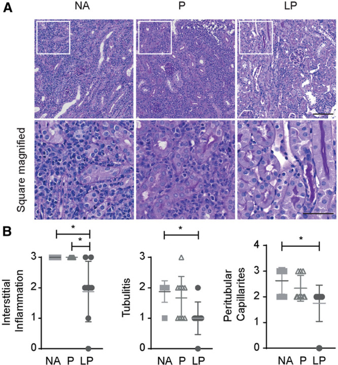
Histological evaluation of LP treatment effect. A, PAS stain of allografts showed dense interstitial leukocyte infiltrates in the tubulo interstitial space in the NA and prednisolone-treated groups. However, liposomal prednisolone reduced the inflammation markedly. The upper images are taken at a 200-fold magnification (bar: 100 µm), the lower images are the enlarged squares of each group to show the differences in infiltrate density (bar: 50 µm). B, Banff classification of treated allografts reveals decreased interstitial inflammation, tubulitis, and peritubular capillaritis in LP vs NA treatment. Interstitial inflammation is also reduced in LP vs P treatment (mean ± SD. * P < 0.05, 1-way analysis of variance with a Tukey multiple comparisons test. NA = no additional treatment; N = 8, P = prednisolone; N = 9, LP = liposomal prednisolone; N = 9). PAS, periodic acid Schiff.
