Abstract
The microbial community in the plant rhizosphere is vital to plant productivity and disease resistance. Alterations in the composition and diversity of species within this community could be detrimental if microbes suppressing the activity of pathogens are removed. Species of the insect-pathogenic fungus, Metarhizium, commonly employed as biological control agents against crop pests, have recently been identified as plant root colonizers and provide a variety of benefits (e.g. growth promotion, drought resistance, nitrogen acquisition). However, the impact of Metarhizium amendment on the rhizosphere microbiome has yet to be elucidated. Using Illumina sequencing, we examined the community profiles (bacteria and fungi) of common bean (Phaseolus vulgaris) rhizosphere (loose soil and plant root) after amendment with M. robertsii conidia, in the presence and absence of an insect host. Although alpha diversity was not significantly affected overall, there were numerous examples of plant growth-promoting organisms that significantly increased with Metarhizium amendment (Bradyrhizobium, Flavobacterium, Chaetomium, Trichoderma). Specifically, the abundance of Bradyrhizobium, a group of nitrogen-fixing bacteria, was confirmed to be increased using a qPCR assay with genus-specific primers. In addition, the ability of the microbiome to suppress the activity of a known bean root pathogen was assessed. The development of disease symptoms after application with Fusarium solani f. sp. phaseoli was visible in the hypocotyl and upper root of plants grown in sterilized soil but was suppressed during growth in microbiome soil and soil treated with M. robertsii. Successful amendment of agricultural soils with biocontrol agents such as Metarhizium necessitates a comprehensive understanding of the effects on the diversity of the rhizosphere microbiome. Such research is fundamentally important towards sustainable agricultural practices to improve overall plant health and productivity.
Introduction
Current agricultural practices are often damaging to the surrounding environment and require large energy inputs in the forms of fertilizers and pest control agents [1]. Therefore, in order to establish increasingly sustainable agricultural practices, the development of techniques designed to decrease these energy inputs is of immediate importance. One way to facilitate these goals would be through the increased integration of beneficial plant microbiomes (i.e. those enhancing plant growth, nutrient use efficiency, abiotic stress tolerance, and disease resistance) in the rhizosphere.
Agricultural plants utilize the complex community of rhizospheric microbes to maintain health and primary production [2]. These microbes vary in their ecological roles, and provide benefits that include the stimulation of plant growth [3], competitive suppression of pathogens through secondary metabolites or spatial restriction [4], increased resistance to biotic and abiotic stress by induced systemic resistance [5], and solubilization and transport of nutrients that would otherwise be unavailable to the plant [2,6].
The diversity of the rhizospheric community gives rise to a complex web of microbial interactions. This suggests a paucity of information regarding the cause of the observed plant phenotype relative to the current microbial community. There are numerous studies where microbial amendments have been applied to the rhizosphere in benefit of the plant. These include rhizobia inoculants for increasing nitrogen fixation [7,8], Azospirillum treatments that are phytostimulatory [9,10], mycorrhizal inoculants that deliver nutrients to the host plant [11–13] and biocontrol inoculants such as Trichoderma harzianum that demonstrate antagonism against fungal diseases [14–16]. However, the extent to which these inoculants affect the microbial (bacteria and fungi) rhizospheric community is influenced by numerous factors and remains an area of great interest and in need of research. Major questions remain regarding how the impact of microbial taxonomic groups can be related to the functional capabilities of the root microbiome.
Metarhizium is a genus of rhizosphere inhabiting, insect-pathogenic fungi that are ubiquitous in soil and currently used as a biocontrol agents against crop pests through crop or seed treatment with conidial formulations [17]. Metarhizium also provides benefits to a variety of host plants including resistance to salt stress [18], increased plant biomass and growth [18,19], stimulation of root growth [17,20], acquisition of insect-derived nitrogen [21,22], and antagonism of plant pathogens [23]. The plant growth-promoting qualities of Metarhizium coupled with its ability to parasitize insects make it an attractive candidate as a rhizospheric inoculant.
Here we grew common bean plants, Phaseolus vulgaris, in unsterilized Metarhizium amended soil, under laboratory conditions. As the plant associating lifestyle of Metarhizium may converge with the insect pathogenic lifestyle to differentially affect the microbiome, such as when insect-derived nitrogen is available for exchange with the plant [21], we also included Galleria mellonella larvae as a treatment. The bacterial and fungal community profiles from both rhizospheric soil and the plant root (rhizoplane and endosphere) were analyzed by Illumina sequencing. A generalized linear model was used to assess whether Metarhizium amendment had a significant effect. A quantitative PCR was performed to confirm the results of Illumina sequencing for the bacterial genus, Bradyrhizobium; identified as being significantly affected by Metarhizium application. To determine whether the described community had the capacity to suppress disease, plants were challenged with the known bean pathogen, Fusarium solani f. sp. phaseoli. An interactive website was created (https://metarhiz-microbiome.shinyapps.io/shiny3/) that allows for user-directed data analysis. This research is a necessary step in understanding the influence that amendment with Metarhizium has on the rhizospheric community under controlled conditions and could help elucidate other potential means of plant growth promotion through secondary interactions with microorganisms within the rhizospheric community.
Materials and methods
Biological materials
Haricot bean seeds (Phaseolus vulgaris; “soldier” variety) were purchased from OSC seeds (Ontario, Canada). The fungal lab strain Metarhizium robertsii (ARSEF 2575-GFP) that expresses green fluorescent protein (GFP), and Fusarium solani f. sp. phaseoli, were grown on potato dextrose agar (PDA) for 14 days to obtain conidia. The transformation of the Metarhizium strain has been previously described [24]. Conidia were harvested with 0.01% Triton X-100 and the suspensions were adjusted to a final concentration of 1.5 x 10⁶ conidia/mL. Wax moth larvae (Galleria mellonella) were purchased from a local bait shop (Missassauga Imports). Unsterilized soil (clay:loam mix) collected from a farm (Pelham, Ontario) in April before any crop had been planted and was sifted to remove large debris and used directly in the experiment. The farm grew corn the previous year and before that the land was fallow for at least 10 years.
Experimental setup
Bean seeds were surface sterilized by immersion in 2% NaOCl for 5 minutes, rinsed with sterile water and then immersed in 15% H2O2 for 20 minutes. Seeds were rinsed a minimum of three times with sterile water until no peroxide remained. A 100uL aliquot of the last water rinse was plated on PDA to ensure sterility. Bean seeds were germinated on water agar and individually placed in 9-cm plastic pots filled with field-collected soil, for a total of 36 potted plants (S1 Table). To eighteen pots, 3 larvae each were buried in the soil below the seedling and of these pots, nine had 5 mL of a 1.5 x 10⁶ conidia/mL suspension of M. robertsii 2575-GFP added to the soil surface, and nine received only water. To the remaining eighteen pots without larvae, nine pots were amended with 5mL conidial suspension, and nine pots received only water. A final nine pots were filled with only the field-collected soil (“bulk soil”; no plant, no treatment). Plants were incubated at 25°C with 50–60% humidity, and under a 16/8-hour light/dark cycle for 14 days.
Plant harvest and DNA extraction
Rhizospheric soil was collected by removing bulk soil from the roots with gloved hands and gently shaking the root above a weigh dish until 1 g was obtained. The soil was sieved through a 1 mm2 mesh and 250 mg of soil was taken from each sample, including the pots without a bean plant, and processed as recommended by the manufacturer for extraction of DNA using the Soil Plus DNA mini kit (Norgen Biotek). The remaining root was washed gently under running tap water to remove residual soil and DNA was extracted from the entire root system using the CTAB protocol described below. Samples were pooled in groups of three for a total of 27 samples (15 soil, 12 root). DNA was quantified using Qubit 3.0™ (Thermo Fisher).
CTAB DNA extraction
The fresh root tissue was weighed and then immediately flash frozen in liquid nitrogen and ground to fine powder using a mortar and pestle. A 2X CTAB solution [2% (w/v) hexadecyltrimethylammonium bromide (CTAB), 100 mM Tris (pH 8.0), 20 mM EDTA (pH 8.0), and 1.4 M NaCl was autoclaved sterile, to which 2% (w/v) of polyvinylpyrrlidone (Mw 40,000) and 1% 2-mercaptoethanol are added] was preheated to 65°C and applied to the root powder at a volume of 5:1 (i.e. 5 mL 2X CTAB per 1 g of fresh root weight) and vortexed for 1 minute. The tube was incubated in a 65°C water bath for 30 minutes. After incubation, the tube was vortexed again for 10 seconds to homogenize the solution, and then a 1 mL aliquot was transferred to a 2 mL tube. One volume of chloroform:isoamyl alcohol (24:1) was added to the supernatant and vigorously shaken to form an emulsion, followed by centrifugation at 14,000 rpm for 2 minutes. This step was repeated once. The supernatant was transferred to a 1.5 mL microfuge tube and 0.7 volumes of isopropanol were added and mixed by inversion to precipitate the DNA. The tube was centrifuged at 14,000 rpm for 5 minutes. The DNA pellet was washed once with 70% ethanol and once with 100% ethanol, being centrifuged at 14,000 rpm for 2 minutes each time. The ethanol was decanted, and the pellet allowed to dry for 10 minutes on the bench top. The pellet was resuspended in 50 μL of 1X TE buffer pH 8.0.
CFU determination of fungal inoculum
To the remaining 0.75 g sieved soil of each sample, 5 mL of 0.01% Triton X-100 was added and the sample vortexed vigorously. A 100 uL aliquot was plated, in duplicate, on CTC [25] agar plates and incubated at 27°C for 5 days. Colonies of M. robertsii were verified by visualization of GFP fluorescence and counted.
Next generation sequencing, sequence processing, and statistical analyses
DNA samples were sent to Microbiome Insights (Vancouver, Canada) for analysis. The 16S ribosomal RNA genes (V4 region) and ITS2 region genes were sequenced on an Illumina MiSeq (v. 2 chemistry) using the dual barcoding protocol [26]. Primers and PCR conditions used for 16S sequencing are identical to those of Kozich et al. [26]; those used for ITS2 sequencing were described by Gweon et al. [27]. Bacterial Raw Fastq files were quality-filtered and clustered into 97% similarity operational taxonomic units (OTUs) using the mothur software package [28]. Sequences were processed and clustered into operational taxonomic units (OTUs) with the mothur software package (v. 1.39.5)[28], following the recommended procedure (https://www.mothur.org/wiki/MiSeq_SOP; accessed Aug 2018). Paired-end reads were merged and curated to reduce sequencing error [29]. Chimeric sequences were identified and removed using VSEARCH [30]. The curated sequences were assigned to OTUs at 97% similarity using the OptiClust algorithm [31] and classified to the deepest taxonomic level that had 80% support using the naive Bayesian classifier trained on the Greengenes taxonomy outline (version 13.8)[32]. A total of 189,365 high-quality bacterial reads was obtained with a final 16S dataset of 16,356 OTUs (including those occurring once with a count of 1) and a read range of 2,442 and 12,606. High quality reads were classified using Greengenes (v. 13.8) [32] as the reference database. There were 914,945 high-quality fungal reads with a final ITS2 dataset containing 3,409 OTUs (including those occurring once with a count of 1) and a read range of 10,613 and 68,269. The processing pipeline was identical as the one used for bacteria, except for the following differences: paired-end reads were trimmed at the non-overlapping ends, and high quality reads were classified using UNITE (v. 7.1) [33] as the reference database. A consensus taxonomy for each OTU was obtained and the OTU abundances were then aggregated at multiple taxonomic levels (genus, family, phylum) (S1 File). Data analysis was performed using R statistical programming language [34]. OTU abundances were summarized with the Bray-Curtis index and a principle coordinate analysis (PCoA) was performed to visualize microbiome similarities and a permutational analysis of variance (PERMANOVA) was done to test the significance of groups. Alpha diversity was calculated using Shannon’s diversity index. For analysis of differential OTU abundances the R package ALDEX2 [35] was used in which technical variation is assessed by Monte-Carlo sampling from a Dirichlet distribution, returning a multivariate probability distribution, from which the centred log-ratio is then calculated. The development version was used as it allows for generalized linear models that test for interactions between two covariates. Welch’s t-tests with an adjusted P value were used for pairwise comparisons. Summarized statistical results are shown in supporting information S3 to S8 Tables. The interactive website (S2 File) allows for examination of the data and download of the R scripts, which is also provided as supporting information (S3 File). Raw sequences are available from the Short Read Archive (NCBI) (Accession: PRJNA558088).
Bradyrhizobium qPCR confirmation and culturing
To confirm the results of Illumina sequencing that detected an increase in abundance of Bradyrhizobium, a real-time PCR was performed using the genus-specific primers 691-709fAP (5’-GTGAAATDCGTAGAKATT-3’) and 907r (5’-CCGTCAATTCMTTTRAGTTT-3’) [36]. A 20 μL qPCR reaction utilized the SensiFAST SYBR no-rox kit (Bioline), 0.125uM of each primer, and 10 ng of DNA from the original extracted samples. A standard curve was created from DNA extracted from a culture of Bradyrhizobium japonicum generously provided by Dr. Edward Topp (Agriculture and Agri-Food Canada). The qPCR protocol followed the standard manufacturer’s conditions and results were analyzed using CFX™ Manager version 3.1 (Bio-Rad). To ensure that the presence of Bradyrhizobium was not from DNA contamination [37] 100 μL aliquots of soil samples (described above in CFU determination of fungal inoculum) were plated on yeast-mannitol selective media [(g L-1): mannitol (10.0); K2HPO4 (0.25); KH2PO4 (0.35); yeast extract (1.7); MgSO4•7H2O, (0.15); NaCl (0.1); CaCl2•2H2O (0.08); Chloramphenicol (0.03); Tetracycline (0.01); Cycloheximide (0.05)] [38] and incubated at 27°C for 14 days. Colony PCR was performed on all colonies that appeared similar to the B. japonicum strain using the universal bacteria primers 27F/1492R in a standard PCR reaction (New England Biolabs) according to manufacturer instructions. The approximately 1500 nucleotide amplicon was sent for Sanger sequencing (TCAG, Sickkids, Toronto) and identified through BLASTn analysis.
Microbiome disease-suppression activity assay
To determine the ability of the soil microbiome to suppress disease, the known bean root rot pathogen, Fusarium solani f. sp. phaseoli was applied via the soil-drench method to bean plants growing in autoclaved- or unautoclaved field-collected soil [39,40]. Small batches of the microbiome soil were autoclaved three times, with a period of 24 hours between cycles (121°C, 15 psi, 60 minutes). The soil-drench application consisted of a final volume of 5 mL of: F. solani, F. solani + M. robertsii, or water. A total of 3.75 x 10⁶ spores was applied for each fungus. Each condition had a total of three biological replicates. Co-inoculation of F. solani with M. robertsii was performed to confirm the ability of M. robertsii to antagonize F. solani [23]. Plants were incubated in the conditions stated above. After 14 days, plants were harvested and the roots were washed and examined for signs of root-rot disease such as discolouration of hypocotyl/upper tap root, lesion formation, and necrosis [41]. Two measures of disease severity were performed as previously described [23]. Root rot disease was scored from 0–1, with 0 indicating no root-rot symptoms and 1.0 indicating greater than 75% of rotted root area [42]. The disease index based on necrotic lesions was scored from 0–5, with 0 = no disease symptoms; 1 = slight browning, or <50% discolouration of the hypocotyl and firm upon pressure from thumb and forefinger, and slight root pruning; 2 = as 1 but >50% surface discolouration; 3 = discoloured hypocotyl and roots collapsing under pressure and extensive root pruning; 4 = darkly discoloured hypocotyl and roots completely collapsed or collapsing easily under pressure and severe root pruning; and 5 = dead or dying plant [43]. The difference between mean values for each disease index was evaluated with an ANOVA and Scheffe’s post-hoc (α = 0.05; n = 3).
Data availability
To explore the data further please see the interactive website at: https://metarhiz-microbiome.shinyapps.io/shiny3/ (created by Dr. Alison S. Waller; S2 File). Here the trends between different treatments for bacterial and fungal OTUs of interest (at the genus, family and phylum level) can be examined. Alternatively, a factor of interest can be selected (e.g. Metarhizium addition, insect or sample type), and a P value and a list of OTUs that have significantly different abundances, based on generalized linear model (GLM) or distance-based Welch’s t-test, can be viewed. The raw data is available for download on this site as well as from the Short Read Archive of NCBI (Accession: PRJNA558088). Matrices of OTU abundance for bacteria and fungi at phylum, family, and genus level are available for download as well.
Results
Metarhizium inoculum is able to establish within native microbiota
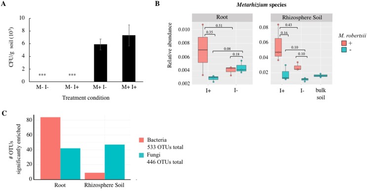
As an internal control measure, colony forming units (CFU) g-1 soil of the fungal inoculum M. robertsii 2575-GFP was quantified, utilizing GFP fluorescence of colonies in order to ensure there was no cross contamination of sample treatments (Fig 1A). M. robertsii 2575-GFP was only detected in pots that received the conidial inoculum. This was necessary to confirm that any Metarhizium detected by Illumina sequencing in uninoculated samples was due to the natural occurrence of this fungus. Fig 1B shows the relative abundance of Metarhizium detected in all conditions as determined from Illumina sequencing. Overall, there was a higher abundance of Metarhizium detected in rhizospheric soil than with the root (rhizoplane and endosphere). Conidial inoculation resulted in a statistically significant increase in the abundance of Metarhizium when G. mellonella larvae were present (Welch’s t-test, P = 0.04).
Fig 1.
Determination of (A) CFU of M. robertsii 2575-GFP recovered from soil of potted bean plant treatments after 14 days, (B) relative abundance of Metarhizium species from Illumina sequencing, and (C) number of bacterial and fungal OTUs enriched in the root and rhizosphere soil compartments. Treatment conditions: M = M. robertsii 2575-GFP; I = Insect, Galleria mellonella larvae. Statistically significant differences in CFU g-1 soil of M. robertsii are shown by asterisks (ANOVA, F = 9.41, P < 0.001; n = 9). Enriched OTUs were determined from the generalized linear model (GLM).
Growth measurements determined that there were no significant differences in the dry weight of plants between any treatment (ANOVA, P = 0.39, n = 9) (Table 1).
Table 1. Dry weight measurements for bean plants.
| Dry weight (g) | |||
|---|---|---|---|
| Treatment | Root | Phyllosphere | Total |
| Bean | 0.11 ± 0.04 | 0.44 ± 0.14 | 0.54 ± 0.15 |
| Bean + insect | 0.11 ± 0.03 | 0.33 ± 0.11 | 0.44 ± 0.10 |
| Bean + Metarhizium | 0.11 ± 0.02 | 0.35 ± 0.09 | 0.46 ± 0.10 |
| Bean + Metarhizium + insect | 0.12 ± 0.02 | 0.34 ± 0.18 | 0.46 ± 0.17 |
Plants were treated with Metarhizium robertsii with and without insect larvae (Galleria mellonela) and grown in field-collected soil for 14 days. There was no significant difference between the conditions (ANOVA, P = 0.39; n = 9).
Identification and diversity of bacteria in treatments
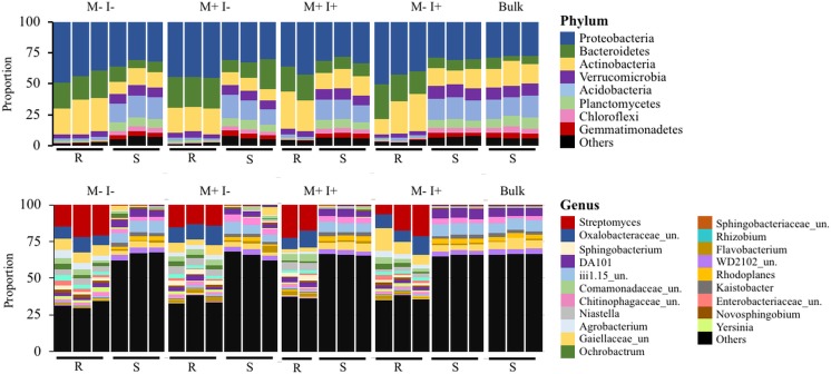
Through Illumina sequencing, a total of 533 bacterial OTUs were identified, with 84 and 9 enriched in the root and rhizosphere soil compartments, respectively (Fig 1C). The list of OTUs and the corresponding taxonomic classification can be viewed in S2 Table. The composition of bacterial phyla and the predominant genera (relative abundance above 1%) of the different treatments can be seen in Fig 2. In all treatments, the major phyla were Proteobacteria, Actinobacteria, and Bacteriodetes (Fig 2). However, there was a larger proportion of Acidobacteria and Verrucomicrobia in soil (~10–20%) compared to root (~2%). At the genus level, rare taxa (<1% abundance) accounted for nearly 70% of all taxa in soil, and approximately 35% in roots. Aside from the large proportion of lower-abundance taxa, Streptomyces, Sphingobacterium and unclassified Oxalobacteraceae and Comamonadaceae predominated in root samples (Fig 2). In contrast, DA101 and unclassified Gaiellaceae, Chitinophagaceae, and Acidobacteria (subdivision 6; iii1.15) were most abundant in soil samples.
Fig 2. Proportional distribution of the major (abundance greater than 1%) bacterial phyla and genera present in P. vulgaris root (R) and rhizospheric soil (S).
M: M. robertsii 2575-GFP. I: Insect, Galleria mellonella, larvae. Bulk soil represents original potting substrate (no bean plant). All conditions have three replicates which are pools of three samples each. “Others” signifies lower-abundance taxa. Un. = unclassified.
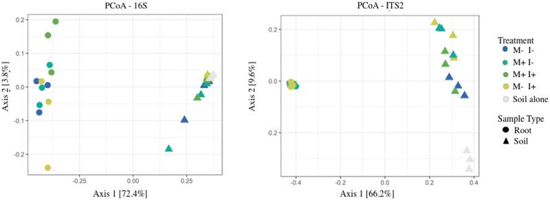
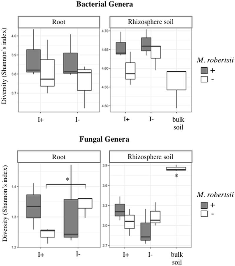
To visualize microbiome similarities between the treatments, a principle coordinate analysis (PCoA) was performed on Bray-Curtis indexed OTUs (Fig 3). The PERMANOVA revealed significant separation between sample type (F = 18.83, P < 0.001) and treatment type (F = 1.82, P < 0.001). The treatments distinctly clustered based upon sample type (soil vs. root) and supported the dissimilar diversity that was visualized in Fig 2. Moreover, ANOVA analysis of the Shannon’s diversity (H’) revealed statistically higher bacterial diversity in the rhizospheric soil than in the root (F = 453.79, P < 0.001). However, the addition of Metarhizium did not result in significant changes to bacterial diversity, nor did any of the treatments (Fig 4, S1 Table). Similar trends were seen at the family and phylum level.
Fig 3. Principal coordinate analysis (PCoA) of bacterial and fungal genera from root and soil compartments.
M: M. robertsii 2575-GFP. I: Insect, Galleria mellonella, larvae. Bulk soil represents original potting substrate (no bean plant). All conditions have three replicates which are pools of three samples each.
Fig 4. Shannon’s alpha diversity index between treatment groups for bacterial and fungal genera.
I = insect larvae (Galleria mellonella). Asterisk denotes a significant difference (t-test, α = 0.05, n = 3).
Metarhizium effects on bacterial communities
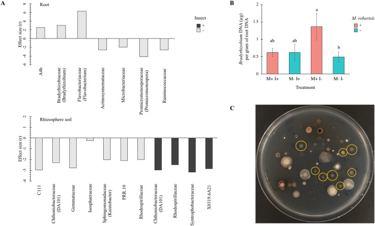
Welch’s t-tests were performed to examine if Metarhizium amendment significantly affected the abundance of specific bacteria (Fig 5). Fig 5 shows the significant (α = 0.05) increase or decrease of bacterial Families (identified genera shown in brackets) after M. robertsii addition, in rhizospheric soil and root tissue. The P values were corrected for false-discovery rate (fdr). Summarized statistical analyses are available in S5 Table.
Fig 5. Bacteria significantly affected by Metarhizium robertsii amendment.
The effect size from Welch’s t-tests using a corrected (fdr) P value (α = 0.05) is shown indicating relative abundance of bacterial Families (genus shown in brackets if known) significantly affected by Metarhizium robertsii amendment in (A) rhizosphere soil and root samples. Light bars = absence of larvae; dark bars = larvae present (n = 3). (B) The significant increase in the abundance of Bradyrhizobium in Metarhizium treated versus untreated root samples was confirmed with a SYBR® green PCR assay using Bradyrhizobium-specific primers. Error bars are standard deviation (n = 3). Conditions with different letters are significantly different (Scheffe’s post-hoc, α = 0.05). M: M. robertsii 2575-GFP. I: Insect, Galleria mellonella. (C) Bradyrhizobium (circled in yellow) cultured from potting soil on selective yeast-mannitol agar.
Rhizospheric soil had a total of 11 (11/517 = 2.1%) bacterial taxa whose abundance was significantly reduced by Metarhizium amendment. There were seven OTUs significantly affected when Galleria larvae were absent (Fig 5A), belonging to a variety of phyla including Actinobacteria, Chloroflexi, Latescibacteria (previously WS3), Planctomycetes, Proteobacteria, and Verrucomicrobia. Only two of these OTUs were able to be identified to genus level and these were Kaistobacter and DA101. Within rhizospheric soil treated with a combination of Galleria larvae and fungal inoculum, there was a total of four bacterial families that were significantly affected (Fig 5A), Rhodospirillaceae, Syntrophobacteraceae, X0319.6A21, and Chthoniobacteraceae (genus DA101). In all cases, the relative abundance of these bacteria decreased significantly after Metarhizium amendment.
In root tissue there was a lower number of bacteria whose abundance was significantly affected by Metarhizium amendment (7/400 = 1.8%) (Fig 5A) and these changes only occurred in the absence of Galleria larvae. Actinosynnemataceae, Microbacteriaceae, Promicromonosporaceae (genus Promicromonospora), and Ruminococcaceae were reduced in root samples of beans grown in Metarhizium-amended soil. The relative abundance of Bradyrhizobiaceae, A4b, and Flavobacteriaceae (genus Flavobacterium) increased after fungal treatment. Flavobacterium was increased approximately 4-fold in Metarhizium-treated roots. An unclassified Bradyrhizobiaceae (genus Bradyrhizobium matched through BLASTn analysis) and unclassified A4b were not detected in root tissue of uninoculated bean plants but were present in root tissue of beans grown in Metarhizium-treated soil (Fig 5A). The increase in Bradyrhizobium was confirmed with qPCR using genus-specific primers [36] in a SYBR® green assay (Fig 5B). Bradyrhizobium was cultured on a selective medium [44] to confirm its detection was not from artificial DNA contamination [37] (Fig 5C).
Identification and diversity of fungi in treatments
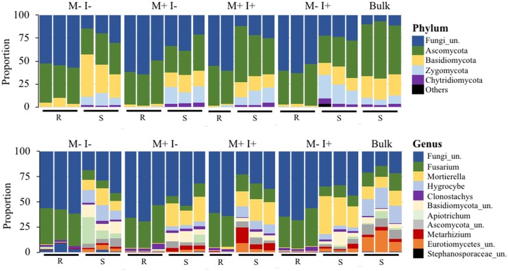
A total of 446 fungal OTUs were identified through Illumina sequencing, with 42 and 47 enriched in the root and rhizosphere soil compartments, respectively (Fig 1C). The list of OTUs and the corresponding taxonomic classification can be viewed in S2 Table. The proportional abundance of the major fungal phyla and genera (abundance greater than 1%) is shown in Fig 6. In root samples, unclassified fungi accounted for approximately half of all OTUs at the phylum level with the remainder being mainly Ascomycota (Fig 6). In rhizospheric soil, the abundance of unclassified fungi was lower than in the root and was in approximately equal proportion to Ascomycota, Basidiomycota, and Zygomycota. Within the distribution of fungal genera, there remained a high proportion of unclassified fungi in root (~61%) and rhizospheric soil (~34%). In addition, root samples were predominated by Fusarium (>30%) and Clonostachys. Rhizospheric soil appeared more diverse in all treatments in comparison to root samples and proportionally different when compared to field-collected soil that was without a bean plant (i.e. original bulk soil) (Fig 6). Fusarium, Mortierella, Hygrocybe, unclassified Basidiomycota/Ascomycota/Eurotiomyctes/ Stephanosporaceae, Apiotrichum, Clonostachys, and Metarhizium were all in high abundance in rhizospheric soil of all treatments. The highest abundances of Metarhizium were unsurprisingly the soil samples from amended treatments, however, Metarhizium was detected in all samples as seen in Fig 1 and the fungal OTU relative abundance matrix (See S1 File).
Fig 6. Proportional distribution of major (abundance greater than 1%) fungal phyla and genera present in P. vulgaris root (R) and rhizosphere soil (S).
M: M. robertsii 2575-GFP. I: Insect, Galleria mellonella. Bulk soil represents samples of the original potting substrate (no bean plant). All conditions have three replicates which are pools of three samples each. “Others” signifies lower-abundance taxa.
Ordination analysis using the abundance profile of fungal genera shows the treatments distinctly clustered based upon sample type, similar to bacterial taxa, in the Bray-Curtis indexed PCoA analysis (Fig 3). The PERMANOVA revealed significant separation between sample type (F = 70.59, P < 0.001) and treatment type (F = 3.83, P < 0.001). Unlike bacteria, fungal taxa of bulk soil and rhizosphere soil, from all treatments, clustered separately from each other indicating that the bean plant altered the fungal community profiles; all treatments with a bean plant had lower diversity (Fig 4). Interestingly, this separation driven by the variance of axis 2 [9.6%] does not result in any further separation of the root fungal communities.
Similar to bacterial diversity, the diversity of fungi in soil (H’ = 2.7–3.9) was higher than that of the root (H’ = 1.2–1.5) (ANOVA, F = 657.71, P < 0.001). Shannon’s diversity revealed complex effects on global fungal diversity depending on treatment and sample type (Fig 4, S1 Table). In root samples, when an insect was present, there was no change in diversity with the amendment of Metarhizium (t-test, P = 0.4), however, without Metarhizium, the addition of larvae significantly decreased diversity (t-test, P = 0.02). In comparison, the addition of larvae did not reduce diversity in rhizospheric soil (Fig 4). Overall, Metarhizium amendment did not significantly alter fungal diversity.
Metarhizium effects on fungal communities
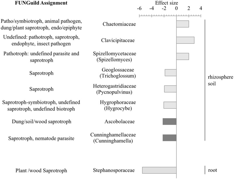
Of the 384 identified fungal genera, pairwise comparisons showed that only 7 OTUs (7/259 = 2.7%) from rhizospheric soil and 1 OTU (1/361 = 0.3%) from root were significantly altered (fdr-adjusted, α = 0.05) upon Metarhizium amendment. Fig 8 shows the effect size, as calculated by distance-based Welch’s t-test, of fungal OTUs that could be classified to a minimum of family level, with a significant change in abundance after M. robertsii addition. FUNGuild assignment revealed that the majority of significantly affect taxa were saprotrophic (Fig 7). A summary of statistical analyses is available in S6 Table.
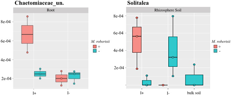
Fig 8. Interaction between the effect of adding Metarhizium and insect larvae as visualized by a box plot for Solitalea (bacteria) and Chaetomiaceae (fungi).
The resultant effect on relative abundance from addition of fungus and insect together opposes the effect of either one applied individually. I = insect (Galleria mellonella) larvae. Bulk soil represents samples of the original potting substrate (no bean plant). Un. = unclassified.
Fig 7. Fungi significantly affected by Metarhizium robertsii amendment.
The effect size from Welch’s t-tests using a corrected (fdr) P value (α = 0.05) is shown indicating relative abundance of fungal Families (specific genus shown in brackets) significantly affected by Metarhizium robertsii amendment in rhizosphere soil and root samples. Light bars = absence of larvae; dark bars = larvae present (n = 3). Fungi were classified into lifestyle guilds using FUNGuild.
In the absence of Galleria larvae, a single fungal OTU from root tissue was significantly reduced by Metarhizium amendment, Stephanosporaceae (t-test, P = 0.01). In root samples from beans treated with a combination of Galleria larvae and Metarhizium, there were no OTUs that could be resolved to family level that were significantly affected.
Metarhizium amendment significantly increased the abundance of fungi in soil (in absence of larvae) from the families Clavicipitaceae, Chaetomiaceae, and Spizellomycetaceae (genus Spizellomyces) (Fig 7). The relative abundance of three unclassified species from the families Hygrophoraceae (Hygrocybe), Geoglossaceae (Trichoglossum), and Heterogastridiaceae (Pycnopulvinus) significantly decreased in rhizospheric soil from plants treated with Metarhizium; Pycnopulvinus was not detected in Metarhizium-amended samples.
Bean plants treated with a combination of Metarhizium amendment and Galleria larvae had a reduction in Cunninghamellaceae (Cunninghamella) (t-test, P = 0.04) and Ascobolaceae (t-test, P = 0.03) from rhizospheric soil (Fig 7). Cunninghamella was represented by a single OTU and BLAST analysis of the corresponding sequence revealed the species, Cunninghamella elegans.
Metarhizium-galleria interaction
In addition to Metarhizium or the larvae amendments affecting microbe abundances, GLM analysis revealed that for some microorganisms there was an interaction when combining both treatments (S2 File, S3 and S4 Tables). In some cases, opposing effects were seen when either Metarhizium or Galleria were added independently compared to when they were combined in the treatment. An example of this is shown in Fig 8 with the bacteria Solitalea in root and an unclassified Chaetomiaceae (fungus) in rhizospheric soil. For Solitalea, when larvae were present the addition of Metarhizium resulted in an increase in abundance, whereas, when there were no larvae, the addition of Metarhizium resulted in a decrease in Solitalea (GLM, P = 0.01) (Fig 8). Similarly, in the root an unclassified Chaetomiaceae increased upon Metarhizum addition if larvae were also present, whereas there was no change in the abundance of Chaetomiaceae with the addition of either Metarhizum or larvae independently (GLM, P = 0.03) (Fig 8).
Microbiome disease-suppressive activity
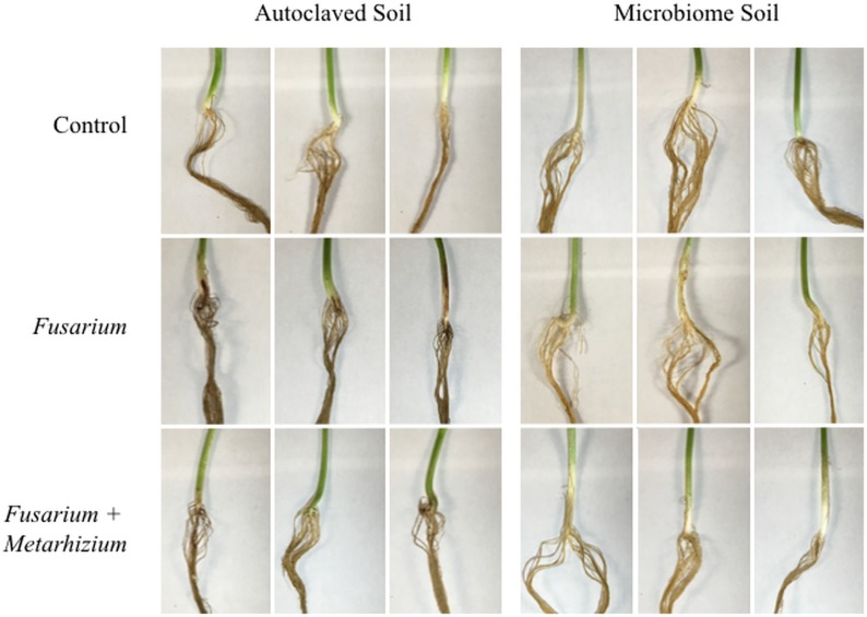
The capability of the microbiome to supress the action of a known fungal pathogen, Fusarium solani f. sp. phaseoli, to which Metarhizium robertsii has been shown to be antagonistic towards [23], was evaluated. Fig 9 shows the hypocotyl and upper root section for each treatment condition, as F. solani causes root-rot in Phaseolus vulgaris [41] and Table 2 shows the calculated disease scores. The hypocotyl of plants grown in autoclaved soil treated with F. solani showed the initial signs of disease. At 14 days, the hypocotyl and upper region of the taproot had reddish-brown, darkly discoloured lesions, indicating signs of necrosis (necrotic lesion index (NLI), 1.600 ± 0.414). These lesions were absent in Fusarium-treated plants that were grown in raw microbiome soil (NLI, 0.155 ± 0.042), indicating suppressive activity by the microbial community. Similarly, plants grown with Metarhizium in addition to Fusarium had noticeably less severe signs of disease (i.e. slight browning of upper tap root and tiny, sporadic lesions on hypocotyl) or completely lacked symptoms, in autoclaved soil (NLI, 0.450 ± 0.376). In microbiome soil, Fusarium-Metarhizium treated plants were free of any disease symptoms and were no different than control plants (NLI, 0.160 ± 0.047). There were no disease symptoms on above-ground tissues of any treatment. There was no difference in the dry weight of plants from any treatment (ANOVA, P = 0.94, n = 3) (S9 Table).
Fig 9. Evidence of disease-suppressive activity of the microbiome.
Bean plants were treated with Fusarium, Fusarium and Metarhizium, or water, and grown for 14 days in microbiome soil or sterilized soil (autoclaved three times). Disease symptoms of root-rot in bean plants is seen as discolouration, lesions, and/or necrosis on the hypocotyl and/or roots. There were no indications of disease on the above-ground tissues. Fusarium: Fusarium solani f. sp. phaseoli; Metarhizium: Metarhizium robertsii 2575-GFP.
Table 2. Disease scoring of bean roots from disease-suppressive assay.
| Disease Indices† | ||
|---|---|---|
| Root rot index‡ | Necrotic lesion index§ | |
| Autoclaved soil | ||
| Bean | 0.005 ± 0.004a | 0.258 ± 0.163a |
| Bean + Fusarium | 0.225 ± 0.117b | 1.600 ± 0.414b |
| Bean + Fusarium + Metarhizium | 0.040 ± 0.039a | 0.450 ± 0.376a |
| Microbiome soil | ||
| Bean | 0.014 ± 0.011a | 0.175 ± 0.065a |
| Bean + Fusarium | 0.020 ± 0.014a | 0.155 ± 0.042a |
| Bean + Fusarium + Metarhizium | 0.020 ± 0.008a | 0.160 ± 0.047a |
† Plants were treated with Fusarium solani f. sp. phaseoli with or without Metarhizium robertsii and grown in microbiome soil or autoclaved soil for 14 days. The average of each disease metric ± the standard deviation, is shown. Values followed by the same letter are not significantly different (Scheffe’s post hoc, α = 0.05; n = 3)
‡Dar et al. (1997) root rot disease index scored from 0 = no disease, to 1.00 = greater than 75% root area rotted.
§Filion et al. (2003) necrotic lesions scored from 0–5; 0 = no disease symptoms; 1 = slightly brown or <50% surface discoloration of the hypocotyl, firm upon pressure from thumb and forefinger, and slight root pruning; 2 = as 1 but >50% surface discoloration; 3 = discolored hypocotyl and roots collapsing under considerable pressure and extensive root pruning; 4 = darkly discolored hypocotyl and roots completely collapsed or collapsing easily under pressure and severe root pruning; 5 = dead or dying plant.
Discussion
M. robertsii is the most abundant Metarhizium species isolated from farmland and open fields in Ontario [45]. Many studies have been conducted to determine its ability to persist in soil or to colonize plants [46–48] in order to determine ways to improve its survivability and thus, its utility as a biocontrol agent. Experimental trials under laboratory conditions often fail to translate to successful application under environmental settings. For example, in sterile laboratory conditions M. brunneum was capable of killing infected wireworms, however when implemented in the field, M. brunneum was suppressed by symbiotic bacteria [49] and rendered ineffective. Moreover, a study comparing endophytic colonization of M. anisopliae and Beauveria bassiana in bean plants showed unpredictable and highly variably levels in non-sterilized soil compared to sterile soil [50] indicating an influence from the microbial community. The success of EIPF agricultural amendments is constrained by a lack of understanding of the complex interactions within the existing microbiome of crops. This dynamic population can include organisms that will compete with or antagonize the applied biological control agent and thus limit its potential. This study presents an important step towards understanding the larger roles played by Metarhizium within its microbial community.
Diversity within a localized microbial community depends upon various mechanisms that restrict domination of the population by only a few competitive species [51]. Alterations that upset these mechanisms, whether biotic or abiotic factors, can lead to a decrease in diversity as a select few species become dominant; if these dominant organism(s) are pathogenic, plant disease develops [51]. However, a decrease in diversity is not always unfavourable. Plants shape the microbiome towards their specific requirements by means of their root exudate and thus rhizospheric soil is typically less diverse than the surrounding bulk soil [8,52,53]. The decrease in diversity of rhizosphere soil compared to bulk soil within this study likely reflects this recruitment of select organisms best suited to the needs of the growing plant.
M. robertsii amendment did not affect diversity and may be due to the presence of naturally occurring strains of Metarhizium. In contrast, fungal diversity of the root decreased when Galleria mellonella larvae were independently added to the soil (Fig 4). The inclusion of an insect to the system was to approximate naturally occurring tripartite Metarhizium-insect-plant interactions. Although G. mellonella is a parasite of beehives, thus not typically found in soil, it has been used consistently as a source of nitrogen for Metarhizium in nitrogen transfer experimentation [54]. Even in field-trials, M. robertsii was able to transfer insect-derived nitrogen to bean plants within 14 days post inoculation [21]. Insect presence in the system altered the overall composition of the microbiome and further amendment with Metarhizium, increasingly changed it structure. Specifically, there was an increase in the fungal species Chaetomium, Trichoderma, and an unclassified Chaetomiaceae which are ecologically important endophytic, plant growth promoting fungi. Chaetomium helps to support tolerance to copper toxicity in maize [55] and is antagonistic to plant pathogens such as apple scab [56]. The plant growth promoting effects of Trichoderma are numerous and include conferring tolerance to salinity stress, in wheat [57], suppression of root knot nematodes [58], induction of plant systemic resistance [59], and increased biomass and yield of soybean [60]. These tripartite interactions between plant-Metarhizium-insect require further experimentation to elucidate the total ecophysiological effects of these community changes within this system.
Metarhizium will persist in the rhizosphere of numerous plant species, often as a mutualistic endophyte [20,21,61]. Benefits of Metarhizium root colonization include tolerance to salt stress [18], increased biomass [19], and antagonism towards a fungal pathogen [23]. In the environment, disease suppression is a phenomenon in which the total microbial community prevents or suppresses the ability of pathogens to grow to sufficient number to infect their host [62]. Suppression arising from the action of a certain organism is “specific” suppression. For example, high abundance of Pseudomonas spp. in soil is suppressive to take-all disease of wheat by the fungal pathogen Gaeumannomyces gramminis var. tritici through the action of the antifungal compound 2,4-diacetylphloroglucinol (DAPG) [40]. Production of secondary metabolites can also act indirectly, such as lytic enzymes from Proteobacteria that specifically target a phytotoxin from F. oxysporum (fusaric acid) preventing its interaction with tomato plants and causing wilt [63]. Cell-free culture extracts from Metarhizium were shown to antagonize the growth of F. solani while the specific bioactive metabolite has yet to be identified [23]. Although non-pathogenic strains exist, F. oxysporum and F. solani are commonly plant root pathogens [14,64–66]. Both species were identified in the bean root by Illumina sequencing (S10 Table), yet all bean plants were healthy and free of disease symptoms (i.e. root discolouration/necrosis, leaf spots; data not shown) [41] which is indicative of disease suppression. However, even species-level identification is often insufficient to identify the lifestyle of a given organism, as evident by the variable FUNGuild classification of the fungi detected in this study. Towards this end, we challenged the microbiome with the known bean-specific pathogen, Fusarium solani f. sp. phaseoli. The phenomenon of disease-suppression by the microbial community was evident as Fusarium was unable to cause hypocotyl and upper root discolouration/necrosis when plants were grown in microbiome soil. Even at this early time point, disease symptoms were apparent in roots grown in sterilized soil; a condition greatly reduced or absent with application of Metarhizium. These results support the work done previously by Sasan and Bidochka (2013), who showed that Metarhizium antagonized F. solani f. sp. phaseoli in dual plate culture, in broth culture and in planta. The formation of root-rot disease symptoms observed by Sasan and Bidochka (2013) was more severe than we observed in our study (disease index 3.93 ± 0.07 and 1.60 ± 0.41, respectively) as their results were obtained after 4 weeks. Given that native Metarhizium spp. were present in all samples, it is likely that Metarhizium plays an important role with respect to disease suppression in this system.
In combination with the action of ‘specific’ suppression, is the collective activity of the soil microbiome in competing with or antagonising pathogens. This type of community activity is termed “general” suppression [39]. A large classification of bacteria noted for their contributions to disease suppression are better known as plant growth promoting bacteria (PGPB); notable genera include Agrobacterium, Arthrobacter, Bacillus, Burkholderia, Caulobacter, Flavobacterium, Pseudomonas, Rhizobium, Serratia, and Streptomyces [67,68]. The majority of members of these genera have been shown to be PGPB however, there still remains the existence of intraspecies variation (symbiotic/pathogenic) that is discernable only by identification of the specific strain [69]. The role of each taxa is discussed as potential means of PGP based upon the referenced information but whose true functionality remains to be proven. The abundance of Flavobacterium, Bradyrhizobium and unclassified bacteria in the family A4b (class Anaerolineae) were significantly increased (Welch’s t-test, α = 0.05) in root tissues after Metarhizium robertsii amendment (Fig 5). Agrobacterium, Arthrobacter, Bacillus, Burkholderia, Caulobacter, Pseudomonas, Rhizobium, Serratia, and Streptomyces were present in all root and soil samples yet remained unaffected by Metarhizium treatment (S2 File). The ecological role of members of the bacterial family A4b remains to be determined however sequencing techniques have detected them in ectomycorrhizal roots [70] and the Anaerolineae lineage of Chloroflexi from which they reside are known anaerobic digesters [71]. Flavobacterium species have been found in diverse aquatic and terrestrial habitats and are frequently one of the most abundant taxa to be detected in rhizospheric soils of numerous plant species [72,73]. Flavobacterium are believed to play an important role in plant growth and protection [73]. Flavobacterium species possess numerous extracellular macromolecular degrading enzymes that facilitate organic matter turnover [74] that can be crucial during the initial stages of plant growth. In addition, these bacteria can synthesize plant-growth stimulating hormones [75] as well as biologically active compounds against plant pathogens, such as Phytophthora capsici of pepper [76]. Flavobacterium was detected in high abundance in all plants which is in agreement with previous reports that Flavobacterium colonization is relatively high during early plant development (14 days) [73,77]. However, given that the abundance of Flavobacterium was significantly increased in roots after Metarhizium amendment (Fig 5), using Metarhizium to facilitate an increase in Flavobacterium root colonization could be a more favorable strategy over directly applying Flavobacterium strains as they are also fish and opportunistic human pathogens [73].
M. robertsii may contribute to general disease suppression and plant growth promotion in concert with PGPB members of the community. Of note, it was found that Bradyrhizobium was significantly increased in roots after Metarhizium amendment (Fig 5). Bradyrhizobium are classically viewed as important PGPB that are capable of nodulation and nitrogen fixation for leguminous plants, however, the dominant ecotype in natural populations do not form these typical symbioses [78,79] and indeed no nodules were observed in this study (data not shown). One hypothesis for the ecological importance of non-nodulating Bradyrhizobium species is the ability to increase plant host fitness by suppressing overproduction of nodules by prolific nodulating strains that would otherwise be energetically costly to the plant [79]. Co-inoculation of nodulating and non-nodulating strains of Bradyrhizobium isolated from Acmispon strigosus revealed that non-nodulating strains competitively colonized A. strigosus and depending on strain combination, could reduce the fitness of nodulating symbionts [79]. The plant growth promoting ability of Bradyrhizobium has also been shown for wild rice, Oryza breviligulata [80], and indicates that their ecological role is greater than their classical taxonomy indicates.
The roots of plant are known to be capable of housing numerous endophytes. Metarhizium amendment increased the abundance of an unclassified Helotiales (family Incertae sedis) and an unclassified Xylariales isolated from roots. Fungi of these orders are typically aquatic hyphomycetes which function to increase nutrient cycling in streams but are now recognized to possess a dual lifestyle as endophytes of terrestrial plants [81,82]. Occupation of plant tissues and utilization of resources by endophytes can competitively exclude pathogenic organisms to the benefit of the host. Antagonists can also have a direct negative effect on plant pathogens. Species of the fungal genus Spizellomyces (Phlyctochytrium) are parasites of nematodes [83], oospores of Sclerospora sorghi [83] and Peronspora tabacina [84], and the potato wart pathogen, Synchytrium endobioticum [85], in addition to their saprotrophic role [86]. Here, an increased abundance of Spizellomyces in soil after Metarhizium amendment was observed. Taken together these results suggest that in addition to the benefits conferred directly by Metarhizium such as promotion of disease suppression (Fusarium root rot) and exchanging insect-derived nitrogen for photosynthate [22,54], Metarhizium may act indirectly by promoting and maintaining an environment rich in numerous PGPB, facilitating up-take of nutrients and minerals, and integrating into the microbial community thus maintaining general disease suppressive activity of the soil.
Metarhizium is a broad-range insect pathogen used as a biological control agent. Its ability to beneficially colonize plant roots and persist in the rhizosphere permit its stability and longevity as a treatment, and further understanding of its role in larger microbiome communities will enhance its use as an agricultural treatment. The plant microbiome has been revealed as an important determinant of plant health and productivity, and next generation sequencing has overcome the limitation of culture-based analyses allowing for comprehensive analyses of plant associated microbial communities. Identifying the specific bacteria and fungi that are linked to beneficial plant properties, and how these inter and intra kingdom interactions relate to plant health, will fuel the development of synthetic communities and disease-suppressive soils for use in agriculture. By focusing this research on multifaceted microbial species, such as endophytic, insect pathogenic fungi, comprehensive treatments can be developed to ensure plant health in numerous ways.
Supporting information
(XLSX)
(PDF)
(PDF)
(PDF)
(PDF)
(PDF)
(PDF)
(PDF)
(PDF)
(PDF)
(PDF)
(PDF)
Acknowledgments
We would like to thank Dr. Edward Topp (AAFC/AAC) for providing a culture of Bradyrhizobium for the confirmation assay.
Data Availability
The raw sequencing data files are available from the Short Read Archive of NCBI (accession number PRJNA558088) as well as the interactive website https://metarhiz-microbiome.shinyapps.io/shiny3/.
Funding Statement
This research was conducted with the assistance of a Natural Sciences and Engineering Research Council of Canada (www.nserc-crsng.gc.ca) Discovery Grant (RGPIN-2014-04496) to MJB. The funders had no role in study design, data collection and analysis, decision to publish, or preparation of the manuscript. This manuscript was published with the assistance of the Brock Library Open Access Publishing Fund awarded to LB.
References
- 1.Shaviv A, Mikkelsen RL. Controlled-release fertilizers to increase efficiency of nutrient use and minimize environmental degradation—A review. Fertil Res. 1993;35: 1–12. 10.1007/BF00750215 [DOI] [Google Scholar]
- 2.Jacoby R, Peukert M, Succurro A, Koprivova A, Kopriva S. The role of soil microorganisms in plant mineral nutrition—current knowledge and future directions. Front Plant Sci. 2017;8: 1617 10.3389/fpls.2017.01617 [DOI] [PMC free article] [PubMed] [Google Scholar]
- 3.Brown ME. Plant growth substances produced by micro‐organisms of soil and rhizosphere. J Appl Bacteriol. 1972;35: 443–451. 10.1111/j.1365-2672.1972.tb03721.x [DOI] [Google Scholar]
- 4.Tan S, Gu Y, Yang C, Dong Y, Mei X, Shen Q, et al. Bacillus amyloliquefaciens T-5 may prevent Ralstonia solanacearum infection through competitive exclusion. Biol Fertil Soils. 2015;52: 341–351. 10.1007/s00374-015-1079-z [DOI] [Google Scholar]
- 5.Saijo Y, Loo EP iian. Plant immunity in signal integration between biotic and abiotic stress responses. New Phytol. 2020;225: 87–104. 10.1111/nph.15989 [DOI] [PubMed] [Google Scholar]
- 6.Nassal D, Spohn M, Eltlbany N, Jacquiod S, Smalla K, Marhan S, et al. Effects of phosphorus-mobilizing bacteria on tomato growth and soil microbial activity. Plant Soil. 2018;427: 17–37. 10.1007/s11104-017-3528-y [DOI] [Google Scholar]
- 7.Maier RJ, Brill WJ. Mutant strains of Rhizobium japonicum with increased ability to fix nitrogen for soybean. Science (New York, N.Y.). 1978. pp. 448–50. 10.1126/science.201.4354.448 [DOI] [PubMed] [Google Scholar]
- 8.Keller KR, Lau JA. When mutualisms matter: Rhizobia effects on plant communities depend on host plant population and soil nitrogen availability. J Ecol. 2018;106: 1046–1056. 10.1111/1365-2745.12938 [DOI] [Google Scholar]
- 9.De Morais TP, De Brito CH, Brandão AM, Rezende WS. Inoculation of maize with Azospirillum brasilense in the seed furrow. Rev Cienc Agron. 2016;47: 290–298. 10.5935/1806-6690.20160034 [DOI] [Google Scholar]
- 10.Dobbelaere S, Croonenborghs A, Thys A, Vande Broek A, Vanderleyden J. Phytostimulatory effect of Azospirillum brasilense wild type and mutant strains altered in IAA production on wheat. Plant Soil. 1999;6: 155–164. 10.1023/A:1004658000815 [DOI] [Google Scholar]
- 11.Klironomos JN, Hart MM. Food-web dynamics: Animal nitrogen swap for plant carbon. Nature. 2001;410: 651–652. 10.1038/35070643 [DOI] [PubMed] [Google Scholar]
- 12.Marschner H, Dell B. Nutrient uptake in mycorrhizal symbiosis. Plant Soil. 1994;159: 89–102. 10.1007/BF00000098 [DOI] [Google Scholar]
- 13.Smith SE, Read D. Mycorrhizal symbiosis. 3rd ed Mycorrhizal Symbiosis Academic Press New York; 2008. [Google Scholar]
- 14.Monteiro VN, do Nascimento Silva R, Steindorff AS, Costa FT, Noronha EF, Ricart CAO, et al. New insights in Trichoderma harzianum antagonism of fungal plant pathogens by secreted protein analysis. Curr Microbiol. 2010;61: 298–305. 10.1007/s00284-010-9611-8 [DOI] [PubMed] [Google Scholar]
- 15.Vinale F, Sivasithamparam K, Ghisalberti EL, Marra R, Woo SL, Lorito M. Trichoderma-plant-pathogen interactions. Soil Biol Biochem. 2008;40: 1–10. 10.1016/j.soilbio.2007.07.002 [DOI] [Google Scholar]
- 16.Elad Y, Chet I, Henis Y. Degradation of plant pathogenic fungi by Trichoderma harzianum. Can J Microbiol. 1982;28: 719–725. 10.1139/m82-110 [DOI] [Google Scholar]
- 17.Sasan RK, Bidochka MJ. The insect-pathogenic fungus Metarhizium robertsii (Clavicipitaceae) is also an endophyte that stimulates plant root development. Am J Bot. 2012;99: 101–107. 10.3732/ajb.1100136 [DOI] [PubMed] [Google Scholar]
- 18.Khan AL, Hamayun M, Khan SA, Kang SM, Shinwari ZK, Kamran M, et al. Pure culture of Metarhizium anisopliae LHL07 reprograms soybean to higher growth and mitigates salt stress. World J Microbiol Biotechnol. 2012;28: 1483–1494. 10.1007/s11274-011-0950-9 [DOI] [PubMed] [Google Scholar]
- 19.Elena GJ, Beatriz PJ, Alejandro P, E LR. Metarhizium anisopliae (Metschnikoff) Sorokin promotes growth and has endophytic activity in tomato plants. Adv Biol Res. 2011;5: 22–27. [Google Scholar]
- 20.Dutta P, Kaushik H, Bhawmick P, Puzari KC, Hazarika GN. Metarhizium anisopliae as endophyte has the ability of plant growth enhancement. Int J Curr Res. 2015;7: 14300–14304. [Google Scholar]
- 21.Behie SW, Bidochka MJ. Ubiquity of insect-derived nitrogen transfer to plants by endophytic insect-pathogenic fungi: An additional branch of the soil nitrogen cycle. Appl Environ Microbiol. 2014;80: 1553–1560. 10.1128/AEM.03338-13 [DOI] [PMC free article] [PubMed] [Google Scholar]
- 22.Behie SW, Zelisko PM, Bidochka MJ. Endophytic insect-parasitic fungi translocate nitrogen directly from insects to plants. Science (80-). 2012;336: 1576–1577. 10.1126/science.1222289 [DOI] [PubMed] [Google Scholar]
- 23.Sasan RK, Bidochka MJ. Antagonism of the endophytic insect pathogenic fungus Metarhizium robertsii against the bean plant pathogen Fusarium solani f. sp. phaseoli. Can J Plant Pathol. 2013;35: 288–293. 10.1080/07060661.2013.823114 [DOI] [Google Scholar]
- 24.Fang W, Pei Y, Bidochka MJ. Transformation of Metarhizium anisopliae mediated by Agrobacterium tumefaciens. Can J Microbiol. 2006;52: 623–626. 10.1139/w06-014 [DOI] [PubMed] [Google Scholar]
- 25.Fernandes ÉKK, Keyser CA, Rangel DEN, Foster RN, Roberts DW. CTC medium: A novel dodine-free selective medium for isolating entomopathogenic fungi, especially Metarhizium acridum, from soil. Biol Control. 2010;54: 197–205. 10.1016/j.biocontrol.2010.05.009 [DOI] [Google Scholar]
- 26.Kozich JJ, Westcott SL, Baxter NT, Highlander SK, Schloss PD. Development of a dual-index sequencing strategy and curation pipeline for analyzing amplicon sequence data on the MiSeq Illumina sequencing platform. Appl Environ Microbiol. 2013;79: 5112–5120. 10.1128/AEM.01043-13 [DOI] [PMC free article] [PubMed] [Google Scholar]
- 27.Gweon HS, Oliver A, Taylor J, Booth T, Gibbs M, Read DS, et al. PIPITS: An automated pipeline for analyses of fungal internal transcribed spacer sequences from the Illumina sequencing platform. Methods Ecol Evol. 2015. 10.1111/2041-210X.12399 [DOI] [PMC free article] [PubMed] [Google Scholar]
- 28.Schloss PD, Westcott SL, Ryabin T, Hall JR, Hartmann M, Hollister EB, et al. Introducing mothur: Open-source, platform-independent, community-supported software for describing and comparing microbial communities. Appl Environ Microbiol. 2009;75: 7537–7541. 10.1128/AEM.01541-09 [DOI] [PMC free article] [PubMed] [Google Scholar]
- 29.Huse SM, Welch DM, Morrison HG, Sogin ML. Ironing out the wrinkles in the rare biosphere through improved OTU clustering. Environ Microbiol. 2010. 10.1111/j.1462-2920.2010.02193.x [DOI] [PMC free article] [PubMed] [Google Scholar]
- 30.Rognes T, Flouri T, Nichols B, Quince C, Frédéric M. VSEARCH: a versatile open source tool for metagenomics. PeerJ. 2016; e2584 10.7717/peerj.2584 [DOI] [PMC free article] [PubMed] [Google Scholar]
- 31.Westcott SL, Schloss PD. OptiClust, an improved method for assigning amplicon-based sequence data to operational taxonomic units. mSphere. 2017;2: e00073–17. 10.1128/mSphereDirect.00073-17 [DOI] [PMC free article] [PubMed] [Google Scholar]
- 32.DeSantis TZ, Hugenholtz P, Larsen N, Rojas M, Brodie EL, Keller K, et al. Greengenes, a chimera-checked 16S rRNA gene database and workbench compatible with ARB. Appl Environ Microbiol. 2006;72: 5069–5072. 10.1128/AEM.03006-05 [DOI] [PMC free article] [PubMed] [Google Scholar]
- 33.Kõljalg U, Larsson KH, Abarenkov K, Nilsson RH, Alexander IJ, Eberhardt U, et al. UNITE: A database providing web-based methods for the molecular identification of ectomycorrhizal fungi. New Phytol. 2005;166: 1063–1068. 10.1111/j.1469-8137.2005.01376.x [DOI] [PubMed] [Google Scholar]
- 34.R Foundation for Statistical Computing. R: a Language and Environment for Statistical Computing. http://www.R-project.org/. 2018.
- 35.Fernandes AD, Reid JNS, Macklaim JM, McMurrough TA, Edgell DR, Gloor GB. Unifying the analysis of high-throughput sequencing datasets: Characterizing RNA-seq, 16S rRNA gene sequencing and selective growth experiments by compositional data analysis. Microbiome. 2014;2: 1–13. 10.1186/2049-2618-2-1 [DOI] [PMC free article] [PubMed] [Google Scholar]
- 36.Morales SE, Holben WE. Empirical testing of 16S rRNA gene PCR primer pairs reveals variance in target specificity and efficacy not suggested by in silico analysis. Appl Environ Microbiol. 2009;75: 2677–2683. 10.1128/AEM.02166-08 [DOI] [PMC free article] [PubMed] [Google Scholar]
- 37.Laurence M, Hatzis C, Brash DE. Common contaminants in next-generation sequencing that hinder discovery of low-abundance microbes. PLoS One. 2014;9: e97876 10.1371/journal.pone.0097876 [DOI] [PMC free article] [PubMed] [Google Scholar]
- 38.Gault RR, Schwinghamer EA. Direct isolation of Bradyrhizobium japonicum from soil. Soil Biol Biochem. 1993;25: 1161–1166. 10.1016/0038-0717(93)90210-3 [DOI] [Google Scholar]
- 39.Schlatter D, Kinkel L, Thomashow L, Weller D, Paulitz T. Disease suppressive soils: New insights from the soil microbiome. Phytopathology. 2017;107: 1284–1297. 10.1094/PHYTO-03-17-0111-RVW [DOI] [PubMed] [Google Scholar]
- 40.Weller DM, Raaijmakers JM, Gardener BBM, Thomashow LS. Microbial populations responsible for specific soil suppressiveness to plant pathogens. Annu Rev Phytopathol. 2002;40: 309–348. 10.1146/annurev.phyto.40.030402.110010 [DOI] [PubMed] [Google Scholar]
- 41.Hagedorn DJ, Inglis D a. Handbook of Bean Diseases. University of Wisconsin-Extension. 1986. [Google Scholar]
- 42.Hassan Dar G, Zargar MY, Beigh GM. Biocontrol of Fusarium root rot in the common bean (Phaseolus vulgaris L.) by using symbiotic Glomus mosseae and Rhizobium leguminosarum. Microb Ecol. 1997;34: 74–80. 10.1007/s002489900036 [DOI] [PubMed] [Google Scholar]
- 43.Filion M, St-Arnaud M, Jabaji-Hare SH. Quantification of Fusarium solani f. sp. phaseoli in mycorrhizal bean plants and surrounding mycorrhizosphere soil using real-time polymerase chain reaction and direct isolations on selective media. Phytopathology. 2003;93: 229–235. 10.1094/PHYTO.2003.93.2.229 [DOI] [PubMed] [Google Scholar]
- 44.Moreira FMS, Gillis M, Pot B, Kersters K, Franco AA. Characterization of Rhizobia isolated from different divergence groups of tropical Leguminosae by comparative polyacrylamide gel electrophoresis of their total proteins. Syst Appl Microbiol. 1993;16: 135–146. 10.1016/S0723-2020(11)80258-4 [DOI] [Google Scholar]
- 45.Wyrebek M, Huber C, Sasan RK, Bidochka MJ. Three sympatrically occurring species of Metarhizium show plant rhizosphere specificity. Microbiology. 2011;157: 2904–2911. 10.1099/mic.0.051102-0 [DOI] [PubMed] [Google Scholar]
- 46.Behie SW, Jones SJ, Bidochka MJ. Plant tissue localization of the endophytic insect pathogenic fungi Metarhizium and Beauveria. Fungal Ecol. 2015;13: 112–119. 10.1016/j.funeco.2014.08.001 [DOI] [Google Scholar]
- 47.Pava-Ripoll M, Angelini C, Fang W, Wang S, Posada FJ, St Leger R. The rhizosphere-competent entomopathogen Metarhizium anisopliae expresses a specific subset of genes in plant root exudates. Microbiology. 2011;157: 47–55. 10.1099/mic.0.042200-0 [DOI] [PubMed] [Google Scholar]
- 48.Fang W, St. Leger RJ. Mrt, a gene unique to fungi, encodes an oligosaccharide transporter and facilitates rhizosphere competency in Metarhizium robertsii. Plant Physiol. 2010;154: 1549–1557. 10.1104/pp.110.163014 [DOI] [PMC free article] [PubMed] [Google Scholar]
- 49.Kabaluk T, Li-Leger E, Nam S. Metarhizium brunneum–An enzootic wireworm disease and evidence for its suppression by bacterial symbionts. J Invertebr Pathol. 2017;150: 82–87. 10.1016/j.jip.2017.09.012 [DOI] [PubMed] [Google Scholar]
- 50.Parsa S, Ortiz V, Gómez-Jiménez MI, Kramer M, Vega FE. Root environment is a key determinant of fungal entomopathogen endophytism following seed treatment in the common bean, Phaseolus vulgaris. Biol Control. 2018;116: 74–81. 10.1016/j.biocontrol.2016.09.001 [DOI] [PMC free article] [PubMed] [Google Scholar]
- 51.Reynolds HL, Packer A, Bever JD, Clay K. Grassroots ecology: Plant-microbe-soil interactions as drivers of plant community structure and dynamics. Ecology. 2003;84: 2281–2291. 10.1890/02-0298 [DOI] [Google Scholar]
- 52.Huang X-F, Chaparro JM, Reardon KF, Zhang R, Shen Q, Vivanco JM. Rhizosphere interactions: root exudates, microbes, and microbial communities. Botany. 2014;92: 267–275. 10.1139/cjb-2013-0225 [DOI] [Google Scholar]
- 53.Broeckling CD, Broz AK, Bergelson J, Manter DK, Vivanco JM. Root exudates regulate soil fungal community composition and diversity. Appl Environ Microbiol. 2008;74: 738–744. 10.1128/AEM.02188-07 [DOI] [PMC free article] [PubMed] [Google Scholar]
- 54.Behie SW, Moreira CC, Sementchoukova I, Barelli L, Zelisko PM, Bidochka MJ. Carbon translocation from a plant to an insect-pathogenic endophytic fungus. Nat Commun. 2017;8: 14245 10.1038/ncomms14245 [DOI] [PMC free article] [PubMed] [Google Scholar]
- 55.Abou Alhamed MF, Shebany YM. Endophytic Chaetomium globosum enhances maize seedling copper stress tolerance. Plant Biol. 2012;14: 859–863. 10.1111/j.1438-8677.2012.00608.x [DOI] [PubMed] [Google Scholar]
- 56.Cullen D, Berbee FM, Andrews JH. Chaetomium globosum antagonizes the apple scab pathogen, Venturia inaequalis, under field conditions. Can J Bot. 1984;62: 1814–1818. 10.1139/b84-245 [DOI] [Google Scholar]
- 57.Zhang S, Gan Y, Xu B. Application of plant-growth-promoting fungi Trichoderma longibrachiatum T6 enhances tolerance of wheat to salt stress through improvement of antioxidative defense system and gene expression. Front Plant Sci. 2016;7: 1405 10.3389/fpls.2016.01405 [DOI] [PMC free article] [PubMed] [Google Scholar]
- 58.Zhang S, Gan Y, Ji W, Xu B, Hou B, Liu J. Mechanisms and characterization of Trichoderma longibrachiatum T6 in suppressing nematodes (Heterodera avenae) in wheat. Front Plant Sci. 2017;8: 1491 10.3389/fpls.2017.01491 [DOI] [PMC free article] [PubMed] [Google Scholar]
- 59.Hermosa R, Viterbo A, Chet I, Monte E. Plant-beneficial effects of Trichoderma and of its genes. Microbiology. 2012;158: 17–25. 10.1099/mic.0.052274-0 [DOI] [PubMed] [Google Scholar]
- 60.Harman GE. Myths and dogmas of biocontrol changes in perceptions derived from research on Trichoderma harzinum T-22. Plant Dis. 2000;84: 377–393. 10.1094/PDIS.2000.84.4.377 [DOI] [PubMed] [Google Scholar]
- 61.Kaushik H, Dutta P. Establishment of Metarhizium anisopliae, an entomopathogen as endophyte for biological control in tea. Res Crop. 2016;17: 375–387. 10.5958/2348-7542.2016.00063.2 [DOI] [Google Scholar]
- 62.Berendsen RL, Pieterse CMJ, Bakker PAHM. The rhizosphere microbiome and plant health. Trends Plant Sci. 2012;17: 478–486. 10.1016/j.tplants.2012.04.001 [DOI] [PubMed] [Google Scholar]
- 63.Toyoda H, Hashimoto H, Utsumi R, H K, Ouchi S. Detoxification of Fusaric acid by a Fusaric acid-resistant mutant of Pseudomonas solanacearum and its application to biological control of Fusarium wilt of tomato. Dis Control Pest Manag. 1988;78: 1307–1311. [Google Scholar]
- 64.Kari Dolatabadi H, Mohammadi Goltapeh E, Mohammadi N, Rabiey M, Rohani N, Varma A. Biocontrol potential of root endophytic fungi and Trichoderma species against Fusarium wilt of lentil under in vitro and greenhouse conditions. J Agric Sci Technol. 2012;14: 407–420. [Google Scholar]
- 65.Gordon TR. Fusarium oxysporum and the Fusarium wilt syndrome. Annu Rev Phytopathol. 2017;55: 23–39. 10.1146/annurev-phyto-080615-095919 [DOI] [PubMed] [Google Scholar]
- 66.Roy KW. Fusarium solani on soybean roots: nomenclature of the causal agent of Sudden Death Syndrome and identity and relevance of F. solani form B. Plant Dis. 1997;81: 259–266. 10.1094/PDIS.1997.81.3.259 [DOI] [PubMed] [Google Scholar]
- 67.Reddy PP. Plant growth promoting rhizobacteria for horticultural crop protection. Springer India; 2014. 10.1007/978-81-322-1973-6_1 [DOI] [Google Scholar]
- 68.Gray EJ, Smith DL. Intracellular and extracellular PGPR: Commonalities and distinctions in the plant-bacterium signaling processes. Soil Biol Biochem. 2005;37: 395–412. 10.1016/j.soilbio.2004.08.030 [DOI] [Google Scholar]
- 69.Marx V. Microbiology: The road to strain-level identification. Nat Methods. 2016;13: 401–404. 10.1038/nmeth.3837 [DOI] [PubMed] [Google Scholar]
- 70.Vik U, Logares R, Blaalid R, Halvorsen R, Carlsen T, Bakke I, et al. Different bacterial communities in ectomycorrhizae and surrounding soil. Sci Rep. 2013;3: 3471 10.1038/srep03471 [DOI] [PMC free article] [PubMed] [Google Scholar]
- 71.Xia Y, Wang YY, Wang YY, Chin FYL, Zhang T. Cellular adhesiveness and cellulolytic capacity in Anaerolineae revealed by omics-based genome interpretation. Biotechnol Biofuels. 2016;9: 111 10.1186/s13068-016-0524-z [DOI] [PMC free article] [PubMed] [Google Scholar]
- 72.Johansen JE, Binnerup SJ. Contribution of Cytophaga-like bacteria to the potential of turnover of carbon, nitrogen, and phosphorus by bacteria in the rhizosphere of barley (Hordeum vulgare L.). Microb Ecol. 2002;43: 298–306. 10.1007/s00248-002-2006-z [DOI] [PubMed] [Google Scholar]
- 73.Kolton M, Erlacher A, Berg G, Cytryn E. The Flavobacterium genus in the plant holobiont: Ecological, physiological, and applicative insights. Microbial Models: From Environmental to Industrial Sustainability Springer, Singapore; 2016. pp. 189–207. 10.1007/978-981-10-2555-6_9 [DOI] [Google Scholar]
- 74.Bernardet J-F, Nakagawa Y. An introduction to the Family Flavobacteriaceae. The Prokaryotes: Volume 7: Proteobacteria: Delta, Epsilon Subclass. 2006. pp. 455–480. doi:10.1007/0-387-30747-8_16
- 75.Umamaheswari T, Anbukkarasi K, Hemalatha T, Chendrayan K. Studies on phytohormone producing ability of indigenous endophytic bacteria isolated from tropical legume crops. Int J Curr Microbiol App Sci. 2013;2: 127–136. [Google Scholar]
- 76.Sang MK, Chun SC, Kim KD. Biological control of Phytophthora blight of pepper by antagonistic rhizobacteria selected from a sequential screening procedure. Biol Control. 2008;46: 424–433. 10.1016/j.biocontrol.2008.03.017 [DOI] [Google Scholar]
- 77.Qin Y, Fu Y, Dong C, Jia N, Liu H. Shifts of microbial communities of wheat (Triticum aestivum L.) cultivation in a closed artificial ecosystem. Appl Microbiol Biotechnol. 2016;100: 4085–4095. 10.1007/s00253-016-7317-y [DOI] [PubMed] [Google Scholar]
- 78.VanInsberghe D, Maas KR, Cardenas E, Strachan CR, Hallam SJ, Mohn WW. Non-symbiotic Bradyrhizobium ecotypes dominate North American forest soils. ISME J. 2015. 10.1038/ismej.2015.54 [DOI] [PMC free article] [PubMed] [Google Scholar]
- 79.Gano-Cohen KA, Stokes PJ, Blanton MA, Wendlandt CE, Hollowell AC, Regus JU, et al. Nonnodulating Bradyrhizobium spp. Modulate the Benefits of Legume-Rhizobium Mutualism. Appl Environ Microbiol. 2016. 10.1128/aem.01116-16 [DOI] [PMC free article] [PubMed] [Google Scholar]
- 80.Chaintreuil C, Giraud E, Prin Y, Lorquin J, Bâ A, Gillis M, et al. Photosynthetic Bradyrhizobia are natural endophytes of the African wild rice Oryza breviligulata. Appl Environ Microbiol. 2000;66: 5437–5447. 10.1128/aem.66.12.5437-5447.2000 [DOI] [PMC free article] [PubMed] [Google Scholar]
- 81.Zhang T, Yao Y-F. Endophytic fungal communities associated with vascular plants in the high arctic zone are highly diverse and host-plant specific. PLoS One. 2015;10: e0130051 10.1371/journal.pone.0130051 [DOI] [PMC free article] [PubMed] [Google Scholar]
- 82.Selosse MA, Vohník M, Chauvet E. Out of the rivers: Are some aquatic hyphomycetes plant endophytes? New Phytol. 2008;178: 3–7. 10.1111/j.1469-8137.2008.02390.x [DOI] [PubMed] [Google Scholar]
- 83.Kenneth RE, Cohn E, Shahor G. A species of Phlyctochytrium attacking nematodes and oospores of downy mildew fungi. Phytoparasitica. 1975;3. [Google Scholar]
- 84.Person L, Lucas G, Koch W. A chytrid attacking oospores of Peronospora tabacina. Plant Dis Report. 1955;39: 887–888. [Google Scholar]
- 85.Karling JS. Parasitism among the chytrids. II Chytriomyces verrucosus sp. nov. and Phlyctochytrium synchytrii. Bull Torrey Bot Club. 1960;87: 326–336. [Google Scholar]
- 86.Lozupone CA, Klein DA. Molecular and cultural assessment of chytrid and Spizellomyces populations in grassland soils. Mycologia. 2002;94: 411–420. 10.1080/15572536.2003.11833206 [DOI] [PubMed] [Google Scholar]
Associated Data
This section collects any data citations, data availability statements, or supplementary materials included in this article.
Supplementary Materials
(XLSX)
(PDF)
(PDF)
(PDF)
(PDF)
(PDF)
(PDF)
(PDF)
(PDF)
(PDF)
(PDF)
(PDF)
Data Availability Statement
The raw sequencing data files are available from the Short Read Archive of NCBI (accession number PRJNA558088) as well as the interactive website https://metarhiz-microbiome.shinyapps.io/shiny3/.
To explore the data further please see the interactive website at: https://metarhiz-microbiome.shinyapps.io/shiny3/ (created by Dr. Alison S. Waller; S2 File). Here the trends between different treatments for bacterial and fungal OTUs of interest (at the genus, family and phylum level) can be examined. Alternatively, a factor of interest can be selected (e.g. Metarhizium addition, insect or sample type), and a P value and a list of OTUs that have significantly different abundances, based on generalized linear model (GLM) or distance-based Welch’s t-test, can be viewed. The raw data is available for download on this site as well as from the Short Read Archive of NCBI (Accession: PRJNA558088). Matrices of OTU abundance for bacteria and fungi at phylum, family, and genus level are available for download as well.


