Abstract
Viral pathogenesis seeks to understand how a virus interacts with its host at multiple levels. Key questions include the source (an infected human, animal, or insect vector), the transmission mechanism, and how the virus is shed and transmitted. Following transmission, pathogenesis is governed by the initial site of replication, whether the virus disseminates within the host, and its tropism for specific tissues and organs. In turn, these steps are dictated by the structure and replication strategy of the virus. In addition to utilizing selected synthetic biochemical pathways in the host cell, viruses frequently reprogram host cells by inducing intracellular signaling pathways that render the cell more permissive. Host–virus interactions also control whether the infection is acute, chronic, latent, or transforming; how the virus interacts with the immune system; and the consequent pathophysiological response of the host. This chapter provides an overview of these basic concepts of viral pathogenesis, with emphasis on the interactions of viruses with their host cells and organisms.
Keywords: Endocytosis, Lectins, Membrane fusion, Pathogenesis, Receptors, Replication
Viral pathogenesis seeks to understand how a virus interacts with its host at multiple levels. Key questions include the source (an infected human, animal, or insect vector), the transmission mechanism, and how the virus is shed and transmitted. Following transmission, pathogenesis is governed by the initial site of replication, whether the virus disseminates within the host, and its tropism for specific tissues and organs. In turn, these steps are dictated by the structure and replication strategy of the virus. In addition to utilizing selected synthetic biochemical pathways in the host cell, viruses frequently reprogram host cells by inducing intracellular signaling pathways that render the cell more permissive to infection. Host–virus interactions also control whether the infection is acute, chronic, latent, or transforming; how the virus interacts with the immune system; and the consequent pathophysiological response of the host. This chapter provides an overview of these basic concepts of viral pathogenesis, with emphasis on the interactions of viruses with their host cells and organisms.
1. Virus–Cell Interactions
1.1. Cellular Receptors and Viral Tropism
Peter Medawar described viruses as “bad news wrapped up in protein,” a succinct summary of the structure of all viruses, in which the nucleic acid genome is enclosed by a protein shell (a capsid) that is sometimes further enwrapped by a lipid membrane from which project one or more viral glycoproteins. However, the news is bad only if it gets delivered: the viral genome must be introduced into the cytoplasm of the host cell, and for this to occur the virus must attach to the cell surface and then penetrate a cellular membrane to deliver its nucleic acid payload along with any viral proteins needed for subsequent replication. The ability of a virus to enter various types of cells is one of the major determinants of viral tropism.
Virus attachment factors. Initial viral attachment often occurs via relatively nonspecific, low affinity interactions with virus attachment factors. Virus attachment factors—in contrast to virus receptors—are cell surface molecules that support virus binding but are not required for virus infection. However, they can make virus attachment and infection more efficient. When infecting cells in tissue culture, the rate-limiting step in infection typically is attachment to the cell surface, as virions are in a large volume of media that minimizes chance encounters with cells. Anything that enhances this step can result in a much greater level of virus infection. The addition of a polycation such as diethylaminoethyl (DEAE)–dextran to virus-containing media typically boosts infection efficiency of tissue culture cells more than 10-fold, by serving as an electrostatic bridge, linking the virus to the cell surface. Glycosoaminoglycans, highly charged molecules on the cell surface, are well known factors that can result in more efficient virus attachment via electrostatic interactions. Multiple copies of viral attachment factors on the cell surface means that even low affinity interactions can result in high avidity binding of virus.
Cell surface lectins (carbohydrate-binding proteins) can also promote attachment of enveloped viruses to target cells as the viral proteins protruding from the viral surface are often heavily glycosylated. A well-studied example is the C-type (for calcium-dependent) lectin DC-SIGN, which binds to high mannose carbohydrates and is expressed on dendritic cells (DCs). The HIV-1 envelope protein (Env) is heavily glycosylated, and most of its N-linked carbohydrates have a high-mannose structure. HIV-1 binds avidly to DC-SIGN, resulting in efficient capture of virus by DCs (Geijtenbeek et al., 2000).
Thus, cell surface attachment factors restrict virus to the two-dimensional surface of the plasma membrane, making subsequent interactions with virus receptors more efficient. However, the importance of attachment factors in vivo is uncertain as the ratio of extracellular space volume to cells is very low and cells are in close spatial proximity. In contrast, virus receptors are critically important for virus infection both in vitro and in vivo.
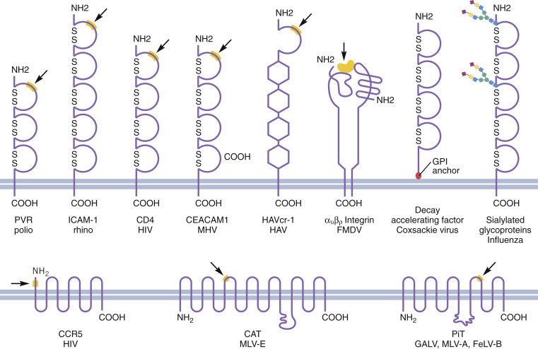
Virus receptors. Virus receptors are host cell molecules, most often glycoproteins on the plasma membrane, that not only bind virions but are often essential for subsequent virus infection (Figure 1 ). Viral receptors are naturally occurring cellular molecules that serve physiological functions for the cell—functions that have nothing to do with infection. Viruses usually bind to their receptors with higher affinity than they do to attachment factors.
Figure 1.

Molecular backbone cartoons of some viral receptors. Receptors diverge widely in their structure and physiological function. The amino and carboxy termini are shown, together with important disulfide bonds and the probable domains that bind virus. αvβ6: integrin chains (integrin dimers serve as receptors for many different viruses); ICAM: intercellular adhesion molecule; CCR5: chemokine receptor 5; CAT: cationic amino acid transporter; CEACAM: carcinoembryonic antigen-related cell adhesion molecule; HAVcr-1: hepatitis A virus cellular receptor 1; PiT: inorganic phosphate transporter; PVR: poliovirus receptor; Viruses: polio: poliovirus; rhino: rhinovirus, major group; FMD: foot and mouth disease virus; HAV: hepatitis A virus; HIV: human immunodeficiency virus; MHV: mouse hepatitis virus (a coronovirus); BLV: bovine leukemia virus; ALV-A: avian leukosis virus A; MLV-E: murine leukemia virus E; GALV: gibbon ape leukemia virus; MLV-A: murine leukemia virus A; FeLV-B; feline leukemia virus B.
Attachment of the virus particle to its cellular receptor is conferred by a virion surface protein, often called the viral attachment protein (VAP). As a rule, there is a single VAP although other viral surface proteins often play an essential role in the steps that follow the initial attachment of virions to the cell surface. For enveloped viruses, the VAP is a surface glycoprotein that oligomerizes to form spikes that protrude from the viral envelope. For nonenveloped viruses, the VAP is one of the surface proteins that form the external structure of the viral capsid. While binding to virus attachment factors is often electrostatic in nature, receptor binding usually occurs via a specific domain on the VAP, most often in the form of a pocket or “canyon” that subsequently interacts with a specific domain on the cellular receptor. These domains can be defined by structural studies and mutations introduced into the cellular receptor protein or the VAP.
Antibodies neutralize virus by binding to the VAP in a way that blocks receptor binding. Since the receptor-binding domain on a VAP often lies in a recessed pocket where it is not directly accessible to antibodies, antibodies that bind to epitopes distant from the pocket can also neutralize the virus. Therefore, viral mutants may escape neutralization without affecting receptor binding. Such escape mutants allow many different virus serotypes to use the same receptor-binding domain of the VAP, as seen for rhinoviruses. Neutralizing antibody escape mutants of this kind are important for the persistence of some viruses, including HIV.
Carbohydrates can also serve as virus receptors. The best studied example is binding of the influenza virus hemaglutinin protein to sialic acid (or N acetyl neuraminic acid), a modified sugar that is found on the tips of some of the branched carbohydrate side chains of glycosylated proteins and glycosphingolipids. Different influenza hemagglutinins bind preferentially to different terminal sialic acid residues, depending on the linkage of the sialic acid to a proximal galactose or galactosamine molecule in the carbohydrate chain. Thus, human-type A influenza viruses bind most avidly to sialic acid α-2,3 galactose configurations while avian-type A influenza viruses bind best to sialic acid α-2,6 galactose. This exquisite specificity of the interaction between the VAP and its cellular receptor helps explain the species tropism of influenza viruses, since different host species express different sialic acid linkages. In turn, this distinction can determine whether an avian or porcine influenza virus can efficiently infect humans (Shinya et al., 2006).
The role of virus receptors in entry. Why are receptors essential for infection whereas attachment factors are not? While virus attachment factors merely support virus binding, virus receptors do something else: they either induce conformational changes in viral proteins that are required for membrane penetration and/or result in delivery of virions to a cellular domain or compartment that is required for entry. This distinction explains why attachment factors are not required for virus infection whereas receptors are required. A host molecule can be considered to be a virus receptor if its elimination from an animal model or one or more cell types prevents infection. The loss of a receptor may not block infection of all cell types since some viruses can utilize more than one receptor. As an example, lab-adapted strains of measles virus use CD46 as a receptor to infect cells, while wild-type strains use CD150 as a receptor to infect lymphocytes, and nectin 4 to infect epithelial cells.
The importance of receptor specificity is exemplified by recent studies of dipeptidyl peptidase 4 (DPPT4), the receptor for MERS-CoV, the cause of a newly emerging infectious disease, Middle East respiratory syndrome (see Chapter 16, Emerging viral diseases). Humans express DPPT4, but mice, hamsters, or ferrets do not and are not susceptible to infection. Molecular studies show that five amino acids in the binding domain of DPPT4 differ between human and hamster cells. Furthermore, DPPT4 on camel cells binds the MERS-CoV attachment protein. This observation provided an explanation for the apparent role of camels as an intermediate host responsible for transmission of this virus to humans (van Doorenmalen et al., 2014). Marmoset cell DPPT4 binds MERS-CoV with affinity similar to that of human cells, and marmosets are an excellent model host for this virus (Falzarano et al., 2014).
1.2. Viral Entry
Viral entry is a multistep process that follows attachment of the virion to the cell surface and results in delivery of the viral genome to the site of replication, either in the cytosol or nucleus. The key step in virus entry is penetration of a cellular membrane. For enveloped viruses, delivery of the viral genome across the lipid bilayer of the virus and a cellular membrane is accomplished by a membrane fusion reaction. For nonenveloped viruses, the viral genome is usually delivered across a cellular membrane by a pore that is formed by protein components of the viral capsid. Virus fusion and penetration proteins exist in metastable states that must be triggered in some way to undergo the needed conformational changes.
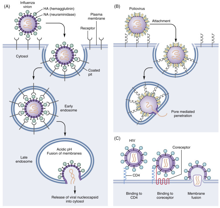
Triggers of virus entry. A number of cellular cues induce the irreversible conformational changes in viral proteins that lead to membrane fusion in the case of enveloped viruses, or membrane penetration in the case of nonenveloped viruses (Figure 2 ). One of the best understood is low pH. Many viruses, after binding to receptors on the cell surface, are endocytosed and delivered to endosomes where the low pH environment induces changes in viral structural proteins that mediate membrane fusion by protonating acidic residues (Helenius et al., 1980). The low pH-dependent entry of many enveloped viruses is exemplified by influenza virus. Nonenveloped viruses may also use this pathway, but instead of eliciting membrane fusion the acidic environment induces structural changes in the viral capsid that result in exposure of hydrophobic domains that insert into the cellular membrane, forming a pore through which the viral genome can pass (Figure 2).
Figure 2.

Pathways of virus entry into host cells. A. Entry of influenza virus. Key events are: attachment of the virion; internalization of the virion by endocytosis; lowering the pH (to <pH 5.5) of the endocytic vacuole leading to drastic reconfiguration of the viral attachment protein (hemagglutinin, HA1 and HA2); insertion of a hydrophobic domain of HA2 into the vacuolar membrane; fusion of the viral and vacuolar membranes; release of the viral nucleocapsid into the cytosol. This cartoon shows a nucleocapsid containing one of the eight genome segments of influenza virus. B. Entry of poliovirus. After binding to cell surface receptors, poliovirus is endocytosed and ultimately delivered to low pH endosomes. There, the low pH environment triggers conformational changes in the viral capsid that result in exposure of hydrophobic domains that insert into the endosomal membrane, forming a protein pore through which the viral genome can exit and enter the cytoplasm. C. Entry of HIV. HIV attaches to the cell surface via binding to various attachment factors such as DC-SIGN. The first required step for entry is binding of the viral envelope glycoprotein to CD4, a type 1 integral membrane protein. This binding event triggers structural alterations in the envelope glycoprotein that induce the exposure of a second receptor binding domain that engages the chemokine coreceptors CCR5 or CXCR4. Coreceptor binding triggers additional changes in envelope that enable it to elicit membrane fusion with the cell membrane.
While many viruses use low pH as a trigger to induce membrane fusion or penetration, other viruses use one or more receptors to trigger needed conformational changes for virus entry. HIV entry is an example of pH-independent entry. The HIV Env binds to CD4, a cell surface protein found on some types of T cells, macrophages, and DCs. CD4 binding induces conformational changes in Env that then enables it to bind to a second receptor, termed a coreceptor (Figure 2). The coreceptors for HIV are the chemokine receptors CCR5 and CXCR4, seven transmembrane domain receptors. Coreceptor binding induces further conformational changes in the Env protein that lead to membrane fusion (Wilen et al., 2012). Viruses can also employ other cellular cues. Binding of Rous sarcoma virus to its cellular receptor induces conformational changes that then make it responsive to low pH, while the Ebola virus glycoprotein must be cleaved by a host cell protease before it can undergo the changes needed for membrane fusion (Sakurai et al., 2015).
Other host determinants of viral tropism. While receptors and triggering mechanisms for virus entry are major determinants of viral tropism, other host factors can also influence cellular susceptibility. A number of enveloped viruses are not infectious when they bud from cells because one of the viral surface glycoproteins requires proteolytic cleavage by a host cell protease to be activated. Viruses that have Class I membrane fusion proteins (retroviruses, orthomyxoviruses, paramyxoviruses) are examples. In such instances, infectious virus is only produced by replication in cell types that express the appropriate protease, which is localized in the secretory pathway. Alternatively, some viral fusion glycoproteins may be cleaved by enzymes in extracellular fluid. In many instances, the degree of susceptibility to proteolytic cleavage is determined by a few amino acids (such as one vs several arginines) at the site of cleavage, so that mutations in 1–2 critical amino acids can alter the tissue tropism of a virus by making it more or less susceptible to this critically important proteolytic cleavage event.
A case in point is Newcastle disease virus, a paramyxovirus of birds. Virulent isolates of Newcastle disease virus encode a fusion protein that is readily cleaved by furin, a proteolytic enzyme present in the Golgi apparatus, so that the protein is activated during maturation prior to reaching the cell surface, and before budding of nascent virions. This makes it possible for virulent strains of the virus to infect many avian cell types, thereby increasing its tissue host range, and causing systemic infections that are often lethal. In contrast, avirulent strains of Newcastle virus encode a variant fusion protein that is not cleaved during maturation in the Golgi, so that nascent virus requires activation by an extracellular protease. The required protease is found only in the respiratory or enteric tracts, thereby limiting tropism to surface cells and conferring an attenuated phenotype on the virus (Panda et al., 2004). Host cell transcription factors can influence the tropism of some viruses. Papillomaviruses replicate in skin and may cause tumors, varying from benign warts to malignant cancer of the cervix. Papillomaviruses commence their replication in germinal cells that are permissive for replication of the viral genomes. However, germinal cells produce proteins that block the transcription of late structural genes of the virus. As the infected basal cells move outward and begin to differentiate, they become permissive for transcription and translation of the papillomavirus structural genes, so that complete infectious virus is only formed in cells that are about to be sloughed. Release of virus from the superficial layers of dead cells promotes the transmission of infection to new sites on the infected person and also to new uninfected hosts (Doorbar et al., 2012).
Physical factors that impact tropism. While the vast majority of human viruses replicate optimally at 37 °C, some mucosal surfaces such as the upper respiratory tract have a lower temperature, about 33 °C. Certain viruses, such as rhinoviruses that replicate in the epithelial cells of the nose and throat, have evolved to replicate optimally at 33 °C. As a result, such viruses are usually restricted in their tissue distribution by their relative inability to replicate at 37 °C, which limits their spread beyond the upper respiratory tract. The harsh environment of the gastrointestinal tract can impact virus tropism as well, due to the acid pH of the stomach, the alkaline pH of the intestine, and the destructive effects of pancreatic digestive enzymes. In general, enterotropism is limited to viruses that can survive these adverse conditions, although there are some exceptions.
1.3. Virus Structure and Replication Can Impact Pathogenesis
There are several links between viral structure and pathogenesis. Large viruses such as poxviruses or filamentous forms of viruses, such as influenza and Ebola, are simply too large to utilize clathrin-coated pits, caveolae, or other commonly used entry routes. Instead, these viruses trigger internalization by activating macropinocytosis—an example of viruses reprogramming cells to assist virus replication (Marsh and Helenius, 2006).
Another important structural feature is the surface of the virion. Enveloped viruses are not stable outside of the human body, and are typically transmitted by transfer of body fluids. In contrast, nonenveloped viruses are much more stable, and many can be transmitted by other mechanisms such as the fecal–oral route—this is how polio and many other GI viruses are transmitted. Hepatitis, from contaminated shellfish for example, is caused by hepatitis A, a nonenveloped virus that is stable outside of the human body. In contrast, hepatitis B and C viruses have envelopes, and are transmitted by sexual contact or by blood. The Caliciviruses that cause outbreaks of diarrhea on cruise ships are nonenveloped, making transmission by fomites much easier and sterilization more difficult.
Viral replication strategies are numerous, but there are several general features that provide links between replication and pathogenesis. DNA viruses that replicate in the nucleus must utilize a host cell pathway to transport their genome to the nucleus. This almost always entails interactions between the virus and the cell’s cytoskeleton. RNA polymerases lack proof reading capability, and have high mutation rates that enable the virus to evolve quickly in the face of new selective pressures, such as the adaptive immune response (see Chapter 17, Virus Evolution).
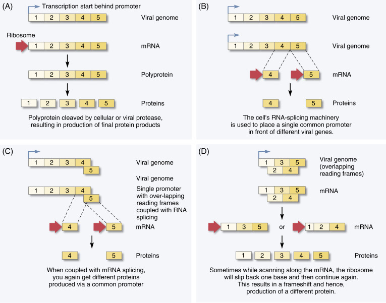
While only about 3% of the human genome actually codes for proteins, a very large fraction of viral genomes encode proteins. Viruses use four basic strategies to pack as much genetic information as possible into the smallest amount of genetic material (Figure 3 ). (1) Polyproteins: Instead of having one promoter for each viral gene, many viruses encode polyproteins. These polyproteins are translated, and cellular or viral proteases then cleave them into individual proteins. (2) Differential splicing: Some viruses use the cell’s machinery to splice their genome, and a single promoter is used to transcribe different viral genes. (3) Overlapping reading frames: By having start codons in several sites, virus genes can overlap. (4) Ribosomal frame-shifting: In some viruses, a ribosome starts translating a protein and then “slips” back one base before starting again. When this happens, it is now in a new reading frame, and thus begins translating a different viral protein. These mechanisms are not mutually exclusive—HIV employs all four, for example. The net effect is to pack an impressive punch within a small genome.
Figure 3.

How viruses pack maximum information into small genomes. A. Many viruses produce one or more large polyprotiens that are cleaved by cellular or viral proteases co- or posttranslationally, resulting in the generation of multiple viral proteins all of which were transcribed via a single promoter. B. Some viruses utilize the cell’s RNA splicing machinery, which makes it possible to place a single, common promoter in front of different viral genes. C. Some viruses use overlapping reading frames, which saves genetic space. D. Ribosomal frame-shifting is used by retroviruses. If the ribosome slips back a base and then proceeds, the reading frame is shifted and a different protein is produced.
2. Routes of Viral Transmission
Most human infections result from transmission of virus from another infected human. However, some viruses are transmitted to humans from animals (bunyaviruses, arenaviruses) or via an insect vector (Dengue, West Nile viruses)—a fact that has been recognized in the case of rabies virus for hundreds of years (Figure 4 ). In these cases, the virus has to interact with pathways present in the animal or insect vector as well in the human host. The transmission process must deliver virus to a specific site that harbors cells susceptible to infection, such as the respiratory or gastrointestinal tracts. Following transmission, the virus typically establishes a primary, local infection. Sometimes, symptoms result from replication at the primary site of infection and virus produced at this site can be spread to other hosts. Other viruses disseminate within the host, being delivered to other tissues and organs that support virus replication. Dissemination can occur by different pathways—lymphatic spread, hematogenous spread, neural spread—and reflects an impressive degree of virus–host adaptation. Upon initiation of infection, the innate and adaptive immune systems are activated and attempt to clear the virus. However, viruses often employ immune evasion strategies, and the balance between immune responses and viral evasion strategies does much to dictate the outcome (see Chapters 4, 5, and 6,Chapter 4Chapter 5Chapter 6 Innate, Adaptive, and Aberrant immunity).
Figure 4.

Historical illustration showing the transmission of rabies. Arabic painting by Abdallah ibn al-Fadl, Baghdad school, 1224.
Courtesy of the Freer Gallery of Art, Washington, DC.
Respiratory tract. This is a common route of infection, with transmission mediated by aerosolized droplets or infected saliva and nasopharyngeal secretions. Droplet size, which is affected by temperature and humidity, plays a major role in determining the anatomic site to which the virus is delivered, with larger droplets lodging in the nose and upper airways and smaller droplets in the alveoli. Innate defenses include mucus and cilia, which trap pathogens and deliver them to the digestive tract, secretory IgA, and alveolar macrophages. Some viruses tend to infect the pharynx—hence pharyngitis; others may infect epithelial cells in bronchioles, causing bronchitis. The site of initial replication can be impacted by the virus inoculum, the site in the respiratory tract to which it is delivered, and the tropism of the virus for distinct cell types within the respiratory system. Avian influenza provides a good example: In humans, there is an anatomical difference in the distribution of sialic acid linkages in the respiratory tract. The α-2,3 linkage preferred by avian influenza is found only deeper in the human respiratory pathway, while the α-2,6 linkage used by human influenza viruses is found in the upper respiratory tract, helping to explain the relative protection of humans from “bird flu.”
Gastrointestinal (GI) tract. Many viruses are transmitted via the fecal–oral route where the environment is challenging—very low pH in the stomach, an alkaline pH in the small intestine, proteases, and bile detergents that will inactivate most viruses. Thus, viruses that infect the GI tract are almost always nonenveloped, and have evolved the ability to survive in the GI environment. In fact, some viruses require these conditions. For reoviruses to infect cells, their outer surface proteins have to be cleaved by host cell proteases which then enables them to bind to M cells, which are found in the epithelium that overlies Peyer’s patches. The bound virus is internalized and transcytosed to the basolateral surface, enabling virus to spread beyond the epithelium (Danthi et al., 2013). There is considerable specialization, as different viruses enter and replicate in different locations throughout the GI tract, from the tonsils to the distal colon.
Urogenital tract. The urogenital tract is the preferred site of entry for a number of viruses, some of which are well adapted to infect epithelial cells and replicate locally while others use this route as a portal of entry to gain access to other tissues. Preexisting lesions can greatly increase the transmission of other viruses via breaches in the epithelium. This is a significant issue with HIV, where other sexually transmitted diseases enhance susceptibility.
Skin and mucous membranes. The intact skin is not a hospitable environment for viruses as it is covered by keratin and a layer of dying cells. As a result, infection via this route will require a break in the skin, which can be mechanical (a scratch), or the bite of an insect vector (a mosquito, tick, sandfly). Many viruses replicate in the dermis, a layer of highly vascularized tissue with fibroblasts and DCs that lies immediately below the epidermis. A smaller number of viruses replicate in the epidermis itself, notably the papillomaviruses. Direct infection of epithelial cells that line mucosal surfaces is more prevalent, though mucus and IgA serve as intrinsic barriers.
3. Viral Dissemination and Movement
Viruses have evolved strategies to usurp host cell pathways to move at both the microscopic scale within and between cells, and the macro scale, within the host.
3.1. Movement on the Microscope Scale
Movement of virions along the surface of a cell. The plasma membrane is structurally heterogeneous. There are tight junctions in polarized epithelial cells, lipid rafts that serve to concentrate some cell surface proteins and exclude others, and projections such as microvilli. This means that virions not only have to bind to the surface of a host cell, but must either bind or migrate to the location on the cell surface that provides molecules or endocytic pathways needed for entry. Many viruses have evolved mechanisms that result in their directed movement on the surface of the plasma membrane (“virus surfing”) until they reach a location that is compatible with entry (Lehmann et al., 2005).
A notable example of this process is provided by Group B coxsackie viruses, nonenveloped virions that must bind to the coxsackie–adenovirus receptor (CAR). However, coxsackie viruses infecting the apical surface of polarized epithelial cells are faced with a conundrum: CAR is strictly localized at tight junctions and is not accessible to virions that attach to the apical plasma membrane. How then can the virus and its receptor interact? On polarized epithelial cells, coxsackie virus first binds to decay-accelerating factor (DAF), a GPI-anchored protein that is relatively evenly distributed on the apical surface of the plasma membrane. Virus binding to DAF triggers the activation of Abl kinase that in turn initiates Rac-dependent actin rearrangements, which result in rapid movement of DAF, with the associated virus, to tight junctions, where it can then bind to its primary receptor and be internalized via caveolae (Coyne and Bergelson, 2006).
Viruses can trigger their own internalization into cells. Many viruses have to be endocytosed by the host cell, and there are many pathways by which this occurs (Marsh and Helenius, 2006). Some viruses enter cells via constitutive endocytic pathways (coated pits/vesicles, caveolae), perhaps assisted by virus-induced cross-linking of cell surface receptors. However, many viruses utilize nonclathrin-dependent pathways, and transduce signals upon binding to the cell surface that causes the cell to engulf the virus by micropinocytosis, a mechanism employed by cells to internalize large particles.
Active dissemination: movement within a cell via microtubules. Once within the cytoplasm, a virus must deliver its genome to the site of virus replication. This can be a considerable distance, particularly if replication occurs in the nucleus. However, the highly viscous nature of the cytoplasm and the extensive cytoskeletal network immobilizes virions in the cytosol. To move within a cell, virions must utilize cellular pathways, often involving the microtubule network (Urnavicius et al., 2015). Interactions with microtubules can be indirect: A virus can be internalized and delivered to a vesicle that itself is transported along microtubules. Alternatively, a number of viruses interact directly with the microtubule machinery to effect retrograde (away from the nucleus) or anterograde (toward the nucleus) transport.
A particularly well-understood example of viral movement along microtubules in both directions is provided by HSV-1 which spreads from its primary site of infection at a mucosal surface by entering local nerve endings. After membrane fusion, the viral capsid is released into the cytosol and interacts with dynein, a motor protein that transports cargo toward the microtubule organizing center that is in close proximity to the nucleus. Viral capsids are thus transported, from the site of entry to a location near the nucleus, in an efficient and rapid manner over distances that can be as long as several feet in the case of retrograde movement within an axon. Following replication, newly assembled virions need to be transported in the opposite direction, which is accomplished by specific interactions to kinesin, a molecular motor responsible for anterograde transport along microtubules (Radtke et al., 2006).
Herpes zoster, like HSV-1, establishes a latent infection in sensory ganglia by moving in a retrograde fashion, from the periphery to the nerve bodies in the ganglia. Upon reactivation, which can be caused by triggers such as stress, newly synthesized particles move in an anterograde fashion along axons, delivering virus to the periphery where it forms a painful rash that involves a single dermatome (area of skin supplied by a single sensory ganglia). Thus, the lesions are typically unilateral, and can keep appearing in the same place (as cold sores do around the mouth).
Viruses can also move efficiently from cell to cell. Just as viruses can utilize cellular pathways to effect directed movement along the cell surface and through the cytoplasm, newly assembled viruses can utilize similar pathways to move to the cell surface and achieve efficient transmission between cells. Poxviruses, after assembling in the cytosol, move along microtubules to the cell surface. Some viruses preferentially bud at the site of cell–cell contact, which results in the formation of a structure termed the virologic synapse, and makes transfer of virus to the new host cell a very efficient process (Mothes et al., 2010).
Another mechanism used by viruses to effect directed transfer to an adjoining cell utilizes surface-based glycosylaminoglycans. In cells chronically infected with murine leukemia virus, newly assembled and budded virus particles are not immediately released, but are bound to glycosylaminoglycans on the cell surface. There is a concentration of viruses to the infected cell periphery, enabling them to be efficiently transmitted to neighboring cells (Mothes et al., 2010).
Finally, some viruses use actin to move between cells. Newly synthesized vaccinia virus particles have actin-binding proteins at one end of the brick-shaped virion. Actin polymerizes at the end of the virus, which propels the virion. The virus can actually be pushed out of the cell by this actin “rocket,” still enshrouded by the plasma membrane. If this projection impales an adjoining cell, the membrane “shroud” then breaks off and the virus finds itself in a new host. A virus that moves in this way from cell to cell, does not enter the extracellular space, and avoids neutralization by extracellular antibodies (Cudmore et al., 1995).
3.2. Long-Range Movement within the Host
Viruses can undergo long-range dissemination within the host, utilizing the lymphatics, the blood, or the peripheral nervous system. HIV is a good example of a virus that can spread via lymphatics as cell-associated virions. During sexual transmission, HIV must cross the epithelium, perhaps through a tear or an abrasion. DCs express lectins—such as DC-SIGN and the macrophage mannose receptor—on their surface, and are among the first cell type that HIV encounters. HIV binds avidly to these lectins, and once bound to DCs, the virus is internalized and retained in an intracellular compartment. Cross-linking of DC-SIGN triggers migration of DCs to regional lymph nodes. Once in the lymph node, HIV is returned to the DC surface and is surrounded by T cells that it can infect. Thus, for all intents and purposes, HIV uses the DC as a “taxi” for transport from a mucosal surface to a more proximal lymphoid organ.
The most common mode of dissemination is via blood (viremia), and blood-borne virus can circulate either cell-free or as cell-associated virions. Poliovirus moves through the lymphatics to reach regional lymph nodes, eventually draining into the blood stream, and reaching the central nervous system (CNS). Because poliovirus circulates as cell-free virions in the plasma, humoral neutralizing antibody can prevent virus from reaching the CNS, and protect against paralytic poliomyelitis. This is the mechanism that underpins the protective efficacy of inactivated poliovirus vaccine.
Selected viruses can spread via the peripheral nervous system. The classical example is rabies virus that does not cause a viremia but only spreads from the point of infection to the CNS and then to the salivary glands. As noted above, viruses that spread via peripheral nerves utilize intracellular transport mechanisms to “hitch a ride” within the neuron.
4. Virus Shedding and Transmission
Virus must be shed to be transmitted to a new, naive host. Viruses may be discharged into respiratory aerosols, feces, or other body fluids or secretions, and each of these modes is important for selected agents. Nonenveloped viruses are more resistant to desiccation and other environmental extremes than are viruses surrounded by lipid bilayers. For this reason, viruses transmitted by the fecal–oral route are generally nonenveloped viruses, while fragile enveloped viruses are usually transmitted by body fluids typically within a short time after release. Viruses that cause acute infections are usually shed intensively over a short time period, often 1–4 weeks, and transmission tends to be relatively efficient. Viruses such as HBV and HIV, that cause persistent infections, can be shed at lower titers for months to years but will eventually be transmitted during the course of a long-lasting infection.
Oropharynx and gastrointestinal tract. Enteroviruses may be shed in pharyngeal fluids and feces. Poliovirus replicates in the lymphoid tissue of the tonsil and in Peyer’s patches (lymphoid tissue accumulations in the wall of the small intestine) whence it is discharged into the intestinal lumen. Other viruses may be excreted into feces from the epithelial cells of the intestinal tract (reoviruses and rotaviruses) or from the liver via the bile duct (hepatitis A virus).
Respiratory tract. Viruses that multiply in the nasopharynx and respiratory tract may be shed either as aerosols generated by sneezing or coughing, or in pharyngeal secretions that are spread from hand to mouth. Often, transmission is via contaminated fomites, such as handkerchiefs, clothing, or toys.
Skin. Relatively few viruses are shed from the skin. Papillomaviruses and certain poxviruses that cause warts or superficial tumors may be transmitted by mechanical contact. A few viruses, such as variola virus, the cause of smallpox, and varicella virus, the cause of chickenpox, that are present in skin lesions can be aerosolized and transmitted by the respiratory route. In fact, it is claimed that the earliest instance of deliberate “biological warfare” was the introduction into Indian tribes of blankets containing desquamated skin from smallpox cases.
Mucous membranes, oral, and genital fluids. Viruses that replicate in mucous membranes and produce lesions of the oral cavity or genital tract are often shed in pharyngeal or genital fluids. An example is herpes simplex virus (type 1 in the oral cavity and type 2 in genital fluids). A few viruses are excreted in saliva, such as Epstein Barr virus, a herpesvirus that causes infectious mononucleosis, sometimes called the “kissing disease.” The most notorious example is rabies virus, which replicates in the salivary gland and is transmitted by a bite that inoculates virus-contaminated saliva. Several important human viruses, such as HBV and HIV, may be present in the semen.
Blood, urine, milk. Blood is an important potential source of virus infection in humans wherever transfusions, injected blood products, and needle exposure are common. In general, viruses transmitted in this manner are those that produce persistent viremia, such as HBV, HCV, HIV, and cytomegalovirus. Occasionally, viruses that produce acute short-term high titer viremias, such as Ebola virus or parvovirus B19, may contaminate blood products. Although a number of viruses are shed in the urine, this is usually not an important source of transmission. One exception is certain animal viruses that are transmitted to humans; several arenaviruses are transmitted via aerosols of dried urine. A few viruses are shed in milk and transmitted to newborns in that manner. The most prominent example is HIV, though visna-maedi virus of sheep and mouse mammary tumor virus can also be transmitted via milk.
Transmission. Enteric viruses are commonly transmitted by oral or fecal contamination of hands, with passage to the hands and thence the oral cavity of the next infected host. Inhalation of aerosolized virus is the major mode of transmission for respiratory viruses. Another significant route is by direct host-to-host interfacing, including oral–oral, genital–genital, oral–genital, or skin–skin contacts. Transmission may involve less natural modes such as blood transfusions, organ transplants, or reused needles. In contrast to propagated infections are transmissions from a contaminated common source, such as food, water, or biologicals. Common source transmission is quite frequent and can produce explosive outbreaks that range in size depending on the number of recipients of the tainted vehicle and the level of virus contamination. One of the most infamous was the World War II epidemic of HBV among military personnel, transmitted by a contaminated batch of 17D yellow fever vaccine.
Sexually transmitted viruses present a special situation, since the probability of spread depends upon the gender and type of sexual interaction between infected host and her/his uninfected contact. For instance, an HIV-infected male is more likely to transmit to a female partner via anal than vaginal intercourse, and that risk is reduced if the male partner is circumcised.
Transmission of arboviruses is complex, since it involves the cycle between an insect vector and a vertebrate host. There are a number of quantitative variables that determine the efficiency of vector transmission. Vertebrate host determinants include the titer and duration of viremia, while insect determinants include the competence of the vector (that is, the ability of the vector to support viral replication in several tissues and shed virus in its saliva) and the extrinsic incubation period (the interval between ingesting the virus and shedding in the saliva), as well as the distinctive feeding preferences of each insect vector. Also, there are a number of alternative patterns of viral maintenance in the vector, including overwintering of virus in hibernating mosquitoes, transovarial transmission of the virus, and venereal spread between male and female mosquitoes.
5. Patterns of Virus Infection
Viruses can cause acute, chronic, or latent infections. Most viruses cause acute, self-limited infections, and most infections in turn are asymptomatic. Symptomatic infections are preceded by an asymptomatic incubation period, the length of which can be related to the inoculum (dose of the virus), the rate of replication, and many other factors. For a virus to initiate a persistent infection, a set of special circumstances must exist that permit the virus to escape the adaptive immune response. The general principles associated with each of these infection patterns are described in Chapter 7, Patterns of Infection.
6. Major Disease Mechanisms
Viruses can cause disease in many ways, which are discussed in subsequent chapters. Here, we will briefly review the major mechanisms involved.
Direct cytopathic effects. Many viruses kill cells, directly by lysis or by inducing apoptosis, and disease can result from loss of parenchymal cells. An example is West Nile virus which infects neurons and induces apoptosis via caspase 3, leading to encephalitis and movement disorders. Another example is Ebola virus. Individuals infected with the Zaire strain of Ebola virus typically develop a hemorrhagic fever, with loss of vascular integrity. The spike protein of Ebola virus appears to be a major culprit; it induces loss of contact with neighboring cells, which plays a role in the vascular leakage and hypotension that are characteristic of fatal Ebola hemorrhagic shock syndrome (Feldman and Geisbert, 2011).
Another example is syncytia formation. Enveloped viruses that bud from the plasma membrane deliver their glycoproteins to the cell surface. These viral proteins can bind to receptors on the surface of adjoining cells, eliciting cell–cell fusion, and syncytia formation. Respiratory syncytial virus, the major cause of viral pneumonia in young children, derives its name from the fact that it can form syncytia not only in tissue culture, but also in the lungs of infected patients.
Disease caused by antibody-mediated immunity. Some viruses, such as hepatitis B, release large amounts of antigen into the blood. Antibodies can bind to the viral antigens, leading to the formation of immune complexes that are deposited in the basement membranes of glomeruli in the kidney, leading to renal dysfunction.
Another example of antibody-mediated diseases is dengue, caused by a mosquito-borne virus that infects millions of people a year. There are four major dengue virus serotypes. Antibodies that neutralize one serotype do not neutralize the others. When a human is infected with a second serotype, antibodies produced against the first serotype bind to, but do not neutralize, the second serotype. The virus is now partially coated with non-neutralizing antibodies, enabling it to bind to cells—such as monocytes and macrophages—that have Fc receptors on their surface. This results in the efficient infection of these cells with massive release of cytokines and subsequent vascular leakage and hemorrhage (Paessler and Walker, 2013). As a consequence, there is a chance that the patient will now develop dengue hemorrhagic fever/dengue shock syndrome, which carries significant mortality.
Disease caused by virus-initiated autoimmunity. Viral antigens are sometimes similar to host antigens so that antibody or cellular responses directed against a pathogen may also cross-react with normal host molecules and cells. It is widely thought that some immunopathological diseases are the result of molecular mimicry, such as the demyelinating diseases like multiple sclerosis (MS) and Guillain–Barré syndrome (GBS). GBS is often preceded by a viral illness. It is hypothesized that a viral peptide presented by the class I pathway resembles a cellular protein (the myelin basic protein), and triggers an autoimmune response that results in demyelination.
Diseases associated with innate immunity and a cytokine “storm.” Recent studies of the reconstructed 1918 strain of influenza virus suggest that the severe disease was due to an exuberant host response in the lungs that led to pulmonary edema and respiratory failure. It is postulated that an excess of proinflammatory cytokines triggered this excessive response (Tisoncik et al., 2012; Peng et al., 2014). Another example is Ebola virus disease, a hemorrhagic fever, accompanied by very high temperature and a clinical crisis. It is postulated that a cytokine storm plays a major role in acutely fatal cases.
Disease caused by virus-induced immunosuppression. Some virus infections can lead to immunosuppression, making the human host susceptible to other infectious agents (see Chapter 6, Aberrant Immunity). Measles virus suppresses the secretion of certain cytokines, leading to transient immunosuppression. Measles kills 150,000 children annually, but deaths are usually not the direct result of the virus, but rather via virus-enhanced infection with other viruses or bacteria.
Disease caused by virus-induced tumorigenesis. Oncogenic viruses are the subject of Chapter 9. Suffice it to say that RNA tumor viruses transform via different mechanisms than those used by DNA tumor viruses. However, in all instances, there is a final common pathway, with a release of the “brakes” on the cell cycle which limit the replication of normal cells. Tumorigenesis involves a multistep process that confers on virus-transformed cells; the ability to grow in the host animal.
7. Reprise
The steps in viral infection begin with infection of individual cells followed by spread through a multicellular host organism. At the cellular level, viruses use attachment factors to associate with the cell surface and then specific cellular receptors to initiate attachment. Entry is a multistep process that involves membrane fusion of enveloped viruses or conformational changes in naked capsids, to deliver the viral genome to either the cytoplasm or nucleus. In general, RNA viruses replicate in the cytoplasm and DNA viruses in the nucleus, with a few notable exceptions, such as poxviruses and retroviruses. The virus uses viral or host enzymes to replicate its genome and for transcription and translation of viral proteins. In many instances, viruses shanghai normal cellular metabolism to support their replication and assembly. Shedding of newly synthesized virions is implemented by lysis of the infected cell to release encapsidated viruses or budding from the plasma membrane to release enveloped viruses. Host cells encode some antiviral proteins and many viruses have evolved countermeasures to evade these intrinsic defenses and to prolong the life of the virus-infected cell by blocking apoptosis or necrosis.
At the level of the host organism, there are many different routes of infection, each of which is characteristic for a specific virus. Viruses usually initiate infection via inhalation, ingestion, or penetration of the skin or mucous membranes. Virions often spread through the lymphatics to the lymph nodes where many of them replicate, to be discharged into the blood as free or cell-associated particles. Blood-borne viruses invade and replicate in various target organs, again characteristic for each virus. Spread from an infected animal or human is usually by release from either the lung, mouth and gastrointestinal tract, mucous membranes, or skin.
Viral infections range from innocuous to lethal. Some viruses are not cytocidal and do not kill infected cells; likewise many viral infections of human or animal hosts occur without apparent illness. However, viruses can cause pathological changes through a variety of mechanisms. These include direct tissue injury such as seen in smallpox, HAV hepatitis, paralytic poliomyelitis, and herpes simplex encephalitis. Virus-initiated immune-mediated diseases include HBV acute hepatitis, dengue hemorrhagic fever, and some cases of influenza pneumonia. Diseases associated with immunosuppression include measles, pneumonia, and congenital rubella. Virus-initiated cancers of humans include cervical cancer (HPV), lymphoma (HTLV-1), and hepatocellular carcinoma (HBV and HCV).
Further Reading
Reviews
- Danthi, P., G.H. Holm, T. Stehle and T.S. Dermody. 2013. Reovirus receptors, cell entry and proapoptotic signaling. Adv. Exp. Med. Biol. 790:42–71. [DOI] [PMC free article] [PubMed]
- Doorbar, J., W. Quint, L. Banks, I.G. Bravo, M. Stoler, T.R. Broker and M.A. Stanley. 2012. The biology and life-cycle of human papillomaviruses. Vaccine 30, Supp. 5:F55–F70. [DOI] [PubMed]
- Feldmann H, Geisbert TW. Ebola hemorrhagic fever. Lancet 2011, 377: 849–862. [DOI] [PMC free article] [PubMed]
- Liang C, Oh B-H, Jung JU. Novel functions of viral anti-apoptotic factors. Nature Reviews Microbiology 2015, 13: 7–15. [DOI] [PMC free article] [PubMed]
- Marsh, M. and A. Helenius. 2006. Virus entry: Open sesame. Cell 124:729–740. [DOI] [PMC free article] [PubMed]
- Paessler, S. and D.H. Walker. 2013. Pathogenesis of the viral hemorrhagic fevers. Annu. Rev. Pathol. Mech. Dis. 8:411–440. [DOI] [PubMed]
- Radtke, K., K. Döhner and B. Sodeik. 2006. Viral interactions with the cytoskeleton: a hitchhiker’s guide to the cell. Cell. Microbiol. 8:387–400. [DOI] [PubMed]
- Taubenberger, J.K. and D.M. Morens. 2006. 1918 Influenza: the Mother of All Pandemics. Emerg. Infect. Dis. 12:15–22. [DOI] [PMC free article] [PubMed]
- Tisoncik JR, Korth MJ, Simmons CP, et al. Into the eye of the cytokine storm. Microbiology and Molecular Biology Reviews 2012, 76: 16–32. [DOI] [PMC free article] [PubMed]
- Wilen, C.B., J.C. Tilton and R.W. Doms. 2012. HIV: Cell binding and entry. Cold Spring Harbor Perspectives in Medicine Volume: 2 Issue: 8 Article Number: a006866. [DOI] [PMC free article] [PubMed]
Original Contributions
- Chen Y-H, Due W, Hagemeijer MC, et al. Phosphatidylserine vesicles enable efficient en block transmission of enteroviruses. Cell, 2015: 160: 619–630. [DOI] [PMC free article] [PubMed]
- Coyne, C.B. and J.M. Bergelson. 2006. Virus-induced Abl and Fyn kinase signals permit coxsackievirus entry through epithelial tight junctions. Cell 124:119–131. [DOI] [PubMed]
- Cudmore, S., P. Cossart, G. Griffiths and M. Way. 1995. Actin-based motility of vaccinia virus. Nature 378:636–638. [DOI] [PubMed]
- Falzarano D, Feldmann H. Delineating Ebola entry. Science 2015, 347: 947–948. [DOI] [PubMed]
- Falzarano D, de Wit E, Feldman H, et al. Infection with MERS-CoV causes lethal pneumonia in the common marmoset. PLoS Pathogen 2014, 10: e1004250. [DOI] [PMC free article] [PubMed]
- Geijtenbeek TB, Kwon DS, Torensma R, van Vliet SJ, van Duijnhoven GC, Middle J, Cornelissen IL, Nottet HS, KewalRamani VN, Littman DR, Figdor CG, van Kooyk Y. “DC-SIGN, a dendritic cell-specific HIV-1-binding protein that enhances trans-infection of T cells.” Cell 100: 587–97. [DOI] [PubMed]
- Helenius, A., J. Kartenbeck, K. Simons and E. Fries. 1980. On the entry of Semliki Forest virus into BHK-21 cells. J. Cell Biol. 84:404–420. [DOI] [PMC free article] [PubMed]
- Mothes, W., N.M. Sherer, J. Jin and P. Zhong. 2010. Virus cell-to-cell transmission. J. Virol. 84:8360–8368. [DOI] [PMC free article] [PubMed]
- Panda, A., Z. Huang, S. Elankumaran, D.D. Rockemann and S.K. Samal. 2004. Role of fusion protein cleavage site in the virulence of Newcastle disease virus. Microb. Pathog. 36:1–10. [DOI] [PMC free article] [PubMed]
- Peng X, Alfoldi J, Gori K, et al. The draft sequence of the ferret (Mustela putorius furo) facilitates study of human respiratory disease. Nature Biotechnology 2014, 32: 1250–1255. [DOI] [PMC free article] [PubMed]
- Rustagi A, Gale M Jr. Innate antiviral immune signaling, viral evasion and modulation by HIV-1. J Molecular Biology 2014, 426: 1161–1177. [DOI] [PMC free article] [PubMed]
- Shinya, K., M. Ebina, S. Yamada, M. Ono, N. Kasai and Y. Kawaoka. 2006. Avian flu: Influenza virus receptors in the human airway. Nature 440:435–436. [DOI] [PubMed]
- Sakurai Y, Kolokoltsov AA, CC, et al. Two-pore channels control Ebola virus host cell entry and are drug targets for disease treatment. Science 2015, 346: 955–960. [DOI] [PMC free article] [PubMed]
- Urnavicius L, Zhang K, Diamant AG, et al. the structure of the dynactin complex and its interacton with dynein. Science 2015, 347. [DOI] [PMC free article] [PubMed]
- Van Doremalen N, Mazgowics KL, Milne-Price S, et al. Host species restriction of Middle East Respiratory Syndrome coronavirus through its receptor, dipeptidyl peptidase 4. J Virology 2014, 88: 9220–9232. [DOI] [PMC free article] [PubMed]
- Yang, Z.Y., H.J. Duckers, N.J. Sullivan, A. Sanchez, E.G. Nabel and G.J. Nabel. 2000. Identification of the Ebola virus glycoprotein as the main viral determinant of vascular cell cytotoxicity and injury. Nat. Med. 6:886–889. [DOI] [PubMed]


