CALVES
Escherichia coli
Escherichia coli is a normal inhabitant of the gastrointestinal tract of warm-blooded animals and ubiquitous in the farm environment. Disease caused by E. coli in calves may present as enteric or septicemic illness and is an important cause of neonatal mortality in dairy calves. Failure of passive transfer and management practices that allow exposure of the neonatal calf to large numbers of E. coli are of central importance in the pathogenesis of disease. A plethora of E. coli serotypes exist on dairy farms. These gram-negative organisms are classified based on various serologic and antigenic parameters, including cell wall or somatic (O) antigens, capsular (K) antigens, pilar or fimbrial (F) antigens, and flagellar (H) antigens. Heretofore, pilus antigens were sometimes classified as K antigens, but recent reference to pilus antigens as F antigens reduces confusion in this area.
Septicemia (Septicemic Colibacillosis, Colisepticemia)
Etiology.
Colisepticemia in neonatal calves can be considered a disease of poor management. Failure of passive transfer is the primary risk factor for this disease. Colostral transfer of immunoglobulins may be compromised by short dry periods, preparturient leaking of colostrum, assumption that a calf has nursed colostrum just because it is left with the dam for 24 hours, primiparous heifers that have poor-quality colostrum, and many other factors. In addition, poor maternity area and poor calf pen hygiene promote exposure of the calf to the multitude of strains of E. coli capable of causing septicemia. Filthy conditions, calving areas that are dirty, wet, overcrowded, or overused, and failure to dip navels are additional factors that predispose to this problem. Sanitation and hygiene with respect to collecting, storing, and administering colostrum are also emerging as important factors in the provision of adequate passive transfer and the prevention of colibacillosis.
Invasive E. coli of many subgroups are capable of opportunistic, septicemic infection of neonatal calves. Various reviews suggest an involvement of a multitude of possible E. coli types. Variations may be explained by geographic or environmental differences.
Calves with less than 500 mg IgG/dl are very prone to septicemic E. coli, and those with 500 to 1000 mg IgG/dl are defined as having partial failure of passive transfer and are also at increased risk. Adequate passive immunoglobulin that ensures at least 1000 IgG mg/dl serum (10 mg/ml serum) or preferably 1600 mg/dl serum is likely to prevent the disease.
Septicemia caused by E. coli most commonly occurs from 1 to 14 days of age. The onset of disease tends to occur earlier in this timewhen frame calves are exposed to high numbers of E. coli soon after birth (i.e., in the maternity pen). Poor or nonexistent transfer of passive immunoglobulins to the calf also hastens the onset of disease. Invasive E. coli may gain entrance through the navel, intestine, or nasal and oropharyngeal mucous membranes. Once invasion and septicemia occur, clinical signs develop rapidly and usually are apparent within 24 hours. Calves with partial failure of passive transfer or those exposed to less virulent E. coli strains may develop more chronic signs of disease over several days.
Septicemic calves shed the causative E. coli in urine, oral secretions, nasal secretions, and later in the feces, provided they survive long enough to develop diarrhea. Thus transmission may occur among communally housed calves, crowded calves, or uncleaned maternity stalls because of the heavily infected secretions of sick and septicemic calves. Because septicemic calves can shed large numbers of the organism before clinical signs are evident, contamination of communal pens and common-use feeding devices (e.g., esophageal feeding tubes) and direct contact with the infected calf or its feces or urine may promote spread of infection. Infected calves allowed to remain in the maternity area will amplify the level of environmental contamination, thereby placing other neonates born in that area at risk. Similar amplification may occur in calf housing areas and reinforces the biosecurity need for spatial and temporal separation between occupants, as well as the appropriate and routine disinfection of calf housing.
Clinical Signs.
Peracute signs of depression, weakness, tachycardia, and dehydration predominate when highly virulent strains of E. coli cause septicemia. Affected calves usually are less than 7 days of age and may be less than 24 hours old. Although often present early on, fever is usually absent by the time obvious clinical signs of disease occur, when endotoxemia and the resultant poor peripheral perfusion often render the animal normothermic or hypothermic. Exceptions to this rule are calves with peracute disease that collapse when exposed to direct sunlight on hot days—such calves can be markedly hyperthermic. Signs of dehydration are mild to moderate in most cases. The suckle reflex is greatly reduced or absent, and the vasculature of the sclerae is markedly injected. Petechial hemorrhages may be visible on mucous membranes and extremities, particularly the pinnae of the ears (Figure 6-1 ). The limbs, mouth, and ears are cool to the touch. Affected calves show progressive weakness and lethargy, often becoming comatose before death. Diarrhea is often seen but may not be apparent in peracute cases.
Figure 6-1.

Ear of 5-day-old Jersey calf demonstrating petechial hemorrhage associated with E. coli septicemia.
Evidence of localization of infection in certain tissues may become apparent in cases that survive the acute disease. Hypopyon may be present, as may uveitis, which is evidenced by miotic pupils with increased opacity to the aqueous fluid (“aqueous flare”). Hyperesthesia, paddling, and opisthotonus (Figure 6-2 ) are signs suggestive of septic meningitis. Lameness may result from bacterial seeding of joints and/or growth plates. Signs of omphalophlebitis may be present. Weakness, poor body condition, and recumbency secondary to weakness or joint or bone pain may be present in chronic cases.
Figure 6-2.

Seven-day-old Holstein calf with opisthotonus associated with E. coli meningitis and septicemia as a result of failure of passive transfer.
Clinical signs of acute septicemia may be difficult to differentiate from those of acute enterotoxigenic E. coli (ETEC) infection because dehydration, weakness, and collapse may be common to both. However, septicemic calves tend to be less dehydrated and have less watery diarrhea than calves with ETEC diarrhea; further, diarrhea tends to develop in the terminal stages of septicemia. Historical data may indicate other neonatal calves have recently shown similar signs or died at less than 2 weeks of age. Other differential diagnoses for acute colisepticemia include asphyxia or trauma during birth, simple hypothermia and/or hypoglycemia, septicemia caused by Salmonella spp., and congenital defects of the central nervous or cardiovascular systems. Polyarthritis caused by Mycoplasma spp. is an important differential diagnosis for septic arthritis secondary to colisepticemia but tends to be seen in older calves, and feeding unpasteurized milk is a significant risk factor for Mycoplasma disease. Salt poisoning, hypoglycemia, congenital neurologic disorders, traumatic injuries, and intoxications (e.g., lead) should be considered as differential diagnoses for meningitis secondary to colisepticemia. We have also seen several herds in recent years with young calves presenting with neurologic signs indistinguishable from meningitis for which the ultimate diagnosis was ionophore toxicity caused by overdosing before feeding milk replacer. Failure of passive transfer and meningitis were not involved.
Ancillary Data.
Calves suffering from peracute E. coli septicemia often have elevated packed cell volumes resulting from dehydration and endotoxic shock. The total white blood cell (WBC) count is variable but is frequently low or within normal ranges. Generally a left shift is observed, and toxic changes (e.g., azurophilic cytoplasm, nuclear hypersegmentation, and Dohle bodies) are often apparent on cytologic examination of blood neutrophils. Plasma fibrinogen concentration is variable. Hypoglycemia is a common finding, and metabolic acidosis, although common, usually is less severe than in calves recumbent as a result of ETEC. In fact, an acid-base and electrolyte determination that does not demonstrate a severe metabolic acidosis in a recumbent, diarrheic, dehydrated calf less than 14 days of age is a grave sign and usually portends septicemia. Severe hypoglycemia would be the only other laboratory finding that might indicate an easily treatable condition. Blood cultures provide the greatest specific diagnostic aid, but results may not be forthcoming in time to help the patient.
Acute, subacute, and chronic septicemic calves may have detectable clinical signs of localization of infection that allow a more definitive diagnostic test (e.g., cerebrospinal fluid [CSF] tap for patients showing signs of meningitis or joint tap to confirm septic arthritis) (Figure 6-3 ). In chronic cases, the serum immunoglobulin concentration (and serum total globulin concentration) may be normal or increased as a result of de novo synthesis of antibodies in response to the well-established bacterial infection.
Figure 6-3.

A 1-week-old calf affected with subacute E. coli septicemia. The calf has fever, diarrhea, dehydration, and a septic carpal joint. The calf had inadequate immunoglobulin levels.
Diagnosis.
Whenever clinical signs suggest the diagnosis, the calf's serum immunoglobulin levels should be analyzed. Although adequate levels of IgG do not rule out the disease, calves with IgG .1600 mg/dl serum based on a single radial immunodiffusion test are unlikely to suffer septicemic E. coli infections. Specific laboratory evaluation of immunoglobulin levels is preferable to field techniques when confirmation of failure of passive transfer (FPT) is essential but may not provide rapid results in the field. Therefore even though dehydration may falsely elevate blood protein levels, these may be useful field tests. Adequate immunoglobulin levels are suggested by serum total protein .5.5 g/dl in clinically ill calves. Serum gamma glutamyl transferase (GGT) activity .50 IU/L is a reliable indicator of adequate passive transfer in ill calves, as is development of visible turbidity in the 18% solution of sodium sulfite turbidity test. Several commercial “quick tests” (e.g., Midlands Bio-Products, Boone, IA) are now available for determining IgG concentration in calves.
Blood cultures provide definitive diagnosis but usually provide this information too late to be of practical value. When multiple calves are affected, however, blood cultures can help to differentiate E. coli septicemia from septicemia caused by other pathogens (e.g., Salmonella spp.); this differentiation is relevant for determining the source of infection and initiation of preventive measures. Further, antimicrobial sensitivity testing of blood culture isolates may aid in directing therapy for subsequent cases. Clinicians and producers should be aware of the differences between specific antigenic strains of E. coli (e.g., ETEC) capable of producing severe disease in calves with adequate passive transfer and the everyday, commensal, and environmental E. coli often associated with sepsis caused by FPT. This is an important distinction, lest clients concentrate preventive efforts and management on specific vaccination programs rather than colostrum and neonatal calf management.
Treatment.
Treatment of peracute E. coli septicemia usually is unsuccessful because of overwhelming bacteremia and endotoxemia in the patient. Signs progress so quickly that most septicemic calves are recumbent and comatose by the time of initial examination. Shock, lactic acidosis, hypoglycemia, and multiple organ failure are common in peracute cases.
If treatment is attempted, correction of endotoxic shock and acid-base and electrolyte abnormalities, effective antimicrobial therapy, and nutritional support are the primary goals. Intravenous (IV) balanced electrolyte solutions should contain dextrose (2.5% to 10%) and sodium bicarbonate (20 to 50 mEq/L if the plasma bicarbonate concentration is, 10 mEq/L) to address hypoglycemia and metabolic acidosis. Adjustments of the concentration of dextrose and sodium bicarbonate in polyionic fluids can be guided by subsequent serum chemistry results. Maintaining normoglycemia in some peracute and acute septicemic calves can be extremely challenging due to consumption of administered glucose by bacteria. Antimicrobials used to treat neonatal septicemia should be bactericidal and possess a good gram-negative spectrum, such as ceftiofur, trimethoprim-sulfa, or ampicillin. Parenteral administration is necessary to achieve effective blood concentrations. Aminoglycosides such as gentamicin or amikacin can be used alone or in conjunction with the synergistically acting beta-lactam antibiotics (e.g., ceftiofur, penicillin, or ampicillin). The use of potentially nephrotoxic aminoglycosides in a dehydrated patient with prerenal azotemia must be weighed against the potential bactericidal activity of the drugs. Given the present concerns regarding aminoglycoside use in food animals, use should be limited to situations in which other antibiotics have proven ineffective. Further, a minimum 18-month slaughter withdrawal must be enforced for calves that receive aminoglycosides. Use of fluoroquinolones (e.g., enrofloxacin, danofloxacin) in dairy calves is currently not permitted under federal law in the United States.
If the previous therapy stabilizes the patient, a transfusion of 2 L of whole blood from (preferably) a bovine leukemia virus (BLV) and bovine virus diarrhea virus persistently infected (BVDV-PI) free cow should be performed because failure of passive transfer is assumed or confirmed. This translates to a dosage of 40 ml of whole blood/kg for the calf. Bovine plasma, which is commercially available, may also be used at the same dosage rate as whole blood. Nutritional support ideally entails frequent feedings of small volumes of whole milk or good-quality milk replacer. Partial or total parenteral nutrition (TPN) may be considered for valuable calves, particularly those with concurrent and significant enteritis. Deep, dry bedding, good ventilation, and good nursing care are essential adjuncts to medical treatment.
Specific sites of localized infection also may require specific therapy. As an example, patients manifesting seizures because of meningitis may require diazepam to control seizures. Calves with septic joints often require joint lavage. In many cases, arthrotomy is necessary to remove fibrin clots from infected joints.
Chronic cases usually are cachectic, have polyarthritis and diarrhea, and have an extremely poor prognosis. Although recumbent, weak, dehydrated, and emaciated, these patients tend to have relatively normal acid-base and electrolyte values, so fluid therapy is of limited value.
Prevention: Colostrum and Management.
Sporadic cases of E. coli septicemia are unfortunate events, but endemic neonatal calf losses resulting from this disease demand a thorough evaluation of management regarding dry cows, periparturient cows, and newborn calves. There are two basic questions that require answers: (1) are newborn calves being fed sufficient volumes of high-quality colostrum soon enough after birth? And, (2) is the environment likely to harbor large numbers of E. coli during the periparturient and neonatal period? In other words, two facets of the dairy operation must be carefully critiqued: colostrum management and the hygiene of the maternity area and neonatal calf pens. A few very basic concepts regarding colostrum should be understood:
-
1.
Maternal immunoglobulin is concentrated in the mammary gland of the dry cow via an active transport mechanism during the last few weeks of gestation. Although IgG1 is the major immunoglobulin transferred, IgG2, IgM, and IgA are found as well. Resultant colostrum contains IgG at much higher concentrations than maternal serum and transfer of maternal antibody into colostrum temporarily decreases maternal IgG1 levels.
-
2.
A minimum of 40 dry days and a maximum of 90 dry days result in the best quality colostrum.
-
3.
Assume dry cows that leak milk before parturition or collection of colostrum have lost the “best” colostrum.
-
4.
Holstein calves must ingest at least 100 g of IgG1 in the first 12 hours of life for adequate passive transfer of immunoglobulins. The immunoglobulin concentration in colostrum deemed “acceptable” ranges from 30 to 50 g IgG/L; obviously if larger volumes of more dilute colostrum are fed, adequate immunoglobulin mass would then be provided. However, most dairy calves simply allowed to nurse dairy breed dams to satiety will not voluntarily ingest an adequate volume of colostrum to meet their required immunoglobulin intake.
-
5.
certain genetic lines of cattle may be prone to low immunoglobulin levels in colostrum. For example, beef cattle tend to have higher levels than Holsteins. These may reflect genetic selection or merely reflect the dilutional effects of the greater milk volume in dairy cattle.
-
6.
A colostrometer (a hydrometer that measures specific gravity of fluid indirectly measures solids and, it is hoped, immunoglobulin concentration) is a common on-farm tool used to assess colostrum quality. Colostrometer readings may be affected by the colostrum temperature; higher temperatures underestimate quality, whereas lower temperatures overestimate quality. Therefore readings should be made when the colostrum is at room temperature (20 to 25° C). That aside, there is considerable overlap in specific gravity readings among colostrums with low and high immunoglobulin concentration. Previous recommendations state the hydrometer should have a colostrum specific gravity reading of .1.050 at room temperature for adequate immunoglobulin levels. However, given the large number of variables that affect colostrum specific gravity (e.g., protein concentration, lactation number, cow breed, and temperature), use of the 1.050 cutoff value will misclassify many (two thirds) poor-quality colostrums as acceptable.
-
7.
Recently a cow-side immunoassay kit (Colostrum bovine IgG quick test kit, Midlands Bio-Products, Boone, IA) has been demonstrated to identify poor-quality colostrums (those with IgG concentrations, 50 g/L) with 93% specificity; in other words, this test appears to be superior to the hydrometer in accurately identifying poor-quality colostrum. Weighing the colostrum is another means of selecting high-quality colostrums. In a large study of Holstein cows, the total volume of first-milking colostrum weighing, 8.5 kg (18.7 lb) was shown to have significantly higher colostral IgG1 concentration than colostrums weighing .8.5 kg. By discarding (or feeding to older calves) Holstein colostrums weighing .8.5 kg, a producer would likely increase the percentage of high-quality colostrums being fed to calves.
-
8.
Pooled colostrum from each cow's first milking may not ensure adequate immunoglobulin content because the poor-quality (dilute) colostrums tend to lower the immunoglobulin concentration of the entire pool and may increase Mycobacterium avium subspecies paratuberculosis and leukemia virus infections.
-
9.
Springing heifers' colostrum has traditionally been considered lower quality than older cows. However, based on immunoglobulin concentration, colostrum from heifers is comparable with cows beginning the second lactation. In theory, the younger heifers have less immunological “experience” than cows, so it is possible the antibody “spectrum” (the number of different antigens to which antibodies are produced) is less in heifers than cows. However, the impact of this theoretical issue on calf health remains unproven. Until contrary data are made available, heifer colostrum should be evaluated on the same basis as cow colostrum.
Given this current summation of colostral quality research, the following recommendations are made for newborn dairy calves:
-
1.
High-quality colostrum cleanly collected from Johne's disease-negative and BLV-negative cows may be stored for use. If not fed within 2 hours of milking, colostrum should be refrigerated in sanitized 1- or 2-L containers until fed; use of larger containers may limit prompt cooling, thereby promoting bacterial overgrowth in colostrum. Fresh colostrum may be refrigerated for 1 week and frozen for 1 year. If frozen, thawing should be performed slowly in warm water. Microwave thawing, if done carefully, can be used to thaw colostrums without overheating and denaturation of colostral antibodies. However, if microwave thawing is used, thawing for short time periods, periodically pouring off the thawed liquid and using a rotating tray may help to limit overheating. Immunoglobulin content of colostrum can be determined by using a validated hydrometer. Colostrum should contain at least 60 g of IgG/L.
-
2.
Calves should receive 4 L of high-quality first milking colostrum during the first 12 hours of life (3 L is often sufficient for Jerseys and Guernseys). The first 2 L should be fed within 1 to 2 hours after birth, and the second 2 L should be fed before 12 hours of life. Many operations have chosen to feed all 4 L by esophageal feeder in a single feeding to larger calves.
-
3.
The exact means of feeding (nipple versus tube) is less important than the timing of colostrum feeding, the volume fed, and the total immunoglobulin mass contained in that volume of colostrum.
-
4.
Passive transfer status can be tested directly by radial immunodiffusion (RID) or indirectly by measurement of serum total protein levels. In healthy, well-hydrated calves 2 to 7 days of age, adequate passive transfer is indicated by a serum IgG concentration .1000 mg/dl and a serum total protein .5.2 g/dl. If the serum sodium sulfite turbidity test is used, use of the 11 endpoint (turbidity in 18% solution) as an indicator of adequate passive transfer status will maximize the percentage of calves correctly classified by this assay.
-
5.
Regular evaluation of passive transfer status of all newborn calves in a herd allows the veterinarian to objectively monitor colostrum management over time. An on-farm testing method that has been scientifically validated (e.g., serum total protein, immunoassay kit, and sodium sulfite turbidity test) should be used.
-
6.
Calves born prematurely or from difficult births may have a variety of physical problems (e.g., swollen tongue) and metabolic disturbances (e.g., hypoxia) that may impact their ability to suckle colostrum and/or absorb immunoglobulins from the gut. Special attention should be paid to these calves to ensure adequate colostrum ingestion, and subsequent testing of serum is recommended to allow early detection and correction of FPT. Calves fed colostrum of questionable quality, those not receiving their first colostrum feeding until after 12 hours of life, and calves of exceptional value also warrant testing for FPT. Calves found to have FPT should receive a 40-ml/kg dose of whole bovine blood or plasma from their dam or from a BLV- and BVDV-PI-negative cow.
-
7.Colostrum can become heavily contaminated with bacteria if good milking hygiene is not practiced at the first milking. Clean teats and udders, clean milking equipment, sanitized storage containers, and sanitized feeding equipment are necessary to limit the possibility of colostrum becoming a culture medium for pathogenic bacteria, including Salmonella spp. and virulent strains of E. coli. McGuirk and Collins have provided goals for bacterial contamination of colostrum:
-
•Total bacteria:100,000 colony forming units (CFU)/ml
-
•Fecal coliforms:10,000 CFU/ml
-
•Other gram-negative bacteria:50,000 CFU/ml
-
•Streptococci (non-Streptococcus agalactiae):50,000 CFU/ml
-
•Coagulase-negative Staphylococcus:50,000 CFU/ml
-
•
-
8.
Esophageal feeders and bottles used to feed colostrum must not be used for older or ill calves and must be disinfected and dried between uses.
-
9.
Colostral supplements are frequently used in place of colostrums. Some of these products may provide IgG concentrations that are reportedly adequate but do not supply IgA, vitamins, growth factors, and so on that are normally present in colostrum and often do not result in serum protein or immunoglobulin levels equal to that of colostrum-fed calves. Some of these products contain very low immunoglobulin mass. Although colostrum replacers containing immunoglobulins derived from serum, milk, colostrum, or eggs provide IgG for the newborn calf, none appear to be equal or superior to natural colostrum when used as a replacement. Use of such products has recently been implemented in certain herds as a tool to limit transfer of infectious agents to the calf via colostrum, such as Mycobacterium avium subspecies paratuberculosis, the causative agent of Johne's disease or bovine leukosis virus.
In addition to colostrum, maternity (calving) pen management practices that predispose to E. coli septicemia must be corrected. The importance of maternity pen hygiene cannot be overstated because no level of passive immunoglobulin transfer can protect completely against gross filth in the environment, and conversely even calves with partial or complete FPT may survive when cleanliness is exceptional. Dry cows should not be kept in filthy environments that allow heavy fecal contamination of the coat and udder. Maternity stalls or calving areas should be cleaned, disinfected, and adequately bedded between uses by different cows.
Newborn calves should be removed from the calving area as soon as possible after birth because they will inevitably incur fecal-oral inoculation as they attempt to nurse. Ideally calves should be moved from the maternity area into individual hutches, without being allowed to contact one another. This may not be feasible on larger dairies with limited manpower. In such situations, a small “safe pen” for calves can be constructed adjacent to the maternity pens. A safe area is a sheltered, fenced-in, well-drained, concrete-floor pen located in or near the maternity area. These typically measure approximately 20 3 20 ft. Walls should be constructed to prevent contact with cows or bedding from the maternity area. This small area can be cleaned and disinfected daily with relative ease, and fresh bedding can be added easily. A large gate to facilitate cleaning with a bucket loader should be installed at one end of the safe pen to facilitate efficient (and therefore regular) removal of all bedding before cleaning and disinfection, which should be rigorous and regularly scheduled. This pen becomes the holding area for all newborn calves in the maternity area. Personnel on the dairy are made responsible for moving newborn calves into the safe pen as soon as possible after birth; use of gloves and footbaths will aid in preventing contaminating newborns with pathogens carried on boots or clothing. The calves are less likely to become rapidly inoculated with maternity area pathogens. The calves are kept here until the calf attendant can provide colostrum and move the calves to hutches. It is critical the safe pen be disinfected regularly and not be used as a long-term housing area for calves, or accumulation of pathogens is inevitable. On large dairies, particularly those experiencing high calf morbidity and mortality problems in the first 2 weeks of life, it may be cost-effective to dedicate one employee to the maternity pen whose sole responsibility is the prompt removal of newborn calves and colostrum administration. Only larger dairies will be able to implement this because 24-hour coverage will be necessary to monitor and care for all calvings.
Enterotoxigenic Escherichia coli
Etiology
ETEC produces enterotoxins that cause secretory diarrhea in the host intestine. Several types of enterotoxins have been identified, and a single ETEC may be capable of producing one or more enterotoxins. Both heat-labile (LT I, LT II) and heat-stable (STa, STb) enterotoxins have been identified in ETEC. In calves, ETEC producing the low molecular weight STa cause the majority of neonatal diarrhea problems.
Pathogenic ETEC must be able to attach to the host enterocytes to create disease. Once adhered, the organism releases enterotoxin, which induces the intestinal epithelial cell to secrete a fluid rich in chloride ions. Water and sodium, potassium, and bicarbonate ions follow chloride, creating a massive efflux of electrolyte-rich fluid into the intestinal lumen. Although some of this fluid is reabsorbed in the colon, the efflux of secreted fluid exceeds the colonic capacity for fluid absorption, and watery diarrhea results.
Because enterotoxins are nonimmunogenic, efforts to control ETEC in calves have centered on inducing antibody against fimbrial proteins. The type I fimbriae that allow pathogenic ETEC to attach to enterocytes are proteins that initially were categorized with capsular (K) antigens. Currently fimbriae are classified as F antigens. Unfortunately even the current literature still refers to K-99, K-88, and so forth, rather than the current designation, F-5 and F-4, respectively. In calves, F-5 (K-99) is the most commonly identified antigenic type and has received the most attention regarding diagnostics and vaccines for calves. However, ETEC possessing other fimbrial antigens or multiple fimbrial antigens including F-41, F-6, and some types still not widely identified are capable of causing diarrhea in calves. Some ETEC possess more than one type of fimbriae, and both F-41 and F-5 types may be isolated from an ill calf. Colostrum possessing passive antibodies against a specific ETEC fimbriae type will protect the newborn calf against that specific F type but will not cross-protect it against others (Table 6-1 ).
TABLE 6-1.
Fimbriae Antigens
| Designations | ||
|---|---|---|
| New | Old | Toxin |
| F 4 | K 88 | LT I |
| F 5 | K 99 | STa |
| F 6 | 987 P | STa |
| F 41 | SIa | |
As for E. coli septicemia, ensuring prompt feeding of adequate levels of colostrum is extremely important to protect calves against ETEC. However, because of lack of cross-protection against various fimbrial antigens, even calves with excellent passive transfer are at risk to ETEC with F types other than those against which the dam has provided colostral antibodies. Colostrum containing antibodies against specific F types will prevent attachment of homologous ETEC to calf enterocytes by coating the fimbriae binding sites. Therefore colostral protection is a local effect of IgG in the gut. To be effective, colostrum containing antibodies against ETEC F antigens must be fed as early in life as possible, lest ETEC colonize the gut before colostrum has been consumed. Although one experimental design showed colostral F-5 antibodies to be effective up to 3 hours after experimental oral challenge with ETEC F-5, it is more practical to assume colostrum should “beat” the ETEC to the gut. Other management factors in addition to colostral feeding are also important in the pathogenesis of ETEC diarrhea. Conditions that allow or encourage buildup of ETEC in the dry cows, in the maternity or neonatal calf facilities, and/or in stored colostrum increase the risk of ETEC diarrhea, as is true with E. coli septicemia. Marrow products may also decrease the risk of ETEC by binding to toxin receptors and/or preventing proliferation of pathogenic bacteria.
Affected calves are usually 1 to 7 days of age, with most cases seen in calves less than 4 to 5 days of age. Calves are most susceptible to F-5 ETEC during the first 48 hours of life and thereafter begin to build resistance to those organisms. Concurrent infection with rotavirus may extend the age of susceptibility to ETEC diarrhea to approximately 10 days of age. In older calves, continued exposure to heavy inocula of pathogenic ETEC may result in intestinal colonization and shedding of the organism in normal or diarrheic stools, thereby facilitating new infections in neonates.
Clinical Signs
These signs vary from mild diarrhea with resultant spontaneous recovery to peracute syndromes characterized by diarrhea and dehydration that progress to shock and death within 4 to 12 hours.
Because of the multitude of ETEC types and variability in their pathogenicity, as well as the influence of passive transfer, individual farms may have sporadic or endemic problems resulting from ETEC. Mild disease is common on many farms and seldom is brought to a veterinarian's attention. These calves have loose or watery feces but continue to nurse (Figure 6-4 ). Spontaneous recovery or apparent response to the farmer's favorite “calf-scour” treatment (usually an oral antibiotic) is the rule. Owners usually call for veterinary assistance only when peracute cases develop, a high morbidity is apparent, calves fail to respond to over-the-counter medications, or mortality in neonatal calves is experienced.
Figure 6-4.

A 5-day-old calf with mild “calf scours” caused by ETEC. The perineum, tail, and hocks are stained by watery or soupy diarrhea. This type of diarrhea could be caused by enteric pathogens other than ETEC, and clinical signs are not specific for diagnosis.
Peracute cases may produce dehydrated, weak, and comatose calves within hours of the onset of the disease. Historically these calves usually have nursed normally and appeared healthy until signs develop. Dehydration and weakness are the predominant signs (Figure 6-5 ). Mucous membranes are dry, cool, and sticky. The suckle reflex is weak or absent.
Figure 6-5.

Peracute ETEC diarrhea patient that is recumbent, extremely dehydrated, and has severe metabolic acidosis.
Most peracute cases show evidence of voluminous diarrhea (Figure 6-6 ), with watery feces coating the tail, perineum, and hind legs. Some calves with peracute disease may not have diarrhea; however, the pooling of fluid in the intestinal lumen creates abdominal distention, and fluid splashing sounds can be detected by simultaneous auscultation and ballottement of the right lower abdominal quadrant. Mild, transient colic may be noted early in the disease course. Bradycardia and cardiac arrhythmia accompany the systemic signs in some peracute cases and result from hypoglycemia or hyperkalemia. Atrial standstill has been documented in some bradycardiac calves with hyperkalemia. Rectal temperatures usually are normal or subnormal if the calf is recumbent.
Figure 6-6.

Peracute ETEC diarrhea with voluminous diarrhea. The calf had a plasma bicarbonate of 6, pH 5 6.98, and K 5 8.4 mEq/L. Following sodium bicarbonate therapy, the calf made a quick recovery.
Because of the profound and peracute signs, differentiation of E. coli septicemia from peracute ETEC infection often is difficult in the field setting. In prodromal, peracute ETEC cases, the presence of massive fluid in the intestine, as evidenced by abdominal contour, simultaneous auscultation and ballottement, and/or abdominal ultrasonography, are key indicators that the characteristic voluminous diarrhea is impending. Further, on resuscitation with IV fluids, ETEC cases typically break with voluminous diarrhea, and provided the concurrent abnormalities in hydration, electrolyte, acid-base, and glucose status are addressed properly with IV fluids, calves with ETEC typically show rapid clinical improvement. In contrast, calves with E. coli septicemia show less voluminous diarrhea, and diarrhea typically develops late in the disease course. Also, unlike ETEC cases, calves with E. coli septicemia typically fail to demonstrate a dramatic clinical response to fluid resuscitation.
Acute cases show obvious watery diarrhea, progressive dehydration, and weakness over 12 to 48 hours. The character and color of the feces vary as well, but feces usually are voluminous, watery, and yellow, white, or green. Such calves may have low-grade fever or normal temperatures and deterioration in the systemic state and suckle response. Continued secretory diarrhea gradually worsens the hydration and electrolyte deficiencies; weight loss is apparent—especially if fluid intake is decreased by reduced suckling.
Translocation of bacteria from the gut into the systemic circulation is an uncommon event when ETEC is the sole agent involved because these organisms do not invade the deeper layers of the gut wall and incite minimal intestinal inflammation. Therefore evidence of localized infection (e.g., hypopyon, arthritis) is uncharacteristic of ETEC infection and more indicative of colisepticemia and/or septicemia secondary to other enteric diseases.
Clinical Pathology
Peracute infections resulting from ETEC cause severe secretory diarrhea that result in a classical metabolic acidosis with low plasma bicarbonate and low venous pH. Hyperkalemia and hypoglycemia also are characteristic. Hyperkalemia results from efflux of K1 from the intracellular fluid in exchange for excessive H1 in the extracellular fluid (ECF). Reduced renal perfusion contributes to retention of K1 in the ECF. Mild hyponatremia and hypochloremia are inconsistently present. Dehydration is generally greater than 8%, and corresponding elevations in packed-cell volume (PCV) and total protein are typical. The WBC count usually is normal, although elevated numbers of WBC may be present because of extreme hemoconcentration, and stress leukograms occasionally are discovered. Leukopenia, left shifts, and toxic cytologic changes in neutrophils are uncommon in ETEC infections, and those findings more likely support septicemia and/or salmonellosis.
Hypoglycemia is more likely to be present if the interval between feedings is prolonged; this finding is not present in all peracute cases. Blood values for a typical case are shown in Table 6-2 . Mild azotemia resulting from prerenal causes (reduced renal perfusion) is common and should be kept in mind when use of potentially nephrotoxic drugs is considered in these patients.
TABLE 6-2.
Laboratory Data From a Typical Peracute ETEC Infection in a 7-Day-Old Holstein Calf
| Item Tested | Electrolyte (mEq/L) | Normal Range |
|---|---|---|
| Na = | 127 | 132-150 |
| K+ = | 8.1 | 3.9-5.5 |
| Cl− = | 104 | 97-106 |
| HCO3− = | 12 | 20-30 |
| Tot CO2 = | 10 | 26-38 |
| Ven pH = | 7.09 | 7.35-7.50 |
Diagnosis
The diagnosis is suggested by the calf's age, physical signs, and laboratory data. Peracute ETEC may be difficult to differentiate from E. coli septicemia and salmonellosis in neonatal calves based on clinical signs alone. Response to appropriate fluid therapy strongly supports ETEC infection, as does confirmation of adequate patient serum immunoglobulins.
Definitive diagnosis requires isolation of an E. coli possessing pathogenic F antigens that allow intestinal attachment in calves having typical clinical signs. When submitting samples for culture, the clinician should indicate that ETEC infection is a possibility and should request typing of E. coli isolates for F antigens (by immunofluorescence, slide agglutination, or polymerase chain reaction [PCR]) and, if available, for enterotoxin (by PCR or, rarely, by ligated gut loop assays). In fatal cases, ETEC can be cultured from the ileum; a section of ileum should be tied off, placed in a sterile container, and transported on ice packs to the laboratory. Isolation of ETEC from diarrheic feces of older calves is generally considered to reflect the presence of the pathogen in the calf population. In such cases, fresh specimens of jejunum and ileum should be examined carefully for histologic evidence of attachment of ETEC to enterocytes. These findings suggest participation in enteric disease by ETEC, rather than simple intestinal colonization by the organism. Obtaining samples for culture before antibiotic therapy, particularly when oral antibiotics are being given, is an important factor in the diagnostic workup of a potential ETEC outbreak.
Histologic examination of fresh samples of ileum and jejunum of affected calves greatly aids in confirming the diagnosis of ETEC infection. Sections of ileum should be cut into 2- to 3-cm lengths, then split longitudinally and swirled in 10% neutral buffered formalin solution to aid in rapid fixation of the mucosa. Samples for histology should not be tied off because this delays fixation of the mucosa. In classic ETEC infection, a dense population of gram-negative rods are found adherent to the mucosa of the ileum.
Because mixed infections with combinations of ETEC, rotavirus, coronavirus, and Cryptosporidium are common, feces and/or intestinal contents should also be analyzed for viral and protozoan pathogens. Salmonellosis also must be included in the differential diagnosis because many types of Salmonella sp. can cause severe diarrhea, dehydration, shock, and acid-base disturbances similar to ETEC. Fever, neutropenia, and a left shift are more commonly observed in Salmonella patients. In addition, enterotoxemia resulting from Clostridium perfringens must be considered, especially in peracute cases with abdominal distention but no diarrhea. Calves with clostridial enterotoxemia may be weak, dehydrated, or “shocky” but seldom have as dramatic a metabolic acidosis as that found in ETEC infections.
Treatment
Appropriate replacement and maintenance fluids constitute the primary therapy of ETEC infection in neonatal calves. Correction of metabolic acidosis and hypoglycemia and reestablishment of normal hydration status are imperative. Calves with peracute signs or those that are recumbent require IV therapy. Calves that can stand but show obvious dehydration, cool and dry mucous membranes, and have a reduced or absent suckle reflex also should initially be given IV therapy. Calves that are ambulatory and have a good suckle response usually can be treated with oral fluids.
Concentrations of required electrolytes based on subjective clinical parameters rather than objective laboratory tests are empiric at best, but sometimes are necessary in field situations. Therefore rules of thumb include:
Recumbent calves 12% to 15% dehydrated—base deficit 15 to 20 mEq/L
Weak calves 8% to 12% dehydrated—base deficit 10 to 15 mEq/L
Ambulatory calves 5% to 8% dehydrated—base deficit 5 to 10 mEq/L
These rules of thumb are not absolute, and chronic low-grade bicarbonate loss and/or increased D-lactate production in the gut may create profound acidosis over a period of days in a calf having only minimal signs of dehydration. A 40-kg calf that is judged 10% dehydrated will need 4 L of fluid simply to address current needs. For all calculations of replacement electrolytes, a 50% ECF will be assumed for neonates. Therefore a 40-kg calf will be assumed to have 40 3 0.5 5 20 L ECF compartment. If this 40-kg calf has a venous plasma bicarbonate concentration of 10 mEq/L, and 25 to 30 mEq/L is the desired normal level, then 15 to 20 mEq of bicarbonate must be replaced in each liter of ECF. Therefore 20 L 3 15 mEq 5 300 mEq (20 L 3 20 mEq 5 400 mEq) would be necessary to correct the bicarbonate deficit associated with the metabolic acidosis.
Total CO2 of venous blood also may be used to calculate base deficits in lieu of HCO3 values.
Much research data and individual clinical opinions exist as to the most appropriate content of initial fluid therapy for ETEC infections in calves. An effective solution, first proposed by Dr. R. H. Whitlock, is formulated by adding 150 mEq of NaHCO3 to 1 L of 5% glucose. This combination is used for the initial 1 to 3 L of IV therapy, depending on severity of measured or suspected metabolic acidosis. Glucose corrects hypoglycemia if present, and both bicarbonate and glucose facilitate potassium transport back into cells, thereby lessening the potential cardiotoxicity associated with hyperkalemia. Some reports minimize the importance of hyperkalemia and suggest using IV potassium in the initial fluid. These workers and others emphasize that dehydrated calves having severe ETEC-induced secretory diarrhea have a total body K1 deficit despite having an elevated ECF [K1]. Although this latter medical fact may be true, it seems risky to tempt fate by administering K1-containing solutions as the initial therapy for a patient known to be hyperkalemic. This is especially true for a patient with bradycardia or arrhythmias because deaths occasionally have occurred when potassium-containing fluids have been given as initial therapy. Once plasma K1 and HCO3 2 levels are quickly improved by the initial 1 to 3 L of 5% dextrose with 150 mEq NaHCO3/L, potassium-containing fluids can be safely used. Balanced electrolyte solutions such as lactated Ringer's solution suffice for maintenance fluid needs, but supplemental NaHCO3 and dextrose may be required to address continued secretory losses and anorexia. Response to treatment usually is dramatic in calves with ETEC secretory diarrhea. Calves initially recumbent usually appear much improved following 2 to 4 L of appropriate IV fluids and usually can stand within 6 hours and begin to nurse within 6 to 24 hours of initial therapy. This type of prompt response strongly suggests a correct diagnosis and tends to rule out septicemia because septicemic calves seldom respond promptly, if at all. Depending on the setting (field versus clinic), maintenance or intermittent IV fluid therapy may be continued or replaced by oral fluids in those calves that quickly regain a suckle response and are eager to eat.
Antibiotic therapy for peracute ETEC infections remains controversial, with current concerns focused on antimicrobial residues in edible tissues and indiscriminate and unnecessary use of antimicrobials leading to resistance. However, in peracute cases, the overlap of many clinical signs with colisepticemia often prompts the clinician to include antimicrobial treatment in the therapeutic regimen. In a Canadian study, diarrheic calves, 5 days of age were found to be at significantly greater risk of bacteremia than older calves; this age range obviously includes calves at risk for ETEC infection. Further, in cases with fever and severe debilitation, the veterinarian is often prompted to consider the possibility of complicating conditions such as bacterial pneumonia. Oral antimicrobial treatment offers the potential benefit of reducing the number of ETEC in the gut, and by reducing the source of enterotoxin, one might reduce the drive for hypersecretion. In his thorough review of the subject, Constable found published evidence supporting the logic and clinical efficacy of amoxicillin trihydrate (10 mg/kg orally every 12 hours) or amoxicillin trihydrate–clavulanate potassium (12.5 mg combined drug/kg orally every 12 hours) for at least 3 days for treatment of undifferentiated calf diarrhea. Repeated use of these products over the long term is likely to induce resistance; therefore long-term efforts must focus on prevention rather than treatment.
Recommended treatments for diarrheic calves with signs of severe systemic illness (e.g., fever, weakness that persists after fluid resuscitation) include ceftiofur (2.2 mg/kg intramuscularly [IM] or subcutaneously [SQ] every 12 hours), amoxicillin, or ampicillin (10 mg/kg IM every 12 hours). Extra-label drug use regulations apply to all of these regimens. Systemic antibiotics usually are continued for 3 to 5 days based on the calf's clinical response, temperature, and character of the feces. Most ETEC that result in high calf mortality have limited antibiotic susceptibility, and sensitivity testing or MIC levels should be determined when the herd history or clinical data suggest high morbidity and mortality from ETEC.
Feces usually remain more watery than normal for 2 to 4 days. If diarrhea persists beyond this time, concurrent infection with other organisms is likely. Other treatments for peracute cases may include flunixin meglumine (1.1 mg/kg IV or SQ every 24 hours) directed against potential endotoxemia, resolution of fever, and reduction of pain associated with fluid-filled bowel. Repeated dosages of this product carry the risk of renal and gastrointestinal injury because continued use of flunixin meglumine interferes with vasodilatory prostaglandin synthesis in the gut and kidney.
Milk or milk replacer should be withheld for no more than 24 to 36 hours, during which time a high-quality oral electrolyte energy source may be fed several times (four to six times) daily. Holding ETEC-infected calves off milk or replacer for prolonged times creates weight loss from inadequate energy intake and places calves at risk of starvation. Even though many oral electrolytes are supplemented with dextrose as an energy source, no commercial oral electrolyte solution provides enough energy for maintenance needs, especially for dairy calves in hutches during winter weather. Weight will be lost, and starvation may occur if these electrolyte solutions are fed as the only ration for more than 1 or 2 days. In highly valuable calves undergoing hospitalization, treatment with parenteral nutrition offers an excellent option to at least approximate maintenance calorific needs while the calf is nil per os (NPO). Calves with ETEC are so significantly catabolic that they will still lose weight despite calorific supplementation with IV lipid and amino acids. Careful monitoring of blood glucose for hyperglycemia and strict attention to aseptic technique, as well as catheter and fluid line maintenance, are important when administering parenteral nutrition. Consequently it is rarely practical outside of a referral hospital.
The alkalinizing potential of oral electrolyte solutions is of great importance, especially when those solutions are utilized as ongoing therapy for peracute cases following initial IV fluids, or when those solutions are used as sole therapy of less severely affected calves having ETEC. Continued HCO3 2 loss accompanying ETEC secretory diarrhea must be anticipated and treated. Therefore oral electrolyte solutions containing bicarbonate are most helpful. The optimal oral electrolyte solutions typically possess 70 to 80 mEq of alkalinizing potential per liter (as bicarbonate or acetate), dextrose, and electrolytes; these should be fed at 4 to 6 L/day. Oral electrolyte solutions that when mixed with water are nearly isotonic are preferred over those that are markedly hypertonic.
Concerns regarding adding oral electrolyte solutions to milk or milk replacers revolve around the alkalinizing solutions' tendency to interfere with abomasal clot formation. Therefore oral electrolytes are fed during separate feedings at least 30 minutes before or after a milk feeding. Calves do not digest sucrose effectively, and addition of table sugar to “home remedy” electrolyte mixtures will reliably worsen fluid and electrolyte losses in diarrheic stools. After 24 to 36 hours of oral electrolyte treatment, calves may be fed small volumes of milk or milk replacer. Calves that respond rapidly to initial fluid resuscitation can be started back on small volumes of milk or milk replacer at an earlier time. During recuperation, calves should be deeply bedded in dry straw or similar bedding material and provided shelter from rain and snow. When milk feedings are resumed, feedings are best performed in small volumes frequently. If this is not possible, total milk or replacer should be divided into two to three daily feedings. Supplemental oral electrolyte solutions can be continued if ongoing fluid and electrolyte losses are assumed to result from continued diarrhea, and these solutions should be fed at intervals between milk or replacer feedings. Unless the calf is hypoglycemic or acidotic, isotonic electrolyte solutions are preferred because they allow a more normal abomasal transit than do hypertonic solutions.
Treatment of acute ETEC infections in calves that are ambulatory and still able to suckle may not require IV therapy. cessation of milk or replacer feeding coupled with substitution of oral electrolyte-glucose solutions for 24 to 36 hours may be sufficient. Bicarbonate loss and resulting metabolic acidosis should not be underestimated, however. It is imperative to use highly alkalinizing electrolyte glucose solutions to provide 4 to 6 L of fluids per day. Parenteral antibiotics are indicated if the affected calf is febrile, and oral antibiotics may be administered when the herd medical history indicates involvement of a highly pathogenic ETEC. Milk or replacer should be restored after 24 to 36 hours, and electrolyte feedings should be used as fluid supplements in the intervals between milk feedings.
Mild ETEC infections seldom require veterinary care. Spontaneous recovery is the rule, and supportive care with oral electrolyte solutions frequently is used by owners in such cases. Use of over-the-counter remedies is widespread among dairy farmers treating mild ETEC infections or nonspecific “calf scours.” Although little scientific evidence is found to justify these products, anecdotal testimonials from farmers exist for oral neomycin, tetracyclines, sulfas, and other antibiotics, oligosaccharides, or protectants. Over-the-counter calf diarrhea products that contain methscopolamine, atropine, or products that reduce intestinal motility are contraindicated and may cause bloat and ileus if overdosed. Bismuth subsalicylate is palatable and can be used safely in calves.
Prevention
This assumes prime importance when a high morbidity, significant mortality, or both occur on a dairy farm. It is not unusual to encounter 70% to 100% morbidity and mortality when virulent strains of ETEC are present. These strains also tend to be resistant to many antibiotics. The usual situation is that the owner tries multiple over-the-counter products on the first few affected calves and then calls for veterinary assistance to select a “better” antibiotic. One or more calves may die or require intensive therapy before a thorough investigation of the problem ensues.
The veterinarian must avoid the temptation to simply provide or suggest a “newer” or better antibiotic if the problem is to be solved. Feces must be submitted from more than one acutely affected calf. If necessary, bull calves should be raised in the identical manner as heifers just to allow them to develop disease and allow early sampling. A qualified diagnostic lab must identify the E. coli as an ETEC stain with attachment antigens and determine antibiotic susceptibility.
Management must be meticulously assessed as to cleanliness of dry cows, colostrum, feeding instruments, maternity areas, and newborn calf facilities. Evidence of successful passive transfer of immunoglobulins must be evaluated in several consecutive calves to rule out E. coli septicemia or poor colostral feeding as the major cause of ETEC infection. Culturing of colostrum at milking and from the bucket or bottle immediately before its feeding can be used to assess the cleanliness of colostrum milking procedures, colostrum storage, and feeding instrument hygiene. Readers are directed to the previous section on colisepticemia for more details on assessment of colostrum management.
If an ETEC with attachment antigens such as F-5 is cultured from the feces of more than one affected calf, preventive measures can be instituted. Management factors including colostral feeding must be emphasized, lest preventive vaccines are looked on by the farmer as a “silver bullet” that obviates any need for management changes. When specific F antigen ETEC are involved, a commercial bacterin containing these F types can be administered to the dry cows 6 weeks and 3 weeks before freshening or at manufacturer's recommended times. Autogenous bacterin manufacturers should be required to show data on endotoxin levels in bacterins because administration of endotoxin-rich vaccines to adult cattle can cause dramatic production losses and/or abortion. Calves born to dry cows in the next few weeks that are unlikely to form sufficient colostral antibodies in response to bacterins may be given commercially available oral monoclonal antibodies (Genecol 99 (E. coli antibodies), Schering-Plough Animal Health Corp., Union, NJ) against F-5 if this is confirmed as the attachment factor for the ETEC in question. Monoclonal antibody products must be given immediately after birth before colostrum is fed. Valuable calves at risk born to these same dry cows also may receive systemic antibiotics for the first 3 to 5 days of life in an effort to prevent infection with the ETEC identified, and selection of appropriate antibiotics should be based on antibiotic susceptibility testing of the causative organism.
Rarely a particular serotype of E. coli other than the F-5 pilus type is isolated from the small intestine of scouring neonatal calves. If the organism subsequently is consistently confirmed as the pathogen (based on samples from multiple affected calves) and commercial dry cow vaccines have not altered the incidence of disease, an autogenous bacterin should be considered. However, the use of autogenous bacterins can only be justified when an absolute diagnosis of a highly pathogenic ETEC has been confirmed by isolates from several affected calves and commercial bacterins fail to stop the disease. Because free endotoxin content may be high in some autogenous vaccines made from gram-negative organisms, the manufacturer should “wash” the preparation to reduce endotoxin content, and data on endotoxin content in the final product should be requested. It is important to resist the temptation to initiate autogenous bacterin production using a nonspecific E. coli isolate obtained from one or more calves that merely had colibacillosis as a result of FPT.
Other Escherichia coli Diarrhea
Etiology
Although less common than ETEC, other forms of E. coli have been identified as causes of clinical calf diarrhea. Enteropathogenic are defined as those capable of attachment and effacement of intestinal cell microvilli. Attaching and effacing (AEEC) are EPEC that do not produce enterotoxins but may produce cytotoxins of various types. They do not possess Shigella-like invasiveness. These organisms have been isolated from calves with diarrhea that have histologic evidence of effacement of microvilli in the cecum, colon, and distal small intestine. cellular degeneration may ensue if the organisms produce cytotoxins. These histologic changes enable differentiation of AEEC from ETEC that attach to enterocytes but do not cause histologic damage. Because the lesions typically involve the large intestine, dysentery and diarrhea may be observed. Malabsorption, maldigestion, and protein loss are characteristic of disease with AEEC or EPEC. Calves from 2 days of age up to 4 months of age may be infected, and other enteric pathogens often are present concurrently.
Shiga-like toxin-producing E. coli (SLTEC) are another type of E. coli that produce hemorrhagic colitis and the hemolytic uremic syndrome in humans. These organisms also have been called EHEC and occasionally have been found in calves. Some of these strains invade the mucosa to reside in the lamina propria of the large intestine and produce a severe hemorrhagic colitis. Ulcerative colitis with hemorrhage may be present grossly and microscopically in necropsy specimens. Those producing Shiga-like toxin (verotoxin) create enterotoxemia, inhibition of protein synthesis, and vascular damage in the involved intestine. Other less common strains of E. coli produce cytotoxic necrotizing factor (CNF), a potent cytotoxin that may be linked genetically to a plasmid that encodes fimbriae and toxins. This plasmid may be found in the same strains of E. coli responsible for calf diarrhea and septicemia in neonatal farm animals.
Clinical Signs
As observed with ETEC diarrhea, dehydration, depression, and weakness are common signs associated with EPEC, AEEC, and SLTEC (EHEC) infections in calves. Dysentery or fresh blood in the feces, when present, suggests severe colitis and distinguishes the disease from ETEC secretory diarrhea. Fever tends to be more common with AEEC and SLTEC because of mucosal damage and erosive or ulcerative damage to the intestine. Diarrhea is profuse in some calves and intermittent but blood and mucus tinged in others. Tenesmus may be observed as a result of colonic inflammation. Blood loss in the feces may be negligible with some AEEC or severe enough to cause anemia and hypovolemic shock in some with SLTEC (EHEC). Dysentery or frank blood in the feces always dictates that Salmonella sp. be ruled out as a cause of the diarrhea because clinical signs of AEEC and SLTEC (EHEC) can resemble closely those found in Salmonella patients. Affected calves usually are 4 to 28 days of age, and morbidity and mortality vary greatly.
Laboratory Data
Calves affected with EPEC, AEEC, and SLTEC have maldigestion and malabsorption and may have protein loss from erosive or ulcerative colonic lesions. Therefore total protein and the albumin fraction of serum may be low. Anemia may be present because of gastrointestinal blood loss. Total WBC counts may be normal or low with a left shift. Although shock and lactic acidosis may create a metabolic acidosis in recumbent patients, calves still standing tend not to have a remarkable base deficit because the pathophysiology of diarrhea is different than the secretory diarrhea of ETEC.
Diagnosis
Fecal cultures that confirm an E. coli possessing cytotoxin, usually without enterotoxin or typical ETEC fimbrial antigens, are necessary to confirm the diagnosis. Categorization and typing of these organisms can only be performed by specialized diagnostic laboratories (Gastroenteric Disease center, Pennsylvania State University, University Park, PA). Because coexisting enteric, bacterial, viral, or protozoan infection is present in most calves with AEEC or SLTEC, feces should be analyzed for rotavirus, coronavirus, ETEC, Salmonella, C. perfringens type C, and Cryptosporidium parvum. If the incidence of diseased calves with diarrhea is found to be high, fecal samples from several acute cases should be evaluated to ensure that the suspected AEEC or SLTEC is in fact the cause of calf diarrhea on this farm.
Treatment
Therapy is similar to that for ETEC infection except that whole blood transfusions of 2 L of blood may be necessary in calves with severe dysentery and fecal blood loss. ceftiofur is the most frequently used parenterally administered antimicrobial for this disease. Broad-spectrum antibiotics such as gentamicin (6.6 mg/kg SQ or IV every 24 hours), amikacin (15 mg/kg SQ or IV every 24 hours), or trimethoprim-sulfa combinations (22 mg/kg IV or orally every 12 hours) are also used because of the microvillus or mucosal damage to the intestine, but these represent extra-label drug use in the United States. Prognosis is guarded for calves with AEEC or SLTEC infections unless intensive care is provided. Colonic, cecal, or distal ileal pathology may be so severe as to cause ulceration or perforation of the intestine in some cases. Because of the gross and histologic intestinal pathology, corticosteroids and prostaglandin inhibitors are contraindicated except when used once, in conjunction with initial shock therapy, because these drugs reduced cytoprotective mechanisms of the bowel.
Because of the maldigestion and malabsorption created by these organisms, oral electrolyte-energy sources may be less useful than in ETEC. These products, however, usually are recommended for at least the first 36 to 48 hours of therapy. Calves continuing to have diarrhea after 48 hours can be returned to milk or replacer feeding but may be candidates for TPN if they are valuable enough to warrant the expense.
Prevention
Because AEEC and SLTEC do not possess typical F-5 fimbriae, commercial dry cow bacterins and monoclonal antibodies against F-5 are unlikely to prevent future outbreaks. Therefore management procedures should be examined carefully and corrected when found deficient. If multiple isolates confirm a single AEEC or SLTEC strain, autogenous bacterins administered twice during the dry period may be considered. Colostral management (hygiene and feeding) and passive transfer of immunoglobulins must be assessed.
If rotavirus, coronavirus, C. parvum, or other enteric pathogens are found to be concurrent problems, these should be addressed from a management and preventative standpoint. The frequent association of these pathogens with AEEC and SLTEC raise concern that these pathogens may be the primary cause of intestinal injury, and AEEC or SLTEC in fact may be secondary.
The veterinarian responsible for herd health must consider the public health concerns associated with some AEEC or SLTEC. Currently the 0157:H7 strain has caused a great deal of bad publicity for cattle because cattle have been blamed as carriers of this organism that may infect people, causing severe colitis and occasionally hemolytic uremic syndrome. Therefore sanitation, disinfection, and careful handling of feces to avoid human exposure are indicated.
Rotavirus
Etiology
Rotaviruses are members of the Reoviridae family and are classified further via complicated division into groups (serogroups), serotypes, and subgroups. The rotaviruses cause diarrhea in multiple species, including humans. Although the rotaviruses share certain antigens and cross-infection of species occurs with some strains, in general resistance is specific, and cross-protection against heterologous strains is poor.
Calves usually are infected by group A serotypes and less commonly by group B serotypes. Initially identified by Mebus and co-workers, the Nebraska rotavirus isolate was used extensively for study and vaccine production. Other group A serotypes have been identified in the United States and abroad. Exposure to rotaviruses apparently is widespread in the cattle population based on serologic surveys. Older calves and adult cattle serve as carriers of the virus, shedding the virus intermittently in feces. In addition, up to 20% of healthy calves may shed rotavirus. As a rule, rotaviruses coexist with other neonatal enteric pathogens such as ETEC and C. parvum in herd calfhood diarrhea outbreaks. Experimental mixed infections of rotavirus with bovine virus diarrhea virus (BVDV) have been shown to result in more severe diarrhea than infection with either of these agents alone, suggesting some synergistic effect in pathogenicity.
Neonatal calves (,14 days of age) are at greatest risk for infection by enteric rotavirus, and most infections occur during the first week of life. Prevalence of infection in neonatal calves born on dairy farms harboring the virus is high, morbidity is high (50% to 100%), and mortality varies greatly. Clinical manifestations of disease and mortality in calves are influenced by several factors, including level of immunity to the virus, magnitude of viral inoculum, viral serotype, concurrent infection of the gastrointestinal tract or other systems, stress, and crowding. Germ-free calves infected by rotavirus have self-limiting diarrhea and rapid recovery. Infected calves in field situations may have inapparent, mild, moderate, or fatal disease. As is true with most enteric pathogens, the younger the patient, the higher the likelihood of severe disease because of losses of water, electrolytes, and body nutrient reserves secondary to diarrhea.
Rotavirus infection is limited to the small intestine and characterized by destruction of villous enterocytes and subsequent replacement of these columnar cells by immature and more cuboidal cells derived from the intestinal crypts. Although these new immature cells are resistant to further viral infection, they are unable to carry out the normal digestive and absorptive tasks necessary for villous enterocytes because of deficient disaccharidase and sodium-potassium ATPase activities. Therefore rotavirus diarrhea is characterized by maldigestion and malabsorption. To further complicate matters, the intestinal crypt cells continue their normal secretory function, which is no longer balanced by absorptive villous function. Thus net secretion outweighs absorption and contributes further to diarrhea. Increasing intraluminal osmotic pressure also may draw further water into the bowel as lactose and other undigested nutrients pass through the gut and are fermented in the colon to volatile fatty acids. Bacterial fermentation of undigested lactose creates both D- and L-isomers of lactic acid; in diarrheic calves, absorption of the slowly metabolized D-isomer may result in accumulation of this acid in the systemic circulation, thereby contributing to the development of metabolic acidosis. Water and electrolyte losses of variable severity occur in affected calves.
The level of local passive immunity conferred to calves by colostral intake somewhat determines the risk and relative severity of infection. Colostrum with a high virus-neutralizing antibody titer (.1:1024) against rotavirus is protective against experimental infection. However, unless colostrum or colostrum/milk combinations with titers this high continue to be fed, this local protection “wears off” within a few days, and the calf becomes susceptible to infection. Colostrum or colostrum milk/combinations with lower virus neutralizing titers may impart partial protection. Feeding of colostrum having very high levels of IgG1 antibodies against rotavirus soon after birth may establish high circulating humoral antibodies against rotavirus. Although this humoral protection will not, by itself, protect a calf from infection, a portion of these IgG1 antibodies are secreted back into the intestine over time and are thought to confer additional local protection against infection.
Clinical Signs
No pathognomonic signs of rotavirus exist in dairy calves that allow differentiation of the disease from ETEC or other enteropathogens. In addition, infections may be subclinical, mild, moderate, or severe based on factors such as inoculum and serotype virulence of virus, immunity of the calf, concurrent enteric or other system infections in the calf, and other stressors.
Depression, reduced suckle response, diarrhea, and dehydration comprise the major clinical signs. Fever, salivation, and recumbency may be observed in some patients. Feces usually are watery and yellow in pure rotavirus enteritis. Because mixed infections are common, however, the color, consistency, and composition of the feces vary greatly.
Signs of depression, dehydration, and shock are more likely to occur in the youngest calves (,5 days of age) and seldom occur in calves more than 2 weeks of age. Recumbent calves usually have profuse watery diarrhea and abdominal distention of the right lower quadrant with fluid-filled small intestine.
Ancillary Data
Laboratory data are not specific enough to aid in the diagnosis of rotavirus enteritis in calves. Severely affected calves will develop a metabolic acidosis with low plasma bicarbonate. Other electrolytes and glucose values tend to be low but vary with severity and duration of disease.
Diagnosis
Diagnosis requires identification of rotavirus particles in the feces of acutely infected calves. Feces should be collected within the first 24 hours of illness and diarrhea. Feces submitted to qualified diagnostic laboratories are examined by electron microscopy to observe viral particles or subjected to testing using a latex agglutination or an enzyme-linked immunosorbent assay (ELISA) test to detect viral antigen. Fluorescent antibody (FA) stains also are available for tissue analysis from fatal cases. Because of the frequency of mixed infections, feces submitted from acute neonatal diarrhea cases should be analyzed for viruses, bacteria, and C. parvum. Feces from more than one acute case in the herd must be tested before staking an entire prevention program on one isolate. Affected calves should be assessed for adequacy of passive transfer of immunoglobulins to rule out FPT.
Treatment
Treatment is nonspecific and generally follows therapy described for ETEC regarding indications and types. Several differences are noted, however:
-
1.
Because of villous enterocyte pathology, the efficiency of absorption of oral electrolyte/energy sources is likely reduced relative to ETEC infections. Obviously this comment is relative, not absolute, because generally less than 100% of the small intestinal villi are damaged. Therefore absorption of some proportion of the glucose, electrolytes, and water that comprise the oral fluids will occur, and aggressive oral fluid therapy (4 to 6 L/day) is still indicated in this disease. Isotonic electrolyte replacements may be preferable unless the calf is hypoglycemic. Electrolyte solutions containing glutamate mixed with yogurt may speed intestinal recovery, although this is not proven in the calf.
-
2.
Maldigestion, as well as malabsorption, will influence the duration of diarrhea and digestibility of milk or milk replacers in viral enteritis patients. Once diarrhea from rotavirus becomes evident, the damage to the intestinal lining has already occurred, and only time and supportive care can allow the intestine to heal. Nutritional support is a critical component of that supportive care—particularly because rotaviral scours may persist for 3 to 7 days. Producers should be counseled that provision of milk or milk replacer is necessary in viral enteritis, even though the maldigestion of the milk nutrients may contribute in part to the pathologic process. Denial (for .24 hours) of milk feeding to a calf with viral diarrhea places the calf at significant risk for cachexia and may lower its resistance to opportunistic disease. Death from starvation may occur in such cases, particularly during times of inclement weather (Figure 6-7 ). To quote Dr. Chuck Guard, “If a calf scours for a week, and all that the calf is fed is oral electrolyte replacer, then that calf will be well hydrated and will have absolutely perfect blood electrolyte concentrations and acid-base balance on the day it starves to death.” Producers should learn to live with the “more-in, more out” rule: The more milk goes in the front end, the more diarrhea comes out the back end. However, this process is not necessarily harmful because digestion and absorption of some fraction of milk nutrients is likely to occur, and these nutrients are necessary to support the tissue synthesis required to return the intestine to normal. Any exacerbation of fluid losses and acidosis that may result from maldigestion of milk nutrients can be offset by aggressive fluid and electrolyte replacement. Ideally the affected calf should be fed small amounts frequently with the addition of lactaid tablets!
-
3.
Maturation of immature villous replacement cells of crypt origin will allow the intestinal tract to return to normal within several days to 1 week in most cases that recover.
Figure 6-7.

A 3-week-old red and white Holstein calf with chronic diarrhea and emaciation caused by rotavirus and Cryptosporidium infection. The calf was normally hydrated and had normal electrolytes but was deteriorating because of malabsorption/maldigestion and cachexia. This is one of the first calves we successfully treated using parenteral nutrition (1982).
IV fluid therapy is necessary for recumbent, extremely dehydrated, or “shocky” patients, and patients that have lost their suckle reflex. IV fluid therapy is best guided by acid-base and electrolyte determinations. If this is not practical or available, however, the most severely affected calves with acute diarrhea should be assumed to have metabolic acidosis, low bicarbonate, high potassium, and low glucose values. Guidelines for fluid therapy are available in the section on treatment of ETEC. Parenteral nutrition may be “life saving” in calves with cachexia.
Although there is no need for antibiotic therapy in pure rotaviral enteritis, the likelihood of mixed infections and the pathologic damage to enterocytes that fosters attachment of bacterial pathogens may be reason enough to treat severely affected calves with systemic antibiotics.
Control
Rotavirus is ubiquitous in cattle populations; therefore management procedures that decrease the magnitude of exposure of neonatal calves to rotavirus must be the focus of preventive efforts. Cleaning maternity pens between deliveries of different cows, immediately removing the calf from the dam (and thus exposure to feces), placing the calf in an individual hutch that has been cleaned and put on a new spot since removal of the last occupant, and feeding the calf from its own nipple bottle or pail rather than a common feeding device all help reduce spread of viral pathogens. Feces from a clinically diseased calf may contain hundreds of millions of viral particles per gram and can contaminate inanimate objects and workers' feet, clothing, and hands to be passed to a naive calf. The use of a safe pen can also be considered (see discussion in section on colisepticemia).
Vaccination of newborn calves or dry cows is somewhat controversial because passive humoral immunoglobulins derived from colostrum probably are not as effective as passive local immunoglobulins derived by continued feeding of colostrum or colostrum/milk combinations that contain high antibody levels against rotavirus. Oral modified-live vaccine (MLV) vaccination of newborn calves before feeding them colostrum has been practiced, but it is somewhat cumbersome for most management teams and risks bacterial infection because colostrum is withheld until several hours after the MLV oral vaccination to prevent inactivation of the vaccine by colostral antibodies. Although this vaccine protocol can induce cell-mediated immunity and secretory IgA and IgM against rotavirus of vaccine serotype, efficacy in field studies has been questioned.
Because colostrum, colostrum/milk combinations, or milk containing virus-neutralizing antibodies .1:1024 will protect the gut from infection by local means, feeding such material to calves for the first 30 days of life usually will prevent rotavirus infection. This also requires that the serotype of rotavirus to which the calves are exposed be the same as that from which the colostral antibodies have been derived. It also requires that management prevent overwhelming exposure of neonatal calves to challenge with this or other combined infections.
Boosting the level of rotavirus antibody in colostrum is a potential means to prevent enteric rotavirus infection if calves are fed adequate to large amounts of colostrum to achieve local protection. If colostrum is only fed for 1 or a few days, the local protective effect will “wear off,” and the calf will become susceptible to rotavirus enteritis. Continued feeding of colostrum is ideal but often not practical. Initial postnatal ingestion of very high antibody-containing colostrum may in fact create high enough humoral antibody levels to create secretory IgG1 antibodies into the gut. Boosting the level of colostral antibodies against rotavirus usually is done by vaccinating the dry cow with MLV or killed vaccines containing rotavirus and coronavirus. Currently the killed products generally are recommended, and the dry cow should be vaccinated 6 and 3 weeks before freshening (or according to manufacturer's recommendations) and subsequently given booster shots each year 4 weeks before freshening. No vaccine or antibody can overcome massive viral challenge, and conversely less concern for passive protection is necessary when management excels at reducing risk for the newborn calf. Given the practical limitations and expense of continued colostrum (or colostrum supplement) feeding of calves, the producer should focus on initial colostrum administration to newborns, maternity pen and hutch hygiene, dry cow vaccination, and controlling spread by fomites and personnel. Incidence of rotavirus diarrhea has been decreased on some farms by mixing some colostrum (10%) with milk or replacer for 30 days.
Being a nonenveloped virus, rotavirus is stable in the environment (6 months in fecal matter) and relatively resistant to the effects of some disinfectants. Decontamination of hutches and maternity pens requires thorough physical effort to remove fecal matter and other organic debris because most disinfectants show reduced, even negligible, activity in their presence. Application of appropriately diluted bleach, a phenolic, or a peroxysulfate disinfectant to a thoroughly cleaned solid surface, with provision of long (.10 minutes) contact time and subsequent sunlight exposure and drying, will effectively reduce the number of infectious rotavirus particles. Heavily soiled areas, such as the ground beneath calf hutches, may need to be stripped down to the packed surface and exposed to sunlight and dry conditions for several days to weeks (depending on weather conditions) before being considered habitable for the next calf.
Coronavirus
Etiology
Based on seroprevalence studies, the bovine coronavirus (BCV) responsible for calf diarrhea apparently is quite prevalent in U.S. cattle herds, as is rotavirus. A closely related strain of BCV has been isolated from feed lot cattle with respiratory and enteric disease. Winter dysentery in adult cattle has been associated with BCV, and the same strain that causes diarrhea in calves has been used to experimentally create winter dysentery in adult cattle. Therefore the upper age limit of susceptibility to infection by this agent is apparently longer than traditionally thought.
Although not as common as rotavirus as a cause of viral enteritis in dairy calves, coronavirus has been identified in neonatal calf diarrhea outbreaks—especially with mixed infections. Affected calves tend to be slightly older than calves infected with pure ETEC or pure rotavirus. They average 7 to 10 days of age at onset, with some observed as late as 3 weeks of age. The virus causes a severe enterocolitis characterized by villous enterocyte destruction in the small intestine and destruction of both ridges and crypts in the large intestine. Maldigestion, malabsorption, and inflammation all contribute to the pathophysiology of coronavirus diarrhea in calves. The virus is cytolytic, and affected villous enterocytes in the small intestine are replaced by cuboidal cells from the crypts, whereas the colonic lesions leave denuded mucosa in affected areas of the colon. The severity of this damage helps explain why coronavirus enteritis, unlike rotavirus, can kill calves even in a germ-free isolation facility. Thus in the natural setting, coronavirus enteritis creates a severe clinical diarrhea and can be associated with mortality .50% when combined with other viral, bacterial, or C. parvum infections.
Clinical Signs
Acute, severe diarrhea, as well as dehydration, reduced appetite or suckle reflex, and progressive depression and weakness are typical, albeit nonspecific, signs of coronavirus infection in calves. Because of the colonic pathology, mucus may be more apparent in feces. Coronavirus is also commonly found in the respiratory tract of young calves, and a pneumonia/enteritis complex may occur in those calves.
Ancillary Data
Coronavirus enterocolitis creates varying degrees of abnormalities in acid-base and electrolyte status also common to E. coli and rotavirus. In severe coronavirus infections or mixed infections that include coronavirus, metabolic acidosis and low plasma bicarbonate are the rule. Potassium values vary with the severity and duration of the diarrhea and acidosis. Hemoconcentration secondary to the diarrhea elevates PCV and total protein values. Leukograms are variable. Although of nonspecific etiology, the acid-base and electrolyte assessment is of greatest value for individual patient management.
Diagnosis
Submission of feces from calves with acute or peracute diarrhea provides the best diagnostic sample from live patients. Feces collected during the first 24 hours of diarrhea are best. Electron microscopy, ELISA, or PCR may be used to detect virus. FA testing of tissue samples obtained from both the small and large intestines is best for necropsy specimens. Because of the cytolytic nature of coronavirus, the virus can disappear rapidly from tissue. Therefore chronically affected calves are not good candidates for sampling.
Treatment
Treatment principles are the same as those previously listed under ETEC and rotavirus treatment. As with rotavirus, oral electrolyte/energy sources may be less efficiently absorbed in coronavirus infections because of enterocyte loss. However, even given these limitations, oral electrolyte-energy sources may contribute to the patient's well-being during the time of intestinal repair. Diarrhea is likely to persist to some degree for 1 week with coronavirus because of the severe enterocolitis. Systemic antibiotics are often indicated to help affected calves cope with secondary bacterial infection of the lung, gut, and other systems.
Control
Every effort should be made to control management factors that predispose calves to infection. These are described in the control of rotavirus. Because coronavirus is an enveloped virus, its persistence in the environment and resistance to disinfectants are considerably lower than those of rotavirus. Dry cows should be vaccinated at 6 and 3 weeks before calving with a killed rota-corona vaccine and boosted each year thereafter at 4 weeks prepartum. Because it is assumed that local antibody is more important than humoral antibody, the feeding of colostrum containing high antibody levels against coronavirus is advantageous, and when possible, such colostrum should be fed for the first 30 days of life. Recently specific antibody products have become available and can be administered to newborn calves at birth. One such product contains K-99 antibodies and coronavirus antibodies (First Defense, Bovine coronavirus–Escherichia coli antibody, bovine origin. Immucell Corporation, Portland, ME) derived from hyperimmune bovine colostrum. In an experimental challenge study with BCV, dairy calves fed a commercial product containing spray-dried bovine serum showed increased feed intake and higher scores for certain clinical parameters as compared with control calves. The expense of such products is considerable, and analysis of the cost and therapeutic benefit is often warranted before use.
Cryptosporidium Infection
Etiology
C. parvum causes diarrhea in neonatal calves that occurs most commonly from 5 to 28 days of age. Cryptosporidiidae are a family of coccidian protozoans grouped with the Sarcocystidae and Eimeriidae families in the suborder Eimeriina. Similar to other coccidia, members of the Cryptosporidiidae family have both sexual and asexual components to their life cycle but differ from other coccidia in having less host specificity. Cryptosporidium spp. are much smaller than Eimeria spp. and are therefore difficult to detect in fecal flotation. Laboratory techniques that use acid-fast stains or immunological techniques greatly aid detection. The true prevalence and pathogenicity of C. parvum in calves have only recently been appreciated.
Following the original description of the parasite in a calf, cryptosporidiosis was thought to be a novel or sporadic infection that most likely affected immunocompromised calves. During the 1980s, it became apparent that the organism was much more prevalent, epidemic to endemic on many farms, and a primary or component cause of neonatal calf diarrhea.
C. parvum can infect calves, lambs, young pigs, people, and other species such as suckling rodents. Public health concerns regarding spread of C. parvum from animals to people are real and require diligence in the diagnosis and management of this parasite. Genetic analysis of human and bovine isolates has revealed two distinct genotypes of C. parvum: genotype 1, which is transmitted solely among humans, and genotype 2, which has a larger host range, including cattle. This distinction is important for investigation of potential zoonotic cases of cryptosporidiosis: if the isolate from the affected person is genotype 2, a bovine or other animal source is possible, whereas an isolate of genotype 1 implicates human-to-human spread. A novel species of Cryptosporidium has recently been isolated from cattle in the northeast United States; the proposed species name is Cryptosporidium bovis. As genomic analysis continues to define the genus Cryptosporidium, further refinements in taxonomy are likely. Its role in diarrheal diseases of calves is not proven.
In cattle, neonates are at greatest risk of infection and disease because age-related resistance seems to be strong; this trend is less evident in humans. Veterinarians, students, technicians, and other individuals involved in handling affected calves, feeding equipment, bedding, or even clothing from in-contact individuals may develop clinical disease if strict hygienic measures are not followed. Immunocompetent hosts usually develop self-limiting diarrhea. However, the organism causes a particularly devastating disease in immunocompromised hosts, wherein persistent infections can occur.
The organism usually infects via the fecal-oral route, but contaminated ground water and contaminated feedstuffs can induce infection. The infective dose of cryptosporidium likely varies among individual animals and people, but the infective dose in a susceptible individual may be less than 100 oocysts. Given that infected calves may shed millions of infective oocysts in each gram of diarrheic stool, there is strong potential on many farms for accumulation of massive infectious challenge.
Sporulated oocysts are readily infective to neonatal calves and release sporozoites that infect primarily the small intestinal (but some colonic) enterocytes by infecting the microvillus brush border. A parasitophorous vacuole that resides adherent to the cell but outside the cytoplasm is formed. The life cycle phases of C. parvum then result in destruction of cells as the parasitophorous vacuoles break to release merozoites that infect other host cells. The subsequent sexual life cycle phase results in formation of oocytes infective to susceptible hosts. Villous atrophy, villous fusion, and inflammation of intestinal crypts ensue. Autoinfection within the intestine occurs, wherein specialized oocysts are released to infect other enterocytes without exiting the host. Clinical signs of diarrhea reflect a mixed pathophysiology of maldigestion, malabsorption, and osmotic effects with or without secretory and inflammatory factors. The autoinfection process has been hypothesized to account for occasional protracted or relapsing cases that can result in cachexia. Damage to the microvilli appears to predispose the calf to combined infections with E. coli, viruses, or Salmonella sp. Therefore it is unusual in dairy calves to find only C. parvum when investigating endemic calf diarrhea. However, because oocyst shedding typically begins with the onset of clinical signs and persists until several days after diarrhea resolves, fecal testing may tend to reveal this pathogen more consistently than rotavirus or coronavirus, which are shed early in the disease course and for a shorter period of time than C. parvum. This reiterates the importance of testing affected calves early in the disease course when investigating the etiology of calf diarrhea. Combinations of enteric pathogens in neonatal calves complicate treatment, worsen the clinical signs and prognosis, tend to result in higher mortality, and predispose to malnutrition. C. parvum may by itself produce severe diarrhea in immunocompromised calves and those exposed to inclement weather and/or poor nutrition.
Clinical Signs
Diarrhea, dehydration, and reduced appetite are the major clinical signs and thus do not differentiate C. parvum infection from bacterial and viral enteropathogens in neonatal calves. Morbidity tends to be greater than 50% in calves less than 3 weeks of age, and mortality is low unless mixed infections occur or supportive treatment is less than adequate. When C. parvum is the only pathogen, diarrhea usually persists for up to 7 days, but most calves do not lose their ability to nurse or their interest in nursing during this time. When mixed infections occur, dehydration, acid-base and electrolyte abnormalities, and dysentery are possible. Malnutrition is a possible sequela to C. parvum infections when poor supportive therapy and poor nutritional quality coexist with the rather chronic diarrhea. Malnutrition is quite common in C. parvum- infected calves raised outside in hutches during winter weather extremes in northern climates. Because these calves normally have greatly increased caloric needs over calves raised at moderate temperatures, maldigestion, malabsorption, and fluid losses greatly compromise their well-being.
Diagnosis
Microscopic identification of C. parvum oocytes is required for positive diagnosis but may require a trained microscopist! In most instances, standard flotation on feces from acutely affected calves is performed, but very fresh necropsy tissue samples of ileum and colon also may be examined following tissue preparation and staining. Acid-fast stains are commonly used to assist in the identification of C. parvum. Immunofluorescence, ELISA, and PCR are all believed to be more sensitive and specific than microscopy. Genetic analysis of bovine or human isolates may be performed to aid in epidemiologic investigations, particularly when zoonotic cases are suspected. Even when C. parvum is suspected and confirmed, mixed infections should be considered and feces submitted for bacteriologic and virologic evaluation.
Treatment
Treatment is supportive and consists of fluids by whatever route indicated by the severity of clinical dehydration. In addition, a high-quality source of nutrients such as whole milk or a quality milk replacer must be fed. If oral electrolyte energy sources are fed during the acute phase of diarrhea, they should not remain the only source of nutrients for more than 24 hours. Thereafter, milk or replacer should be fed at least twice daily and oral electrolyte/energy sources fed between milk feedings to compensate for the fluid losses caused by C. parvum diarrhea. During cold or extreme winter weather, hutch-sheltered or neonatal calves left outside should receive milk or high-quality replacer at least three times daily if twice-daily feeding fails to maintain body condition or C. parvum diarrhea, maldigestion, and malabsorption interfere with efficient utilization of nutrients.
Antibiotics are not necessary, although they may be indicated in mixed infections that include bacterial pathogens. Many drugs have been tested for efficacy against Cryptosporidia, but none have been found to be completely effective or economically justifiable. Standard coccidiostats are ineffective with the exception of lasalocid at doses so high as to be toxic to calves. Recent work in Europe and Canada has demonstrated that treatment with halofuginone lactate will reduce oocyst shedding and delay the onset of diarrhea in calves, but like many other investigated therapies, it does not significantly impact the incidence or severity of diarrhea in treated calves compared with controls. Paromomycin, nitazoxanide, azithromycin, and a few other drugs have shown some activity against C. parvum, and ongoing research to benefit AIDS patients will continue to drive discoveries in this area. These drugs could potentially be used in valuable calves with cryptosporidiosis. Halofuginone (100 μg/kg) for 7 days is approved in several countries for the preventative treatment of cryptosporidiosis in calves. In the near future, immunization of cows with subunit vaccines may increase the effectiveness of passive transfer of antibodies in limiting the severity of this disease in calves.
Control
Because treatment is not possible, prevention assumes supreme importance. Unfortunately many dairy farms fail to effectively control C. parvum once environmental contamination becomes extreme. Although diseased calves serve as the primary source of environmental contamination, oocysts are also spread by movement of laborers, equipment, and animals. Given the low dose of oocysts necessary to cause disease, the morbidity rate can become unacceptably high. Therefore control requires a careful, open-minded reexamination of all management practices related to calf rearing, including maternity pen hygiene, colostrum management, cleaning of feeding equipment, hutches, and the ground surrounding hutches, labor allocation, and the order by which laborers feed and handle calves.
First, calf facilities should include individual calf arrangements rather than grouping. Ideally newborn calves should be separated from the dam at birth and moved to a cleaned and disinfected calf hutch on new dry ground or concrete. Placing a calf in a hutch on the same ground as that used for the previous occupant will not work because C. parvum oocysts can persist for months in such areas, and the ground beneath used calf hutches is often heavily contaminated. Bedding from within and around hutches should be completely removed and disposed of, the ground stripped bare, and the bare ground allowed several days to weeks of sunlight exposure under dry conditions before being considered habitable for another calf. Moving cleaned hutches to new ground or placing them on concrete slabs that can be cleaned, disinfected, and allowed to dry between calves is the best technique. Because cryptosporidium oocysts are highly resistant to the effects of almost all disinfectants, hutches and feeding equipment must be vigorously and thoroughly scrubbed with soap and water and rinsed well with hot water to physically dislodge oocysts. Drying and ultraviolet light are relatively effective against the oocysts; therefore more hutches should be made available than would be occupied at any given time; a 20% vacancy rate for newly scrubbed hutches will often allow ample time for sun exposure and drying of recently emptied hutches. A peroxygen-based disinfectant (Virkon-S, Antec International, Sudbury, Suffolk, United Kingdom) has been shown to reduce the infectivity of Cryptosporidium oocysts under experimental conditions and is currently used in some veterinary hospitals to disinfect thoroughly cleaned surfaces. Newer generation, peroxygen-based compounds have also shown promise for reduction of oocyst viability.
Calves should have individual feeding implements, and removal of manure from the hutch should be done in such a way that calves are not exposed to manure from neighboring calves or hutches. When doing chores, laborers should move from young calves to older calves and from healthy to sick calves to limit spread of oocysts.
Salmonellosis (Calves)
Etiology
Salmonellosis as a sporadic cause of diarrhea has been long recognized in cattle, but intensive management systems have contributed to endemic disease in dairy calves, veal calves, and adult dairy cattle. Currently salmonellosis ranks as one of the two most important bacterial causes of diarrhea in adult dairy cattle (M. avium subspecies paratuberculosis being the other) and has surpassed E. coli in this respect in calves on many operations. Salmonella spp. are gram-negative, aerobic and facultative, anaerobic and facultative intracellular pathogens that cause a wide spectrum of clinical disease, ranging from peracute septicemias to inapparent carrier infections. Recent taxonomic and nomenclature descriptions have become confusing, but the pathogenic serotypes encountered in cattle are all classified within one subspecies of Salmonella, namely, Salmonella enterica subspecies enterica. For example, Salmonella typhimurium, as most veterinarians know it, is now precisely defined as Salmonella enterica subspecies enterica serotype Typhimurium. However, for simplicity and to avoid confusion, only the generic name followed by the serotype will be used throughout this text. Antigenic identification has been based on O (somatic or cell wall), H (flagellar), and Vi (virulence) antigens. Most current serogroups are divided by O antigens and listed by capital letters (e.g., A, B, C, D, and E). Salmonella type B—usually Typhimurium— has historically been the most common cause of enteric salmonellosis in calves and cattle in the northeastern United States, but types C (e.g., S. Newport, S. Infantis, and S. Montivideo) and E (e.g., S. Anatum, S. Muenster) are now commonly diagnosed. Most cattle isolates are Salmonella of types B, C, and E, which are not host-specific, or Salmonella Dublin (type D), which is host-adapted to cattle.
Other recent developments in bovine Salmonella research include a herd survey by Dr. Loren Warnick et al indicating that approximately 4% of cows and calves on New York dairy farms are shedding Salmonella. Multiple risk factors include free stall housing, access to surface water, eating forage from fields where manure has been applied, herd size in the study, presence of diarrhea, and recent antimicrobial treatment in cows. A small but concerning percentage of Salmonella isolates from other studies have been multidrug-resistant (MDR) isolates. These MDR isolates have included several serotypes such as Typhimurium DT104 and Newport. These MDR strains are resistant to tetracyclines, sulfas, and most beta-lactams (via the CMY-2 gene).
Some broad characterization of the clinical syndromes associated with certain serotypes can be made. With group B infections such as S. Typhimurium, as well as with many group E infections, a herd outbreak of diarrhea and septicemia may occur in adults and calves. Abortion or early embryonic death may occur as a result of acute endotoxemia and shock. However, few cattle appear to develop a chronic carrier state, and as the population develops immunity to the agent, clinical signs often dissipate within 1 to 2 months. Infections with group C Salmonella are more difficult to characterize because infection and clinical disease may persist in the herd for variable periods of time, suggesting the potential role of carriers in maintaining the organism over the long term. Infection may also be perpetuated over the long term by environmental contamination or by group C Salmonella continuously cycling through rodents, birds, or insects. Infection with the host-adapted S. Dublin (the most common group D isolate from cattle) is characterized by establishment of a higher percentage of carrier animals in the population. Once S. Dublin is established in the population, most adults experience asymptomatic infection and may serve as shedders (even in milk), and pneumonia, septicemia, and acute death become the primary manifestations of disease in calves. Some calves that develop infection and survive will become long-term carriers. S. Dublin is not common in northeastern dairies. Sporadic abortions may occur with S. Dublin.
Calves with acute, chronic, or carrier intestinal infections shed varying levels of organisms in their feces; this serves as the major source of infection to naive herdmates via fecal-oral transmission. Calves with peracute or acute disease often are septicemic and may shed organisms from other secretions such as saliva and urine. Fecal-oral transmission is the norm for types B, C, and E Salmonella; infection of the distal small intestine, cecum, and colon ensues. Mucosal injury causes maldigestion, malabsorption, and loss of protein and fluid. A secretory component to the diarrhea also is thought to contribute to further electrolyte and fluid depletion. S. Dublin is unique in that respiratory signs may predominate, and transmission may occur by various secretions and feces. Adult dairy cattle may be carriers and harbor S. Dublin in the intestine or mammary gland. Milk, colostrum, or feces from infected or carrier S. Dublin cows can be infective to calves. Clinical epidemics of many Salmonella types including S. Dublin are common in calves in the northeastern United States and other parts of the country. Geographic differences in serogroup prevalence do occur, but widespread transport of calves or adult cows and herds assembled from distant locations has tended to negate geographic limits for various Salmonella serogroups.
Factors that adversely affect the normal enteric flora tend to favor growth of Salmonella, which are common, albeit low, components of the gastrointestinal flora of carrier or “normal” cattle. Parturition, transport, concurrent disease, anesthesia, and withholding of feed and water are just a few of the stresses that cause intestinal ileus, reduced host immunity, and/or shifts in enteric bacterial populations that induce proliferation of Salmonella. In calves, antibiotics that alter the intestinal flora may also favor the growth of Salmonella. Once shedding of large numbers of organisms occurs in a carrier animal, naive calves are at increased risk if crowding, poor sanitation, use of common feeding implements or housing location, concurrent diseases, or stress are present. Both humoral and cellular immune mechanisms are involved in resistance to Salmonella. Calves persistently infected with BVDV are at high risk for developing acute salmonellosis with exposure to the organism.
Clinical Signs
Fever and diarrhea are the hallmark signs of Salmonella types B, C, and E in dairy calves. Fever may precede clinical signs of diarrhea but seldom is detected before calves begin to show diarrhea and appear ill. Fresh blood and mucus in the feces (some calves may have blood in feces before diarrhea) also are common with Salmonella enteritis (Figure 6-8 ). Blood-stained mucus or whole blood clots may be apparent based on the severity of infection and Salmonella type. Clinical signs associated with septic physitis, arthritis, meningitis, and pneumonia may be seen in some calves. Sporadic or endemic disease may occur, and although calves from 2 weeks to 2 months are most commonly affected, those of any age may develop the disease. Newborn calves deprived of colostrum that were purchased from different farms may have clinical signs within the first 3 days. Tremendous variation in clinical severity of disease exists based on the virulence and infecting dose of the Salmonella, and the age, immune status, and existence of concurrent disease in the calf. Type E Salmonella, such as S. Anatum, tends to cause mild signs of diarrhea and fever with variable morbidity and low mortality, whereas types B and C are more likely to cause high morbidity and variable mortality based on strain and exposure dosage of organisms. Neonatal calves have a greater risk of death caused by Salmonella because of septicemia and fluid losses leading to severe dehydration and electrolyte imbalances (Figure 6-9 ).
Figure 6-8.

Blood in the stool from a calf with salmonellosis.
Figure 6-9.

One day's death toll of neonatal calves from a dairy farm suffering high mortality in cattle of all ages during an epidemic caused by a highly virulent S. typhimurium strain.
Peracute septicemia resulting from Salmonella types B, C, and D may cause death before diarrhea becomes obvious. These calves rapidly dehydrate into their intestinal tract, have abdominal distention as a result of filling of the small and large intestine and sometimes forestomach, and die secondary to bacteremia and endotoxemia induced by release of cell wall products of the gram-negative infective agent. Bacteremic calves shed large numbers of Salmonella in other bodily secretions and feces and quickly contaminate premises. Acute cases caused by types B, C, and E show classical acute diarrhea—often with fresh blood and mucus in the feces, as well as fever and dehydration. Feces are foul smelling (septic tank odor) and vary in color and consistency, with the most virulent strains causing profuse watery diarrhea with whole blood clots present. The infected calves are frequently bacteremic, and pneumonia, arthritis, physitis, and meningitis may occur.
Acute disease in calves usually is associated with high morbidity and variable mortality that depends on the strain of Salmonella. Fecal contamination of the environment is especially problematic when calves are housed in group housing, raised slatted stalls or crowded areas, or when born in a stall used both as a maternity pen and sick cow stall. Milk from adult cows shedding Salmonella in their feces or mammary gland and contaminated feeds or feeding devices are common sources. Chronic infection leads to chronic or intermittent diarrhea, weight loss, hypoproteinemia, and failure to thrive. Some chronically infected calves typically evolve from epidemics of acute salmonellosis in dairy calves and thus enhance the risk of exposure for naive herdmates.
Acute infection associated with S. Dublin may be much harder to diagnose because diarrhea may not be the principal sign. Fever, depression, and respiratory signs may be most obvious in acute S. Dublin infections in calves. Although diarrhea may be present, it is seldom the predominant clinical sign, which may lead to erroneous assumption of calf pneumonia. Fever and depression unresponsive to antibiotics may be observed. Abortions may also occur on the premises. Calves infected with S. Dublin are typically 4 to 8 weeks of age.
Laboratory Data
Peracute or acute infection with Salmonella types B, C, and E has variable effects on the patient's leukogram. A degenerative left shift with neutropenia and band neutrophilia is considered classical for severely affected animals, but it is not consistent, and many patients have neutrophilia or normal leukograms. Although blood may be present in the feces, hemoconcentration tends to mask mild anemia resulting from blood loss. PCV is normal or elevated because of dehydration. Total protein and albumin concentrations are usually normal or low because of protein loss into the gut and malabsorption. Renal function may be compromised by dehydration, reduced renal perfusion, endotoxemia, or nephritis secondary to bacteremia with renal infection. Peracute and acute infections cause inflammatory, secretory, and malabsorption-maldigestion types of diarrhea and result in metabolic acidosis and hyponatremia and hypochloremia. These electrolyte changes are particularly common in instances when the calf loses salts and water in the diarrheic stool but is only allowed access to water for rehydration. Potassium may range from high (peracute) to low (subacute, chronic) depending on severity and duration of diarrhea, subsequent fluid losses, and acid-base status.
Acute S. Dublin infection seldom results in profound acid-base abnormalities but may lead to mild electrolyte loss (Na, Cl, and K) and hypoproteinemia. The leukogram in S. Dublin- infected calves is extremely variable and reflects duration of infection. Acute cases may be neutropenic with a left shift, severely neutropenic, or have normal WBC counts. Subacute or chronic S. Dublin infections have a mild to moderate neutrophilia or stress leukogram.
Diagnosis
Regardless of the type or strain of Salmonella, isolation of the organism, coupled with history and clinical signs, confirms the diagnosis. Fecal cultures are the standard test necessary to identify types B, C, and E, whereas fecal, blood, transtracheal wash, or lung tissue samples may be necessary to identify S. Dublin. Fecal samples submitted from suspect calves should be sent to qualified diagnostic laboratories equipped to culture enteric pathogens. When neonatal calves are involved, the laboratory should be forewarned that Salmonella and E. coli are suspected. When affected calves vary from neonatal to several months of age, Salmonella is more likely than E. coli because the latter tends to more commonly affect only neonates. Sample handling is pivotal in reaching a definitive diagnosis, and practitioners should familiarize themselves with their local diagnostic laboratory requirements and recommendations for maximizing the chances of a positive culture from feces, environmental samples, or postmortem tissues. Salmonella are quickly overgrown by many other fecal organisms, and preenrichment or the use of specific selective transport media may be indicated for samples obtained in the field.
Calves that die peracutely should be necropsied and cultures obtained from the ileum, cecum, or colon. In addition, the mesenteric lymph nodes, gallbladder, and heart blood should be cultured. Calves that die following respiratory and enteric signs should have lungs and gut cultured for S. Dublin.
Although pathologists associate salmonellosis with gross enteric lesions such as diphtheritic membranes in the distal small intestine or large intestine, it must be emphasized that peracute Salmonella types B or C and acute S. Dublin infections often cause minimal demonstrable gross lesions. This fact has been borne out by observing necropsy specimens from many calf mortality epidemics and dictates routine bacteriologic assessment rather than empiric gross determination of etiology.
Subacute or chronic cases may have fibrinonecrotic or diphtheritic membranes scattered throughout the large and distal small intestine (Figure 6-10 ). Petechial hemorrhages and edematous mesenteric lymph nodes are other gross pathologic findings in some cases.
Figure 6-10.

Classical fibrinonecrotic or diphtheritic membrane lining the intestine of a calf that died from subacute S. Typhimurium enterocolitis.
Treatment
Fluid and antibiotic therapy is the cornerstone of treatment for calves with salmonellosis. Decisions as to route of fluid administration are based on physical signs, severity of the diarrhea, and economic considerations. As with E. coli infections, calves that are “shocky,” unable to rise, severely dehydrated, and those that have no suckle response should be given IV fluids. Calves that are ambulatory, able to suckle, and are only moderately dehydrated usually can be managed with oral and possibly SQ fluids. Peracute and acute salmonellosis caused by types B, C, or E may result in a metabolic acidosis similar to that found in ETEC infections. However, losses of Na1 and Cl2 tend to be more severe in salmonellosis than those found in ETEC-infected calves. Bicarbonate-rich solutions are indicated in peracute Salmonella infections and should be considered when profound depression or shocklike signs accompany peracute diarrhea. Following correction of metabolic acidosis, balanced electrolytes may be used IV or oral fluids substituted (see section discussing treatment of ETEC). Oral electrolyte-energy solutions are helpful but limited by the maldigestion, malabsorption, and inflammatory lesions in the patient's intestinal tract. Diarrhea tends to persist longer in Salmonella than ETEC infections and may become chronic if the intestine is permanently damaged.
Whole blood transfusion occasionally is necessary because of fecal blood losses and more commonly necessary as a result of severe hypoproteinemia associated with albumin loss from the inflamed intestine. Whole blood (free of BLV and BVDV) is sometimes more economical than plasma for calves having severe hypoproteinemia.
Severe peracute infections that result in shock may necessitate one-time administration of corticosteroids or flunixin meglumine in conjunction with IV fluids. Continued or repeated use of full dosages of either of these products warrants caution because their side effects on the gastric mucosa and renal vasculature appear to be augmented by volume depletion.
Antibiotic therapy for calves having salmonellosis is somewhat controversial and deserves comment.
Reasons not to use antibiotics:
-
1.
Fear of creating antibiotic-resistant strains that may present a risk to humans and animals in the future.
-
2.
Although antibiotic therapy may aid clinical recovery, it does not stop fecal shedding or positively affect the duration of fecal shedding!
-
3.
Salmonellae are facultative intracellular organisms, and antimicrobial penetration into the infected host cell is often limited even for the antimicrobials that show in vitro efficacy against the organism.
Reasons to use antibiotics:
-
1.
Bacteremia is common with salmonellosis of any type in neonates and is very common with S. Dublin.
-
2.
Veterinarians cannot always predict which calves are septicemic and which are only endotoxemic when faced with signs of shock and severe diarrhea.
-
3.
Although intracellular penetration of infected host cells by antibiotics may be limited, adequate blood concentrations of an effective antibiotic may limit spread of infection from the gut to other tissues by acting on the organisms that are free in the blood and ECF.
-
4.
Clinical impressions suggest a shorter course of disease and higher recovery rate when antibiotics are used.
-
5.
Secondary infections (e.g., joints) are possible in severely ill calves with salmonellosis.
Antibiotic therapy is justified for calves with peracute or acute signs that suggest overwhelming infection. Calves having mild signs, calves that are asymptomatic, and those with chronic disease do not appear to benefit from antibiotic therapy. Given the characteristically unpredictable nature of antibiotic susceptibility of Salmonella, selection should be based on culture and sensitivity. Currently many strains are resistant to beta-lactam antibiotics, macrolides, and tetracyclines but are frequently sensitive to aminoglycosides and trimethoprim-sulfas. Antibiotic therapy should be maintained at least 5 to 7 days for peracute and acute salmonellosis and is more likely to be necessary in type B and C infections than in type E. A decision not to use antibiotics for calves with salmonellosis is easier to enforce when mortality is low or nonexistent. This same decision is impossible to enforce when high mortality occurs because owners will not tolerate such losses and will demand antibiotics or change veterinarians in the hope of saving sick calves. Ultimately decisions on antibiotic use must be based on humane considerations weighed against the public health concerns related to induction of resistance in an organism with demonstrated zoonotic potential. Wholesale treatment of calves at risk and the use of oral antibiotics such as tetracycline for all calves in a group are contraindicated because these techniques are more likely to be ineffective or lead to antibiotic resistance. Antibiotics should be considered as potential components of the treatment regimen, with aggressive fluid and electrolyte replacement, good nursing care, and maintenance of adequate nutrition as primary considerations. Experience suggests that resolution of a salmonellosis problem on a dairy requires far more critical and influential decisions than antibiotic selection for individual cases.
Control
Although an individual calf sporadically becomes infected with Salmonella sp. as a result of stress or FPT, endemic infection is more the rule in dairy and veal operations. Infected calves shed large numbers of organisms into the environment, and contamination is worsened by the fluid characteristic of feces in diarrheic calves. Infection spreads quickly when calves are grouped in confinement or crowded into pens. Fecal contamination of feed, water, or feeding devices is common, and septicemic calves may shed organisms in body secretions and feces. S. Dublin– infected calves may shed organisms from body secretions, feces, or the respiratory tract. Inapparent or subclinical infections are common and represent a constant source of environmental contamination.
Cleanliness and disinfection of housing units are extremely important to the control of salmonellosis because a primary determinant of the severity of infection appears to be the magnitude of challenge or infective dose. Many severe outbreaks have resulted from contamination of feed, and this area deserves particular scrutiny in the herd investigation because this route serves as a very efficient means of oral inoculation of new hosts.
Detection of the carrier state by serologic or milk antibody testing has historically been possible for S. Dublin but at the time of writing is no longer available. Detection of carrier animals is difficult because of inapparent infections, variable patterns of shedding, and the failure of negative cultures to completely rule out a carrier state. Recovered animals continue to shed type B, C, and E organisms for 3 to 6 months, or more in some instances, and S. Dublin- infected animals may shed forever. Therefore control measures based on detection and elimination of carriers are more easily instituted when small groups are infected.
Vaccination against Salmonella sp. is controversial. Because cell-mediated immunity is a major factor in host resistance, killed bacterins that stimulate only humoral immunity give questionable protection. Vaccination of dry cows with specific Salmonella sp. bacterins may protect neonatal calves somewhat for the first 2 to 3 weeks of life but probably not thereafter. Autogenous bacterins developed from the specific Salmonella sp. involved in an epidemic may be more helpful in this regard. Killed vaccines administered to neonatal calves have not performed well in research trials, primarily because calves appear to respond poorly to the oligosaccharide side chain antigens that comprise the protective antigens. Both commercial and autogenous bacterins must be used with caution because anaphylactic or endotoxic reactions are possible and are thought to represent an inherited hypersensitivity to endotoxin or other mediators. Aromatic-dependent S. Typhimurium and S. Dublin strains have been used as MLVs in calves and appear promising because they stimulate both cellular and humoral immune responses. In addition, subunit vaccines utilizing siderophore receptors and porins as antigens have shown promise in reduction of salmonella shedding in poultry and are now available for use in adult cattle.
Immunization of calves with commercial J-5 vaccines has been shown to reduce mortality from salmonellosis in an experimental challenge study; however, in a large field trial, J-5 immunization of calves did not affect survival to 100 days.
Control of epidemic salmonellosis in dairy calves entails the basic principles of infectious disease control. Isolation of active cases, hygiene, disinfection, education of handlers, and perhaps culling or depopulation may be required. Whole herd epidemics are frightening experiences that lead to tremendous public health concern. It is critical that the producer understands that once an outbreak is well established, control measures may mitigate the severity of the outbreak but often fail to immediately bring resolution. Patience, persistence, and communication are important.
Methods of Salmonella control in calves include:
-
•Establish diagnosis via culture and sensitivity; conduct investigation of the premises
-
A.Several affected animals should be cultured in epidemics to confirm a common pathogen, although most clinically ill animals will be culture-positive on a single sample.
-
B.Cultures of feces must always be performed.
-
C.Colonic contents and mesenteric lymph nodes may be cultured from necropsy submissions.
-
D.If S. Dublin is suspected, blood, tracheal wash, and/or lung tissue samples may be cultured in addition to feces.
-
E.Carefully examine herd medical records, and conduct an inspection of the premises to characterize the spatial and temporal characteristics of the problem. Is there a common sick cow pen/maternity stall?
-
F.Critique flush water flow patterns and traffic patterns of personnel and vehicles.
-
G.Trace feedstuffs (including colostrum, milk, milk replacer, and water, as well as solid feeds) from their storage, preparation, transport to the animals, and delivery to the animal. Consider the possibility of contaminated feed, feed storage areas and transport equipment, feeding utensils, or feed bunks/buckets as potential sources of oral inoculation of healthy animals. Culture, then disinfect, accordingly.
-
H.It is often useful to culture milk or milk replacer, water, and dry feeds for Salmonella spp. on their preparation or initial storage, during transport in containers, and after they are placed in the final container and presented to the calf (i.e., in a bucket or nipple feeder). This helps to identify potential sites of contamination and amplification of the organism. Producers often forget that milk and milk replacer are excellent culture media for Salmonellae, and small inocula can become tremendous pathogen loads as the organisms replicate in feeds.
-
A.
-
•Isolate infected animals
-
A.This measure is relative and imperfect because some infected animals may not appear ill. However, calves with fluid feces, fever, dehydration, and the like should be isolated because they are shedding billions of organisms into the environment.
-
B.Salmonella control is often a numbers game, and reduction in pathogen load requires inspection of all facets of calf handling and colostrum/milk feeding. Carefully scrutinize each and every step of the process of calf handling from parturition (maternity area hygiene) through placement in the hutch or pen. Often the first material ingested by the newborn calf is directly from the maternity environment, immediately after birth, and before consuming colostrum. In a recent outbreak on a Colorado dairy, a colleague of the author, Dr. Rob Callan, isolated Salmonella Infantis from the nose and mouth of a newborn calf less than 5 minutes after being born. This calf had been pulled several yards across the ground of the maternity pen to a separate area where calves are then fed colostrum. In this situation, Salmonella was likely ingested well before consuming colostrum, greatly increasing the risk of infection. While maternity pen hygiene is essential for minimizing exposure to calves, prompt removal of calves to individual calf hutches can also aid in minimizing exposure. Washable industrial wheelbarrows and wheeled bins make for excellent transfer vehicles for moving calves from the maternity area to designated calf housing. Alternatively, a calf “safe pen” can be considered (see previous section on colisepticemia).
-
C.All feeding and cleaning implements should be scrubbed with soap and hot water, then disinfected between uses, and never used on healthy calves.
-
D.Distance can be an effective buffer for reducing pathogen load to calves. When possible, calves can be moved to new, well-protected ground that is well removed and upwind from adult cows and protected from water flow from the main operation.
-
E.If labor is spread thin, it may be easiest for the producer to hire additional labor or reallocate personnel such that certain person(s) is/are solely dedicated to the husbandry of calves. Disinfection and good hygiene take time, and personnel with multiple time demands often fail to fully and persistently implement good sanitation practices when handling calves.
-
F.Identify and remove all animals persistently infected with BVDV from the herd.
-
A.
-
•Therapy for infected animals
-
A.Fluids to maintain hydration, acid-base balance, and serum electrolyte concentrations.
-
B.Additional protection from weather stress.
-
C.Maintain adequate nutrition.
-
D.Treat with appropriate antibiotics when indicated.
-
A.
-
•Physically clean environment, improve hygiene, and disinfect premises
-
A.Separate calves from adult cattle (particularly critique flush water flow patterns and maternity pen hygiene because these are common sites of spread of infection from adults to calves).
-
B.Clear maternity stalls and disinfect between calvings. Consider use of a “safe pen” for calves (see previous section on colisepticemia) and cleanable, plastic wheeled bins or wheelbarrows for transfer of calves out of the maternity area.
-
C.Do not house young calves in a group.
-
D.Disinfect with a disinfectant approved to kill Salmonella sp. after physically cleaning organic debris from surfaces.
-
E.Being an opportunist, Salmonella spp. often flourish to cause disease in cattle populations under conditions of suboptimal nutrition or reduced immune function. Poor transition cow management, a high prevalence of animals persistently infected with BVDV, and alterations in feed intake brought about by temperature extremes or poor bunk management are examples of the “intangibles” that often determine whether Salmonella infection becomes problematic on a given operation.
-
A.
-
•Educate the farm owner and workers regarding public health concerns
-
A.Farm workers, calf handlers, and their families frequently become infected by Salmonella sp. during calf epidemics, and workers must be educated on how to minimize this risk.
-
B.Insist on handlers wearing separate footwear and coveralls when handling infected calves. Allocate labor to prevent cross-infection from diseased calves to healthy calves. If certain personnel are responsible for all calves, have those individuals handle healthy calves first. Disinfect boots, hands, and implements. Be very careful about hygiene.
-
C.Do not drink raw milk from adult cows if any signs of enteric disease or abortion have been observed in the herd.
-
A.
-
•
Recognize that recovered animals will shed intermittently or constantly for some time and thus represent significant risk to uninfected animals and people. Therefore ongoing hygiene, disinfection, and surveillance are necessary.
-
•
Immunization with modified live (preferred) or killed vaccines (commercially available orautogenous) should be considered as an adjunct measure to be used only when the aforementioned management changes do not result in satisfactory abatement of the problem. Producers should be reminded that immunity is finite, and increased herd-level immunity brought about by the use of a vaccine is unlikely to succeed over the long term if initiated without meaningful changes in husbandry and hygiene.
-
•
Feeding prebiotics or oligosaccharides may be beneficial.
Clostridium perfringens––Enterotoxemia
Etiology
Enterotoxemia caused by C. perfringens type C is a commonly fatal disease that occurs in dairy and beef calves. Enteric disease caused by types A, B, and D has been reported in calves but is far less common. Neonates are most commonly affected, although disease losses in older calves (usually, 3 months of age) can be significant. C. perfringens type A is a gram-positive anaerobic bacterium that is part of the normal intestinal flora of vertebrates. Intake of large quantities of soluble carbohydrate and/or protein is considered a risk factor for the development of type C enterotoxemia; the organism undergoes explosive growth under such conditions, creating a “superinfection” of the enteric lumen and producing exotoxins (termed major lethal toxins) that cause the majority of damage to host tissues. The exact reason for infection often is difficult to determine in sporadic cases but usually can be linked to “pushing” calves nutritionally when endemic problems are observed in a herd. Feeding of large volumes of milk or milk replacer, especially in the form of large meals, appears to be a common triggering factor. Heavy grain feeding, foraging on grain crops, sudden access to high-quality forage, or overfeeding following a period of hunger are also considered risks.
Beta toxin is the principal major lethal toxin of type C, although variable amounts of alpha toxin are produced by this organism. Beta toxin induces necrosis of enterocytes in the small intestine, thereby allowing toxin access to the deeper layers of the gut wall, which creates extensive submucosal necrosis and intraluminal hemorrhage. Alpha toxin is a phospholipase that destroys lecithin within host cell membranes and membranous organelles. Terminally, multisystemic signs of disease can result from absorption of the major lethal toxins and from other gut-origin toxins and/or organisms from the damaged gut into the bloodstream.
Beta toxin is a protein that is inactivated by exposure to trypsin. Thus the lethal effects of beta toxin may be exacerbated in neonates because of either low pancreatic trypsin production or the presence of trypsin inhibitors in colostrum. When calves ingest large volumes of milk or concentrate, the calf's pancreatic enzymes may be sufficiently diluted to prevent inactivation of beta toxin; alternatively, the organism may proliferate to a degree that the massive amounts of toxin released simply exceed the limited amount of trypsin in the gut.
Clinical Signs
Signs of enterotoxemia are acute or peracute and consist of colic, abdominal distention, dehydration, depression, and diarrhea. Sudden death or such rapid progression of signs that the calf is not observed to be ill before death can occur in peracute infections. Colic and abdominal distention usually precede diarrhea, and although the feces are loose, they are never as voluminous or watery as those found in calves with ETEC or salmonellosis. Feces in some enterotoxemia calves contain obvious blood and mucus (Figure 6-11 ). Acute cases characterized by abdominal distention and colic may mimic intestinal obstructions unless diarrhea develops to rule out obstruction. Ballottement of the right lower quadrant reveals increased fluid in the small intestine. Progressive dehydration, depression, abdominal distention, and shock ensue unless intensive therapy is instituted. Neurologic signs are observed occasionally in the terminal stages of fatal cases of type C enterotoxemia. Affected calves usually have been in excellent condition and are often reported to have been vigorous eaters.
Figure 6-11.

A 5-week-old Holstein with acute and severe hemorrhagic enteritis caused by C. perfringens type C. The calf recovered after intensive therapy with IV administered antibiotics (penicillin and ceftiofur), IV fluids, clostridium antitoxin, blood transfusion, flunixin meglumine, gastroprotectants, and transfaunation.
Ancillary Data
Blood work seldom is helpful or specific in enterotoxemia patients. Hemoconcentration is a given, but the leukogram and serum chemistry may be normal. In subacute cases, the serum albumin may be low because of intestinal losses and some loss into the peritoneal cavity. Hyperglycemia and glycosuria have been purported to be diagnostic but are more indicative of C. perfringens type D in lambs—not cattle. Any stressful disease may result in hyperglycemia and glycosuria in neonatal ruminants, and these findings are not pathognomonic for enterotoxemia in calves.
Acid-base and electrolyte data are not dramatically abnormal. Enterotoxemia calves do not usually have a severe metabolic acidosis as calves severely affected with acute ETEC or Salmonella tend to have.
Diagnosis
Other than the physical signs, there are few clues to assist in the diagnosis of enterotoxemia. For fatal cases, necropsy findings often are quoted as diagnostic. However, they seldom are, and necropsy is used primarily to rule out other diseases. In field situations, it may be impossible to obtain meaningful samples and have them reach a diagnostic laboratory in time to be helpful. All dead animals have some C. perfringens in their intestines, so the relative numbers and toxin types must be assessed to diagnose accurately the type of C. perfringens present and attach significance to the organism. Intestinal enzymes tend to break down alpha and beta toxins within hours of death. C. perfringens type A may proliferate in the gut and invade tissues within a short period after death—especially in warm weather. In fact, postmortem enteric proliferation of C. perfringens type A may be so extensive that it masks the presence of type C when luminal contents are cultured. The absolute diagnosis of enterotoxemia caused by type C organisms requires culturing C. perfringens from the gut, genotyping to determine that the isolate is type C, demonstration of gross or histologic lesions, and, if available, testing to identify beta toxin from the intestine of fatal cases. In the less common type B and type D enterotoxemias in cattle, absolute diagnosis requires demonstration of epsilon toxin of type D in addition to identification of the organism by culture and genotype. Genotypic analysis is usually performed by multiplex PCR (mPCR), although mouse protection testing can also be used.
Calves with acute enterotoxemia must be diagnosed primarily based on clinical signs of colic, abdominal distention with fluid distention of the small intestine, dehydration, diarrhea, and a rapidly progressive course. Ancillary data, if available, can help rule out other differential diseases. Progressive shock secondary to abomasal perforation with diffuse peritonitis can be ruled out by transabdominal ultrasound and paracentesis. Acid-base and electrolyte determinations on venous blood and fecal cultures help rule out ETEC and acute salmonellosis because enterotoxemia calves seldom have a profound metabolic acidosis.
Feces or enteric contents may be cultured and assayed for toxins. Toxin identification is laborious and difficult because the toxins are labile and may be rapidly degraded. Proper sampling, storage, and shipment to a qualified laboratory are necessary.
Treatment
Supportive treatment requires IV fluids (crystalloids and colloids such as Hetastarch 5 to 10 ml/kg) with appropriate electrolytes and glucose to rehydrate the calf. Ideally IV potassium or sodium penicillin (44,000 U/kg IV every 6 hours) should be given for the first 24 to 48 hours of therapy but can then be replaced by procaine penicillin (44,000 U/kg IM every 12 hours) if the calf is improved. Calves that are in shock may also be given dexamethasone or flunixin meglumine (0.5 to 1.1 mg/kg IV) as one-time treatments.
Resolution of clinical signs is gradual and slow. Abdominal distention sometimes takes days to resolve, and diarrhea tends to be sporadic rather than voluminous. Recovering calves have variable appetites primarily based on their degree of abdominal distention and hydration status. Recovery in successful cases may require fluid and antibiotic support for up to 7 days. Progressive intestinal ulceration and subsequent perforation have been observed as an occasional complication in recovering calves. Therefore repeated use of nonsteroidal and steroidal drugs is contraindicated to avoid further damage to the intestinal tract. Prolonged ileus and failure of abomasal emptying may evolve as a problem in recovering calves. If conservative therapy with IV fluid support and antibiotics fails to resolve this problem and the patient becomes more distended after drinking milk or electrolytes, metoclopramide (0.1 to 0.25 mg/kg SQ every 8 to 12 h or as a continual infusion) may be helpful to increase abomasal emptying and relieve abdominal distention. Administration of histamine antagonists may also be considered as described by Ahmed et al in calves with poor abomasal emptying as a means of trying to increase luminal pH and lessen the chances of mucosal ulceration (cimetidine 50 to 100 mg/kg orally every 8 hours, ranitidine 10 to 50 mg/kg orally every 8 hours or preferably ranitidine 1.5 mg/kg IV every 8 hours). Antitoxins are available commercially, and although they may be of use early in the course of the disease, efficacy of these products is difficult to determine. Blood and/or plasma transfusions may be needed due to intestinal damage.
Control
Presentation of excessive amounts of starch, sugar, or soluble protein into the stomach and/or intestine is considered pivotal in the development of enterotoxemia; thus all potential influences on this pivotal event must be considered when formulating a preventive plan. Evaluation of ration net energy, fiber content and forage length, bunk space, animal hierarchy within a pen, feeding frequency, the rate and magnitude of changes in ration between successive production groups, and feed mixing practices is essential to identify and correct problems with carbohydrate overload and/or slug feeding. Prevention of enterotoxemia in calves requires consideration of environmental or management factors that may trigger ingestion of larger than normal volumes of milk or replacer. Decreasing the volume of milk fed per feeding by increasing the frequency of feedings has met with some success. Milk and milk replacer should be fed at or near body temperature to prevent induction of ileus. For pasture-fed animals, turnout onto a new pasture should be very gradual (e.g., day 1, 15 minutes of grazing; day 2, 30 minutes; day 3, 1 hour; day 4, 2 hours, and so on).
Vaccination with C. perfringens toxoids is indicated for herds that have experienced sporadic or endemic enterotoxemia. When successful diagnostic tests confirm a specific type, toxoids obviously should contain that type. When specific types have not been identified, types C and D toxoid usually are suggested because type C is the most commonly identified in calf enterotoxemia.
All dry cows and heifers should be vaccinated twice, 2 to 4 weeks apart (or according to manufacturer's recommendations); thereafter yearly boosters should be given 1 month before calving; and calves should be vaccinated with the same vaccine at 8 and 12 weeks of age. Immunization of neonatal calves has been utilized for enterotoxemia control in problem herds. However, no change in antibody titers to C. perfringens toxins has been demonstrated in immunized calves (immunized at, 7 weeks of age) or lambs (immunized twice up to 6 weeks of age) that received colostrum from vaccinated dams.
Type A Enterotoxemia and Abomasitis
Etiology
Sporadic cases of enterotoxemia associated with C. perfringens type A have been reported in calves. Abomasitis, abomasal tympany and bloat, and ulceration of the abomasum have also been linked to C. perfringens type A. It is uncertain whether the Clostridium organism is the cause of this condition, and C. septicum, Salmonella, and most recently Sarcina sp. have been implicated.
Abomasitis is a sporadic disorder of neonatal to weanling calves, lambs, and kids. This disease is characterized by diffuse, hemorrhagic to necrotizing inflammation of the abomasal mucosa, frequently involving the deeper layers of the abomasal wall in severe or chronic cases. Intramural emphysema and edema of the abomasal wall may be present. Abomasal ulceration and perforation may occur in a subset of affected animals.
A variety of putative etiologies for this disease exist, including primary bacterial or fungal infection, immunosuppression, and pica; trauma from coarse feed or trichobezoars; and vitamin/mineral deficiencies. In 1987, investigators at Kansas State University detected C. perfringens types A and E in stomach contents of affected calves and the following year reproduced the disease experimentally by intraruminal inoculation of C. perfringens type A in calves. The ability of this organism to produce gas is considered to contribute to the gastric dilation and intramural emphysema evident in affected animals. More recently S. Typhimurium DT104 was isolated from the abomasal wall of midwestern veal calves with abomasitis. Although authors of earlier case reports associated copper deficiency with abomasitis and abomasal ulcers in beef calves, Roeder and colleagues demonstrated that, in the absence of copper deficiency, abomasitis could occur spontaneously and be induced experimentally. Thus although copper deficiency may act as a contributory factor for abomasitis and enteric disease of calves, it does not appear to be a requisite factor for either condition.
Clinical Signs
Clinical signs include lethargy, abdominal tympany, colic, bruxism, fluid distention of the stomach, diarrhea, and death. Although the number of case studies on abomasitis is few, on review of the available literature, the case fatality rate appears to be very high (75% to 100%). Typically significant signs of tympany and colic precede diarrhea, which is usually low in volume.
Treatment
Treatment of enterotoxemia caused by C. perfringens type A is similar to that used for types C or D. Antitoxin for types C and D has unknown efficacy in treatment of type A cases. For abomasal tympany or abomasitis, IV fluid therapy, plasma therapy, parenteral antibiotic therapy, and antitoxin administration as for enterotoxemia are warranted in the initial medical management. Orogastric tube passage and decompression may be helpful in some cases; elevation of the calf's forequarters while the tube is placed may be helpful in releasing gas. Oral antibiotics such as penicillin or tetracycline may be helpful in reducing the rate of intraluminal gas production. Decompression of the abomasum via percutaneous ventral abomasocentesis has been described, and intraluminal injection of antibiotics could be performed after decompression. Laxatives appear to be of limited benefit in such cases, and large doses of magnesium oxide/hydroxide laxatives are likely contraindicated because they may exacerbate metabolic alkalosis seen in early stages of the disease, induce hypermagnesemia, and simply pull more fluid into the gut lumen.
A large, right-sided tympanic resonance in an ill calf may be a case of abomasal or cecal volvulus, and surgical exploration is indicated if initial medical management does not result in resolution of tympany. Similarly, a left-sided tympanic resonance may reflect left displacement of the abomasum (LDA), and given the apparent high rate of ulceration of the abomasum associated with LDA in calves, surgical exploration is warranted in cases that do not respond to medical management. Abomasotomy may be indicated for refractory cases of abomasal tympany. Abomasotomy allows for removal of luminal foreign bodies such as hairballs and removal of putrefying milk, both of which may prevent a satisfactory response to medical management.
Prevention
In dairy calves, poor milk hygiene, intermittent feeding of large volumes of milk, and feeding cold milk or milk replacer, often via bucket, have been empirically incriminated as potential contributory factors for abomasal tympany, ulceration, and abomasitis. Epidemiologists at The Ohio State University are currently conducting a practitioner survey study on abomasitis and abomasal tympany, and potential preventive strategies for this disease may be forthcoming. Anecdotal reports indicate that increasing the frequency of milk or milk replacer feeding and decreasing the volume fed at each feeding, as well as maintaining milk or replacer at body temperature until it is fed, may reduce the incidence and severity of this condition. A vaccine (Clostridium perfringens type A toxoid, Novartis Animal Health, Larchwood, IA) that induces high antibody titers against alpha toxin, a primary virulence factor of C. perfringens type A, has recently been released into the U.S. market for prevention of diseases in cattle caused by this organism. As a dry cow vaccine, this product may increase colostral titers against alpha toxin, but the efficacy of this product in reducing calfhood diseases caused by C. perfringens type A is currently undetermined.
Other Possible Infectious Causes of Calf Diarrhea
Table 6-3 lists other possible infectious causes of diarrhea that have been documented in calves.
TABLE 6-3.
Other Reported Infectious Causes of Calf Diarrhea (Significance of These Organisms Is Unknown)
| Organism | Signs | Treatment | Reference |
|---|---|---|---|
| Giardia | Often no signs. High morbidity, low mortality. Diarrhea in 2- to 12-wk-old calves | Fenbendazole 15 mg/kg bwt for 3 days | Claerebout E, et al: New therapeutic approaches to Cryptosporidiosis and Giardiosis. In ACVIM Proceeding, 2006. |
| Campylobacter sp. (jejuni, fecalis, or coli) | Generally no disease. Diarrhea with blood and mucus | IV and oral fluids. Tetracycline or erythromycin | Morgan JH, Hall GA, Reynolds J: The association of Campylobacter species with calf diarrhea. |
| In Proceedings: 14th World Congress on Diseases Cattle, 1986, pp. 325-330. | |||
| Clostridium difficile | Possible diarrhea in calves < 2 wk of age, although experimental inoculation did not cause diarrhea | Supportive |
Diarrhea and Emaciation Caused by Milk Replacer Feeding
Etiology
There has been a great deal of change in composition and formulation of modern day milk replacers compared with early milk replacers produced during the 1950s and 1960s. Like adult cow rations, milk replacers may be formulated on a least-cost basis for ingredients—especially those comprising the crude protein fraction because this is the most expensive component.
Milk proteins have been the preferred source of proteins for milk replacers, and pasteurized skim milk powder (low heat prepared and then spray dried) is ideal. Unfortunately the price of skim milk has risen to a point where it is no longer economically possible to include it as the total source of proteins in most milk replacers. Most milk proteins are now derived from whey protein concentrate, dried whole whey, dried whey products that are byproducts of cheese manufacturing with casein and fat extracted, and/or spray-dried plasma (often from other species, e.g., porcine).
Other protein sources such as modified soy protein, soy protein concentrate, soy protein isolate, and special processed soy flour also have been utilized. Special processing of these soy protein sources by heat or chemical is necessary to deter allergic gastroenteritis in calves ingesting these. Such processing reduces antinutritional factors (ANFs) in soy proteins and allows a soluble product. Moreover, soy proteins seldom are fed as the entire protein source and often comprise less than 50% of total protein, thus allowing their inclusion and successful use for milk replacer protein sources.
The total protein content of a milk replacer should be a minimum of 20%, with most current minimal recommendations being 22%. Milk proteins should compose as much of the protein as possible, and processing of the proteins should not damage the nutrient by subjecting it to high temperatures or other factors.
Fat content of milk replacers is another source of controversy between feed companies and nutritionists. Countless feeding trials have been conducted to show that each company's product is the perfect feed. However, in northern climates, there is no question that a 20% or higher fat content is best based on field observations.
The fiber level in milk replacers is a rough correlation to plant origin sources of protein in some instances. With the advent of acceptable soy protein sources, however, fiber levels cannot be the sole means of evaluation. Early milk replacers with soy flour or other soy source added could be judged somewhat by crude fiber because each 0.1% crude fiber suggested 10% of the protein to be of plant origin. Inclusion of modified soy protein, soy protein concentrate, and soy protein isolate, however, does not increase the fiber content significantly. Therefore crude fiber is not of great value when evaluating current milk replacer protein content.
Yet another controversial aspect of milk replacer feeding involves physiologic “clotting” in the calf abomasum. Milk fed by conventional means causes reflex esophageal groove closure and diversion into the abomasum rather than forestomach. In the abomasum, milk quickly is separated into a casein and milk fat coagulum and a liquid component, whey. Chymosin (rennin) and pepsin in the presence of calcium and hydrochloric acid assist this separation. The whey, which contains lactose, protein, immunoglobulins, and minerals, passes into the duodenum for digestion, whereas the casein/fat coagulum is digested slowly. For many years, it was thought that milk replacers had to “clot” in the abomasum or else they were inferior and caused diarrhea and poor growth. Because only milk or skim milk feeds have casein and whey components, tests for clotting were most applicable to those milk replacers with skim milk as the source of protein. In essence, tests for clotting were designed to detect skim milk origin whey proteins that had been heat denatured by excessive temperatures during processing or drying and therefore would not clot. Because most current milk replacers have a high composition of whey protein or soy-origin protein, they do not clot, yet appear to be well digested.
In addition to the composition of milk replacers, practitioners should be familiar with the common errors associated with milk replacer feeding. The amount of milk replacer fed may or may not be enough for maintenance and growth of suckling calves. Similarly the dilution may be too great or the owner may be skimping because of economic pressures. Replacer should be reconstituted at approximately 12.5% solids (similar to whole milk) and fed at least twice daily for a total of at least 10% to 12% body weight. Cold weather extremes and northern winter housing will necessitate higher volumes or a third or fourth feeding each day. Some manufacturers do not recommend enough milk replacer to meet maintenance and growth requirements. Therefore recommended total amounts may be erroneous. Another problem with some milk replacers is the high sodium content, which may cause neurologic signs if free water is unavailable!
In the past, it has been commonplace for newborn calves to receive colostrum until 3 to 4 days of age and then be switched to replacer or whole milk. This is no longer so frequently practiced, and milk replacer may be fed as early as day 2 of life in many settings. Yet another common feeding error for farmers using milk replacer is not increasing the amount fed as the calf ages. In other words, the calf receives 1 cup of milk replacer in 2 quarts of water twice daily at 4 days of age and is still being fed the same amount at 4 weeks of age. Only through a step-by-step discussion with the owner and by careful observation can the veterinarian detect and correct some calf feeding problems.
High-quality calf starter grains can mask the effect of a poor-quality milk replacer; some authors believe that up to 50% to 75% of calf weight gain before weaning may result from high-quality calf starter intake. Milk replacers containing antibiotics or decoquinate are advertised widely, but their value is difficult to assess because studies yield contradictory results.
Calf diarrhea, emaciation, or both can result from errors in milk replacer feeding. The preceding discussion lists some of the common problems in milk replacer composition and feeding. The true “etiology” of milk replacer-related calf mortality varies but would include:
-
•
Poor-quality milk replacer (i.e., one with less than 22% protein, 20% fat, or poor-quality or overprocessed protein source)
-
•
Feeding at the wrong dilution
-
•
Feeding the wrong amount (usually not enough)
Clinical Signs
Calves suffering malnutrition from poor-quality milk replacer appear thin, have dull hair coats with patchy alopecia, usually have diarrhea that coats their perineum, tail, and hind legs, and are hungry. Affected calves have a normal or subnormal temperature unless an opportunistic infection (e.g., pneumonia) causes a fever in the terminal stages. Calves have no body fat and are weak. Owners complain about calf mortality that usually occurs at 3 to 6 weeks of age and attribute death to diarrhea. Calves may die suddenly but often remain hungry and willing to nurse even if recumbent 1 to 2 days before death. All calves in the preweaning group look thin. The owner may report that the calves look good for the first week but then seem to deteriorate. Calves that survive to weaning often do well on high-quality solid feeds and regain condition.
Ancillary Aids
If calves are dying as early as 3 weeks of age, enteric pathogens and parasites must be ruled out by submission of either fecal samples from live animals or feces and gut samples from necropsy samples. It may be necessary to assess blood selenium and vitamin E values from calves that become recumbent. Total protein values may be low because of persistent low protein intake or fecal losses associated with enteritis, a result of poor-quality protein sources. Blood work is normal unless a stress leukogram exists. Assessment of adequacy of passive transfer is also prudent.
Necropsy confirms malnutrition based on serous atrophy of fat in the epicardial grooves, omentum, and perirenal areas. Gut contents are often fluid, reflecting either poor digestion of nutrients in the cachectic state or opportunistic, secondary enteric infections of the compromised host. Pneumonia may be present as a concurrent condition.
Diagnosis
Diagnosis usually can be made by inspection of the calves coupled with a careful history and evaluation of the milk replacer and feeding procedures. Differential diagnoses include infectious causes of diarrhea, coccidiosis, and selenium deficiency. Calves that are less than 3 weeks of age require careful consideration of infectious enteric bacterial, viral, or protozoan pathogens, whereas older calves between 3 weeks of age and weaning require consideration of coccidiosis and salmonellosis. The owner will be adamant that an infectious disease is responsible because so many calves appear to be affected by ill thrift.
Improper preparation and mixing are occasional factors that augment malnutrition. Careful reading of the instructions on milk replacers and/or consultation with a nutritionist affiliated with the manufacturer may reveal that mixing at hot temperatures (104 to 106o F) is required for complete solubilization of fat in the replacer; subsequent cooling to body temperature is necessary for acceptance by the calf. In such cases, greasy residue may be detected in the mixing vessel as well as in the bottles or buckets following ingestion by the calf.
Inspection of the whole group of calves is very helpful—especially when the veterinarian routinely observes the calves at monthly visits. The sight of a whole group of malnourished but hungry and bright nursing calves in a barn that usually has well-conditioned calves almost guarantees that the owner has switched to a new (cheaper) milk replacer. Calves “eat until they die” and appear hungry even though they are in poor condition. Necropsy of fatal cases confirms serous atrophy of fat and allows other diseases to be ruled out following submission of appropriate samples.
Prevention
Correction and prevention merely require the feeding of a high-quality milk replacer at proper dilution and in proper quantities. The owner must be convinced that milk replacer is not the place to save pennies. In fact, given the increased costs associated with calf losses in such cases, it can be stated that the most expensive milk replacer a producer can buy is often the cheapest one. Milk replacer is never as good as whole milk for calves; therefore whenever possible, owners should be encouraged to feed calves whole milk that is at least 22% crude protein and 20% crude fat (dry matter basis) unless there is a problem with Johne's disease, Salmonella, Mycoplasma, or leukosis in the herd. Milk discarded because of antibiotic residues is not ideal and should not be fed to group-housed calves. It carries an increased risk for transmission of several contagious, infectious diseases unless pasteurization is performed. Many owners need to reassess the costs of feeding milk versus milk replacer because feeding proper quantities of high-quality milk replacer may be nearly as expensive as whole milk and can never be as good a diet. Use of a pasteurizer for feeding waste milk to calves has been shown to be of economic benefit on larger dairies. Pasteurization of waste milk is worthy of consideration for those operations that routinely produce calves for sale as replacement stock because milk-borne transmission of infectious agents of concern (e.g., Mycoplasma spp.) may be reduced.
Soured colostrum and pickled colostrum provide other excellent sources of feed for calves, but their storage problems and potential for spreading pathogens frequently discourage farmers from using these feeds.
If the veterinarian has made a diagnosis of diarrhea and emaciation due to milk replacer issues and feeding of pasteurized whole milk cannot be done, the following instructions can be followed:
-
1.
Ensure adequate colostral feeding to ensure passive transfer of immunoglobulins during the first 12 hours of life.
-
2.
Feed colostrum for the first 3 days of the calf's life at 10% to 12% body weight, but only if sure the cow is not shedding M. paratuberculosis or M. bovis.
-
3.Begin feeding a high-quality milk replacer on day 4 at 10% body weight:
-
•Minimum 22% protein—most or all of milk origin if possible
-
•Minimum 20% fat
-
•Minimal crude fiber
-
•
-
4.
After the first week, quantity can gradually be increased to maintain 10% to 12% body weight intake.
-
5.
Be sure dilution factors for milk replacer are correct to mimic the total solids of milk.
-
6.
All calves should have fresh water and a high-quality calf starter available at all times. Feed starter in small amounts initially until the calf begins to eat well enough that the starter does not spoil in the feeder. High-quality hay can be available in small quantities starting at 2 weeks of age.
-
7.
Regularly monitor the preparation and feeding temperature of milk replacer.
An excellent calf starter, adequate feed intake, and good management may mask the effects of a poor-quality milk replacer. This is why some farms seem to have “starving” calves on a specific milk replacer, whereas others seem to achieve acceptable growth with the same product.
Summary for Neonatal Calf Diarrhea (,14 Days of Age)—Diagnostic Protocol
Table 6-4 gives a diagnostic plan for herd neonatal diarrhea problems.
TABLE 6-4.
Diagnostic Plan for Workup of Herd Neonatal Diarrhea Problems (<14 Days of Age)
| Management | Patient |
|---|---|
|
|
| Generalities | |
| l. If FPT, ignore E. coli unless same organism confirmed also on non-FPT calves. | |
| 2. Whenever possible, more than one calf should be sampled before blaming the whole herd problem on a single isolate. | |
| 3. Only fresh cases are worth sampling. | |
| 4. If patient older than 2 weeks, consider poor replacer rather than infectious diseases. | |
Coccidiosis
Etiology
Coccidiosis has become one of the most serious problems encountered in raising dairy calves when the calves are grouped and housed in mini free stall barns equipped with lock-in head gates. This style of management currently is very popular because it decreases labor requirements.
Although up to 20 species of Eimeria may infect cattle, E. bovis and E. zuernii are considered the major pathogenic species. Sporulated oocysts that are infective for calves and cattle arise from oocysts passed in the feces of cattle with patent infections. Moisture and cool conditions are conducive to sporulation, whereas extreme heat and dryness are detrimental. Oocysts can remain viable for more than 1 year in favorable conditions that include moisture and absence of temperature extremes. Fecal contamination of feedstuffs, water, or hair coats allows ingestion of infective oocysts by cattle. Conditions that favor fecal contamination of feed and water exist when calves are grouped in mini free stalls or bedded packs. Calves may ingest feces containing oocysts from feed bunks that become contaminated when calves come running up to the bunk when being fed and then splash manure into the bunk or waterers, from calves licking themselves and ingesting feces or fecal-stained hair, or from browsing on a contaminated bedded pack under the roofed area of the housing unit.
Coccidia are quite host-specific intracellular parasites that complete both the asexual and sexual phases of reproduction within the host, but, as mentioned previously, sporulation occurs outside the host. Ingested sporulated oocysts excyst in the host, release sporozoites that invade host cells (central lacteals of ileal villi for E. bovis, connective tissue cells of ileal lamina propria for E. zuernii), and grow to schizonts (meronts) that release merozoites that then infect epithelial cells in the cecum and colon. Second-generation schizonts then form in these cells and subsequently release merozoites that begin the sexual phase of the reproductive cycle by invading yet another host epithelial cell and become microgamonts (male) or macrogamonts (female). Microgamonts release microgametes that seek host cells containing macrogametes (matured macrogamont); fertilization takes place; and a zygote forms and matures to an oocyst that then is released by rupture of the host cell. Invasion of cells and subsequent release of merozoites and oocysts incite varying degrees of pathology to the epithelium of the cecum and colon of affected calves. The magnitude of enteric pathology appears to be related to the dose of oocysts ingested. Small doses of ingested oocysts may result in inapparent infection and eventual induction of immunity. Large doses are more likely to result in clinical disease.
Oocysts are observed in feces (patent infection) approximately 17 to 20 days following infection with E. bovis and 16 to 17 days with E. zuernii. The numbers of oocysts in the feces do not always correspond with the degree of enteric pathology or clinical signs because even asymptomatic animals may shed fairly large numbers of oocysts. Conversely, some calves become severely ill before the majority of protozoa complete their life cycle and produce oocysts; therefore fecal oocyst counts may be relatively low despite the serious pathology present in the large intestine.
Recovered calves are thought to be relatively immune to reinfection by the same species of Eimeria but at risk for infection by other species. Factors that have a negative influence on the calves' immune competence enhance the pathogenicity of coccidia. Therefore calves exposed to coccidia oocysts and simultaneously subjected to stress, poor nutrition, exogenous corticosteroids, concurrent inflammatory diseases, or acute or persistent BVDV infection would likely show severe signs of coccidiosis.
Clinical Signs
Classical textbook signs of acute coccidiosis in calves include diarrhea containing mucus and blood, tenesmus, depression, and reduced appetite. Rectal prolapse may occur secondary to proctitis and prolonged tenesmus. Affected calves appear dehydrated, thin, and have poor hair coats. Milder cases merely show mild diarrhea without systemic signs and many cases are subclinical.
In fact, the textbook signs of acute coccidiosis seldom occur in dairy calves. The predominant signs of coccidiosis in group-raised dairy calves are loose manure, poor condition, poor growth rates, and poor hair coats (Figure 6-12 ). The feces seldom contain blood or mucus and tend to have a pea soup consistency. Feces stain the perineum, tail, and hocks of typical cases. Although most calves in the group are infected, only those with severe infection show dramatic signs. Coccidiosis is a perfect example of the “weak sister” law in parasitology—this law states that when a group of animals are parasitized, the most seriously affected bring attention to the problem and act as a signal that the entire group needs treatment. Dairy calves and heifers occasionally show textbook signs of blood- and mucus-stained feces, tenesmus, and inappetence. Tenesmus can be severe and sufficient to prevent the patient from concentrating on eating or drinking.
Figure 6-12.

Typical signs of coccidiosis in dairy calves. Some of the calves are well grown and have normal hair coats, whereas others (especially the heifer that is not in a lock-in stanchion) are undergrown, have a rough, dry unshed hair coat, and are thin. Many have looser feces than normal for their diet, and the hindquarters are stained by loose feces.
Coccidiosis is a major disease in group-raised dairy calves and heifers. Heifers raised in confinement groups require prophylactic treatment for coccidiosis, or growth rates can be severely compromised. The age of onset for clinical signs varies. Theoretically it is possible that calves will show signs by 3 weeks of age based on life cycles of E. zuernii and E. bovis. Fortunately this seldom occurs unless newborn calves are put in contaminated environments such as group housing arrangements or hutches that have not been cleaned since previous occupancy by infected calves. In general, coccidiosis becomes a problem for dairy calves at weaning when they are grouped. Weaning and grouping of calves that were previously housed individually induce stress. This stress, combined with an environment that fosters fecal contamination of feedstuffs, water sources, and hair coats, creates an ideal situation for coccidiosis. Therefore clinical signs of coccidiosis are most commonly seen in 8- to 16-week-old calves raised in mini free stall or automatic lock-in facilities. Occasional outbreaks have been observed in 12- to 18-month-old heifers as well and rarely in milking age animals. It would be assumed that older animals showing signs of coccidiosis had never developed resistance to the Eimeria sp. involved.
Morbidity is higher than expected because many infected animals remain subclinical or show mild signs. Mortality is low unless the problem is neglected, if severe oocyst loads exist, or if concurrent disease affects the coccidiosis patients.
Nervous coccidiosis has been well described in Canada and the northern United States. Although this form has been observed primarily in beef calves, it may occur in dairy calves as well. Heavy loads of coccidia coupled with severe winter weather seem to be contributing factors to nervous coccidiosis. Affected calves can show a variety of neurologic signs, including (but not limited to) severe tremors, nystagmus, and recumbency. Opisthotonos may be observed and confuse the diagnosis with that of polioencephalomalacia. The mortality rate is high for calves with nervous coccidiosis.
Diagnosis
Clinical signs coupled with fecal flotation to confirm high numbers of coccidia allow a positive diagnosis. The diagnostic limitations of fecal oocyst numbers must be kept in mind:
-
1.
Diarrhea may precede the highest oocyst counts by a few days in acute cases because merozoite damage to the colonic epithelium may cause diarrhea before full patency and maximal oocyst shedding to occur. In other words, an animal severely affected by coccidiosis may have relatively low or even zero oocyst counts on a given fecal sample. Necropsy and histopathology may be necessary to confirm the diagnosis in such cases.
-
2.
Nonpathogenic species of coccidia may artificially elevate oocyst counts as they traverse the intestinal tract.
-
3.
Healthy animals may have oocysts in their feces.
In general, oocyst counts of .5000/g of feces are considered significant when coupled with clinical signs. Several calves should be sampled to confirm the diagnosis because severely affected groups of calves tend to show higher oocyst counts as a population.
The major differential diagnoses are salmonellosis, BVDV infection, endoparasitism as a result of nematodes, and poor nutrition. These diseases should be ruled out when response to treatment for coccidiosis fails to correct the problem. Nervous coccidiosis dictates a much broader differential diagnosis, including polioencephalomalacia, Histophilus somni (Haemophilus somnus) meningoencephalomyelitis, lead poisoning, and many other neurologic diseases. A CSF tap would be an essential test to rule in or out some of the differential diagnoses.
Calves that die from acute, severe coccidiosis may or may not have gross pathologic lesions in the cecum and colon. Severe infections may cause a diphtheritic membrane from sloughed mucosa, blood, and fibrin. Whole blood clots occasionally are found in the colon, and the mucosa of the cecum, colon, and rectum may be thickened. Small white spots (schizonts) may be apparent on close inspection of the mucosa of the ileum or colon. Microscopic lesions mainly reflect colonic damage secondary to second-generation schizonts and sexual phases. Inflammation, sloughing of epithelial cells, cellular infiltrates, and alteration of the appearance of infected epithelial cells to a less columnar shape may be observed. Schizonts of E. bovis tend to be located in the villous tips, whereas E. zuernii schizonts are located adjacent to the muscularis layer.
Treatment and Prevention
Treatment and prevention of coccidiosis in calves entail orally administered coccidiostatic or coccidiocidal agents, some of which also are used as prophylactic treatment when exposure of calves to coccidia is likely. Amprolium, monensin, lasalocid, and decoquinate are the drugs used most commonly to treat groups of affected or at-risk calves. These drugs are added to feed or water at the rates listed in the chart (Table 6-5 ).
TABLE 6-5.
Drugs that Aid in Prevention and Treatment of Coccidiosis
| Drug | Name | Use | Dose |
|---|---|---|---|
| Amprolium | Corid* | Prophylactic | 5 mg/kg body weight (bwt) for 21 days |
| Therapeutic | 10 mg/kg bwt for 5 days | ||
| Monensin | Rumensin† | Prophylactic | 16.5-33.0 g/ton feed continuously, or 1.0 mg/kg bwt for 28 days |
| Lasalocid | Bovatec‡ | Prophylactic | 1 mg/kg bwt continuously |
| Decoquinate | Deccox§ (several products available) | Prophylactic | 0.5 mg/kg bwt for 30 days |
| Sulfamethazine | Therapeutic | 140 mg/kg bwt loading dose, then 70 mg/kg bwt for 5-7 days | |
| Sulfadimethoxine | Several products available | Therapeutic | 55 mg/kg bwt loading dose, then 27.5 mg/kg bwt for 5-7 days |
Corid (amprolium), Merial Ltd., Duluth, GA.
Rumensin 60 (monensin sodium), Elanco Animal Health, Greenfield, IN.
Bovatec (lasalocid), Alpharma, Inc., Fort Lee, NJ.
Deccox (decoquinate), Alpharma, Inc.
Ionophores such as monensin and lasalocid are fed continuously in many calf-raising operations where coccidiosis is known to exist; the same is true for decoquinate. Manufacturer's warnings, dosages, and withdrawal times must be observed and are subject to change. Decoquinate is not toxic to young calves, but ionophores may be. Although various sulfa drugs were the first treatments for coccidiosis in animals, they are not used at present except to treat small groups or individual calves so sick they may not be eating or drinking well enough to ingest therapeutic dosages of drugs added to their feed or water. When sulfa drugs are used for treatment, it is beneficial to treat simultaneously with amprolium at treatment levels. Individual calves that are severely dehydrated may require supportive fluids and, rarely, blood transfusions if colonic hemorrhage has been severe. Tenesmus may be so severe and persistent as to require epidural anesthesia to allow the calf or heifer to rest, eat, and drink.
Although the aforementioned drugs are used widely for prophylaxis in calves at risk for coccidiosis, they should not be thought of as the only means of control. Management practices that allow dirty environments, manure buildup, feeding on ground level, feed and water contaminated by manure, and crowding should be corrected. If calves are kept in a clean environment, manure should be scraped away daily to prevent “splashing” of feces into bunks, troughs, waterers, and all over calves' bodies. If premises are cleaned and disinfected between consecutive groups of calves, the risk of coccidiosis is lowered tremendously. Unfortunately many farmers would rather rely on a drug placed in the feed than do the required cleaning. Many of the coccidiostats are only effective for as long as fed, so calves become at risk if medicated feed is discontinued. Therefore anticoccidial drugs usually are included in the ration from weaning through breeding age rather than based on manufacturer's recommendations (e.g., 30 days). Increasingly, calf starters and milk replacers including coccidiostats are being marketed for dairy calves. Although these drugs may not be necessary in preweaned calves, it is possible that clinical coccidiosis could occur as early as 2 to 3 weeks of age. Although the rumen is poorly developed in the neonatal calf, lasalocid fed from day 1 improved gain and also counterbalanced coccidial infection (experimentally induced) before weaning. certainly if management conditions allow preweaning coccidiosis, mixed infections of the gastrointestinal tract would be possible in the 2- to 4-week-old calf. C. parvum, rotavirus, coronavirus, E. coli, or Salmonella sp. could be involved. Obviously mixed infections could worsen the pathology.
Nematodes
Etiology
Intestinal nematodes are an important concern for pastured calves and growing heifers. Although current trends make pasturing of young dairy calves and heifers uncommon, consideration of intestinal parasite burdens is worthwhile for confined heifers and essential for growing heifers on pasture. A basic understanding of parasites' life cycles and the geographic incidence of the various intestinal parasites is essential when making recommendations to owners. Pastured heifers require planned parasite control programs that include management and anthelmintic components.
The major nematode parasites of the abomasum include Ostertagia ostertagi, Trichostrongylus axei, and Haemonchus placei. O. ostertagi (brown stomach worm) is the most important nematode in cattle because of its ability to undergo hypobiosis or arrested development of the L4 stage within the abomasum of infected young cattle. Arrested larvae reside in the lumen of gastric glands during seasons of the year that would likely interfere with the parasites' existence outside the host. Therefore larvae acquired during late fall and early winter at temperatures found in northern climates persist in the host as inhibited larvae for weeks to months before maturing in late winter and spring. In southern temperate zones, larvae acquired during the spring become inhibited and finally mature in late summer or early fall. The biologic purpose of O. ostertagi hypobiosis is to avoid exposure of eggs and larval stages to weather not conducive to survival of the parasite. Therefore harsh winters are avoided in the north, as are hot dry summers in southern zones. In the abomasal wall, hypobiotic larvae are apparently not targeted by the immune system for elimination, even in previously exposed adult cattle. When arrested larvae emerge from the abomasal glands, they tend to do so with a vengeance that creates severe abomasal pathology and illness known as ostertagiasis type II. Maturation and emergence of large numbers of inhibited L4 larvae cause acute anorexia, weight loss, hypoproteinemia, and severe diarrhea as a result of abomasal mucosal injury, increased abomasal pH because of parietal cell dysfunction, and hyperplasia. The mortality rate is high, although prevalence usually is low. The resultant greatly thickened and nodular abomasal wall has caused pathologists to describe the gross lesion as “Moroccan leather” in appearance. Because ostertagiasis type II occurs in the late winter and spring in northern zones and late summer or fall in southern areas, parasites may not be considered as the cause of illness in affected heifers. Inhibited larvae also are resistant to many commonly used anthelmintics. Ostertagiasis type I is more classically typical of nematode infections because pastured heifers acquire significant loads of larvae that mature to adults over approximately 3 weeks. Type I infections occur during peak pasture seasons and can result in diarrhea, weight loss, hypoproteinemia, or simply poor weight gain.
H. placei and other species found less commonly in cattle also possess the ability to undergo hypobiosis to avoid temperature extremes detrimental to survival outside the host. H. placei is pathogenic as a result of blood sucking that can lead to severe anemia.
T. axei may cause injury to the abomasal mucosa that leads to hypoproteinemia, altered digestion and defense mechanisms, and diarrhea. Gangylonema spp. also live in the abomasum and forestomach but are not thought to be major pathogens. Small intestinal nematodes include other Trichostrongylus spp., Cooperia spp., Nematodirus helvetianus, Bunostomum phlebotomum, Toxocara vitulonum, and several other parasites. Bunostomum phlebotomum and the Cooperia spp. are bloodsuckers that damage the intestinal mucosa and create anemia. N. helvetianus is an extremely hardy, weather-resistant parasite that causes diarrhea in cattle when present in great numbers.
Large intestinal nematodes include Oesophagostomum radiatum, Chabertia ovina, Trichuris discolor, and Trichuris ovis. O. radiatum causes multiple inflammatory reactions in the cecum and colon of cattle. This inflammation, subsequent nodule formation, and hemorrhage cause inflammatory bowel disease that result in diarrhea, weight loss, and hypoproteinemia in heavily parasitized animals. Subsequent long-term pathology and full or partial immunity allow O. radiatum nodules to become necrotic or calcified. These chronic lesions are grossly apparent in the bowel serosa and are responsible for the parasite being labeled the “nodular worm.” Intussusceptions sometimes occur at the site of chronic O. radiatum lesions, and therefore pathologists theorize that the lesions may disrupt or alter normal intestinal motility.
Trichuris spp. occasionally are identified as the cause of severe diarrhea in heifers. Trichuris spp. are known as “whipworms” and concentrate in the cecum of cattle hosts.
The aforementioned nematodes all possess pathogenicity of varying degrees to young animals or mature animals not previously exposed to parasites. Pastured animals, especially those pastured on lands that are grazed every year, are at risk. The first pasture season of an animal's life presents the greatest risk. Thereafter, partial or full immunity develops to protect animals during their adult years. Fortunately anthelmintics are available to counteract these parasites. Anthelmintics used appropriately, combined with pasture management, allow dairy heifers to be pastured successfully. All authorities agree that parasites are detrimental to calves and heifers—especially those on pasture. Much controversy exists, however, when the topic of worming adult dairy cattle is discussed. Natural immunity (at least for cows that grazed pasture and acquired exposure to parasites as heifers) should protect adult cattle previously exposed to parasites. Attempts to demonstrate milk yield difference between wormed and nonwormed dairy cattle have given mixed results, and much debate exists on the relative merits of adult cow deworming.
Clinical Signs
As with any parasitic infestation or infection, overt clinical signs may be present only in a few animals within a group. However, all animals in the group will harbor parasite loads. Mild nematode levels simply deter normal growth and gain rates in heifers without causing clinical signs of disease. Moderate levels of nematodes cause some animals in the group to show variable amounts of diarrhea, weight loss, poor hair coats, decreased appetite, hypoproteinemia, and anemia. Heavy nematode levels cause acute appearance of these signs and a greater prevalence of animals showing signs within the group. Appetite depression is consistent, responsible for weight loss or lack of weight gain, and, as yet, is unexplained. The predominant types of nematodes in each herd will dictate the signs observed. For example, in ostertagiosis type II, acute hypoproteinemia, severe diarrhea, and inappetence would be observed in some animals within the group, and the time of year would not coincide with atypical parasitic problem. If Haemonchus spp., Bunostomum spp., or Cooperia spp. predominates, anemia could be the major sign.
Ancillary Data
Anemia caused by blood loss and hypoproteinemia characterized by hypoalbuminemia are the major abnormalities detected on complete blood count and serum biochemistry. Abomasal pH increases as acid production decreases secondary to parietal cell dysfunction. Pepsinogen is not activated completely to pepsin, a proteolytic enzyme, because this activation requires a low abomasal pH. Therefore increased plasma pepsinogen levels may be demonstrated when severe abomasal pathology exists as a result of O. ostertagi or less commonly T. axei. Few, if any, veterinary diagnostic laboratories in North America routinely offer serum pepsinogen analysis on a commercial basis, however. Eosinophilia may or may not be present in WBC differentials and is not an essential aid to diagnosis. Fecal flotation and larval culture provide the definitive diagnostic tools. Very severe, acute infections with profuse diarrhea may have significant electrolyte losses of Na1, Cl2, HCO3 2, and K1.
Diagnosis
A definitive diagnosis requires identification of worm eggs or larvae in the feces of cattle having signs consistent with nematodiasis and ruling out other infections or toxic diseases. When diarrhea is a major sign, salmonellosis, BVDV infection, coccidiosis, and toxicities that result in diarrhea must be ruled out.
Treatment and Control
Minimizing pasture contamination by parasite eggs and larvae is a major portion of parasite control for dairy heifers. Heifers should be wormed before turnout and then at 3 and 6 weeks following turnout (or 3 and 8 weeks after turnout if ivermectin is used). This schedule helps to reduce recently ingested worm burdens before mature females begin to contaminate pastures with eggs, and the second treatment should kill ingested overwintered larvae that contaminate the pasture. The two-treatment program helps prevent the dramatic L3 load that generally occurs in northern climate pastures during late summer and early fall. Migration of L3 from manure to herbage occurs earlier during wet summers than dry ones, whereas L3 loads tend to peak in the fall in northern climates. Although it may be ideal to select an anthelmintic specifically directed against the major nematodes present on each farm, it is more practical to select broad-spectrum anthelmintics that kill most types of nematodes. Available anthelmintics for nonlactating or nonbreeding age dairy cattle are listed in Table 6-6 . Label recommendations and changes in status of approval may occur with any of the drugs listed in this table. Therefore the practitioner should always verify that label approval exists for dairy animals. Pasture rotation with worming before movement to new pasture is another management tool seldom practiced with dairy operations because of limited acreage.
TABLE 6-6.
Anthelmintics Approved for Nonlactating Dairy Animals
| Drug | Dose | Slaughter Withdrawal Time (days) | Spectrum/Comments |
|---|---|---|---|
| Albendazole | 10 mg/kg PO | 27 | Gastrointestinal nematodes, including hypobiotic Ostertagia L4, lungworms, Moniezia (tapeworms), adult liver flukes (Fasciola) |
| Not for use in female dairy cattle of breeding age; potentially teratogenic if administered in early pregnancy | |||
| Doramectin injectable | 200 μg/kg SQ | 35 | Gastrointestinal nematodes, including hypobiotic Ostertagia L4, lungworms, grubs, sucking lice, mites |
| Not for use in female dairy cattle of breeding age or in veal calves | |||
| Doramectin (pour-on) | 500 μg/kg topically | 45 | Gastrointestinal nematodes, including hypobiotic Ostertagia L4, lungworms, grubs, sucking and biting lice, mites |
| Not for use in female dairy cattle of breeding age or in veal calves | |||
| Eprinomectin (pour-on) | 500 μg/kg topically | 0 | Gastrointestinal nematodes, including hypobiotic Ostertagia L4, lungworms, grubs (Hypoderma), sucking and biting lice, mites, and horn flies |
| Effective when applied to wet cattle | |||
| Fenbendazole | 5 mg/kg PO | 8-13 (depending on product) | Gastrointestinal nematodes (adult), lungworms |
| 10-15 mg/kg PO | 8-13 (depending on product) | Gastrointestinal nematodes, including hypobiotic Ostertagia L4, lungworms, tapeworms | |
| Ivermectin (injectable) | 200 μg/kg SQ | 35 | Gastrointestinal nematodes (adult and larvae), including hypobiotic Ostertagia L4, Dictyocaulus (lungworm), grubs (Hypoderma), sucking lice, and mites |
| Not for use in female dairy cattle of breeding age or in veal calves | |||
| Ivermectin (pour-on) | 500 μg/kg topically | 48 | Gastrointestinal nematodes, including hypobiotic Ostertagia L4, Dictyocaulus (lungworm), grubs (Hypoderma), sucking and biting lice, mites, and horn flies |
| Not for use in female dairy cattle of breeding age or in veal calves | |||
| Levamisole | Varies with formulation | 7 days | Gastrointestinal nematodes (adult), lungworms |
| Not for use in female dairy cattle of breeding age | |||
| Moxidectin (injectable) | 200 μg/kg SQ | 21 days | Gastrointestinal nematodes (adult and larvae), including hypobiotic Ostertagia L4, Dictyocaulus (lungworm), grubs (Hypoderma), sucking lice, and mites |
| Not for use in female dairy cattle of breeding age or in veal calves | |||
| Moxidectin (pour-on) | 500 μg/kg topically | 0 | Gastrointestinal nematodes, including hypobiotic Ostertagia L4, Dictyocaulus (lungworm), grubs (Hypoderma), sucking and biting lice, mites, and horn flies |
| Not for use in veal calves | |||
| Morantel tartrate | 0.44 g/100 lb PO | 14 days | Gastrointestinal nematodes (adult) |
| Oxfendazole | 4.5 mg/kg PO | 7 days | Gastrointestinal nematodes, including hypobiotic Ostertagia L4, lungworms, tapeworms |
| Not for use in female dairy cattle of breeding age | |||
| Clorsulon | 7 mg/kg | 8 days | Fasciola hepatica adults and late immature larvae |
| Clorsulon and Ivermectin | 1 ml/110 lb SQ | 49 days | Gastrointestinal nematodes (adult and larvae), including hypobiotic Ostertagia L4, lungworms, Fasciola hepatica adults, grubs (Hypoderma), sucking lice, and mites |
| Not for use in female dairy cattle of breeding age or in veal calves |
(Data Assembled from Information Given on Manufacturer's Labels)
Although worming and parasite control are definitely beneficial to pastured heifers, the economic benefits of worming adult lactating dairy cows are controversial. Worming programs for lactating cattle may be justified if the cattle are pastured for a significant time each year. Worming would primarily benefit first calf heifers and newly acquired cattle that perhaps had not been pastured as heifers—thus not likely to have parasite resistance. Dairy herds with a high percentage of first calf heifers and utilizing pasture for a portion of the year probably can justisfy anthelmintic treatment of lactating cattle.
Economic justification may be lacking for use of anthelmintics in lactating cows from totally confined herds whose heifers never are pastured. Available anthelmintics for lactating dairy cattle are listed in Table 6-7 .
TABLE 6-7.
Anthelmintics Approved for Lactating Dairy Cattle (Assembled from Manufacturer's Labels)
| Drug | Dose | Withdrawal Period for Slaughter and Milk (days) | Spectrum |
|---|---|---|---|
| Eprinomectin (pour-on) | 500 μg/kg topically | Meat = 0 | Gastrointestinal nematodes, including hypobiotic Ostertagia L4, lungworms, grubs (Hypoderma), sucking and biting lice, mites, and horn flies |
| Milk = 0 | |||
| Effective when applied to wet cattle | |||
| Fenbendazole | 5 mg/kg PO | Meat: 8-13 (depending on product) | Gastrointestinal nematodes (adult), lungworms |
| Milk: 0 | |||
| Morantel tartrate | 0.44 g/100 lb PO | Meat =14 | Gastrointestinal nematodes (adult) |
| Milk = 0 | |||
| Moxidectin (pour-on) | 500 μg/kg topically | Meat = 0 | Gastrointestinal nematodes, including hypobiotic Ostertagia L4, lungworms, grubs (Hypoderma), sucking and biting lice, mites, and horn flies |
| Milk = 0 |
Trematodes
Etiology and Signs
Liver flukes are a greater problem in beef than dairy cattle, but certain geographic areas harbor flukes and their intermediate hosts, thereby representing risks for pastured dairy heifers and cows. Fasciola hepatica is found in certain areas in the Gulf Coast and western United States. Cattle, along with sheep, are the primary definitive hosts of F. hepatica and shed eggs with feces. Eggs require a moist environment to hatch miracidia, which find a snail intermediate host. Following a complicated reproductive cycle in the snail, the parasite eventually produces metacercaria, which are ingested by the host. Metacercaria invade the duodenum, and immature flukes then penetrate the gut and seek out the liver, where they penetrate the capsule, migrate in the parenchyma, and eventually reside in the bile ducts. This wandering through the gut and liver parenchyma creates a great deal of pathology, which in heavy infestations may cause hypoproteinemia, anemia, reduced appetite, peritonitis, clostridial diseases such as bacillary hemoglobinuria (C. hemolyticum), or black disease (C. novyi).
Fascioloides magna, the deer liver fluke, is a large fluke that can infect cattle and sheep. The fluke is found along the Gulf Coast, the Great Lakes region, and the northwestern United States. Although cattle can be infected, the resulting parenchymal cyst does not allow egg release, thus making cattle dead-end hosts. In deer, the natural host, thin-walled parenchymal cysts are formed that allow eggs to emerge into bile ducts. F. magna is particularly vicious in sheep because continued migration without encystment is the rule. Because infections are not patent in cattle, liver condemnation or gross postmortem lesions are the major consequences of F. magna infection.
Dicrocoelium dendriticum occurs in many geographic regions around the world and in a few areas in the northeastern United States (including the central New York region of our practice) and Canada. The life cycle is complex, with both the land snail (Cionella lubrica) and the black ant (Formica fusca) necessary for transmission back to a definitive host, such as cattle, sheep, goats, horses, pigs, and people. Metacercariae (in ants) ingested by host cattle penetrate the duodenum and directly enter the bile ducts where they usually cause little detectable illness. However, heavy infestations can occasionally inflame or obstruct biliary ductules and the gallbladder. Most cases are self-limiting.
Fasciola gigantica is limited to tropical regions and will not be discussed here.
Paramphistomum, the rumen fluke, is thought not to be highly pathogenic in the United States but can cause illness in tropical zones. Ingested metacercariae may encyst in the duodenum or migrate to the duodenum wall where they stay for weeks before migrating upstream to become adults in the rumen and reticulum. Acute paramphistomiasis, which is rare in the United States, refers to duodenal pathology created by large populations of wandering and invading immature flukes.
Diagnosis
Identification of fluke eggs in the feces is possible for F. hepatica and D. dendriticum. Paramphistomum spp. eggs can be confused with F. hepatica, especially because both are operculated, but F. hepatica eggs are slightly smaller and stained yellow. Routine sampling of feces from 10 to 15 at-risk animals is indicated when F. hepatica is suspected. An ELISA serologic test also has been devised to diagnose F. hepatica lesions. Necropsy specimens are very helpful whenever fluke infestations are suspected.
Treatment
Table 6-6 also lists the available drugs to counteract fluke infestation in nonlactating dairy heifers. Albendazole, clorsulon, and clorsulon-ivermectin are effective against adult and late immature F. hepatica. Albendazole is effective against F. magna and D. dendriticum. Control measures include avoidance of pastures harboring intermediate hosts or killing intermediate hosts such as snails with molluscicides. Veterinarians practicing in endemic fluke regions should be familiar with diagnosis and control measures if their clients pasture heifers.
Cestodiasis
Moniezia benedeni is the small intestinal tapeworm of cattle and is thought to be nonpathogenic. Oribatid mites are the intermediate hosts, and following ingestion of infective cysticercoids by cattle, the worms mature over 2 months and then are shed spontaneously several months later. Treatment with albendazole is effective when necessary. Other tapeworms such as Taenia saginata, the beef tapeworm of humans, Taenia hydatigena (adults in dogs), and Echinococcus granulosus are not major parasites of dairy cattle and will not be discussed here.
Cryptosporidium muris
Cryptosporidium muris causes gastric cryptosporidiosis in young and adult cattle. The organisms, which have larger oocysts than C. parvum, are found in the peptic glands of the abomasum. The significance of these organisms is unknown, but infection apparently causes hypertrophy or thickening of the abomasum and the mucosal folds within. The same author has identified the organism by histopathology, fecal flotation, and acid-fast staining of fecal smears.
Papular Stomatitis
Etiology
A parapoxvirus indistinguishable from the virus of pseudocowpox and also possibly related to the contagious ecthyma virus of sheep and goats is the cause of papular stomatitis in calves. The disease is spread by contact between infected and noninfected calves or common feeding devices or containers. The virus is quite contagious in young calves and also can cause lesions on the skin of humans working with calves. Such zoonotic infection creates lesions similar to the milker's nodules of pseudocowpox in people. Papular stomatitis virus lesions on the muzzle and in the oral cavity of calves frequently are confused with erosions caused by BVDV. It was first described in the United States in 1960.
Signs
A raised papule on the muzzle or nares is the most commonly observed lesion because of the external nature of the lesion (Figure 6-13 ). Papules on the palate, tongue, or lips probably are more common but are less likely to be observed (Figure 6-14 ). Although red and raised on the external mucosa, the lesions may appear crusty or brownish-yellow in the oral cavity and have roughened edges (Figure 6-15 ). Oral cavity lesions may be flat and therefore confused with erosions. Some papules develop a necrotic white center that sloughs, leaving an ulcerated area within the raised papule. Papular stomatitis lesions may cause an affected calf to show mild salivation and reluctance to nurse or eat. Such signs usually lead to examination of the oral cavity and subsequent diagnosis. Many, if not most, cases are asymptomatic and go undiagnosed.
Figure 6-13.

Typical raised circular lesions of papular stomatitis on the muzzle and lips of a calf.
Figure 6-14.

Circular papular stomatitis lesions on the ventrum of a calf's tongue.
Figure 6-15.

Papular stomatitis lesions caudal to the incisor teeth and ventral to the tongue in a calf. The lesions are brownish-yellow in color, slightly raised, and have rough edges.
Calves from several weeks to several months of age are most commonly affected, but the disease has also been observed in older growing cattle. Lesions also occur in the esophagus and forestomach mucosa but are only detected in those locations during necropsy examination. Such lower alimentary lesions may be confused with infectious bovine rhinotracheitis (IBR)– or BVDV-induced lesions.
Severe illnesses or immunodeficiencies frequently allow extensive proliferation of papular stomatitis virus and subsequent advanced lesions. When such severe lesions occur, they are clinically suspected to be BVDV infections. In fact, concurrent BVDV infection in immunocompetent calves or poor-doing BVDV-PI calves will greatly accentuate papular stomatitis lesions when the virus is present. Similarly, IBR virus and chronic bacterial infections predispose to worsening of existing papular stomatitis virus. Clinical errors are most common, however, when BVDV-PI calves with a superinfection with cytopathic (CP)-BVDV or chronic concurrent diseases develop amplification of papular stomatitis lesions. Most spontaneous, uncomplicated papular stomatitis lesions resolve within several weeks in immunocompetent animals and are diagnosed by inspection. Severe cases may require biopsy and viral culture to differentiate the lesions from BVDV. Intracytoplasmic eosinophilic inclusions are typical in the cytoplasm of degenerating cells.
Although the disease may be fatal, most early reports that cited extensive lesions and fatalities probably represented concurrent BVDV infections.
Treatment and Prevention
No specific treatment or method of prevention exists other than to minimize spread by housing calves separately and not using common feeding devices or buckets.
INFECTIOUS DISEASES OF THE GASTROINTESTINAL TRACT—ADULTS
Actinobacillosis
Etiology
Actinobacillus lignieresii, a gram-negative pleomorphic rod, is a normal commensal organism in the oral flora of cattle. Injuries to the oral mucosa or skin that become contaminated with A. lignieresii may develop soft tissue infection characterized by an initial cellulitis that evolves into a classical pyogranulomatous infection that can be confused with neoplasia or actinomycosis. Sulfur granules, which are yellow-white cheesy accumulations containing the organism, develop in pus or pyogranulomatous soft tissue lesions associated with A. lignieresii infection.
“Wooden tongue,” a soft tissue infection of the tongue of cattle, is the classical example of A. lignieresii infection in cattle, but soft tissue granulomas developing around the head, neck, or other body areas are common as well. Granulomas of the esophagus, forestomach, and occasionally other visceral locations also are possible. Lymphadenitis, lymph node abscesses, and infectious granulomas originating from lymph nodes may follow soft tissue infections of the oral cavity or pharynx. Extremely fibrous feed material has been incriminated as the cause of mucosal injury that allowed opportunistic A. lignieresii infection as a herd problem in dairy heifers. Direct inoculation of the organism into mucosal wounds can occur in the oral cavity, esophagus, and forestomach. Inoculation probably occurs from oral secretions or saliva when soft tissue wounds of the skin are infected at sites distant from the oral cavity.
Signs
Acute wooden tongue in cattle appears as a diffusely swollen, firm tongue that fills the oral cavity. Firm or fluctuant intermandibular swelling usually accompanies the inflammatory enlargement of the tongue (Figure 6-16 ). Salivation is observed, and the swollen tongue may protrude from the oral cavity. Anorexia is relative or complete because the tongue has reduced mobility and may be injured by the teeth if chewing is attempted. Fever is present in acute infections but frequently absent in subacute or chronic cases. Distended salivary ducts appearing as ranulae may be observed ventral to the tongue.
Figure 6-16.

Acute wooden tongue with intermandibular cellulitis and salivation.
Chronic wooden tongue lesions consist of pyogranulomatous masses and fibrosis of the tongue. Weight loss and salivation are common in chronically infected cattle.
Atypical A. lignieresii infection is characterized by granulomas, pyogranulomas, or lymph node abscesses. Serous or mucoid nonodorous pus may drain from abscessed infections. Granulomas are raised, red, fleshy to firm in consistency, and contain sulfur granules. Granulomas may occur in the oral cavity, esophagus, forestomach, or other visceral locations. In addition, external granulomas have been observed in the nares, eyelids, face, pharyngeal region, neck, limbs, and abdomen (Figure 6-17 ). Infection of abomasopexy toggles or sharp incision sites also has produced granulomas (Figure 6-18 ).
Figure 6-17.

Atypical actinobacillosis granuloma in the jugular furrow of a cow secondary to a perivascular administration of concentrated dextrose solution.
Figure 6-18.

Actinobacillosis granuloma at the site of an abomasopexy incision.
Diagnosis
Excision or biopsy of granulomas to provide material for bacterial culture and histopathology is the only means to confirm a diagnosis of A. lignieresii. Clinical appearances and the presence of sulfur granules are suggestive of diagnosis but not specific because the masses must be differentiated from actinomycoses, botryomycosis, neoplasia, parasitic or foreign body granuloma, and exuberant granulation tissue. Acute wooden tongue can be diagnosed by aspiration of fluid-distended salivary ranulae, the tongue itself, or intermandibular phlegmon when present. Aspirates are submitted for cytology and culture. Chronic wooden tongue lesions are best diagnosed by biopsies submitted for both culture and histopathology.
Treatment
Wooden tongue and other lesions of A. lignieresii respond to systemic sodium iodide therapy. IV sodium iodide (20% solution) is an extremely irritating preparation that should be administered IV only by a veterinarian. IV sodium iodide is administered at a dose of 70 mg/kg body weight. This dose is repeated at 2- to 3-day intervals until iodism occurs. Alternatively, oral organic iodide can be fed (1 oz/450 kg body weight, daily) following initial IV therapy until iodism occurs. Signs of iodism include serous lacrimation, seromucoid nasal discharge, and scaly dandruff-like skin appearing on the face and neck of treated cattle. For acute wooden tongue lesions, response to iodine therapy is usually dramatic. Subacute lesions respond more slowly, and chronic lesions carry a guarded prognosis. Sulfonamides, tetracycline, or ampicillin also may be useful and can be used alone or in conjunction with iodine therapy for severe A. lignieresii infections of the tongue.
Treatment of A. lignieresii granulomas consists of debridement or debulking (if the involved anatomic area allows surgical intervention), coupled with medical therapy as outlined previously. Recurrent or severe lesions can be treated with combined therapy as discussed previously. Cryosurgical treatment has been combined with surgical debulking of some A. lignieresii granulomas and appears most effective when mushrooming granulomas attached to a narrow skin base are selected for therapy.
The exact reason that iodides are effective in A. lignieresii infection is not known. Suggested mechanisms include penetrance into granulation tissue and destruction of organisms, simple decrease in the granulomatous response, and combinations of activity against A. lignieresii and the granulomatous inflammatory response. Iodides are unlikely to cause abortion in cattle, although some commercially available preparations of injectable sodium iodide are labeled with a warning that forbids their use in pregnant cattle.
Actinomycosis
Actinomyces bovis, a gram-positive filamentous organism (Figure 6-19 ) that can assume many forms, is the cause of lumpy jaw in cattle and occasionally causes granulomatous infection in other areas of the body. In young cultures, diphtheroid organisms are observed, but in older cultures and crushed preparations of sulfur granules obtained from pus, the organism may be filamentous, branching, coccoid, club-shaped, or diphtheroid. Infection with A. bovis typically results in formation of “sulfur granules” in pus or infected tissue. These so-called sulfur granules contain large numbers of the organism. In older literature, the term actinomycosis implied granulomatous infections containing sulfur granules and did not differentiate A. lignieresii or staphylococcal infections from those caused by A. bovis.
Figure 6-19.

Gram stain of aspirate from lumpy jaw lesion. Gram positive branching filamentous rods are characteristic of Actinomyces bovis.
The organism is difficult to culture to the degree necessary for bacterial susceptibility testing. This fact contributes to the dearth of scientific information regarding the appropriate therapy for lumpy jaw in cattle. Many other species of Actinomyces have been studied in people, and comparative information from these studies suggests that A. bovis is much more resistant to antibiotic therapy than human isolates such as Actinomyces israeli.
Lumpy jaw is a debilitating disease of cattle resulting from infection of the mandible or maxilla by A. bovis. The organism has been described as a normal inhabitant of the oral flora and digestive tract of cattle. It is assumed that infection of bones and teeth occurs following injury to the oral mucosa by fibrous feeds or dental eruption (this may be a reason that lumpy jaw seems most common in young adult cattle) and subsequent inoculation of A. bovis. The organism also may penetrate around the alveoli of the teeth or contaminate skin wounds in common feed and water troughs. Dr. Rebhun observed one herd with an epidemic of lumpy jaw, with 7 of 60 cows affected. The point source cow had a large, draining, lumpy jaw lesion, and all cows ate silage twice daily from a feed bunk made of coarse boards. Discharge of the organism from the point source cow certainly contaminated the sideboards and bunk. Whether the organism had gained access through the oral cavity, injury to the oral cavity by wood splinters, or skin puncture from wood slivers on the sideboards could not be ascertained. Lumpy jaw usually is a sporadic infection but, as in the aforementioned herd, can be an epidemic or endemic herd problem.
Rarely A. bovis causes infectious granulomas on soft tissues similar to those caused by A. lignieresii. Granulomas caused by A. bovis have been identified in the trachea, testes, and digestive tract.
Clinical Signs
Early A. bovis infections of the mandible or maxilla appear as warm, painful swellings consisting of distinct edema overlapping a firm, painful, bony swelling (Figure 6-20 ). Such early infections easily could be confused with a traumatic injury. Over a period of weeks, however, bone enlargement becomes obvious and soft tissue edema much less apparent. Salivation and some difficulty in eating may be observed, but inappetence and weight loss seldom are a problem in early cases. Once the infection is established in bone, the swelling becomes hard and painful (Figure 6-21, Figure 6-22, Figure 6-23 ). Severe cases will have distortion of the teeth anchored in the affected bone when the mouth is examined. External swelling is merely the “tip of the iceberg” as established by skull radiographs that confirm severe osteomyelitis with multifocal radiolucencies caused by rarefaction of bone. Pyogranulomatous infection of bone and associated soft tissues evolves in untreated cases, and these animals will have granulomas develop at the site of draining tracts through the skin or into the oral cavity (Figure 6-24 ). Because of distortion, malocclusion, or loss of teeth, eating becomes more difficult for severely affected cows. Salivation, reduced appetite, hesitant attempts to chew, and dropping food from the mouth may be observed. Oral mucosal or tongue lacerations may be apparent. Draining tracts discharge copious quantities of serous or mucopurulent pus that should be considered infectious to other cows.
Figure 6-20.

Early lumpy jaw lesion consisting of an edematous soft tissue swelling overlying a painful, firm, bony swelling on the mandible.
Figure 6-21.

Severe A. bovis infection of the mandible with ulceration of the skin and granulomatous proliferation.
Figure 6-22.

Actinomycosis of the mandibular symphysis region in a cow.
Figure 6-23.

Actinomycosis of the maxilla.
Figure 6-24.

Advanced lumpy jaw with draining tracts.
Diagnosis
Absolute diagnosis requires a tissue core biopsy or fluid aspirate to identify the causative organism. Core biopsies also allow histologic confirmation. Radiographs confirm osteomyelitis with multiple radiolucent zones and proliferation of periosteal bone (Figure 6-25 ). Radiographs also help differentiate lumpy jaw from bony neoplasia, tooth root infections, fractures, sequestra, and, when the maxilla is involved, sinusitis.
Figure 6-25.

Skull radiograph of cow affected with advanced lumpy jaw. Osteomyelitis and characteristic multifocal radiolucencies are present.
Treatment
Recommended treatment for lumpy jaw usually includes sodium iodide, but this treatment is often not effective and should be considered an adjunct, at best, to appropriate antibiotic therapy.
Discussion of treatment also must allow for the tremendous variation in severity of osteomyelitis caused by A. bovis. Basketball-sized lesions are unlikely to respond to any therapy, whereas early lesions may be resolved successfully by several protocols. Therefore treatment is best instituted early. Long-term antibiotic therapy is necessary for well-established infections, and this fact makes owners reluctant to treat cows that do not appear ill and are still producing well. Ironically when this same untreated cow eventually becomes ill as the lesion enlarges, many owners will then want something to be done.
Streptomycin or penicillin-streptomycin combinations have been the drugs of choice for lumpy jaw; unfortunately streptomycin and penicillin-streptomycin combinations no longer are available for use in cattle in North America. Therefore other treatments will need to be considered. In one comparative study, erythromycin was active against A. bovis when mean inhibitory concentration (MIC) of 0.06 to 0.12 was achieved. Therefore erythromycin may be a good choice. Isoniazid can be used at 10 to 20 mg/kg orally every 24 hours for 30 days, and rifampin can be used at 20 mg/kg orally every 24 hours for 30 days or used at 5 to 10 mg/kg orally every 24 hours and combined with procaine penicillin at 22,000 U/kg SQ every 24 hours for 30 days. Isoniazid may cause abortion, should be used with caution in pregnant cattle, and represents extra-label drug use in the United States. Because antibiotic therapy necessitates prolonged administration and may involve extra-label use of drugs, the implication of such therapy should be discussed with owners before instituting therapy.
Current recommendations include penicillin 22,000 U/kg once daily and sodium iodide IV (30 g/450 kg) administered once or repeated at 2- to 3-day intervals until iodism occurs. Alternatively, organic iodides can be fed at 30 g/450 kg body weight once daily until iodism occurs. Duration of therapy is dependent on the severity of the lesion and response to therapy. Recently we have also been injecting antibiotic-coated (penicillin or erythromycin) beads into the lesion. Long-term antibiotic therapy has resulted in a surprising cure in a few advanced cases.
Surgery has been suggested and still is used by some as treatment for lumpy jaw of the maxillae. Surgical debulking or removal of large pyogranulomas projecting from the skin of advanced cases may reduce the size of the lesion. Surgical debulking may incite severe hemorrhage. In addition, the affected bone may be further compromised or fractured if overzealous debridement and curettage are performed. Once again, the external swelling or masses are just the tip of the iceberg. Loose teeth may require extraction, and fistulous tracts may be flushed with iodine solution as ancillary aids.
Vesicular Stomatitis
Etiology
Vesicular stomatitis virus (VSV) causes lesions that may be indistinguishable from those of foot-and-mouth disease (FMD) in cattle and pigs. Horses also may be infected with VSV, whereas FMD does not occur in horses. The causative virus of VSV is a member of the genus Vesiculovirus in the family Rhabdoviridae, and two distinct serotype groups—Indiana and New Jersey types—are recognized. Each of these major types may be further subdivided into subtypes. The New Jersey VSV type tends to be more pathogenic in cattle. VSV is considered endemic in parts of South America, central America, and the southwestern United States. In recent years, many livestock operations in the intermountain West have been quarantined following detection of VSV infection in cattle and/or horses. VSV usually occurs during the summer and fall, but one large epidemic developed during the late fall and early winter in California during 1982 and 1983. The general trend for cases to occur in the summer and fall is suggestive of an arthropod vector. Recent attention has focused on a species of midge, Culicoides sonorensis, as a likely vector for VSV because viral replication in multiple tissues of this insect has been documented following experimental VSV infection. Ingestion of infected grasshoppers has also been shown to induce infection in cattle. Other insects, such as blackflies and sandflies, may act as mechanical vectors for transmission among animals. Many different animals can be infected by VSV, including sheep, goats, wildlife, birds, and insects.
During outbreaks, the virus spreads rapidly from infected animals through secretions and aerosol transmission. Intact skin is not penetrated by VSV, but abrasions or injuries allow infection through the skin and may explain the spread of teat lesions through infected herds by milking machines. When epidemics occur, morbidity is high—especially in dense populations of animals—but mortality usually is low.
Clinical Signs
After VSV is introduced to susceptible animals, clinical signs of varying intensity occur. Classical signs include salivation, fever, lameness, and teat lesions. Fever may precede the more obvious signs because viremia is short-lived. Blanched lesions of the oral mucosa, coronary band, and teats evolve into vesicles that rupture and then slough the involved mucosa to leave denuded surfaces. Within the oral cavity, the lips, tongue, gums, or other areas may be involved. Obvious problems with mastitis occur when teat lesions are widespread. Many animals in some outbreaks have minimal or subclinical lesions that escape detection. Once the disease is signaled by a few animals with obvious lesions, physical examination of other animals on the premises frequently will reveal small erosions or ulcers resulting from earlier infection. As with many diseases, severity of disease varies greatly based on serotype of VSV, density of the population infected, concurrent diseases, and other factors. Many of these signs cannot be distinguished from FMD lesions.
People working with VSV-infected animals or with VSV in laboratories can become infected asymptomatically or develop signs of fever, muscular aches and pains, and possible lip blisters.
Infected cattle usually recover; mortality rates are low. However, economic losses resulting from decreased production are profound. Oral lesions cause infected cows to eat less, thereby affecting production. Lameness, if present, further deters appetite and access to feed. Teat lesions represent the most disastrous consequence of VSV because mastitis can easily follow incomplete “milk-out” as a consequence of the painful teat lesions. Therefore although mortality of the natural disease is low, the cull rate and economic losses can be catastrophic to dairy farmers.
Diagnosis
Whenever a diagnosis of vesicular stomatitis is possible, state and federal regulatory veterinarians should be alerted for help in diagnosis and ruling out FMD.
Treatment and Control
Common sense measures such as milking cows with teat lesions last, using aggressive disinfection practices in the milking parlor, reducing animal density, attempting isolation of clinical cases, and reducing stress are the only means of treatment. Softened feeds may be more easily ingested by affected animals. Secondary bacterial infections of lesions may necessitate occasional use of antibiotics.
Control measures and containment must be left to regulatory veterinarians. On Colorado dairies that experienced repeated annual outbreaks, the rate and scope of spread on infected dairies have been curtailed when aggressive insect control measures were initiated soon after detection of the disease. Humans working with infected cattle should wear gloves and perhaps masks.
Bluetongue
Bluetongue (BTV) is an Orbivirus transmitted by Culicoides gnats from infected to noninfected ruminants. BTV is almost uniformly asymptomatic in cattle. Cattle and wild ruminants are thought to be reservoirs of BTV, whereas sheep suffer a more apparent clinical disease characterized by fever, edema, excessive salivation, frothing, and hyperemia of the nasal and buccal mucous membranes. These acute signs in sheep progress to crusting, erosions, and ulcers of the mucous membranes. In addition, affected sheep are lame, resulting from both coronitis and myositis. Pregnant ewes infected with BTV may abort, resorb, or subsequently give birth to lambs with congenital anomalies such as hydranencephaly, cerebellar lesions, spinal cord lesions, retinal dysplasia, and other ocular anomalies or skeletal malformations. The aforementioned clinical signs for sheep summarize a “classical” case, but BTV can be subclinical in sheep and cattle.
In fact, tremendous variation in clinical manifestations and consequences are possible following BTV infection. One reason for this variation is the multitude of BTV serotypes (at least 24) that have been identified. In addition, there are at least nine serotypes of epizootic hemorrhagic disease viruses—a similar orbivirus that primarily affects whitetail deer but can affect cattle causing BTV-like signs—that have been identified. In the United States, four to five BTV serotypes and two epizootic hemorrhagic diarrhea virus (EHDV) serotypes have been identified. BTV-positive cattle are not found in the northeastern United States unless the cows were transported from other regions.
Control of the disease is difficult for several reasons:
-
1.
The biologic vector, Culicoides spp., is difficult to control, and the virus may overwinter in the larval form of these insects.
-
2.
Infection in cattle, goats, and possibly other wild ruminants can remain subclinical, but infected hosts can act as reservoirs of disease. Sheep are the major species to show clinical signs of disease.
-
3.
Multiple serotypes require specific testing rather than group antigen testing for best detection.
-
4.
Some strains of BTV may cause only subclinical infections in cattle—thereby not arousing clinical suspicion of disease.
Transmission of virus from infected to susceptible animals by Culicoides sp. has been studied and results in seroconversion. Laboratory or experimental infections in cattle usually do not result in clinical illness, but some reports of field outbreaks describe obvious clinical illness with signs similar to those in sheep with BTV. Therefore even though most BTV in cattle is thought to be subclinical, certain husbandry or environmental conditions or strains of BTV in field outbreaks appear capable of causing clinical disease. Sunlight may enhance or worsen the clinical signs when sheep are infected with BTV.
Clinical Signs
Most cattle infected with BTV are asymptomatic. When clinical signs are observed in field outbreaks, mucosal and skin lesions predominate. Hyperemia and oral vesicles that ulcerate may involve the mucous membranes of the mouth or tongue. The muzzle may undergo similar vesicular changes that lead to a “burnt” appearance with a dry cracked skin that may slough. Salivation is common, and swelling of lips may occur. Fever is present. Stiffness or lameness is common as a result of both myositis and coronitis. Coronary band hyperemia, ulceration, necrosis, exudates, or sloughing may occur. The skin of the neck and withers may become thickened, exudative, and painful. Therefore depending on the clinical signs demonstrated, the differential diagnosis for a bovine case of BTV infection might include BVD, vesicular stomatitis, FMD, malignant catarrhal fever (MCF), IBR, rinderpest, and bovine papular stomatitis. Cattle infections with EHDV are thought to be rare but can cause signs and fetal consequences similar to BTV. Obviously given the overlap of clinical signs of some BTV infections in cattle with important foreign animal diseases, regulatory officials must be contacted following detection of such lesions.
Reproductive consequences are rare in infected cattle but include fetal death, fetal resorption, abortion, persistent infection of immunotolerant fetuses, and congenital defects such as hydranencephaly, skin disorders, ocular disorders, and skeletal lesions. Infected bulls may become temporarily sterile. Cattle infections with EHDV are thought to be rare but can cause signs and fetal consequences similar to BTV.
Most cattle with clinical signs recover but may carry the virus for prolonged periods, and others suffer prolonged lameness or poor condition.
Diagnosis
Clinical signs aid diagnosis and require differentiation from photosensitization, BVDV, MCF, IBR, VSV, and FMD. If sheep reside on the premises, clinical signs may be obvious. Regulatory veterinarians should be consulted immediately when BTV is suspected because the differential diagnoses include exotic diseases.
Absolute diagnosis can be achieved by BTV isolation from heparinized blood, fetal specimens, spleen, or bone marrow. Samples should not be frozen. In an experimental challenge study, BTV could be isolated from the blood of infected cattle for up to 49 days postinoculation; however, infected cattle maintained a level of viremia infective for Culicoides spp. for a maximal duration of 3 weeks. Caution should be used in interpreting these results, however, because different strains of BTV may behave differently in cattle. Monoclonal FA, virus neutralization, or molecular probes may identify serotypes of virus isolated from blood or tissues. The blood of infected cattle remains positive for viral nucleic acids on PCR for a much longer duration (nearly 4 months) than for virus isolation.
An outer-coat protein, VP2, is responsible for causing virus-neutralizing antibody against BTV and EHDV in infected animals. Inner-core protein VP7 is a serogroup-specific antigen as are the nonstructural proteins NS1 and NS2. The older complement fixation tests and serum neutralization antibody tests primarily detected group antigens. An immunodiffusion test has also been used, but currently a competitive ELISA is recommended. The ELISA test is quantitative and is serotype-specific through incorporation of monoclonal antibody.
In utero infection resulting in persistent infection of immunotolerant animals can be confirmed only by viral isolation—probably best performed by collecting precolostral blood so that maternal passive antibody in colostrum does not confuse the situation. Precolostral blood for ELISA testing may allow diagnosis of in utero infection and seroconversion of immunocompetent fetuses with or without congenital anomalies. For adult cattle, virus isolation or paired ELISA tests on serum to confirm an increasing titer are required for diagnosis.
Treatment
If clinical cases even exist, treatment would be symptomatic. Affected cattle should be kept out of sunlight if possible.
Control
As previously mentioned, control is extremely difficult. Regulatory veterinarians should be consulted. Although a stable positive serum antibody titer only indicates past infection, the economic implications of a positive titer are profound. Export markets, embryo transfer potential, sale of bulls to bull studs, and sale of semen to various markets are negated by positive BTV antibodies in healthy cattle. Geographic incidence of BTV antibodies varies greatly, with serologic evidence of infection in the Northeast being quite rare but relatively common in the southern states, Great Plains, and West.
Pharyngeal Trauma
Etiology
Natural or iatrogenic pharyngeal trauma commonly results in gastrointestinal and respiratory consequences in affected cattle. Coarse or fibrous feeds, awns, and metallic foreign bodies occasionally cause pharyngeal punctures or lacerations, but the most common cause of pharyngeal trauma in dairy calves and cattle is iatrogenic injury. Inappropriate, rough, or malicious use of balling guns, paste guns, esophageal feeders, magnet retrievers, Frick speculums, and stomach tubes are the usual causative instruments. Failure of laypeople to lubricate implements, judge appropriate depth of the oral cavity, or hold the animal's head and neck straight when administering oral medication are the most common errors. Purely rough or sadistic treatment also is common.
Acute pharyngeal injury or trauma can have many sequelae. Small punctures may have few acute consequences but eventually result in cellulitis or pharyngeal abscesses. Most acute injuries result in both local and systemic effects. Local effects include pain, reluctance or inability to swallow, salivation, and cellulitis. Systemic effects reflect damage to vagal nerve branches in the pharynx. Such nerve damage can affect rumen motility, eructation, swallowing, and predispose to inhalation pneumonia. Bloat resulting from failure of eructation reflects direct or inflammatory injury to vagal nerve branches controlling the complex act of eructation, which requires coordinated activity of larynx, pharynx, and proximal esophageal muscle.
Often cattle that suffer pharyngeal injuries had primary illnesses, the treatment of which included administration of oral medications. Therefore early clinical signs of pharyngeal injury may be thought to represent failure of response or worsening of the primary condition. Pharyngeal trauma is common in dairy cattle and is underdiagnosed as a cause of illness.
Clinical Signs
The chief complaint in cattle with pharyngeal trauma is anorexia and a suspected abdominal disorder that has not responded to medication (including orally administered medications). Direct tissue trauma is quickly complicated by cellulitis or phlegmon of the retropharyngeal tissue. Clinical signs usually include salivation, a ‘'sore throat” as evidenced by an extended head and neck, fever, fetid breath, soft tissue swelling in the throat latch, and localized or diffuse pharyngeal pain (Figure 6-26 ). Most cattle are unable or unwilling to eat. Dysphagia may be present in severe cases and may lead to dehydration because of inability to drink. Other gastrointestinal signs caused by varying amounts of direct or indirect damage to vagal nerve branches may occur; these include bloat, rumen stasis, or signs of vagus indigestion. Megaesophagus is an infrequent complication. Respiratory complications include nasal discharge associated with dysphagia, inspiratory stridor, and inhalation pneumonia. Subcutaneous emphysema is present in some patients as air is sucked into the retropharyngeal area and dissects subcutaneously. Although subcutaneous emphysema sometimes is limited to the retropharyngeal area, usually air dissects down the neck and reaches the thorax or locations further caudal.
Figure 6-26.

Anxious expression, extended head and neck, salivation, and soft tissue swelling in the throat latch region of a cow with pharyngeal trauma.
Pharyngeal trauma usually only occurs in one animal in the herd, but herd epidemics have been associated with mass medication (Figure 6-27 ).
Figure 6-27.
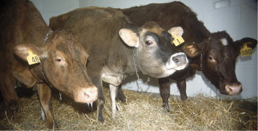
Three representative Jersey cows from a herd with an epidemic of pharyngeal trauma associated with mass medication delivered by an owner. Salivation, extended head and neck because of sore throats, anxious or depressed appearance, dyspnea, inhalation pneumonia, bloat, and subcutaneous emphysema occurred to varying degrees in the affected cattle.
Diagnosis
Frequently the clinical signs, coupled with a manual examination of the oral cavity to palpate the pharyngeal laceration, are sufficient for diagnosis. Most injuries are in the caudal pharyngeal region dorsal to the larynx. Severe lacerations also may damage the soft palate or proximal esophagus. Administered boluses or magnets may still be embedded in the retropharyngeal tissues in some cases. An oral speculum and focal light examination also may allow a view of pharyngeal injuries.
Endoscopy and radiology are very helpful ancillary aids, especially when a manual examination of the oral cavity is inconclusive or when the size of the animal—as with a calf—precludes manual examination. Endoscopy usually allows a view of pharyngeal injuries, but diffuse swelling of the pharynx, larynx, and soft palate sometimes interferes with this procedure. Radiographs are diagnostic of pharyngeal trauma in most cases because air densities and radiolucent retropharyngeal tissues are readily apparent. Pharyngeal foreign bodies and embedded boluses or magnets also are apparent with radiographs (Figure 6-28 ).
Figure 6-28.
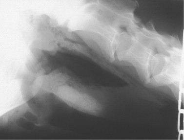
Radiograph of the pharyngeal region of a cow that suffered pharyngeal trauma, laceration, and foreign body deposition of a sulfa bolus delivered with a balling gun. A large radiolucent area and tissue emphysema are apparent ventral to the cervical vertebrae, and the bolus can be seen embedded between the air density dorsally and the trachea.
Treatment
Broad-spectrum antibiotics, analgesics, and supportive measures such as IV fluids are the major components of therapy for pharyngeal trauma. Whenever possible, it is best to avoid any oral medications. However, sometimes gentle passage of a stomach tube to provide an economical means to hydrate a patient with dysphagia is necessary. Most small pharyngeal lacerations respond to antibiotics such as ceftiofur (2.2 mg/kg IM every 24 hours), tetracycline (9 mg/kg, IV every 24 hours), ampicillin (6.6 to 11.0 mg/kg IM or SQ every 12 hours), or other broad-spectrum combinations. Penicillin is not a good initial choice because it seldom is able to control the expected mixed infection. Judicious use of analgesics such as flunixin meglumine (0.5 to 1.1 mg/kg IV every 24 hours) aids patient comfort, relieves the “sore throat,” and may allow an earlier return of appetite. Resolution of dysphagia, when present, is an important positive prognostic sign because the patient can now drink effectively and hydrate herself. Resolution of fever is another positive prognostic sign but may be misleading if temperature decreased because of concurrent therapy with nonsteroidal antiinflammatory drugs (NSAIDs).
Nursing procedures and ensuring access to fresh clean water and soft feeds such as silage or gruels of soaked alfalfa pellets are helpful. In severe cases, placement of a rumen fistula may be necessary to allow for direct placement of mashes or liquids directly into the rumen, thereby bypassing the damaged and painful tissues. Antibiotic therapy should be continued 7 to 14 days or longer depending on response to treatment and healing of the pharyngeal wound. Foreign bodies, boluses, or magnets embedded in retropharyngeal locations must be removed.
Prognosis is good for most cases but is guarded for cattle having large lacerations, soft palate lacerations, proximal esophageal lacerations, inhalation pneumonia, and vagus indigestion.
Prevention
Veterinarians should educate laypeople on how to safely administer oral medications. Stomach tubes, speculums, esophageal feeder tubes, and balling guns should be inspected after each use to identify any sharp edges or “burrs” that may incite future injury. Proper size speculums, tubes, and so on should be based on the animal's size and not “one size fits all.”
Alimentary Warts
Etiology
Papillomas and fibropapillomas are observed sporadically in the oral cavity, esophagus, and forestomach of dairy cattle. Oral lesions may occur on the hard palate, soft palate, or tongue. Bovine papilloma virus (BPV) of various types is the suspected cause of these lesions. The genome of BPV2 has been found in such lesions, but the complete virus itself may not be present. These lesions tend to be present in adult animals rather than calves. BPV2 has also been demonstrated in association with urinary bladder tumors in cows with chronic enzootic hematuria.
Jarrett and co-workers have also found a high incidence of papillomas and carcinomas of the upper alimentary tract and forestomach in cattle ingesting bracken fern in Scotland. A BPV labeled BPV4 has been identified in these lesions.
Signs
Papillomas and fibropapillomas of the mouth, esophagus, and forestomach create no clinical signs unless they interfere with eructation. Occasional warts at the cardia or distal esophagus act as a ball valve to interfere with eructation and cause chronic or recurrent bloat, leading to signs of vagus indigestion (Figure 6-29 ). Such lesions may be precursors for carcinomas, but this has not been proven except when associated with the ingestion of bracken fern.
Figure 6-29.
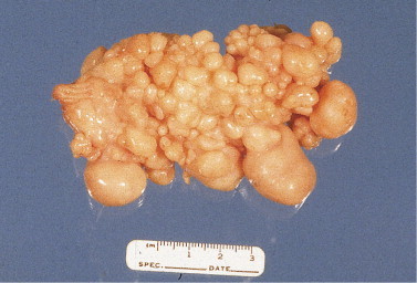
A fibropapilloma that was surgically removed via rumenotomy from a 2-year-old cow with chronic bloat.
Diagnosis
Inspection, endoscopic biopsy, and histopathology are the means of diagnosis. Rumenotomy may be necessary to confirm lesions at the cardia.
Treatment
Lesions are not treated except when discovered during rumenotomy in cattle with failure of eructation. In such cases, removal is curative.
Salmonellosis
Etiology
Much of the discussion regarding salmonellosis has been addressed in the section on calf diarrhea. Salmonella sp. causes enterocolitis that varies tremendously in severity in adult cattle. Septicemic salmonellosis may result in abortion or shedding of the causative organism into milk. The organism also may be found in milk secondary to environmental contamination and subsequent mastitis. This latter route appears to be typical of S. Dublin mastitis and possibly to lesser degrees for other types.
Salmonella spp. are facultative intracellular organisms that can hide in macrophages, be distributed along with these cells, and occasionally cause bacteremia following invasion of the intestine. Fecal-oral infection is the most common route of infection, but other mucous membranes can be invaded by some serotypes. Following ingestion of Salmonella organisms, a cow may or may not become clinically ill. Factors that determine pathogenicity include:
-
1.
Virulence of the serotype
-
2.
Dose of inoculum
-
3.
Degree of immunity or previous exposure of host to this serotype
-
4.
Other stressors currently affecting the host
The first two factors relate to the serotype of Salmonella involved. Because more than 2000 serotypes have been described, it is not surprising that a great deal of variation exists in the clinical signs, prevalence, morbidity, and mortality. Because Salmonella spp. often act as opportunistic pathogens, management, nutritional, and environmental factors that adversely impact the cow's defenses are often at play when the disease becomes problematic on a given operation.
Salmonellosis was primarily a sporadic disease in dairy cattle in the northeastern United States until the 1970s. A single cow within a herd may develop the disease secondary to septic metritis, septic mastitis, BVD, or other periparturient disorders. Infection seldom spreads to other cows. However, in recent decades, larger herds and increased use of free stall housing have changed the clinical epidemiology of salmonellosis, such that herd outbreaks with variable morbidity and mortality are now the rule. Free stall housing creates a nightmarish setting for diseases such as salmonellosis that are spread by fecal-oral transmission. Stressors include such things as concurrent infection with other bacterial or viral pathogens, transportation, parturition, poor transition cow management, gastrointestinal stasis or disturbance of the gastrointestinal flora by recent feed changes, heat or cold, and recent anesthesia or surgery.
Another contributing factor to herd infections is contaminated ration components fed to dairy cattle. Protein source supplements and animal byproduct components may be contaminated with Salmonella sp. Improperly ensiled forages that fail to reach a pH, 4.5 can harbor Salmonella sp. Birds shedding Salmonella can contaminate cut forages or feed bunks to infect adult cattle. This latter pathogenesis has been suspected in several herd outbreaks of type E, but birds also could transmit other types of Salmonella by acting as either biological or mechanical vectors. Farm implements used to handle manure or haul sick or dead animals can be a very efficient means of spreading Salmonella if these are used to haul feed, bedding, or healthy animals. The spreading of liquid manure on fields in addition to no-plow planting of crops has caused an increase in forage contamination.
Herd epidemics with an acute onset and high morbidity should be investigated as point source outbreaks of feed or water contamination. Chronic, endemic problems may represent spread of infection by carrier cattle to susceptible or stressed herdmates who then propagate the herd problem by shedding large numbers of organisms in feces during acute disease. It is not unusual to have a herd outbreak in lactating cows without an outbreak in young calves or vice versa.
In the northeastern United States, types B, C, and E are responsible for most herd endemics. Most type B isolates are S. Typhimurium of varying virulence and antibiotic susceptibility. Type C includes S. Newport and S. Litchfield. Type E usually is S. Anatum. In general, types B and C Salmonella are more virulent than type E, but because of the multitude of existing serotypes, it is impossible to generalize further. Type D, most of which are S. Dublin, are common in the western United States. A summary of the typical characteristics of diseases induced by the various serogroups is presented earlier in this chapter in the section on salmonellosis in calves.
S. Dublin is largely host adapted to cattle, whereas other types are nonhost adapted. A particularly frightening characteristic of S. Dublin infection is that infected cows remain carriers for a long time or even forever. Some shed consistently, others intermittently, and others are “latent” carriers that shed only when stressed. S. Dublin also causes mastitis, which tends to be subclinical and persistent. Mastitis caused by S. Dublin is thought to originate from environmental contamination of the udder by feces from infected cattle rather than septicemic spread to the udder. Infection of calves by S. Dublin is common in the western United States and has begun to appear in the eastern and midwestern United States. Infected calves shed large numbers of organisms, frequently are septicemic, and have respiratory signs coupled with fever that confound the diagnosis and mislead veterinarians unfamiliar with this disease. Other than S. Dublin– infected cattle, most cattle infected with nonhost-adapted serotypes such as S. Typhimurium are thought to shed the organism for less than 6 months. However, latent carriers or chronic infection may occur occasionally, and chronic S. Typhimurium mastitis has been documented following an enteric epidemic.
Salmonella spp. are capable of attachment to, and destruction of, enterocytes. Pathogenic serotypes gain access to the submucosal region of the distal small intestine and colon where their facultative intracellular characteristics guard them against normal defense mechanisms of naive cattle. From this location, the organisms enter lymphatics and may commonly create bacteremia in calves. As with most facultative intracellular bacteria, the host's cell-mediated immune system is essential for effective defense. Diarrhea caused by Salmonella sp. is primarily of inflammatory origin with lesser contributions (in some serotypes) by secretory mechanisms. Because mucosal destruction occurs, maldigestion and malabsorption contribute to the diarrhea, and protein loss into the bowel is significant when virulent strains infect cattle. Severe inflammation of the colon is common with resultant fresh blood in the feces or dysentery.
Clinical Signs
As in calves with salmonellosis, adult cattle infected with Salmonella sp. have enteric disease of greatly varied severity. Type E organisms usually cause mild diarrhea, dehydration, fever for 1 to 7 days, and a clinical situation that resembles winter dysentery in that affected cattle appear neither severely dehydrated nor toxic. As a rule, fresh blood is seen less commonly in the feces of type E infections than in type B and C infections. However, the same type E organisms may overwhelm cattle stressed by concurrent infections or metabolic disease caused by altered defense mechanisms or preexisting acid-base and electrolyte abnormalities.
Fever and diarrhea are expected in salmonellosis consistently, although fever may be absent or have preceded the onset of diarrhea by 24 to 48 hours. This prodromal fever has been confirmed in hospitalized animals that acquired nosocomial salmonellosis. These patients were found to have fever without any signs of illness 24 to 48 hours before developing diarrhea subsequently confirmed as Salmonella types B or C. Fever ranges from 103.0 to 107.0 ° F (39.4 to 41.7° C) and correlates poorly with other clinical signs as regards severity of illness. However, detection of fever in sick or apparently healthy cows during a herd outbreak is an extremely important aid to diagnosis of an infectious disease rather than a dietary indigestion. Diarrhea is consistent, at least in animals with clinical disease, and may appear as loose manure, watery manure, loose manure with blood clots, or dysentery (Figure 6-30 ). Endotoxemia and dehydration accompany diarrhea when virulent strains are encountered or when enteric invasion and bacteremia exist. Anorexia usually accompanies the onset of diarrhea and may be transient in mild cases or prolonged in patients with severe diarrhea and endotoxemia. Feces from cows with types B and C salmonellosis often are foul-smelling, containing blood and mucus. Whenever diarrhea with fresh blood and mucus is observed in cattle, salmonellosis should be considered. Recently fresh cows are very susceptible to infection during herd epidemics, and errors in transition cow management often amplify the impact of disease on these cows. Environmental factors such as heat stress tend to amplify the clinical signs and increase morbidity and mortality. Recording temperatures in apparently healthy cows during a herd outbreak may confirm fevers in some that are about to develop diarrhea or may represent subclinical infections. Concurrent infection with Salmonella sp. and BVDV following the purchase of herd additions can lead to devastating mortality. Dr. Rebhun observed one herd with this combination of acute infections that lost 35 of 130 adult cattle within 7 days (Figure 6-31 ). The usual mortality rate in herd outbreaks of S. Typhimurium is approximately 5% to 10%.
Figure 6-30.
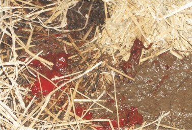
Fresh blood clots mixed with feces of a cow that had a type C Salmonella sp. enterocolitis.
Figure 6-31.
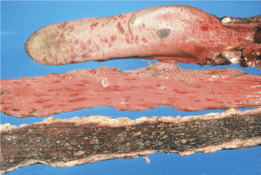
Necropsy specimens from a cow having concurrent BVDV and salmonellosis. The tongue (top) shows multiple BVDV erosions; the esophagus (middle) shows multifocal linear BVDV erosions; and the colon (bottom) shows severe inflammatory colitis with mucosal necrosis caused by salmonellosis.
(Photo courtesy Dr. John M. King.)
Abortions are common, especially when serotypes B, C, or D cause infection and can occur for several reasons:
-
1.
Septicemia with seeding of the fetus and uterus causing fetal infection and death
-
2.
Endotoxin and other mediator release that cause luteolysis via prostaglandin release and apparent alteration in hormonal regulation of pregnancy
-
3.
High fever or hyperthermia brought about by concurrent fever and heat stress during hot weather
Cows may abort at any stage of gestation, but as with many causes of abortion, expulsion of 5- to 9-month fetuses are most likely to be observed by dairy personnel.
Salmonella sp. may be found in the milk of infected cattle. With types B, C, and E organisms, this contamination of milk may represent septicemic spread of the organism to the mammary gland, environmental fecal contamination of the milk and milking equipment, or both. Herds infected with S. Dublin have chronic mastitis in a percentage of cows infected by this organism. Mastitis caused by S. Dublin may be subclinical, and environmental contamination of quarters has been shown to be a more likely cause than septicemic spread to the udder. Occasional cows have chronic mastitis with Salmonella spp. other than S. Dublin. Quarters that shed organisms and feces from infected cows create major public health concerns for farm workers and milk consumers. Contaminated milk is a major risk for the entire dairy industry and reasons enough to investigate every herd outbreak of diarrhea in dairy cattle with appropriate diagnostic tests. Whereas proper pasteurization reliably eliminates the organism from milk, raw milk should not be consumed.
Diarrhea and illness caused by salmonellosis are common in farm workers and families whenever herd outbreaks occur. It is the veterinarian's obligation to inform clients and workers regarding the public health dangers of salmonellosis and to direct sick farm workers or family members to physicians for treatment.
Ancillary Aids and Diagnosis
Hematology and acid-base electrolyte values are valuable ancillary aids for individual or valuable cattle but are seldom diagnostic because of the great variation in clinical illness. Fecal cultures are the “gold standard” of diagnosis, and samples from several patients in the early stages of the disease should be submitted to a qualified diagnostic laboratory. Isolates should be typed and antibiotic susceptibility determined. Unlike salmonellosis in horses, Salmonella spp. can usually be cultured from even a “watery” fecal sample from cattle with salmonellosis.
Peracute salmonellosis associated with virulent serovars tends to create a neutropenia with degenerative left shift in the leukogram and metabolic acidosis with Na1, K1, and Cl2 values all lowered in affected adult cattle. Elevations in PCV, blood urea nitrogen (BUN), and creatinine can be anticipated in those patients with severe diarrhea. Total protein values initially may be elevated because of severe dehydration but are just as likely to be normal or low because albumin values decrease quickly as a result of the severe protein-losing enteropathy. BUN and creatinine may be elevated simply because of prerenal azotemia or because of acute nephrosis resulting from septicemia/endotoxemia.
Just as fever precedes the onset of diarrhea in some patients, so may the expected neutropenia with left shift. This has been documented in some cattle that acquire nosocomial hospital infections, although it is unlikely to be detected in field outbreaks because cattle yet unaffected with diarrhea seldom are sampled. Cattle with less than overwhelming acute salmonellosis may have neutropenia, normal WBC numbers, or neutrophilia. Recovering cattle tend to have a neutrophilia.
Sodium, potassium, and chloride tend to be low in most cattle having severe or prolonged diarrhea. As mentioned, peracute severe salmonellosis will result in metabolic acidosis as a result of massive fluid loss and endotoxic shock, but most adult cattle with nonfatal diarrhea do not develop significant acidosis.
Differential diagnosis of salmonellosis in adult cattle is brief if limited to diseases causing fever and diarrhea. BVDV infection and winter dysentery would be the primary differentials. Herds with serotypes such as type E causing relatively mild signs of fever and diarrhea require differentiation from winter dysentery (depending on the time of year), BVDV infection, and indigestions. Herds suffering mortality associated with very virulent type B or C infections must be differentiated from BVDV infection. For cases of more chronic diarrhea, subacute ruminal acidosis, internal parasites, Johne's disease, eosinophilic enteritis, lymphosarcoma, chronic peritonitis, and copper deficiency should be considered. Viral isolation should be attempted from the buffy coat of ethylenediaminetetraacetic acid (EDTA) blood samples or necropsy specimens to rule out BVDV infection and fecal cultures from multiple patients or necropsy samples evaluated for presence of Salmonella sp. Infections caused by Campylobacter sp. and Yersinia sp. occasionally have been reported in adult cows with fever and diarrhea. The significance and disease incidence associated with these organisms are unknown.
Classical gross necropsy lesions of diffuse or multifocal diphtheritic membranes lining the region of mucosal necrosis in the distal small bowel and colon are present in subacute and chronic cases. In peracute cases, however, minimal gross lesions other than hemorrhage and edema may exist within the involved bowel and enlarged mesenteric lymph nodes. The more acute the death, the less likely gross lesions will be observed. Fibrin casts sometimes are found in the gallbladder and are considered pathognomonic for salmonellosis by some pathologists.
Treatment
Supportive treatment with IV fluids is necessary for patients that have anorexia, depression, and significant dehydration. Individual patients may be treated aggressively following acid-base and electrolyte assessment. However, outbreaks in field settings seldom allow extensive ancillary workup, and fluid therapy is administered empirically. Use of balanced electrolyte solutions such as lactated Ringer's solution is sufficient for most cattle. Cattle having severe acute diarrhea and .10% dehydration are likely to have metabolic acidosis and may require supplemental bicarbonate therapy. For example, a 600-kg patient judged to be 10% dehydrated and mildly acidotic (base excess 5 25.0 mEq/L) should receive 60 L of balanced fluids for correction of dehydration. Rehydration alone may decrease the lactic acid and correct the metabolic acidosis. (The only times that bicarbonate therapy is absolutely needed for correction of acidosis in dairy cattle are for severe rumen acidosis, enterotoxigenic E. coli, or other enteric infections causing excessive production of D-lactate.) If balanced electrolyte fluid therapy does not correct the metabolic acidosis, the cow may need to be treated with bicarbonate. This hypothetical cow has 200 kg or L of ECF (0.3 [ECF] 3 600), and the base deficit of 25.0 mEq/L implies that each liter of her ECF is in need of 5.0 mEq/L HCO3 2. Thus 1000 mEq NaHCO3 (0.3 3 600 3 5) could be added just to make up the existing deficit, and more NaHCO3 would likely be necessary to compensate for anticipated continued losses. This example readily highlights the feeling of helplessness that veterinarians and herd owners experience when a virulent serotype causes serious dehydration in more than a few cows. Placement of a catheter in an auricular vein may prevent catheter damage from head catches, a common problem with jugular catheters on dairies. Auricular or jugular vein catheter placement may allow for repeated administration of IV fluids and repeated IV administration of flunixin meglumine. Hypertonic saline (7.5 times normal) administered at 3 to 5 ml/kg followed by 10 to 20 gallons of oral electrolyte solution, either consumed voluntarily or given by orogastric tube, is a highly practical method of fluid resuscitation in a field setting. This method has become commonplace and is a time- and labor-efficient way of addressing dehydration in grade cattle. Administration of hypertonic saline into smaller-diameter veins, such as the auricular vein, may result in phlebitis and catheter failure. When multiple animals merit oral fluid administration during an outbreak of salmonellosis or any other enteric disease, or if the same equipment is to be used for drenching of other cattle, laypeople should be aggressively educated as to the possibility of cross-contamination and the need for disinfection between uses. As a crude rule of thumb, cattle that show no voluntary interest in drinking following rapid IV administration of 3 to 5 ml/kg of 7.5 times normal saline solution should provisionally be given at best a guarded prognosis and are mandatory candidates for large-volume oral fluid drenching.
Oral fluids and electrolytes may be somewhat helpful and much cheaper than IV fluids for cattle deemed to be mildly or moderately dehydrated. The effectiveness of oral fluids may be somewhat compromised by malabsorption and maldigestion in salmonellosis patients but still should be considered useful. Cattle that are willing to drink can have specific electrolytes (NaCl, KCl) added to drinking water to help correct electrolyte deficiencies.
Antibiotic therapy is controversial. Its opponents warn of the potential for emergence of resistant strains that may present great risk for people and animals in the future. Evidence for this phenomenon is sparse except for long-term feed additive antibiotics, and one could argue that other species, including humans, represent similar risks when treated with antibiotics. Further opposition states that systemic antibiotics prolong the excretion of Salmonellae in the feces and may not shorten the clinical course of purely enteric disease. However, discerning those animals with infection limited to the gut wall from those animals with gut wall and systemic infection is never easy.
Proponents of antibiotic therapy remind us that salmonellosis frequently induces bacteremia (although this is most common in calves), thereby risking septicemic spread of the organism. Clinical differentiation of septicemia versus endotoxemia without septicemia is not easy unless localized infection appears in a joint, eye, the meninges, or lungs. In other words, clinicians can seldom accurately predict all salmonellosis patients that are truly septicemic. In addition, appropriate antibiotic therapy reduces the total number of organisms shed into the environment by counteracting septicemic spread that allows all bodily secretions, not just feces, to harbor the organism. These points should be considered by veterinarians and probably dictate against the use of antibiotics in salmonellosis patients having mild to moderate signs (e.g., low grade fever, diarrhea, and mild dehydration). Except for valuable cattle that are seriously ill with salmonellosis, systemic antibiotics are seldom administered to adult cows with salmonellosis in the Cornell Hospital.
Therefore antibiotics are sometimes used when patients appear moderately to severely ill and show signs of fever, dehydration, and profuse diarrhea or dysentery. These patients usually have elevated heart and respiratory rates, are weak, and appear endotoxemic or septicemic. Given the unpredictable nature of antimicrobial susceptibility of Salmonella, antimicrobial therapy should be guided by culture and susceptibility results. Withdrawal periods should be observed for any nonlabel usage of antibiotics. Antibiotics should be continued 4 to 7 days in patients that are improving.
NSAIDs, especially flunixin meglumine, may be helpful for “antiendotoxic” effects and blockage of various mediators of inflammation and shock. Cattle may be started on 1.1 mg/kg body weight IV every 24 hours and then tapered to 0.50 mg/kg body weight every 24 hours, or the medication may be discontinued after 1 to 2 days. If repeated IV administration is not practical, SQ administration is vastly preferred over IM administration, which may result in marked muscle damage. Overdosage or administration of repeated doses of flunixin may cause abomasal or renal pathology, and IM administration may induce myoglobin release that augments the renal adverse effects. Corticosteroids are contraindicated except as a one-time dose of water-soluble corticosteroid for a gravely ill patient in shock. Prednisolone sodium succinate is preferred in this instance.
Isolation of patients with salmonellosis is ideal, albeit difficult, in field settings. Whenever possible, cattle with diarrhea should be confined to an area of the barn away from the rest of the herd. Workers must be educated regarding mechanical transmission of infected feces and other discharges from infected to uninfected cattle. Workers should also be educated regarding the zoonotic implications inherent with salmonellosis.
Prevention and Control
Herd epidemics appear to be increasing in frequency based on confirmed isolations from multiple cow outbreaks identified from New York and the rest of the northeastern United States. Conditions that contribute to an increasing incidence of epidemic salmonellosis include larger herd size, more intensive and crowded husbandry, and the trend for free stall barns with loose housing, which contributes to fecal contamination of the entire premises. Other major contributing factors include the use of feedstuffs that may be contaminated with Salmonella sp. and spreading contaminated manure on unplowed fields. Outbreaks caused by types C and E Salmonellae have been caused by contaminated feed components, and type E also has been spread by birds that are carriers of the organism.
When salmonellosis has been confirmed in a herd, the following control measures should be considered:
-
1.Conduct an epidemiologic investigation to help determine the source.
-
•Commodities barn/feed storage and handling: Inspect and document source(s), and obtain samples of commodities for culture. Are there other dairies in the area with similar problems? Who hauls the feed onto the farm, and in what? Is this vehicle or trailer used solely for feed transport (not animals, bedding, or manure)? On the farm, how is the feed handled? Is the feed-hauling equipment used for other purposes (e.g., carcass hauling, bedding removal)? Are there other animals or a large population of birds with exposure to the feeds?
-
•Water sources: Is there likely fecal contamination? What are the containers used to haul water to pastured cattle, and how/by whom are they handled?
-
•Manure handling: Equipment used and destination? What is the flow pattern of flush water? Are the personnel involved in manure handling later handling animals or their feed? Is the manure being spread on unplowed crop fields? Flow patterns of labor, vehicles, water, bedding, and movement of sick and healthy cattle on the dairy should be critiqued.
-
•Introduction of new animals: Are newly purchased animals quarantined and cultured? How are cattle taken to shows handled on return? Has bulk tank milk been tested for BVD?
-
•Management of cows in the sick pen and maternity pen: Too often, these two sets of cattle are managed and housed together, creating ideal circumstances for infection of fresh cows and heifers. Physical separation and careful allocation of personnel and equipment to each group should be reviewed.
-
•
-
2.
Isolate obviously affected animals to one group if possible.
-
3.
Treat severely affected animals.
-
4.Institute measures to minimize public health concerns.
-
•No raw milk should be consumed.
-
•Workers and milkers should wear coveralls, disposable or rubber boots, gloves, and perhaps masks when milking or cleaning barns. Workers and milkers should be encouraged to wash well after work or before eating. Disinfectant footbaths should be placed at exits and entrances to the barn and parlor (for humans and beast), and these footbaths should be maintained regularly.
-
•
-
5.
Physically clean the environment, improve hygiene, and disinfect premises (see also the section on calf salmonellosis). Pressure spraying to physically remove organic matter is very helpful before disinfection. Because removal of organic debris is incomplete on some surfaces, use of a disinfectant that retains its activity in organic debris and that has documented efficacy against Salmonella is optimal. Because shedding is likely to occur from recovered cattle for some time, ongoing efforts at improved hygiene are in order. In particular, protect dry cows and disinfect maternity areas.
-
6.
Following resolution of the outbreak or crisis period, a mastitis survey should be conducted that includes bulk tank surveillance. If any Salmonellae are recovered, culture of the whole herd is indicated to identify carrier cows that should be culled immediately. For S. Dublin outbreaks, all cattle should be screened by milk culture and, if available, serologic testing performed to detect carriers that should be culled.
If an epidemic continues despite all of the above guidelines, autogenous bacterins may be considered. Although efficacy and safety of autogenous bacterins are (justifiably) questioned, many practitioners have claimed excellent results when all other measures fail to stop ongoing endemic infections when freshening cows become ill, abortions continue to appear, or calves continue to become ill because of salmonellosis. At the time of this writing, a new siderophore receptor/porin vaccine derived from S. Newport has just become licensed in the United States for use in dairy cattle (SRP vaccine, Agri-Labs, St. Joseph, MO). It remains to be seen what impact this product will have on salmonellosis in the modern dairy industry. The efficacy of J-5 vaccines in salmonellosis control in adult cattle is unknown; in a large field trial, J-5 immunization of calves did not affect survival to 100 days. Unfortunately it is difficult to evaluate the efficacy of vaccines used to control endemic salmonellosis in field settings because improvement may be attributed to the vaccine but influenced by herd immunity or alterations in management. (For a further discussion of vaccinations for salmonellosis, see the section on calf salmonellosis.)
Prevention is best accomplished by maintaining a closed herd and culturing new feed additives and components before using them in the ration.
Hemorrhagic Bowel Syndrome (Jejunal Hemorrhage Syndrome)
Hemorrhagic bowel syndrome (HBS) is a newly emerging, highly fatal intestinal disease that has been recognized most frequently in adult dairy cows in the United States. Recently reports of HBS in Canadian dairy and beef cattle have been published. Other names given to HBS include jejunal hemorrhage syndrome, bloody gut, dead gut, and clostridial enteritis. HBS is characterized by sudden, progressive, and occasionally massive hemorrhage into the small intestine, with subsequent formation of clots within the intestine that create obstruction. Affected areas of the intestine become necrotic, and affected cows appear to suffer from the combined effects of blood loss, intestinal obstruction, and devitalization of bowel. The disease is seen most commonly in adult dairy cows early in lactation, although cases occasionally occur in late lactation or the dry period.
Etiology
The cause of HBS is currently unknown, and no consistent predisposing factor has been identified. The majority of HBS cases occur during the first 4 months postpartum. In a large survey of American dairy producers, the median parity for cows affected by HBS was reported to be the third lactation, and the median number of days in milk for affected cows was 104 days. During this period, dairy cows experience physiologic stress associated with peak milk yield. In addition, the rations fed during this stage of production are rich in energy and protein and fiber-depleted relative to rations fed later in lactation. These factors have been proposed to place cows at greater risk for HBS, but the events that lead up to the development of this disease remain undetermined.
The gross and histologic features of HBS have been described in a few reports. Gross lesions are usually segmental or multifocal in distribution in the small intestine, primarily in the jejunum with occasional involvement of the duodenum or ileum. Affected segments show purple or red discoloration of the intestinal wall, with distention of affected segments caused by intraluminal casts or clots of blood (Figure 6-32, Figure 6-33 ). The intestine orad to these lesions may be distended with fluid and gas, indicating obstruction of affected segments. Fibrin accumulation on the surface of affected intestine may be evident, and affected segments may rupture antemortem or postmortem. The blood clot in affected segments is often tenaciously attached to the mucosa, and manual removal of the clot often results in “peeling off” of the surrounding mucosa (Figure 6-34 ). On histologic examination of affected bowel, HBS appears to be a segmental, necrohemorrhagic enteritis, with submucosal edema, mucosal ulceration, transmural hemorrhage, and neutrophil accumulation evident in affected areas. Sloughing of mucosa in affected areas may also be present.
Figure 6-32.
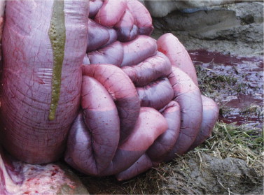
Fresh field autopsy performed within minutes of death on a mature Holstein cow with HBS. Note the purplish discoloration and gas production throughout the small intestine. There was diffuse jejunal involvement with death occurring as a result of blood loss.
Figure 6-33.
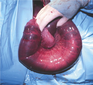
Intraoperative picture of mature Brown Swiss cow with HBS. In contrast to the cow in Figure 6-29, this animal demonstrated the rather more common involvement of just a segment of jejunum with a blood clot obstructing an approximately 12-inch section of bowel.
(Photo courtesy Dr. Liz Santschi.)
Figure 6-34.
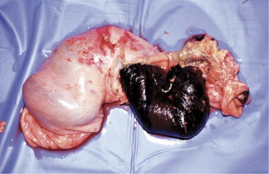
Resected section of jejunum cut open to show tenacious intraluminal blood clot from an adult Holstein with HBS.
Several reports indicate an association between C. perfringens type A and HBS. This association is based on the following observations: (1) affected cows have positive fecal cultures for this organism; (2) C. perfringens type A can be readily isolated in heavy growth from blood clots in the jejunum of affected cows; (3) there is microscopic evidence of intestinal necrosis associated with a dense intraluminal population of large, gram-positive bacteria; and (4) other enteric pathogens associated with hemorrhagic enteritis are rarely identified in tissues or enteric contents of affected cows. In addition, based on anecdotal evidence, reduced monthly incidence of HBS has occurred following administration of an autogenous C. perfringens vaccine to adult cows on certain dairies. At present, data from controlled studies are not available for evaluation of the effect of such vaccines on the incidence of this disease.
C. perfringens is a large, gram-positive, anaerobic bacillus that is considered to be ubiquitous in the environment and in the gastrointestinal tract of most mammals. The rate of isolation of the organism from the gastrointestinal tract of cattle may be enhanced by high grain diets. Genetic classification of C. perfringens is performed by mPCR. Type A usually produces alpha toxin, although different isolates may produce different quantities of this toxin. Alpha toxin is a calcium-dependent phospholipase that is capable of cleaving phosphatidylcholine in eukaryotic cell membranes. Additionally, the recently discovered beta2 toxin may be produced by C. perfringens type A. Beta2 toxin is also a lethal toxin, and strains of C. perfringens with the cpb2 gene produce variable amounts of beta2 toxin in vitro.
In two studies, C. perfringens type A and/or type A 1 beta2 was isolated from feces and/or intestinal contents of 28 of 32 cows with HBS. These bacteriologic findings are concordant with those of other reports. In the past, veterinary microbiologists have been reluctant to consider C. perfringens type A as an important disease-causing pathogen of livestock because this organism is part of the normal flora of the cow's intestine. Furthermore, this organism proliferates rapidly in the intestine after death, making isolation from necropsy specimens of questionable diagnostic significance. Because C. perfringens types A and A 1 beta2 can be isolated from the gastrointestinal tract of apparently healthy animals, the diagnostic significance of isolation of these organisms from animals with enteric disease is increased if the corresponding toxins can be detected in gastrointestinal contents or blood. In a recent study, C. perfringens types A and A 1 beta2 were isolated from multiple sites of the intestinal tract of HBS cows at a significantly higher rate than unaffected herdmates (cows with LDA). In addition, intraluminal toxin production was demonstrated in the intestine of HBS cows but not in the intestine of control herdmates with LDA.
It is unclear at present whether enteric proliferation of and intraluminal toxin production by C. perfringens type A occur as part of the primary insult to the intestine, or if these processes occur secondary to another disease or triggering factor. Hemorrhage into the intestine from another cause could, in theory, initiate secondary proliferation of the ubiquitous C. perfringens because this organism is likely to rapidly multiply when large quantities of soluble protein or carbohydrate is presented to the intestine. In other words, blood certainly could act as a very rich culture medium for this organism. Once the organism proliferates, however, the toxins that it releases during rapid growth could contribute to the degradation of the intestinal wall that is so characteristic of HBS. This destruction of the intestinal wall in sections of the gut affected by HBS is likely to contribute to the subsequent shock and peritonitis that is evident in so many affected cows.
Investigators at Oregon State University have focused on characterizing the role of Aspergillus fumigatus, a fungus that can be found in livestock feeds. Genetic material of this fungal agent can be detected in the blood and intestine of affected cattle but not in unaffected cattle. Two hypotheses can be presented regarding the possible participation of A. fumigatus in HBS: (1) as a primary contributor to the intestinal lesion; or (2) as an agent that impairs the cow's immune system, thereby facilitating or inciting whatever disease process triggers HBS. Anecdotal reports suggest that the incidence of HBS can be reduced on dairies following the introduction of a feed supplement (Omnigen AF, Prince Agri Products, Quincy, IL) into the ration. Controlled studies on the efficacy of this product for HBS prevention are pending. This product has recently been demonstrated to improve certain indicators of immune function in the WBCs taken from immunosuppressed sheep.
Clinical Signs
Cows are rapidly debilitated by the combined effects from sudden and massive hemorrhage into the small intestine. As a result, affected cows may simply be found dead or dying. A rapid pulse and rapid respiratory rate are commonly found in affected animals, and the mucous membranes are pale. The cow's extremities are often cool, and the rectal temperature is often below normal; the loss of blood into the intestine and the resulting shock contribute to these findings. In this sense, affected cows can resemble milk fever cases. Unlike milk fever, however, the feces of affected cows are dark, tarlike, and may contain dark red to black clots of digested blood (Figure 6-35 ). As clots form in the affected segments of the intestine, the intestine often becomes obstructed, causing some cows to show abdominal distention, reduced fecal output, and signs of colic. Glucose can often be detected in the urine of affected cows, indicating a severe stress response.
Figure 6-35.
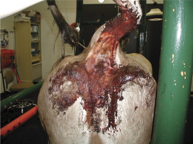
Perineum of mature Brown Swiss cow demonstrating the admixture of fresh and digested blood clots typical of HBS.
When viewed from behind, the abdominal contour is typically round or pear-shaped in the standing animal. Progressive distention is often appreciated in the lower right abdomen, presumably resulting from accumulation of multiple loops of blood-filled small intestine in the ventral abdominal cavity. Scattered, low-pitched “pings” may be evident in the lower right abdomen. This progressive abdominal distention distinguishes cases of HBS from cases of bleeding abomasal ulcer. Occasionally motility is reduced throughout the gastrointestinal tract, and affected cows can appear bloated. In our experience, rectal examination often does not reveal distended loops of intestine because the blood-filled segments of intestine seem to sink to the ventral abdomen, thereby becoming beyond the reach of the examiner. However, small intestinal distention was palpable per rectum in six of eight cows in a Canadian study.
Ultrasonography can be used to visualize intestinal distention and clot formation within loops of affected bowel. A 3.5- or 5.0-MHz, sector- or linear-array probe is placed on the abdominal wall at the lower aspect of the right side. Dilated loops of intestine can often be seen, and on occasion, material consistent with the appearance of clotted blood can be seen within the distended loops (Figure 6-36 ).
Figure 6-36.
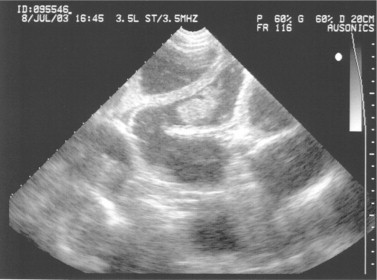
Transabdominal ultrasound image of lower right quadrant of a cow with HBS, demonstrating variably distended loops of small intestine and one small, nonadherent, hyperechoic intraluminal blood clot.
Differential diagnoses include intussusception, intestinal volvulus, enteritis, and abomasal ulcer. Cows with an abomasal ulcer may show melena and shock but rarely develop the progressive abdominal distention characteristic of HBS. Cattle with enteritis continue to pass significant quantities of feces, particularly following treatment with fluids and calcium salts, whereas cattle with HBS usually do not. Further, once hydration, electrolyte balance, and normocalcemia are restored by fluid therapy, cattle with enteritis typically show resolution of any mild abdominal distention that might have developed as a result of ileus. Differentiation of HBS from intussusception and intestinal volvulus requires exploratory laparotomy.
Treatment
Successful treatment of this disease is difficult. Occasional, anecdotal reports exist of successful treatment with fluids, laxatives, antiinflammatory drugs, and antibiotics; however, it appears that such treatment successes are quite rare. Cows treated with medical support alone almost inevitably develop ileus, intestinal necrosis (tissue death) with subsequent peritonitis, and shock. Death of affected cattle occurs within several hours to 1 to 2 days after the onset of clinical signs.
At surgery, multiple inflamed segments of jejunum, ileum, or rarely duodenum are found. The serosal surface of affected segments is often dark purple to black in color. The affected segments of intestine are friable and turgid with luminal blood, and the casts of clotted blood within the lumen of the intestine impart a gelatin-like feel to the affected bowel (Figure 6-37 ). Involvement of multiple segments of jejunum and/or ileum is frequently found, which eliminates the option for intestinal resection and anastomosis. Techniques for surgical management of HBS cases to date include manipulation of the affected intestine so as to break down the obstructing clots, enterotomy and removal of the offending clots, and resection and anastomosis of affected segments. Common reasons for poor surgical outcome include discovery of multiple segments of nonviable bowel, septic peritonitis, and bowel rupture during intestinal manipulation. Also, if the initial surgical procedure is completed successfully, affected cows may develop repeated clotting and recurrent obstruction of the intestine after surgery. Of 22 cows affected with HBS presented to a university veterinary hospital over a 3-year period, only 5 (23%) survived; four of these survivors were treated surgically. In other studies, treating HBS cases with surgery, medical management, or both has resulted in little success.
Figure 6-37.
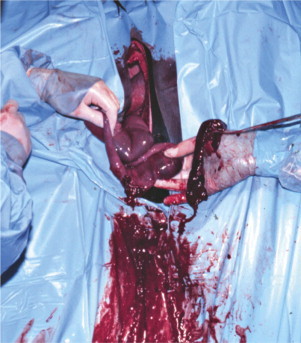
Intraoperative image of enterotomy site being used to manually remove and massage obstructing blood clots out of the jejunum in a cow with HBS.
(Photo courtesy Dr. Ryland Edwards.)
Prevention
Preventive strategies for HBS remain somewhat speculative at present, given the lack of understanding about the pathogenesis of this disease. In addition, controlled studies on the clinical efficacy and economic impact of particular preventive measures have not been completed. Nonetheless, potential risk factors for clostridial overgrowth in the intestine of ruminants have been identified in previous studies, and strategies to reduce those risks might, at least in theory, provide benefits in HBS control. Similarly, the potential role of pathogenic fungi in HBS warrants careful consideration when designing preventive strategies. In short, until more refined information regarding the cause of HBS is published, it may be best to first consider all proposed causes or risk factors (e.g., bacteria, fungi, and reduced host disease resistance) and take measures to mitigate these potential risk factors. In so doing, one should consider: (1) identifying and correcting management and environmental factors that might impair cow immunity, (2) performing a careful partial budget analysis of the cost of specific preventive measures, and (3) deciding on which specific corrective measure(s) might be most justified for a particular dairy.
To begin, a thorough analysis of transition and fresh cow management should be performed to identify problems with cow comfort, hygiene, nutrition, and disease control that might impact disease resistance during the apparent period of greatest risk for HBS, which is the first 3 to 4 months of lactation. Ration formulation and mixing should be reviewed as well, with due consideration given to such issues as effective fiber and soluble carbohydrate content and their potential dietary influences on gut flora. Feed bunk and pen management should be carefully critiqued to ensure that feed intake is consistent; efforts should focus on identifying and correcting management problems that cause “slug feeding” (e.g., pen overcrowding, poor parlor throughput, and infrequent feeding) and that predisposes to subacute rumen acidosis. Silage management, commodity storage, and feed preparation should be examined to determine whether spoilage and mold formation are problematic. Because these critical areas impact numerous facets of cow health other than HBS, identification and correction of problems in these areas will likely provide an overall benefit to cow health. Finally, potential use of feed additives or vaccines directed against specific, potential contributory pathogens should be considered carefully, with the costs of the proposed interventions and their potential efficacy weighed against the prevalence and costs of the disease.
Bovine Virus Diarrhea
Etiology and Background
The disease commonly referred to as bovine virus diarrhea (BVD) was first described by Olafson et al in 1946. This initial disease was highly infectious, contagious, and imparted high mortality. The causative organism was later isolated and so began the prolific long-term research into this pathogen of cattle. The initial clinical descriptions of BVD by Fox were of a severe disease characterized by high fever, diarrhea, mucosal lesions, and leukopenia. However, throughout the period of 1950 to 1975, the disease was largely disregarded in parts of the United States—including the northeast—because serologic surveys suggested that most adult cows had serum neutralization titers against BVDV. These results were interpreted to mean that BVDV frequently infected cattle as a subclinical or mild infection and was of little clinical significance. A direct consequence of this thinking was a nearly complete lack of interest in vaccination of dairy cattle against BVDV. The major clinical evidence of BVDV during the years 1950 to 1975 was sporadic subacute or chronic infection in one or more heifers on a farm. These affected animals usually were between 6 and 24 months of age; they developed diarrhea, typical mucosal lesions, fever, weight loss, and survived in poor condition for a variable time before death. Because of the sporadic appearance of such cases, these animals were thought to be immunodeficient and therefore susceptible to BVDV. This theory was tenable for single-case infections but became less believable when four to six heifers on one farm developed similar signs because the likelihood of multiple immunodeficient animals on one farm seemed small.
During that time, the use of modified-live BVDV (ML-BVDV) vaccines occasionally preceded the development of signs of BVD in a group of heifers by 1 to 4 weeks. Although this further discouraged the use of BVDV vaccines, it was explained as an unfortunate circumstance and likely that the heifers had already been incubating field virus. These subacute or chronic cases—usually in heifers—were often called “mucosal disease” because of the easily observable oral erosions and gastrointestinal lesions often found at necropsy, as well as the characteristic clinical signs of fever, weight loss, and diarrhea. Virologic limitations at many diagnostic laboratories during this period added further confusion to the disease clinically referred to as BVD or mucosal disease. Diagnosis was based primarily on serum neutralization titers and FA procedures on tissue samples rather than viral isolation. Current knowledge helps explain why so many of these clinically obvious BVD patients had low or nonexistent serum neutralization (SN) titers against BVDV. Further, the FA techniques used were poor tests that gave erratic results. Therefore in many cases over this time period, a textbook example of clinical BVD could not be confirmed as BVDV infection.
Reproductive and fetal consequences of the virus were studied during these years (1950 to 1975), and the implications of BVDV in reproductive failure were questioned clinically but seldom confirmed. The virus was shown to be a potential cause of abortion and congenital anomalies such as cerebellar hypoplasia and ocular defects. Absolute diagnosis of BVDV infection as a cause of clinical reproductive, gastrointestinal, or other system disease was made difficult by limited laboratory capabilities.
The past 20 years have brought both a wealth of research regarding the virus and the reemergence of BVDV as a major pathogen in cattle. The virus had been classified as a pestivirus within the Togaviridae family because of similarities with hog cholera virus and the virus of Border disease. Recent reclassification finds BVDV as a member of the genus Pestivirus within the family Flaviviridae. BVDV is classified in vitro into one of two “biotypes,” cytopathic (CP-BVDV) or noncytopathic (NCP-BVDV), based on how each biotype affects cell cultures. CP-BVDV causes vacuolation and death of certain cell lines within days of inoculation into cell culture, whereas NCP-BVDV inoculation into cell culture results in inapparent infection. NCP-BVDV is the more prevalent biotype in cattle. It serves as the parent virus from which, following genetic recombination, CP-BVDV arises.
In addition, a multitude of “strains” or heterologous isolates exist within each of the BVDV biotypes. The exact number of strains or genetic variation in the virus is not known, but the implications regarding clinical variations and effective immunization against these multiple strains constitute the major current concerns for BVDV. Further, the strain of virus used to complete a research study may or may not have implications for cattle exposed to a heterologous strain in the “real world.” Some strains may be capable of causing congenital anomalies, whereas others cause severe gastrointestinal injury. Therefore the strain chosen for study may have a profound outcome on the study results.
Through genetic sequencing, BVDV can be further classified according to one of two major genotypes (commonly called “types”): 1 and 2. Type 1 strains are considered the classic genotypes banked since the 1950s. Type 2 BVDV was first detected by genetic sequencing of isolates from severe clinical cases in adult cattle and calves in the northeastern United States and eastern Canadian provinces in 1993 to 1994. There are currently 11 recognized subgenotypes of BVDV type 1 (designated 1a, 1b, 1c, and so on) and two subgenotypes of type 2. Viral isolates within a given subgenotype are closely related in nucleotide sequence, sharing .90% sequence homology. Although severe clinical disease was characteristic of the outbreak of type 2 BVDV in the early 1990s, it should be emphasized that virulent strains of type 1 exist.
Perhaps the most important discovery about BVDV has been the identification and explanation for cattle persistently infected (PI) with BVDV. Animals with BVDV-PI and having little or no SN antibody against the homologous strain were recognized and later produced experimentally by infecting fetuses between 40 and 120 days gestation with NCP-BVDV. These workers were able to cause the PI state by directly infecting fetuses in seropositive dams (58 to 125 days) or infecting seronegative dams carrying fetuses (42 to 114 days) with NCP-BVDV. For unknown reasons, PI cannot be caused by experimental challenge with CP-BVDV.
A brief review of the PI condition is warranted here. Fetuses that are exposed to NCP-BVDV between the approximate ages of 40 and 125 days gestation may become PI with this strain of virus. These animals are immunotolerant of that NCP strain because they consider those viral antigens to be self. Such PI fetuses have several potential outcomes: being born normal and growing to adulthood normally; being born apparently normal but succumbing to disease before 1 year of age; or being born weak, small, or dead. However, if a PI animal is challenged by a heterologous CP-BVDV, severe disease may ensue, and in such instances, PI animals usually succumb with signs of acute, subacute, or chronic BVD. Apparently the immunotolerance of the PI animal to its homologous NCP-BVDV renders it unable to mount functional immunologic defenses against certain CP-BVDV strains. This scenario of infection by CP-BVDV in NCP-BVDV-PI animals was assumed by many previous researchers to be the only way animals could get the characteristic “mucosal disease” or fatal clinical BVD. Further, this “superinfection” of PI animals by CP-BVDV strains appeared to explain the outbreaks of BVD that followed use of modified-live BVDV vaccines.
More recent studies have shown that animals that develop naturally occurring BVDV-PI often harbor antigenically similar CP and NCP viruses. Genetic studies of these viruses have revealed that insertion of novel RNA into the NCP-BVDV can cause conversion into the CP-BVDV biotype. In other words, a PI animal may develop fulminant CP-BVDV infection from genetic reassortment of its own virus, from transfer of genetic material from a heterologous strain to its own virus, or from exposure to an entirely novel CP or NCP strain. In those instances, classic “mucosal disease” may develop in the PI animal.
“Mucosal disease” is often considered as a separate entity from “BVD” by clinicians and researchers. Dr. Rebhun believed strongly that mucosal lesions do not dictate a separate, uniformly fatal entity that is necessarily distinct from BVD, and that signs of BVD follow the biologic bell-shaped curve. True, it has been proven that certain CP-BVDV strains can cause superinfection of PI animals, resulting in fatal disease. This fatal disease may follow an acute, subacute, or chronic course and is frequently characterized by fever, diarrhea, weight loss, mucosal ulcerations of the gastrointestinal tract, digital lesions, and/or dermatologic lesions. However, clinical experience has shown that naive cattle can have mucosal lesions caused by NCP-BVDV infection, yet subsequently survive and form SN titers against this strain. Clinical experience also has shown that fatal BVD has occurred solely as a result of virulent strains of NCP-BVDV and that PI animals are not the only animals that die when exposed to certain CP- or NCP-BVDV. In short, the presence of mucosal lesions is not predictive of death or survival, nor of the PI status. Although the signs of BVD may be more obvious or more profound in superinfected PI than in non-PI animals, the same disease is present.
Similarly it has been tempting to be “clear-cut” when explaining temporal variation in consequences of fetal exposure to BVDV. Exposure to infected semen may prevent implantation or result in embryonic failure (for reasons that are unclear) until the dam develops immunity against the virus. Infection of the fetus before day 40 may or may not result in fetal death or infertility. Some work suggests embryonic death is likely during this time, but some cattle (or some cattle infected with some strains of virus) can conceive despite acute infection created by oral or IV routes.
Fetuses that are infected with NCP-BVDV before 125 days of gestation are at risk for PI. Fetuses exposed to NCP-BVDV strains between 90 and 180 days may also develop congenital anomalies such as cerebellar hypoplasia, ocular lesions, and many other problems. Because of the overlap between possible PI and congenital lesions, a calf born with a congenital lesion may be either PI or possess a precolostral titer against the BVDV that infected it in utero. Fetuses exposed to NCP-BVDV after 180 days of gestation are thought to either form antibodies against the virus and survive or be aborted. CP-BVDV strains apparently do not cause PI when pregnant seronegative cows are infected before fetal immunocompetence. Fetal infection by CP-BVDV may cause fetal death, abortion, or the subsequent birth of healthy calves having precolostral antibodies against the infecting CP-BVDV. Congenital lesions may also result from in utero CP-BVDV infections.
The major concern raised by PI animals is constant dissemination of virus because these animals remain a reservoir of BVDV within the herd and shed large amounts of virus in secretions and excretions. Although non-PI herdmates can be vaccinated against BVDV, potential risk to fetuses and young calves remains a concern for herds harboring PI animals. Put simply, PI animals may shed so much virus that the finite immunity in herdmates can be overwhelmed, resulting in infection of non-PI, immunocompetent, and previously exposed and/or immunized herdmates. Exposure of pregnant herdmates to asymptomatic PI animals is a well-established means of perpetuating endemic BVD infection in both dairy and beef herds.
PI explains many heretofore confusing aspects of clinical problems created by BVDV but does not explain the profound variations and patterns of clinical disease caused by BVDV. This variation is more likely explained by multiple strains of NCP-BVDV and CP-BVDV, some of which appear to have a degree of organ specificity. Obviously previous exposure of cattle to BVDV through natural exposure or vaccination, other diseases that exist concurrent to BVDV exposure, age and genetics of the cattle, and the strain of BVDV all have a great influence of the clinical picture created when a group or herd of cattle are exposed. There is no question, however, that within each herd having detectable clinical disease associated with BVDV, the specific clinical signs of disease are repeatable. For example, herds with abortions as a common finding will continue to see abortion, and herds with calves affected with congenital lesions will continue to see such calves without necessarily having cows affected with high fever and diarrhea. Other herds will experience high calf mortality associated with BVDV, and yet others will have recently fresh cows developing high fevers. Thus it is unusual to see multiple clinical situations within a single herd experiencing disease caused by BVDV. A specific “set” or pattern of signs is more typical, and clinicians never should underestimate the ability of BVDV infection to assume multiple appearances. Future research may allow further distinction of BVDV strains capable of producing specific clinical signs such as thrombocytopenia, specific congenital anomalies, abortions, or gastrointestinal disease. The disturbing implications of multiple BVDV strains—each possibly possessing individual pathogenicity—center on the consequential potential need for vaccines that can protect cattle and their fetuses against the heterogenous array of BVD viruses.
Clinical Signs
A multitude of clinical signs are possible in cattle exposed to BVDV. Frequently it is emphasized that most naive cattle or calves experimentally infected with BVDV show little if any evidence of illness yet seroconvert and develop neutralizing antibodies against the infecting strain of BVDV. Such subclinical infection and absence of overt disease also may occur in field situations. However, many other factors such as age of the animal, concurrent diseases or stresses, relative exposure, dosage, strain and biotype of BVDV, herd and individual cow immune status from previous exposure to BVDV via natural or vaccination means, and presence or absence of PI cattle in the herd must be considered in field situations. As discussed previously, herds experiencing clinical disease because of BVDV will tend to establish a specific pattern of signs rather than variable signs. Clinicians must keep an open mind when considering BVDV as a cause of disease because the signs may be so variable. New signs of BVDV continue to emerge and will continue to do so as more strains evolve. Much of the current experimental work with BVDV has been done with a limited number of strains. These laboratory strains may or may not cause signs similar to wild or field strains. certain field strains seem capable of causing specific clinical signs. For example, a field strain of NCP-BVDV (genotype 2) found to cause thrombocytopenia was able to create thrombocytopenia in experimentally infected cattle. However, it is obvious that not all strains of BVDV cause thrombocytopenia.
The reported dearth of clinical signs in cattle acutely infected with BVDV is further questioned now that many references to support this theory are quite dated. In addition, the strains responsible for subclinical infections as evidenced by these serologic surveys may or may not be as prevalent currently as they were 20 to 30 years ago. Clinical signs will be described based on field outbreaks that have been confirmed as BVDV infections.
Acute Illness
Classical signs of fever and diarrhea are possible in naive but immunocompetent calves or adult cattle infected with certain strains of BVDV. Fever and depression usually precede the onset of diarrhea by 2 to 7 days, and fever is frequently biphasic. This biphasic fever starts as high fever (105.0 to 108.0° F/40.6 to 42.2° C) that diminishes over several days only to recur 5 to 10 days after the original fever. Diarrhea and gastrointestinal erosions may be observed during or after the second fever spike, or the patient may recover without showing further signs. Oral erosions will be present in only 30% to 50% of the infected cattle, so absence of oral erosions does not rule out BVDV. Outbreaks of BVDV are most common in 6- to 10-month-old heifers but in naive populations could occur at any age. A high incidence of clinical disease (mostly high fever) has been seen in recently fresh cows being reintroduced into the milking herd that had a PI animal.
Initial clinical signs in addition to fever include slight to moderate depression and reduced appetite and production. Cattle with very high initial fever often show tachypnea and may be erroneously diagnosed as having a “viral pneumonia.” The tachypnea usually is simply a physiologic response to allow loss of heat caused by fever. If a second fever wave occurs, the clinical signs tend to worsen as appetite and milk production plummet. If gastrointestinal lesions develop, the cow's appetite is completely suppressed. Few diseases cause the severe degree of anorexia apparent in acute BVDV patients with fever, diarrhea, and gastrointestinal lesions.
Oral erosions and digital lesions (described below) are the only “lesions” of BVDV visible to clinicians seeking signs of the disease. Because many, if not most, acutely infected cattle show lesions in neither area, clinicians must maintain an index of suspicion based on other signs (e.g., fever, diarrhea) and examine as many affected animals as possible. In some herds having this form of BVDV, only recently fresh cows develop signs, and these affected fresh cows are observed sporadically rather than as an epidemic. Morbidity and mortality levels vary with the classical acute illness but usually range from 10% to 30%. Occasional catastrophic outbreaks with much higher mortality rates are still encountered in naive or highly stressed groups of cattle. When present, oral erosions are much less obvious than those observed in pathology texts or in chronic or classic mucosal disease (Figure 6-38 ). Focal or multifocal erosions can occur anywhere in the oral cavity and are most common on the hard or soft palates. Hyperemia and erosive changes on the papillae near the lip commissures are sometimes apparent. The papillae may be blunted, shortened, or simply have erosions on the apical portion, causing these areas to appear much more pink or red than the bases (Figure 6-39 ). Erosions at the gingival area adjacent to the incisor teeth may occur but sometimes are difficult to interpret because of the natural pink appearance of the gingiva adjacent to the teeth. Close inspection of this area will distinguish sloughing epithelium and erosions from the normal healthy pink mucosa (Figure 6-40 ). Both the dorsal and ventral surfaces of the tongue should be examined carefully for ulcers (Figure 6-41 ). Slight to moderate salivation may be observed in cattle with oral erosions, and grinding of the teeth may indicate pain caused by other gastrointestinal lesions. Digital lesions are infrequent in adult cattle experiencing acute BVDV infection, but, when present, they appear as coronary band hyperemia, exudation and erosion, or interdigital erosions. Lameness is a distinct sequela to such lesions. The character of the feces in BVDV patients with diarrhea varies from simply loose to watery, and blood or mucus may be apparent in severe cases or in those having thrombocytopenia. Tenesmus may develop secondary to profuse diarrhea and rectal irritation and may be confused with signs of coccidiosis. Leg edema and dermatitis may be noticeable in some PI animals.
Figure 6-38.
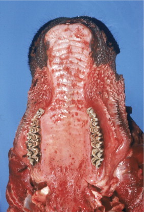
Extensive erosion on the soft and hard palate regions of a heifer that died from chronic BVDV infection. This heifer was persistently infected with BVDV. Oral erosions in most field cases of BVDV infection involving naive cattle are not this dramatic or extensive.
(Photo courtesy Dr. John M. King.)
Figure 6-39.
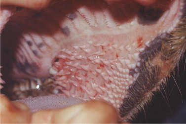
Hyperemia and erosion of the mucosa of the papillae near the lip commissures of an acutely infected naive cow. The papillae in the middle of the region are eroded, inflamed, and more pink or red than unaffected papillae. Such papillae may or may not appear “blunted.”
Figure 6-40.
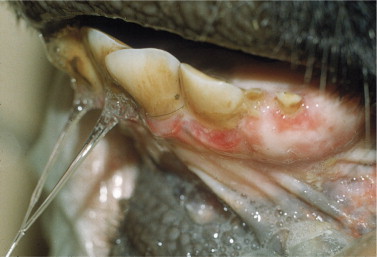
Distinct erosions of the mucosa adjacent to the incisor teeth of an acutely infected cow from a herd outbreak of BVDV.
Figure 6-41.
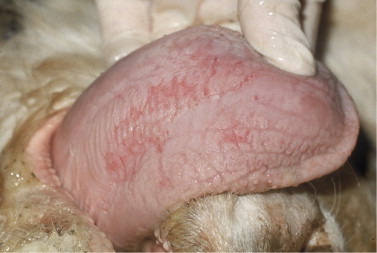
Erosions on the ventral surface of the tongue in a superinfected PI-BVDV heifer.
Immunocompetent seronegative cows exposed to strains of BVDV capable of causing classical acute signs usually seroconvert and survive. However, some seronegative non-PI cows exposed to these viruses become seriously ill and may die. Some NCP-BVDV strains possess sufficient pathogenicity to kill adult, immunocompetent, seronegative cattle. This fact was highlighted by the 1994 epidemic of BVD in Ontario and the northeastern United States. Therefore a cow or calf does not have to be PI to be killed by a field strain of BVDV. Fatal consequences of BVDV (other than superinfection of PI animals) can occur directly as a result of BVDV-induced thrombocytopenia with subsequent hemorrhage, electrolyte, fluid, and protein losses caused by severe diarrhea, and other causes. Most commonly, however, fatal consequences of BVDV are secondary to opportunistic pathogens creating concurrent infection during BVDV viremia. Even immunocompetent healthy cattle suffer profound alterations in cellular defense mechanisms during the time between onset of BVDV infection and humoral antibody production or recovery. Most healthy cattle exposed to BVDV infection survive this time uneventfully, but less fortunate ones may develop pneumonia, mastitis, metritis, or other bacterial infections while viremic. Temporarily altered cellular immunity affects lymphocytes neutrophils, macrophages, and may predispose to bacteremia or alter clearance of circulating microbes. The clinical consequence of this temporary lapse in cellular defenses is an inability of such patients to overcome routine infections. Cattle infected with BVDV experimentally may or may not be exposed to other routine infections, whereas cattle naturally infected with BVDV are subject to multiple stresses and infections. During the period of viremia and altered cellular defense, dairy calves and cows may succumb to IBR virus, other enteric pathogens (especially Salmonella sp.), bacterial mastitis, bacterial pneumonia, and other infections. High mortality has been observed when BVDV and Salmonella sp. concurrently infect groups of calves or cows. Recently assembled herds or purchased groups of replacement heifers may trigger severe disease by introducing a new strain of BVDV to a resident herd. Immune responsiveness returns to normal as BVDV infection wanes and serum neutralization titers against the virus increase. Therefore seronegative immunocompetent cattle infected with BVDV do not have any residual or permanent immunodeficiency following resolution of the infection and seroconversion. Both increased severity of concurrent disease and lack of responsiveness to conventional therapy for that disease may be seen during the window of time that a patient is viremic with BVDV. Concurrent infections such as IBR or pneumonia caused by Mannheimia haemolytica in animals viremic with BVDV may be so severe as to mask the underlying BVDV because signs of illness or postmortem lesions incriminate respiratory pathogens as the cause of illness. Failure of these more obvious infections to respond to conventional therapy should raise the index of suspicion regarding BVDV infection. For example, a severe outbreak of M. haemolytica pneumonia masked underlying BVDV in a herd that had recently added 20 replacement heifers. Cultures obtained from affected cattle from tracheal wash and necropsy confirmed M. haemolytica sensitive to several antibiotics. The indicated antibiotics had been used to treat affected animals, but the expected clinical response was not obtained. Mucosal lesions subsequently were found in a few of the fatal cases, and a NCP-BVDV was isolated from the blood buffy coat of several affected animals.
In addition to altered cellular immune responsiveness, acute BVDV usually causes a leukopenia characterized by lymphopenia and sometimes neutropenia. Therefore not only are WBC functions diminished but also their absolute numbers are as well. Leukopenia increases the risk of opportunistic bacterial infection, and neutropenia seems to be associated with increased severity of concurrent diseases.
BVDV also attacks lymphoid tissues such as the spleen, lymph node germinal centers, and Peyer's patches and can infect lymphocytes and macrophages (Figure 6-42 ).
Figure 6-42.
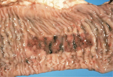
Necrosis of Peyer's patch in necropsy specimen of fatal BVDV infection.
(Photo courtesy Dr. John M. King.)
Combining all the aforementioned negative effects on host immunity helps explain why some non-PI cattle die during acute BVDV infection. Some would argue that these cattle in fact die from Mannheimia sp., Salmonella sp., or whatever secondary infection overwhelms the animal during the transient altered immunity caused by acute BVDV infection rather than from BVDV itself. The net effect, however, is mortality, and some BVDV strains can kill or contribute to the death of seronegative, immunocompetent, adult cattle.
Thrombocytopenia associated with type 2 acute BVDV infection has been observed in adult dairy cattle, dairy calves, and veal calves. Although platelet counts, 100,000/μl are abnormal, clinical evidence of bleeding seldom is observed unless the platelet count is, 50,000/μl. Conditions such as stress, injections, trauma, or insect bites that may contribute to clinical signs of bleeding in thrombocytopenic clinical patients may not be present in experimental models. Thrombocytopenia associated with bleeding causes blood loss anemia, which is highly fatal unless treated with fresh whole blood transfusions. Thrombocytopenia occurs as a result of viral infection and destruction of megakaryocytes in bone marrow. Dysfunction of circulating platelets may contribute to clinical signs of impaired coagulation. Field outbreaks of acute BVDV with thrombocytopenia are characterized by one or more of the affected cattle having signs of epistaxis, bloody diarrhea, bleeding from injection or insect bite sites, ecchymoses and petechial hemorrhages on mucous membranes, or hematoma formation (Figure 6-43 ). Not all infected cattle show signs of bleeding, and the magnitude of thrombocytopenia varies greatly. In addition, inapparent infection with subsequent seroconversion may occur in some herdmates. However, when bleeding is associated with other clinical signs such as diarrhea, fever unresponsive to antibiotics, gastrointestinal ulceration, and leukopenia, then BVDV should be strongly suspected. Platelet counts and isolation of BVDV from mononuclear cells in whole blood confirm the diagnosis. Other causes of bleeding can be ruled out by coagulation panels, including assessment of fibrin degradation products.
Figure 6-43.
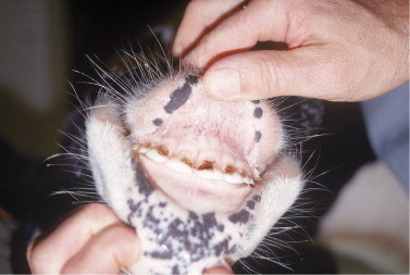
Petechiation and severe intestinal bleeding (PCV 10) in an 8-month-old heifer having thrombocytopenia associated with acute BVDV infection. After a blood transfusion, the heifer recovered.
Acute BVDV infection of naive, non-PI calves may cause inapparent infection with seroconversion or clinical signs that include fever and diarrhea of varying severity. The greatest risk for calves with acute BVDV infection is concurrent infection with other enteric or respiratory pathogens. Transient reduction of cellular immune function and defense mechanisms during BVDV viremia predispose to and worsen concurrent infection. Therefore diarrheic neonatal calves (,2 to 3 weeks of age) can have acute BVDV infection masked by identification of encapsulated E. coli, Salmonella sp., rotavirus, coronavirus, or C. parvum (Figure 6-44 ). Similarly calves up to several months of age may have overt respiratory disease caused by Mannheimia sp., H. somni, or respiratory viruses that are isolated from tracheal wash or necropsy specimens. In all of these situations, concurrent BVDV should be suspected when the severity of disease, morbidity, and mortality seem excessive for the identified pathogens. Naive, non-PI calves born to seropositive cows should acquire passive antibody protection against homologous strains for 3 to 12 months. However, this passive protection may or may not protect against heterologous strains and may not be protective if calves receive less than adequate amounts of colostrum. In addition, overwhelming exposure to BVDV may override any passive protection in some instances. Seronegative calves are at risk at all times. Whenever severe calf mortality associated with enteric or respiratory pathogens occurs, BVDV should be considered and ruled in or out by viral isolation from blood, necropsy tissue samples, or tracheal wash samples.
Figure 6-44.
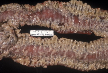
Concurrent S. Typhimurium and BVDV-induced intestinal lesions in a neonatal calf.
(Photo courtesy Dr. John M. King.)
Persistent Infection
PIs caused by NCP-BVDV arise from fetal infections occurring before 125 days of gestation. Such calves are born seronegative and PI if the dam is PI. Alternatively, a PI calf can be born to a non-PI, immunocompetent dam—the sole requirement is BVDV infection that creates viremia in the dam of sufficient magnitude to cause transplacental infection. PI calves may be transiently seropositive if the dam (PI or not) was infected during pregnancy and passed antibodies to the calf through colostrum; PI dams may generate colostral antibody titers to heterologous strains of BVDV.
Calves may appear normal at birth, grow normally, and become productive members of the herd. This situation is perhaps the most frightening because such PI cattle are not easily detected and continue to harbor and shed homologous BVDV through body secretions. Apparently healthy PI cattle also reliably reproduce PI offspring that subsequently act as reservoirs of infection for herdmates. PI calves or cattle that are clinically normal may develop signs of acute or chronic (“mucosal disease”) BVD if exposed to heterologous strains of CP-BVDV through natural exposure, administration of modified live BVDV vaccines, or genetic recombination of their homologous BVDV strain. In fact, the source of RNA that causes conversion to CP from NCP biotype may even be derived from the RNA of the animal's own cells.
At one time, it was assumed that all heterologous strains of BVDV would cause fatal infections in PI cattle because such cattle would not recognize these strains as foreign. It also was assumed that CP-BVDV strains were necessary to cause disease in PI animals because many workers found both CP- and NCP-BVDV in cattle having chronic or mucosal disease. Not all heterologous strains of BVDV cause PI animals to develop illness, however. Experimental inoculation of PI cattle with certain CP-BVDV strains not only may fail to produce disease but also may be associated with seroconversion against the heterologous CP-BVDV and continued failure of seroconversion against the homologous NCP-BVDV. This situation and that of the PI calf that attains passive-colostrum origin antibodies from its dam constitute two reasons that a PI animal could have serum-neutralizing antibodies against BVDV.
Apparently healthy PI animals often remain in the herd, produce PI offspring, and represent significant sources of perpetuating infection for herdmates and fetuses. Some PI calves are born weak, small, or die shortly after birth. Weak calves that survive generally succumb to enteric or respiratory pathogens within the first few weeks of life. Clinical signs and gross necropsy findings may not suggest BVDV infection, and death is attributed to enteritis or pneumonia of varying causes. This clinical scenario allows BVDV to escape detection unless blood or tissue samples are submitted for viral isolation or antigen detection. Some workers have observed domed skulls and finer-than-normal maxillary shape (“deer noses”) in PI calves that are weak, small at birth, and often do not thrive (Figure 6-45 ).
Figure 6-45.
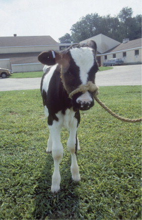
A 6-week-old BVDV-PI calf with poor growth and abnormally developed skull.
The intermediate clinical view for PI calves falls somewhere between the apparently healthy PI calf that remains healthy, and the calf that is obviously weak, small, or nonviable at birth. This intermediate type is apparently normal at birth but dies before 2 years of age. The cause of death in such PI animals is variable. Recurrent or chronic infections are the hallmark of these calves. Enteritis, pneumonia, ringworm, pinkeye, ectoparasites, or endoparasites may affect such calves, and they may persist or respond poorly to therapy. Unexplained pneumonia and/or diarrhea in a single growing heifer on a farm should arouse a suspicion of PI in that animal. Poor growth and stunting compared with herdmates is obvious in these PI animals (Figure 6-46 ). Because chronic bacterial, parasitic, or fungal infections typify many of the PI calves in this category, the integrity of immune responses must be questioned. Although PI animals initially were thought to have complete immunocompetence except for the “self” BVDV that they harbor, complete immunocompetence seems unlikely in all cases. There may be a variable expression of cellular or secretory immunity, and other factors, such as the exact time of in utero infection and the strain of NCP-BVDV, may play roles in relative immunocompetence. At least some PI animals appear to have reduced lymphocyte and neutrophil function.
Figure 6-46.
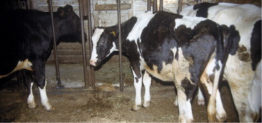
BVDV-PI yearling heifer that is stunted and has not grown well. The heifer is stanchioned between two healthy herdmates of the same age.
In addition to apparently heightened susceptibility to a variety of opportunistic pathogens, PI animals in this category can succumb to superinfection with CP-BVDV (Figure 6-47 ), as discussed previously for classical mucosal disease. In fact, PI animals in this category (e.g., chronic disease, poor-doers, less than 2 years of age) compose the majority of “classic BVD,” “chronic BVD,” or “mucosal disease” cases. Signs of BVD tend to be profound with diarrhea, poor condition, dehydration, mucosal lesions, and sometimes leg edema (Figure 6-48 ) and skin and digital lesions. The course of disease is highly variable—some cases die rapidly, whereas others linger on as poor-doers. The major differential diagnoses for chronic poor-doer BVDV-PI animals are bovine leukocyte adhesive deficiency (BLAD) and chronic internal abscessation because all of these conditions yield similar gross clinical appearances.
Figure 6-47.
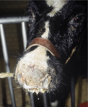
“Classic” mucosal disease in a 6-month-old heifer. After contact with “outside” cattle, an entire group of replacement heifers developed fever, diarrhea, and dermatitis, but this heifer that was a PI animal was the only one that died.
Figure 6-48.
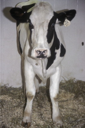
A 12-month-old heifer with persistent fever and edema of all four legs caused by vasculitis and BVDV-PI.
Congenital Lesions
Some BVDV infections may only become apparent after the birth of calves with congenital lesions. Once again, the individual pattern of disease or set of signs within a specific herd may be unique to that herd. Adult cattle may experience subclinical infection that results in abortion or congenital anomalies such as cerebellar hypoplasia, cataracts, retinal and optic nerve degeneration, hydranencephaly, hypomyelinogenesis, brachygnathism, varying degrees of hairlessness, and other congenital lesions (Figure 6-49, Figure 6-50, Figure 6-51 ; see also Figure 12-15). It must be mentioned here that BVDV is not responsible for all congenital cataracts; there are many other causes. Although a plethora of types of congenital lesions are possible, only one or two may appear in a single herd and will be repeated in affected calves born over a period of weeks or months. Usually several consecutive calves are affected with the same type of congenital lesion. For example, in a herd that Dr. Rebhun investigated, brachygnathism and cataracts typified the congenital lesions, whereas in other herds, other ocular lesions or cerebellar hypoplasia may predominate. The strain of infecting BVDV certainly may play a role in determining the anatomic area of congenital defects because some strains seem to possess a degree of organ specificity. Both CP and NCP strains are capable of inducing fetal anomalies. Most congenital lesions are thought to indicate in utero infection between days 75 to 150 of gestation. Overlap between this time and the period for persistent infection (40 to 125 days) exists. Therefore calves born with congenital lesions may be PI or may be seropositive in precolostral blood samples depending on exactly when the in utero infection occurred and whether NCP or CP BVDV caused the congenital lesion. Calves with congenital lesions should be tested to determine whether they are PI—especially if the congenital lesions are not life threatening and the owner would like to keep the animal.
Figure 6-49.
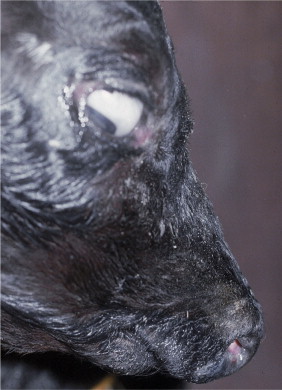
Brachygnathism in a calf associated with in utero BVDV infection.
Figure 6-50.
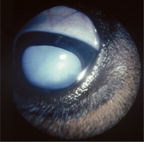
Diffuse cataract (bilateral) in a calf that was infected by BVDV during the mid-trimester of gestation.
Figure 6-51.
Optic nerve degeneration and chorioretinal scarring apparent as hyperreflective zones dorsal to the optic disc in a calf infected by BVDV during the mid-trimester of gestation.
Reproductive Signs
In addition to fetal congenital defects, BVDV may cause a variety of reproductive consequences. Abortion always is a possibility when in utero BVDV infection occurs (Figure 6-52 ). Abortion has been observed or caused (experimental infections) at most stages of gestation with CP-BVDV and is possible in the midtrimester or last trimester as a result of NCP-BVDV. Fetuses may be infected several weeks or months before abortion in some instances. Mummification also is possible following in utero BVDV infection.
Figure 6-52.
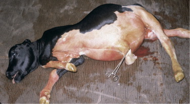
Aborted fetus from a BVDV-infected cow.
Perhaps the greatest concerns for future BVDV research revolve around effective protection of the fetus from BVDV. The temporal relationships among PI, congenital lesions, and, to a lesser degree, abortion or mummification seem to have been worked out. However, the consequences of early in utero fetal infection (0 to 40 days) are not as well known.
Acutely infected immunocompetent bulls and PI bulls shed BVDV in semen. Insemination with infected semen will cause infection and subsequent seroconversion in seronegative cattle. Cattle infected by such semen tend not to conceive until establishing immunity and seroconversion. Oophoritis has been detected several weeks following experimental infection, and ovarian dysfunction may be responsible for the impaired fertility seen in some infected cow populations. Semen is a possible source of infection for herds and probably has been the occasional cause of reduced fertility and other BVDV problems in herds. Frozen semen also has been shown to be capable of BVDV transmission to susceptible cattle. Although PI bulls may have detectable abnormalities of semen, these are not consistent, and standard semen testing should not be used in lieu of viral isolation or antigen detection to identify infected bulls. Some immunocompetent (non-PI) bulls may shed the virus in the semen for an extended time. Most commercial bull studs now routinely screen incoming bulls for PI status or virus shedding before semen collection for AI purposes.
Intrauterine infusion of BVDV at the time of insemination was shown to cause susceptible cows to have early reproductive failure, low pregnancy rates, high return rates, and seroconversion. Reproductive failure appeared as a result of failure of fertilization. However, when either seronegative or seropositive cattle were infected orally or nasally rather than intrauterine, conception was not affected. Thus the consequences of maternal exposure to BVDV at the times of breeding, fertilization, implantation, or early gestation remain somewhat unknown when infection by routes other than intrauterine occur.
Fluids containing BVDV-contaminated fetal bovine serum used for embryo transfer also can serve as a source of infection in susceptible cattle and reproductive failure. The potential consequences of PI embryo donors or PI recipients currently dictate testing of animals to be used for these purposes in embryo transfer.
Diagnosis
In classic cases with fever, diarrhea, mucosal lesions, and digital lesions, diagnosis may be made with confidence based on the clinical signs. Unfortunately this represents a distinct minority of the cases. Clinicians must remember that even in epidemic acute disease, 50% of infected cattle may have detectable lesions on clinical examination. In addition, because most cattle infected by strains of BVDV have subclinical or mild infections, signs suggestive of BVDV may be absent. Specific physical examination findings are limited to oral mucosal lesions and digital lesions. Such lesions may be obvious in superinfected PI animals having all of the signs of severe BVD (“mucosal disease”) but may be subtle or absent in seronegative animals experiencing acute BVDV infection. Mucosal lesions also may lag behind nonspecific early signs of fever, depression, and reduced milk production. Whenever BVDV infection is suspected, a methodical examination of the oral cavity—aided by focal light illumination—is essential if subtle erosions are to be found. Lesions can be in any area of the oral cavity, but focal erosions of the hard and soft palates, tongue erosions, erosions at gingival border of the incisor teeth, and blunted hyperemic papillae that are eroded at the tip are most commonly seen. Digital lesions are even less common than oral mucosal lesions in field outbreaks of BVDV. When present, coronitis and interdigital erosions are most common. Laminitis usually is observed only secondary to chronic coronitis in PI animals suffering superinfection. Although not widely practiced, endoscopy to see the esophageal mucosa might allow detection of typical linear erosions that are quite common in both acute and chronic infections.
Persistence of high fever or biphasic high fever occurring over more than 7 days is found in many acute BVDV infections. Initially the affected animal may not appear seriously ill and may be thought to have a “respiratory virus.” If, however, fever persists and is unresponsive to antibiotics, these same cattle may show more overt anorexia, depression, and dehydration after several days. Diarrhea and mucosal lesions are more common at this time. Few diseases of dairy cattle cause the profound and complete anorexia observed in BVDV-infected cattle having mucosal and gastrointestinal lesions. Oral erosions, esophageal erosions, forestomach erosions, and lower gastrointestinal lesions contribute to patient pain, discomfort, and subsequent anorexia (Figure 6-53 ). Salivation and bruxism also may be observed in these patients.
Figure 6-53.
Multifocal linear erosions of the esophageal mucosa caused by acute BVDV infection.
Bleeding associated with fever and diarrhea in several calves or cows should raise the suspicion of thrombocytopenia associated with acute BVDV infection; it necessitates confirmation of both BVDV infection through isolation and thrombocytopenia through taking platelet counts. Similarly herd reproductive problems such as abortions, mummified fetuses, or dramatically reduced conception rates should be grounds for ruling BVDV in or out as a potential cause. Congenital malformations or lesions in one or a series of calves born within a few weeks or months also should dictate BVDV as part of the differential diagnosis.
Laboratory work may suggest BVDV infection but is not reliable as a sole diagnostic aid. For example, leukopenia characterized by lymphopenia is present in most calves and cattle suffering acute infection with BVDV. Many of these animals are neutropenic as well. Fever of unknown origin coexisting with persistent leukopenia should raise suspicion of acute BVDV but could be mimicked by other diseases.
Without question, the most difficult cases of BVDV infection occur when acute BVDV breaks out concurrently with other pathogens such as Mannheimia sp. pneumonia, Salmonella sp. enterocolitis, or viral respiratory infections such as IBR or BRSV. In such outbreaks, morbidity and mortality may be exceedingly high, and physical findings and lesions at necropsy are predominated by the non-BVDV diseases. Cattle with acute BVDV infection may have had little time to develop pathognomonic gross lesions consistent with BVDV before dying from their concurrent diseases because the transient immune suppression of cellular defense mechanisms during acute BVDV infection frequently predisposes to concurrent opportunistic infection and alters normal immune responses to these opportunists. Necropsy findings in such cases identify overwhelming bronchopneumonia (Mannheimia sp.) respiratory pathology consistent with IBR or BRSV, or severe enterocolitis caused by Salmonella sp. Lesions consistent with BVDV infections may be absent or only present in a minority of the fatal cases. The temptation for the clinician and pathologist is to accept these gross lesions as sufficient evidence of the primary cause and thus fail to submit samples for viral isolation.
Similarly some PI calves or yearlings that are chronic poor-doers and have chronic pneumonia, ringworm lesions, chronic or intermittent diarrhea, chronic parasitism, chronic pinkeye, or other lesions that have not responded to conventional therapy may be written off as having illness caused by the more obvious bacterial infection if viral lesions are not present or missed. Once again, diagnostic testing to demonstrate the presence of BVDV in the animal is essential for positive diagnosis.
Although high mortality calf diarrhea outbreaks are more typically caused by E. coli, rotavirus, coronavirus, cryptosporidium, and Salmonella sp., occasional outbreaks may have concurrent BVDV infection, and viral isolation should be a part of the diagnostic material submitted from both live and necropsied calves in such cases. The differential diagnosis for BVDV infection is lengthy and depends somewhat on the clinical signs present in the affected herd. Acute infections characterized by diarrhea and fever must be differentiated from salmonellosis and other causes of enteritis by bacterial fecal cultures and blood cultures. Abortion epidemics must be differentiated from other bacterial, viral, and protozoan causes of abortion. When hemorrhages are present along with signs of fever and diarrhea, BVDV must be differentiated from bracken fern intoxication, disseminated intravascular coagulation (DIC), or other coagulopathies and certain mycotoxicoses.
Other mucosal diseases such as BTV and vesicular diseases—both endemic and exotic—must be considered in unusual cases and may necessitate consultation with federal regulatory veterinarians if confusion exists as to the definitive diagnosis.
Concurrent bacterial, viral, or parasitic diseases may confuse or mask the presence of BVDV infection. Whenever multiple animals fail to respond to conventional therapy for suspected or confirmed bacterial infection, the possibility of BVDV infection should be investigated. Weak or unthrifty PI calves must be differentiated from bacterial septicemia, selenium deficiency, and enteric pathogens. The source of illness in chronic “poor-doers” or unthrifty PI calves or yearlings with multiple problems must be differentiated from BLAD, chronic internal abscesses, malnutrition, parasitism, and chronic pneumonia or enteritis.
Because of the variability in clinical signs of BVDV infection, the only absolute proof of BVDV infection is diagnostic testing to demonstrate the presence of virus in tissues or blood. Tracheal wash samples may contain virus in some live calves, and tissues such as intestine, lymph nodes, spleen, and lung may demonstrate virus on necropsy specimens. The presence of virus in blood can be confirmed through submission of whole blood samples for viral isolation from the buffy coat or for detection of viral genetic material through PCR. Antigen-capture ELISA (AC-ELISA), which detects virus in serum, can also be used on adults and calves over 6 months of age. AC-ELISA is considered less reliable in younger calves because colostral antibody may bind to the virus in the blood and limit the ability of antibodies on the ELISA plate to bind to, and therefore detect, the virus. Further, AC-ELISA may not be able to detect the low levels of viremia in some acute infections, so this test may lack sensitivity relative to other viral detection methods for acute cases. In young calves with colostral antibodies against BVDV, PCR on whole blood is the preferred diagnostic test. Alternatively, skin biopsy (usually ear notches) in formalin or kept cold in saline (check with the diagnostic laboratory) can be submitted for immunohistochemical staining, virus isolation, AC-ELISA, or PCR. Ear notch testing is the preferred test for PI animals because virus antigen is consistently found in the ear skin of those animals at any age. In acute infection of immunocompetent adults, detectable viremia persists for up to 2 weeks. On rare occasions, acutely infected, immunocompetent animals may remain viremic up to 30 to 40 days. The period of viremia tends to be much shorter in subclinically infected animals.
Serology, despite limitations in PI animals, may be helpful when seroconversion can be demonstrated following illness; many animals possess titers .512 following a recent herd epidemic. Paired sera can be obtained at a 14-day interval from animals with clinical signs and/or their penmates; serologic testing for both type 1 and type 2 BVDV should be performed. Obviously serum titers representing neutralizing antibody levels may be greatly influenced by vaccinations and natural infection. Antibody titers from recently infected, immunocompetent animals are often indistinguishable from vaccination-induced titers.
Positive viral isolation coupled with low or nonexistent neutralizing antibody levels suggest acute infection (immunocompetent animal) or persistent infection with BVDV (immunotolerant animal). Generally, immunocompetent animals will seroconvert and clear viremia within 2 to 4 weeks, whereas PI animals remain viremic with low or nonexistent titers to the homologous strain.
PI animals can be detected by virus isolation or PCR performed on whole blood, and skin biopsies can be submitted for immunohistochemistry, virus isolation, AC-ELISA, or PCR. A positive result on any of these tests may simply reflect acute infection in a normal animal, so PI status is technically confirmed by repeat testing and detection of the virus 3 to 4 weeks after the initial positive result. Animals confirmed as PI should be culled or well isolated from the remainder of the herd because they serve as a constant source of high viral challenge for their herdmates. Again, on occasion, acutely infected, immunocompetent (non-PI) animals can remain viremic for 30 to 40 days. Delaying the second test for 6 weeks may be preferred if the tested animals are of particularly high value; in such cases, false incrimination of an animal as PI would result in significant financial loss. Such animals should be considered PI until proven otherwise by the second test and well isolated from their herdmates. Methods for screening the herd for PI animals are discussed further below in the section on prevention.
Treatment
Cattle with mild clinical disease associated with acute BVDV infection do not require specific therapy but should be offered fresh feed and water and not be subjected to any exogenous stress, transport, or vaccinations. Cattle with specific problems such as diarrhea may require oral or IV fluid therapy if continued diarrhea coupled with relative or absolute anorexia causes dehydration. Clinically ill animals (i.e., those with fever, depression, diarrhea, and dehydration) should be subjected to no extraneous stress and may benefit from prophylactic bactericidal antibiotics to minimize the potential for opportunistic bacterial infections such as pneumonia. Calves with acute BVDV infection are more likely to require supplemental fluids and electrolytes.
In cattle with clinical evidence of bleeding caused by thrombocytopenia, benefit may be derived from fresh whole blood transfusions. Usually 4 L of whole fresh blood collected from a BLV-negative, non-PI-BVDV donor is adequate unless blood loss has caused life-threatening anemia. Other affected cattle in these herds with thrombocytopenic strains of BVDV may merely be observed if clinical bleeding is not apparent. Such cattle should not be subjected to surgical procedures, parenteral injections, or crowding and should have insect populations controlled to avoid multiple insults that could cause clinical bleeding. Clinical bleeding seldom occurs unless platelet counts are, 50,000/μl and trauma to skin or tissues is excessive. Diarrhea may become bloody in some patients in these herds because inflammatory gastrointestinal lesions may sufficiently irritate the colon to cause bleeding.
Despite the fact that most acute BVDV infections are subclinical, this does not hold true for all field epidemics, and some immunocompetent animals do develop severe illness as a result of acute infection with various BVDV strains and thus may benefit from symptomatic therapy. Acute BVDV can be fatal to immunocompetent calves and adult cattle. Death caused by BVDV does not implicitly confirm superinfection of PI animals. This is especially true when complications such as thrombocytopenia or secondary bacterial or viral infections befall an immunocompetent cow with transiently depressed cellular immune responses resulting from acute BVDV infection.
Corticosteroids and NSAIDs are contraindicated in cattle suffering acute BVDV infection because both categories of drug further predispose to digestive tract erosion and ulceration. Animals that are ingesting feed and water can be treated with judicious doses of aspirin as an antipyretic, but even aspirin reduces cytoprotective prostaglandins in the gastrointestinal tract and kidney to some degree.
Prevention
Effective control of BVDV infection in dairy cattle requires four fundamental steps:
-
•
wImprovement of herd immunity through immunization
-
•
Identification and removal of PI animals within the herd
-
•
Screening of new animals for PI status before introduction into the herd
-
•
Implementation of biosecurity practices to prevent fetal exposure to BVDV; in other words, prevention of future PI animals
-
1.
Improvement of herd immunity through immunization
The goals of a BVDV immunization program on dairies include (a) prevention or reduction in severity of acute disease in adults and young stock; (b) prevention or reduction of the rate of fetal infection in pregnant heifers and cows; and (c) enhancement of colostral immunity for protection of newborns. Two fundamental challenges to effective vaccination for BVDV exist. First, the broad antigenic diversity of BVDV makes it difficult to create a vaccine that induces immunity to all of the potential strains that a herd may encounter over time. Second, the massive amounts of virus shed by PI animals may result in infection even in immunized cattle. The importance of this second issue cannot be overemphasized. Exposure to viremic animals compromises any effort at complete protection of a herd through immunization, and elimination of PI animals from the herd is the vital step in BVDV control.
As of 2004, more than 180 United States Department of Agriculture (USDA)-licensed BVDV vaccines were commercially available. The practitioner must choose between modified-live and inactivated (killed products). Both have advantages and disadvantages, summarized below:
MLVs—advantages:-
•Activation of cellular and humoral immunity
-
•Long duration of immunity
-
•Good, albeit incomplete, fetal protection; greater than inactivated vaccines
MLVs—disadvantages:-
•Potential for transient immunosuppression
-
•Potentially unsafe for administration to pregnant cattle (not for all products—some have documented safety for use in pregnant animals)
-
•Colostrum-derived antibodies may block immune response in calves
-
•Potential for transient ovarian infection and transient impairment of fertility (not to be used in cattle immediately before breeding)
-
•May induce acute disease in PI cattle—this should not be a deterrent to use, however.
Inactivated (killed) vaccines—advantages:-
•Not immunosuppressive
-
•No risk of fetal or ovarian infection (safe for administration to pregnant cattle and immediately before breeding)
Inactivated (killed) vaccines—disadvantages:-
•Primarily activate the humoral immune response (less cell-mediated immunity)
-
•Require more frequent administration (boostering)
-
•Shorter duration of immunity
-
•Variable fetal protection (field versus vaccine strain heterogeneity may limit efficacy)
In the past, MLV vaccines were suspected of inducing clinical disease, including persistent infection, because of insufficient attenuation of the virus used in the product and/or live virus contamination of vaccine reagents. With greater testing procedures available for extraneous virus testing, as well as higher standards for reagents such as fetal bovine serum for cell culture systems, the risk of live virus contamination of MLV has been greatly reduced over the past. To ensure that these precautions are used for a given product, safety data and quality control procedures for vaccine production should be requested from manufacturers of MLV vaccines.
A single recommendation for vaccination for dairy herds is unlikely to be uniformly accepted as optimal. This likely reflects different practitioners having different experiences with a variety of different vaccines. It is likely that this variation in professional opinion is because practitioners have been observing a variety of BVDV strains challenging a variety of herds over time. The following guidelines are recommended by us and others:
Replacement heifers (separated from pregnant cows and heifers): Immunize with a MLV product, ideally containing type 1 and type 2 strains, at 5 to 6 months of age and again 60 days before breeding. This schedule allows replacement heifers to receive two doses of vaccine before they become pregnant and limits potential problems caused by transient ovarian infection by vaccine virus.
Adult cows: Administer a MLV vaccine with a label claim for safety in pregnant cattle, once annually 2 to 4 weeks before breeding.
Immunosuppression following MLV BVDV vaccination has been documented, so immunization should be timed to occur during periods of relatively low stress and low pathogen challenge.
If killed products are to be used, manufacturer's recommendations should be followed regarding the timing of the priming and booster immunization; the interval for these immunizations is typically 2 to 3 weeks. To maximize antibody spectrum, a product containing inactivated type 1 and type 2 virus should be used, or at least one giving demonstrative cross protection against both. Cows and heifers should receive a booster immunization before breeding, in midgestation/midlactation, and for lactating animals, again at dry-off. Killed products may be optimal for administration to newly purchased, pregnant heifers and cows of unknown previous vaccination status.
Adequately vaccinated cattle should impart passive antibody protection to calves through colostrum—at least against homologous strains of BVDV. This passive protection probably dissipates between 3 and 8 months of age in most instances. Therefore the timing of initial active immunization of calves against BVDV is somewhat controversial. If killed products are used, calves born to vaccinated cows should probably be vaccinated three times—at 12, 14, and 18 weeks of age. Manufacturer's recommendations as regards appropriate intervals between dosing should be followed, but all calves or older cattle of questionable immune status must be vaccinated at least twice to establish primary immunity. Semiannual boosters are then recommended.
Modified live vaccines are more likely to be blocked by maternal-derived (colostral) antibody in calves, so delaying administration of the first dose until 5 to 6 months is recommended. If protection of younger calves is desired, killed products may be administered at an interval determined by the label.
A common error in vaccination programs is to give a single killed vaccine to first-calf heifers that have never received previous adequate primary immunization. Management deficiencies allow this mistake to occur more commonly than we realize. Do not assume that dairy farmers have “done it right” and always make directions for use clear-cut when selling vaccine to owners. With more widespread use of computerized records, documentation of proper timing of immunization can be implemented by making BVDV vaccination a recorded “health event” for all cattle.
As dairies become larger, the gap between management and the cow-side worker has widened. What the manager perceives to be the standard operating procedure for vaccination and what occurs when cows and calves are vaccinated may be vastly different. Therefore it is imperative the veterinarian take an active role in training workers on proper vaccine storage, handling, and administration. Personal observation of immunization practices often allows the veterinarian to detect and quickly correct problems. Because labor forces on dairies may turn over rapidly, repeated training sessions on this topic are often required. When necessary, the veterinarian should be willing to assume the role of long-term educator of the workforce.
-
•
-
2.
Identification and removal of PI animals from the herd
The prevalence of PI animals is thought to vary greatly from dairy to dairy. Data from a few large prevalence studies indicate that PI cattle typically represent approximately 2% or less of the cattle population. Because the virus may be transmitted vertically, even closed herds may have PI animals.
In the past, serologic screening of the herd after immunization was used to attempt to identify PI animals—the concept was these animals, being immunotolerant to BVDV, would tend to have low titers, and low-titered animals could then be targeted for testing by virus isolation to confirm PI status. However, in light of the fact that PI animals may mount an immune response to heterologous field or vaccine strains, this method is unlikely to accurately identify all PI animals and is not currently endorsed.
Tests that detect the presence of virus in the live animal are considered necessary for accurate identification of the PI state. Initially all animals in the herd, regardless of age or apparent health status, should be included for testing. Tests include virus isolation or PCR on whole blood and AC-ELISA, IHC, PCR, or virus isolation on skin biopsies. AC-ELISA should not be used on calves under 6 months of age, owing to potential problems with colostrum-derived antibody interference with viral detection. To prevent confusion between acutely viremic immunocompetent animals and PI animals, it may be necessary to repeat testing on any positive animal 3 to 4 weeks (or, for valuable animals, 30 to 40 days) after the first positive test.
To reduce testing costs on large numbers of animals, certain laboratories offer testing on pooled samples—for example, skin biopsies from multiple animals can be placed together in saline, and PCR can be run on the pooled sample to detect the presence of BVDV. Alternatively, composite milk samples can be pooled together and checked for the presence of virus by PCR or virus isolation. Bulk tank samples or string samples can also be used to screen large numbers of lactating cows. Current recommendations state that pooled milk samples should represent fewer than 400 animals to optimize chances of detection of PIs. It is best to check with the regional veterinary diagnostic laboratory for the preferred number of samples to be pooled, shipment requirements, and so on. Obviously a positive result on a pooled sample would require follow-up testing of the constituent individuals to identify the viremic or PI animal(s).
On rare occasions, infected bulls may shed virus only in the semen. Therefore to cover this rare, yet complication-rich scenario, bulls that test negative for virus in blood or skin biopsy should ideally have their semen screened by virus isolation or PCR.
Ethical considerations regarding the fate of PI animals are worthy of mention. Sale of animals known to be PI at livestock auctions is simply unethical because these animals serve as virus-producing machines that expose many other animals, causing potentially devastating disease on the farms of the unknowing purchasers. The most ethical practice is to euthanize these animals on the premises, although otherwise healthy animals may be considered for slaughter. To our knowledge, no studies currently exist on the persistence of BVDV in properly composted carcasses, but data on the survivability of viruses related to BVDV indicate that long-term environmental persistence is unlikely. Alternatively, carcasses of PI animals can be removed from the premises for rendering.
Once all animals in the herd have been tested and PI animals removed, testing should focus on the calves born to gestating, non-PI females. Once these calves have been tested and PIs removed, testing should focus on new introductions, show animals, semen and embryos, and heifers raised off-site (see numbers 3 and 4 below). The producer must understand, however, that introduction of a novel strain of BVDV onto a farm may result in fetal infection in non-PI, pregnant females, warranting eventual testing of their offspring.
-
3.
Screening of new animals for PI status before introduction into the herd
Reducing risk of introduction of BVDV is best accomplished by avoiding the purchase of untested cattle. Without question, the greatest disasters resulting from acute BVDV have followed the purchase of assembled cattle from sales to increase herd size. These purchased animals may be PI. Alternatively, they may be acutely infected—in either case, they represent sources of new virus to the herd. In addition, if newly purchased cows and/or heifers were exposed to BVDV and became viremic in early pregnancy, they may be carrying PI fetuses. Therefore for optimal herd protection, purchased adults should be tested before introduction into the herd; later, the offspring that they were carrying at the time of purchase should be tested for PI status because PI calves can be born to immunocompetent dams. Whenever possible, new herd introductions should be tested and well isolated from the remainder of the herd for 4 to 6 weeks—during this period, any PI animals in the group of new introductions can be identified and removed, and any acutely infected animals can be given adequate time to recover. Any contact between isolated new introductions and the remainder of the herd, even at fence lines, should be avoided during this period.
Tests to detect virus should be used on newly purchased cattle. Virus isolation, PCR, immunohistochemistry, or AC-ELISA on blood or skin biopsies can be used. Pooling of samples can be considered when large numbers of animals are to be introduced (see number 2 above). Collection of samples for testing for other diseases in newly purchased stock (e.g., Johne's disease, Mycoplasma spp. mastitis) can be performed at the same time.
-
4.
Implementation of biosecurity practices to prevent fetal exposure to BVDV
Fetal infection leading to the PI state is a critical control point because PI animals represent a massive source of viral challenge for the herd. Even with good vaccination practices, all immunity is finite, and overwhelming viral challenge could theoretically lead to transplacental passage of virus even in immunized, pregnant females. Therefore protection of pregnant cows and heifers from exposure to high viral challenge is a critical goal of BVDV control within a herd biosecurity program.
Contact with cattle outside the herd should be eliminated or minimized, even at fence lines. Cows and heifers in the first trimester of pregnancy should be considered the most susceptible to creation of the fetal PI state. These animals should be located on the farm in the area that is most protected from contact with outside cattle, new introductions, and show cattle. Pen allocation and pen milking sequence should be critiqued and, if necessary, changed to maximize protection of these animals. Contact of these animals with ill cattle should be minimized whenever possible.
Heifers raised and bred at heifer-raising operations warrant particularly careful scrutiny in a BVDV control program. Heifers from multiple herds are often raised on such operations, and viral challenge from PI animals or acute BVDV infections on that operation could easily induce fetal infection in pregnant heifers. Therefore young heifers should be tested before transport to heifer-raising operations; if this is not feasible, prompt testing after arrival on such operations is warranted. In addition, all calves born to heifers raised off-site should be considered potential PI animals and tested after birth.
Cattle taken to shows should be considered another source of novel virus on a farm. In the ideal world, show cattle should be tested and confirmed to be non-PI before being taken to shows or sales—this is simply a good ethical practice intended to protect other animals and producers. Show cattle should also be well immunized to limit the likelihood of them developing acute infection while off the premises. Given the shortcomings of vaccines in protecting against the tremendous number of strains of BVDV, even well-vaccinated show cattle should be considered potentially exposed to novel BVDV at shows or sales and ideally kept isolated from the home herd for 4 to 6 weeks on return. If exposed at shows, pregnant show cows and heifers may experience viremia of sufficient magnitude to induce fetal infection, and their calves should be subsequently tested for PI status.
Most reputable sources of semen, embryos, and fetal calf serum have BVDV testing strategies in place. However, nothing should be taken for granted, and the individuals or companies providing bull semen and/or embryos should be requested to provide documentation of their current control programs and quality control measures on reagents. Control of BVDV in embryo and semen production operations has been recently reviewed. In short, acutely or persistently infected bulls, embryo donors, and embryo recipients are potential animal sources of BVDV. Animals within these populations that shed large amounts of virus may cause fetal infection in others, so BVDV testing of all animals with which bulls, embryo donors, and embryo recipients come in contact during semen/embryo collection, transfer, and pregnancy is necessary. Rarely, infected bulls may shed virus only in semen (i.e., test negative on blood or skin IHC), and semen testing by PCR or virus isolation is considered optimal. All animal-origin reagents used in embryo transfer or in vitro fertilization (including semen and oocytes) should be screened for the presence of BVDV.
Winter Dysentery
Etiology
The etiologic agent responsible for winter dysentery has remained elusive for as long as the disease has existed. In the northern hemisphere, the disease is characterized by explosive herd outbreaks of diarrhea between the months of October and April. Campylobacter fetus jejuni long was suspected as a cause, but Koch's postulates never were confirmed. MacPherson, however, was able to infect susceptible cattle using filtered feces from infected cows and therefore believed a virus was involved. Bovine coronavirus has been demonstrated in feces and colonic epithelium of affected cattle, and the same strain that causes diarrhea in calves has been used to experimentally create winter dysentery in adult cows. In Europe, Breda virus (Torovirus) has been associated with winter dysentery outbreaks.
Winter dysentery is of economic importance primarily because of production losses both during the acute outbreak and because some cows do not return to previous production levels for the remainder of that lactation. Death losses are minimal but do occur—almost always in first-calf heifers that develop hemorrhagic diarrhea.
The disease is highly contagious and can spread easily from an affected herd to unaffected herds via fomites—both inanimate and animate. Veterinarians, milk tank drivers, inseminators, salespeople, and other farm visitors frequently are blamed for spreading the disease. Newly purchased cattle and cattle attending shows during the fall, winter, or early spring also can be infected and instigate a herd outbreak. Herds experiencing winter dysentery subsequently appear immune for 2 to 3 years, based on clinical impressions that many herds have an outbreak every third year. Relative age-related resistance is observed, but this protection is incomplete. Cattle infected for the first time tend to have more severe clinical signs than those previously affected.
Clinical Signs
Signs include acute diarrhea in 10% to 30% of the cows within a herd, followed by similar signs in another 20% to 70% of the animals within the ensuing 7 to 10 days. The diarrhea is explosive and appears semi-fluid, dark brown in color, has a pea-soup consistency, is malodorous, and forms bubbles as puddles of manure are formed. Most affected cows have decreased appetite, production losses of 10% to 50%, and become mildly to moderately dehydrated. Some develop cool peripheral parts and sluggish rumen motility suggestive of hypocalcemia. Severely affected animals—especially first lactation cattle experiencing the disease for the first time—have hemorrhagic enterocolitis with dysentery and fresh blood clots in the feces. Tenesmus may be present in these animals, and blood loss anemia may develop. A soft moist cough is apparent in several of the affected animals, but the lungs auscult normally in these cattle. Fever usually precedes clinical signs by 24 to 48 hours, and experienced clinicians will detect fever in apparently healthy herdmates that have not yet developed diarrhea. Fevers are mild, ranging from 103.0 to 105.0° F (39.44 to 40.56° C). Some cattle have mild fever 103.0 to 104.0° F (39.44 to 40.0° C) accompanying the onset of diarrhea. Herd production decreases commensurate with incidence and severity of disease. Affected cows, especially those in mid or late lactation, may never return to previous production levels for the remainder of their current lactation.
Diagnosis
Winter dysentery must be differentiated from dietary diarrhea, coccidiosis, BVDV, and salmonellosis. Dietary diarrhea seldom causes fever and usually is associated with feed changes. Coccidiosis can cause diarrhea, dysentery, whole blood clots, and tenesmus in heifers and first lactation cows but seldom affects multiparous cows. Fecal smears and flotation allow a diagnosis of coccidiosis. Usually BVDV infection causes leukopenia, higher fever, more prolonged disease, and could be ruled out by viral isolation and paired serum samples. The most likely differential diagnosis is salmonellosis caused by type E or mildly pathogenic types of B or C Salmonella spp. Salmonellosis of these types can cause fever—frequently preceding the onset of diarrhea—and a variable number of animals may develop diarrhea that contains blood, fibrin strands, or mucus. A neutropenia with left shift in the leukogram would suggest acute salmonellosis but is not a constant finding. Fecal culture obtained from several acute cases is the only way to rule out salmonellosis.
Confirmation of the presumptive diagnosis requires demonstration of bovine coronavirus in feces by electron microscopy, ELISA, or PCR. Immunohistochemical staining of colonic tissue may be used on necropsy specimens in acute cases.
Treatment
For most affected cattle, supportive treatment with oral astringents remains the time-tested mode of therapy. Occasional high-producing cattle require parenteral calcium solutions to counteract secondary hypocalcemia or treatment of ketosis secondary to reduced appetite. Oral fluids and electrolytes may be necessary for moderately dehydrated cattle. All cattle should have access to salt and to fresh water.
Severely dehydrated cows occasionally require IV fluid therapy, and first-calf heifers that become anemic because of blood loss require fresh whole blood transfusions in some instances. Cattle with tenesmus may necessitate epidural anesthesia to allow rest and reduce rectal and colonic irritation.
Treatment usually is only necessary for 1 to 5 days, by which time most affected cows have recovered their appetites and normal manure consistency. Unfortunately the disease often dwindles through the herd for 7 to 14 days, such that new cases are still appearing at a time when most cattle are recovered. Although there is no proven efficacy to preventive measures, practicing sound herd biosecurity regarding new herd introductions and show cattle may reduce the likelihood of outbreaks (see descriptions in section on BVDV). Further, disinfected boots and equipment should be required for all visitors to a dairy, as well as clean outer garments. The efficacy of immunization with commercially available coronavirus vaccines is currently unknown.
Campylobacter jejuni
Etiology
Campylobacter jejuni, formerly Vibrio jejuni, is a gram-negative, curved to spiral motile rod capable of causing enterocolitis in many species, including humans, in which the organism is one of the major causes of bacterial enterocolitis. C. jejuni may be present in the normal intestinal flora of many domestic animals and people but is found with greater incidence in diarrhea patients. Because of its ubiquitous nature, the significance of isolation of C. jejuni from diarrheic feces sometimes is hard to interpret. However, isolation of C. jejuni coupled with failure to isolate other pathogens such as Salmonella sp., Clostridium sp., or enteric viruses should be considered significant.
Diarrhea and other clinical signs vary from inapparent or mild to fulminant and may be influenced by concurrent diseases, stress, inoculum, strain of C. jejuni, other enteric pathogens, and other factors. This variation is highlighted by the reported profound differences observed between experimental infection of gnotobiotic calves and experimental infections of calves with normal gastrointestinal flora. Gnotobiotic calves had mild catarrhal enteritis with minimal clinical signs, whereas fever, chronic diarrhea for up to 2 weeks, and some degree of dysentery were observed in nongnotobiotic calves. Although most experimental infections have been completed in calves, adult cattle are thought to be susceptible as well. Infection in people can occur at any age.
The strain of C. jejuni and other factors may influence the site of colonization within the intestine, but most strains affect both the small intestine and colon. C. jejuni produces a cholera-like enterotoxin that is an important component of pathogenicity. The organism is mucosa associated but does not appear to be invasive—at least in experimental studies.
Clinical Signs and Diagnosis
Mild or inapparent cases yield little or no detectable signs. Clinical patients with severe signs of diarrhea, fever, dehydration, anorexia, cessation of milk production, and dysentery tend to be sporadic or only represent a low percentage of cattle within a herd. Adult cows and calves are at risk.
The signs are nonspecific and require differentiation from those indicating salmonellosis, coccidiosis, BVDV infection, and other enteric pathogens. Isolation of C. jejuni from diarrheic feces and ruling out other enteric pathogens are essential for diagnosis. Enterocolitis resulting from C. jejuni seldom is confused with winter dysentery because of low morbidity in the former contrasted with high morbidity in the latter. Salmonellosis represents the primary differential diagnosis.
Treatment
Calves or cattle with severe diarrhea or dysentery may require 7 to 14 days for recovery. Some diarrhea may persist despite improved vital signs in recovering patients. Oral or IV fluids may be required, and it is best to select fluids following blood acid-base and electrolyte analysis. In humans, antibiotics such as erythromycin, tetracycline, and aminoglycosides are most effective, whereas penicillin, ampicillin, cephalosporins, and trimethoprim-sulfa combinations appear ineffective. Antibiotic susceptibilities are not known for C. jejuni infections in cattle and would be best determined by fecal culture and susceptibility results.
Control
Because animals and animal products, such as unpasteurized milk and improperly cooked meat, usually are blamed for C. jejuni enterocolitis in humans, a positive diagnosis of C. jejuni diarrhea in cattle justifies public health concerns. Infected cattle may throw off the infection over several weeks or may remain carriers. Obviously many cattle (and people) are asymptomatic carriers so that wide-scale herd testing serves no purpose. However, veterinarians should advise caution in handling infected cattle and avoidance of unpasteurized milk on farms where the problem is confirmed. C. jejuni will grow in milk and may arrive in milk from a septicemic spread but is more likely to contaminate milk because of environmental contact.
Enterotoxemia in Adult Dairy Cattle
Etiology
Enterotoxemia thought to be caused by C. perfringens has been observed as a sporadic cause of acute death in adult dairy cattle. Some herds have had endemic problems with more than one cow being found dead or agonal over a few months. Premonitory signs are not observed, and, as in calves, the condition is believed to be related to diets exceptionally rich in protein and energy. C. perfringens organisms present in the intestinal tract take advantage of such rich diets and proliferate to produce excessive exotoxins—especially beta-toxin. Although C. perfringens and toxin have been identified from acute necropsy specimens in adult cattle, it is not known whether a particular strain of the organism is responsible for the adult cow disease.
Signs
Signs are minimal, and most affected cows are found down, agonal, or already dead. Diarrhea may be observed or the animal simply may have abdominal distention, colic, and depression. Another clinical syndrome is one that causes fever, anorexia, small-volume diarrhea, and death, with severe abomasal edema found at necropsy.
Diagnosis
Fresh necropsy specimens must be obtained if C. perfringens is suspected as a cause of acute death. Necropsy lesions, as in calves infected by C. perfringens type C, may be minimal but might include small intestinal fluid distention, serosal hemorrhages, and an edematous mesentery. Feces and small intestinal content should be cultured for C. perfringens; if possible, luminal contents should be tested for the presence of toxins. Samples should be transported in a cooled but unfrozen state. A complete necropsy to rule out other causes of acute death is imperative because the diagnosis of C. perfringens enterotoxemia is made by exclusion of other diseases such as peracute, virulent salmonellosis.
Treatment and Control
Treatment is seldom possible, but if specific C. perfringens organism and toxins are identified, vaccination with appropriate toxoids would be indicated.
Malignant Catarrhal Fever
Etiology
MCF has been observed in domestic and wild ruminants worldwide and is caused by a group of gamma herpesviruses. It is a severe lymphoproliferative disease characterized by high fever, corneal edema, mucosal erosions, and lymph node enlargement in clinically affected animals. Lymphocytic vasculitis of a variety of tissues is the classic microscopic lesion. Many ruminant species and pigs are susceptible to MCF viruses, and losses have been incurred on dairies, feed lots, ranches, game farms, zoos, and deer meat-raising facilities. In Africa, the causative agent has been isolated and identified as alcelaphine herpesvirus-1 (AHV1). The term alcelaphine relates to the subfamily of Bovidae, Alcelaphinae, in which wildebeest, hartebeest, and topi are classified. These species are thought to be the reservoir. The virus apparently is highly cell associated but can be spread during stress such as parturition or shipment and may be free in fetal fluids or young of wildebeest. At those times when the virus is released, it becomes infectious for cattle.
In other parts of the world, including the United States, MCF is termed “sheep-associated” because sheep appear to be the most likely reservoir of infection. Ovine herpesvirus type 2 (OvHV2) has been identified from ruminants with MCF using PCR, and seroconversion of cattle with MCF to OvHV2 can be demonstrated using competitive inhibition ELISA (CI-ELISA). However, the virus has not yet been isolated and grown in tissue culture, so this virus is currently considered a likely, but not definitive, cause of the disease. Sheep-associated MCF occurs in cattle, bison, pigs, deer, elk, and moose.
Most sporadic or epidemic MCF in cattle has been associated with proximity to sheep. Infection is widespread in North American sheep, and ovine infection is almost always asymptomatic. Cattle and sheep do not have to interact or be in common pastures for the disease to appear. Cases can be observed at any time of year. In addition, some cattle that develop MCF have no historical direct or indirect exposure to sheep. Asymptomatic, persistent infections with OvHV2 in cattle may occur, and these infections may or may not develop into clinical MCF. Hence, the incubation period for this disease has been difficult to ascertain, with infected cattle developing disease weeks to months after exposure. This may explain why some cases do not seem linked to exposure to sheep. In a recent study, most dairy cattle exposed to OvHV2 under natural conditions (close proximity to a sheep feed lot) developed asymptomatic infection rather than overt signs of MCF.
Clinical Signs
Most cattle affected with MCF have dramatic clinical signs of multisystemic inflammatory disease. A great deal of variation is possible, however, and has caused many authors to categorize MCF based on predominant signs. Such categorization is difficult because significant overlap and intermediary clinical situations occur frequently. Sporadic cases are most common in cattle, but herd epidemics have been described in several areas of the United States.
Fever is common in all cases and is high (105.0 to 108.0° F/40.56 to 42.2° C). Peracute, acute, chronic persistent, and chronic intermittent cases all have fever that usually persists as long as signs are observed. Lymphadenopathy is another finding that is common to most cases. All other signs result from a severe vasculitis that affects many organs but may affect some organs more than others in individual patients. Vasculitis is profound and histologically associated with lymphocytic infiltrates that occasionally can be so extensive as to suggest lymphoreticular neoplasia. Vasculitis affects the gastrointestinal tract, central nervous system, eyes, urinary system, liver, skin, upper respiratory tract, and other areas.
Peracute cases may die within 1 to 2 days because of overwhelming viremia and vasculitis of all major organs and yet have minimal clinical signs other than fever, lymphadenopathy, depression, and prostration. Terminal neurologic signs are possible as a result of central nervous system vasculitis.
The classical “head and eye” form of MCF is most common in sporadic cases. This form is characterized by persistent high fever (105.0 to 108.0° F/40.56 to 42.22° C), lymphadenopathy, severe nasal and oral mucosal lesions, ocular lesions, and remarkable depression (Figure 6-54 ). In acute cases, extensive mucosal lesions may make it appear as though the animal has had its mucous membranes burned. Frequently the muzzle and large regions of the oral mucosa appear hyperemic and have a blanched necrotic epithelium that sloughs to leave erosions and ulcers if the patient survives long enough for this to occur. The muzzle may appear dried or sunburned, and the superficial epithelium subsequently may slough away. Sloughing of the nasal mucosa may result in diphtheric crusts that occlude the airways. Salivation and copious nasal discharge are typical findings. On occasion, oral and nasal lesions are present in the caudal aspects of the mucosal surfaces, out of visual range during physical examination.
Figure 6-54.
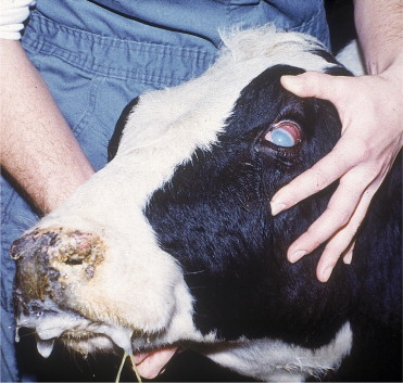
Adult Holstein cow with the head and eye form of MCF.
Bilateral ophthalmitis results from vasculitis throughout the eyes that spares only the choroid in most cases. Corneal edema is the most common lesion and occurs because of inflammatory changes and exudative cellular deposits on the corneal endothelium that disrupt this layer, thereby allowing overhydration of the corneal stroma. The corneal edema typically begins at the limbus within 2 to 5 days after the onset of fever. The corneal edema then rapidly spreads to the center of the cornea. This centripetal spread of edema distinguishes the ocular features of MCF from contagious keratoconjunctivitis (pinkeye). A severe anterior uveitis, scleritis, conjunctivitis, and retinitis usually coexist. As in other regions of the body, mononuclear cell infiltrates appear in the eyes. Depression is profound because of central nervous system vasculitis, and CSF confirms a dramatic inflammation characterized by increased protein values and mononuclear cell pleocytosis. Other neurologic signs are possible. Skin lesions and inflammation of the coronary bands and horn basal epithelium also are possible in those patients that survive more than a few days. The clinical course for most “head and eye” MCF cattle is 48 to 96 hours, although some cases may survive for a longer time and a few had even been reported to survive.
Acute MCF also may cause severe enterocolitis with diarrhea being a predominant sign. Such cases also are febrile and can have some degree of mucosal lesions, ocular lesions, and other organ involvement. This “enteric form” is again a relative designation because patients frequently have other detectable lesions in addition to diarrhea. However, severe diarrhea may be the most apparent sign and thus may confuse the diagnosis of MCF with BVDV, rinderpest, or other enteric diseases. In bison, enteric signs tend to predominate in acute cases.
Mild forms of MCF also have been observed. The broad spectrum of potential and observed clinical signs in MCF also makes it likely that some cattle suffer subclinical mild disease, recover, and respond immunologically to the causative virus. Although previously considered to be a highly fatal disease, up to 50% of MCF cases may survive the acute disease to either recover or become chronic cases.
A rare acute form of the disease presents as a severe hemorrhagic cystitis with hematuria, stranguria, and polyuria. Cattle having this form of acute infection have high fever and only survive 1 to 4 days. Although the most striking clinical signs are limited to the urinary system, histologic evidence of vasculitis and lymphocytic infiltration are generalized on necropsy study.
Chronic MCF is characterized by a long clinical course—usually weeks—of high fever, erosive and ulcerative mucosal lesions, bilateral uveitis, papular or hyperkeratotic skin lesions, lymphadenopathy, and digital lesions (Figure 6-55, Figure 6-56 ). Mucosal lesions tend to be severe, slough tissue, and cause salivation and inappetence (Figure 6-57 ). Some chronic cases recover only to relapse weeks to months later. Such cases appear healthy between episodes, but recurrence of fever and mucosal, ocular, and skin lesions is debilitating. It is rare for chronic MCF cattle to recover completely and survive.
Figure 6-55.
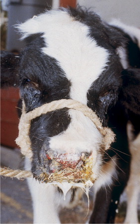
A 6-month-old Holstein bull with chronic MCF.
Figure 6-56.
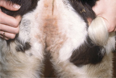
Papular dermatitis in the escutcheon region in a calf with chronic MCF.
Figure 6-57.
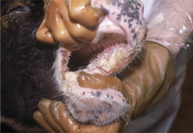
Chronic necrotic oral and lingual lesions in a yearling heifer with chronic recurrent MCF.
Diagnosis
Given the wide variability of possible clinical signs of MCF in cattle, differential diagnosis could include many diseases. Head and eye lesions could be confused with severe IBR respiratory and conjunctival infections because corneal edema can occur in some severely affected IBR conjunctivitis cases. However, IBR usually is epidemic and affected animals have characteristic mucosal plaques present on the palpebral conjunctiva and nasal mucosa. Most mucosal diseases such as BVDV, BTV, VSV, and FMD may need to be considered depending on the duration, location, and severity of signs. Cattle having severe diarrhea but minimal mucosal lesions could be confused with BVDV or rinderpest infections.
Acute bracken fern intoxications, bacillary hemoglobinuria, and other causes of hematuria may be considered in acute MCF characterized by hemorrhagic cystitis. Acute or subacute mucosal lesions that cause sloughing of muzzle epithelium could be confused with primary or secondary (hepatic) photosensitization.
When ocular lesions are present in MCF patients, the diagnosis is made easier because none of the other mucosal diseases cause severe uveitis and ophthalmitis. As mentioned, IBR conjunctivitis can have corneal edema in severe cases, but intraocular inflammation does not occur with IBR. There are no ocular lesions in acute or chronic postnatal BVDV infections. The acute mucosal lesions of MCF also are unique in classic cases. The oral mucosa is diffusely inflamed and appears as though the patient drank boiling water. The muzzle mucosa appears burnt, crusty, or eroded in these same patients. Unfortunately these classic mucosal lesions do not occur in all MCF patients, and patients with multifocal erosions or ulcers can be more difficult to differentiate from those with BVDV and other mucosal diseases.
High fever (105.0 to 108.0° F/40.56 to 42.22° C) that persists through the entire clinical course is characteristic of most acute MCF cases. Chronic MCF cases also will have persistent fever that may or may not be as high as that found in acute cases.
Nervous signs suggest a diagnosis of MCF because central nervous system involvement is rare with other mucosal diseases. However, high fever and terminal prostration are common in fatal cases of most mucosal diseases and could be confused with neurologic signs.
Clinical diagnosis of MCF is best supported by CSF analysis. The characteristic CSF mononuclear cell pleocytosis and elevated protein value found in MCF patients is useful whenever the patient's clinical signs dictate consideration of differential diagnoses. Confirmation of sheep-associated MCF requires demonstration of viral genome in the blood through PCR analysis of white cells obtained from a whole blood (EDTA) sample. Depending on the primers used, PCR for OvHV2 is specific for that virus and will not detect the genome of AHV1. Alternatively, CI-ELISA can be used to detect MCF antibodies, but this test may not be positive at the time of initial clinical signs. CI-ELISA detects antibodies to both OvHV2 and AHV1 and cannot currently distinguish between wildebeest-and sheep-associated MCF.
Histopathology allows detection of pathognomonic diffuse vasculitis with lymphocytic infiltrates in many organs, including the gastrointestinal tract, urinary tract, liver, adrenals, central nervous system, skin, and eyes. Necrotizing vasculitis is present in lymphoid tissues.
Prevention
Prevention of MCF centers on limiting exposure to infected wildebeest and sheep. Airborne transfer of OvHV2 is suspected to occur over a distance of more than 70 meters, so segregation of cattle from sheep by greater distances may be protective. For cattle and bison herds, Callan recommends a separation distance of 1 mile from sheep. Carrier (asymptomatically infected) cattle may be identified by CI-ELISA on serum and PCR on whole blood. The CI-ELISA test detects seroconversion in exposed individuals that may, owing to varying viral loads in blood, be intermittently negative by PCR on whole blood. Alternatively, acutely infected animals may be positive on PCR but negative on CI-ELISA owing to the delays inherent in generation of an immune response. Therefore application of both tests may provide optimal sensitivity for detecting infected cattle. Although OvHV2 DNA can be detected in milk, nasal secretions, and ocular secretions of asymptomatic and clinically affected cattle, this viral DNA appears to be cell-associated and does not pose a significant risk for horizontal transmission. Transmission from cattle or bison to other animals has not been demonstrated and is considered likely to be a rare event, if it occurs at all.
Johne's Disease (Paratuberculosis)
Etiology
M. avium subspecies paratuberculosis (MAP), the cause of paratuberculosis—better known as Johne's disease—is an acid-fast, gram-positive, intracellular bacterium. The organism is extremely hardy and can remain viable in the environment for up to 1 year, given sufficient moisture and cool temperatures. Extremes of heat and dryness decrease viability.
Johne's disease is a chronic insidious disease of cattle characterized by a majority of subclinical infections with no evidence of infection. Debilitated animals having chronic diarrhea, weight loss, and hypoproteinemia during the terminal stages of the disease represent a small minority of infected animals (,5%). Typically infection occurs most commonly through fecal-oral contamination during the early perinatal period. Infected fecal material is ingested by calves either when nursing the dam or by licking environmental materials contaminated with manure. Older calves have a more variable outcome following infection, and larger doses of MAP are required to cause infections that lead to later onset of clinical signs. Furthermore, young adult or adult cattle seem to have even greater age-related resistance. However, this resistance is relative rather than absolute, and some experimental infections of older calves and adults have been reported. Factors including concurrent diseases, genetics, environment, and other stressors may contribute to increased susceptibility to infection. Although difficult to define, certain breeds and genetic lines within these breeds have been thought to be susceptible to Johne's disease. Guernseys, for example, appear to have inherent susceptibility. Unfortunately it is difficult to separate true genetic weakness from massive exposure to organisms in infected herds.
In addition to the fecal-oral route of infection, in utero infection of fetuses harbored by infected dams is possible with an estimated 25% of fetuses from dams with clinical signs infected in utero.
Similarly, infected macrophages in milk from infected cows have been suggested as a source of infection for calves. The percentage of infected cows that shed M. paratuberculosis in milk varies with the stage of infection. In utero and milk containing MAP occur more commonly in heavy shedders and cattle with clinical signs of Johne's disease. Semen and reproductive tracts from infected bulls also have yielded M. paratuberculosis, but semen rarely appears to be a source of infection. Infection of susceptible calves may occur throughout the small intestine with less colonization of the large intestine. The ileum is commonly believed to be the most predisposed site, but in some cattle the ileum may have little or no MAP, whereas the mid-jejunum will be heavily colonized. Following initial uptake of MAP by the macrophages in the mucosa, the organism elicits a granulomatous response in the mucosa extending to the submucosa. Over time, MAP spreads to the regional lymph nodes and eventually becomes systemic in advanced stages of the disease. The extent of infection varies greatly among individual cattle. In addition, the incubation period is extremely long, often requiring 2 to 3 years up to 10 years in some cases from time of infection to the development of clinical signs. Several points deserve emphasis.
-
1.
Most infected cattle never develop clinical signs before being culled from the herd. Factors that contribute to clinical disease versus asymptomatic infection are not known but probably include organism dose, age at infection, nutrition, concurrent diseases, stresses, and genetics. Infected cattle shed MAP in their manure and transmit the disease to herdmates by MAP contamination of the environment. Herd infection prevalence varies from 20% to 100% in heavily infected herds. Despite this rather high incidence of infection, it is unusual to see clinical signs in more than 5% to 10% of adult cows in the herd per year. Johne's disease has been shown to persist in some herds for more than 10 years with no overt clinical signs of infection.
-
2.
It is widely accepted that cattle that develop clinical signs shed large numbers of organisms and represent the greatest threat to contaminate the environment. Recently described super-shedders may shed MAP in higher concentrations (1 to 5 million CFU/g of manure) than cattle with clinical disease. Potentially these animals represent the greatest source of environmental contamination and reservoir for possible transmission to herdmates. Most super-shedders are asymptomatic with no evidence of diarrhea or weight loss, yet excrete huge numbers of MAP organisms into the environment.
-
3.
Passive shedding of MAP may occur when noninfected cattle ingest manure contaminated forage or water. With a super-shedder in the herd, ingestion of as little as 5 ml of manure contamination in forage may result in passive shedding and give rise to a positive fecal culture for a previously uninfected cow. The risk of misclassification of such cattle must be considered when control programs include fecal cultures of all adult animals. Recent experience suggests this phenomenon may represent 50% of all culture-positive cattle in the herd when a super-shedder is present.
Because most infected cattle are asymptomatic and clinical cases may have decreased production and be culled before a final diagnosis is made, the true incidence of Johne's disease is hard to estimate. The 1996 National Animal Health Monitoring Survey (NAHMS) estimated 20% of dairy herds were infected. In herds with more than 400 cows, the prevalence increased to 40%. Current unofficial estimates suggest more than 75% of dairy herds in the United States are infected with MAP, with an individual dairy cow prevalence of between 5% and 10% depending on method of detection.
Regional surveillance of Pennsylvania and, indirectly, other areas of the northeastern United States confirmed a dairy cow prevalence of up to 7.3% in many areas. With ever increasing dairy herd size, largely attributed to purchase of cows of unknown status, the herd prevalence will continue to increase. In herds in which 10% of the culled cows have clinical Johne's disease, the average loss exceeds $220 per cow or $22,000 for a herd with 100 milk cows. Without question, this disease is of tremendous economic importance to the entire cattle industry and especially to the dairy industry.
Clinical Signs
Although most cattle infected with MAP remain asymptomatic, cattle with clinical signs signal the diagnosis and alert both veterinarian and herd owner to the possibility of a herd-wide problem. However, if the cow with clinical signs was purchased, the magnitude of the herd problem may not be as extensive. Clinical signs consist of chronic diarrhea, progressive wasting and loss of condition, and eventually ventral pendent edema, especially in the submandibular area as a result of hypoproteinemia. Temperature and vital signs are normal. The diarrhea classically has been defined as pea soup in consistency and often forms bubbles because of the rather liquid consistency. However, given today's laxative diets, the diarrhea observed in a Johne's disease patient is best described as looser compared with herdmates. Initially the manure of an infected cow is formed but loose, then becomes pea soup-like, and finally, in advanced cases, a very watery consistency. Appetite and attitude remain normal in early cases, but milk production and body condition deteriorate because of progressive protein loss. Abomasal displacement is another observed complication in cattle with moderate to severe Johne's disease. The exact cause of displacement is unknown, but gastrointestinal stasis caused by hypocalcemia and reduced dry matter intake may contribute to the condition.
Moderate to advanced clinical cases have obvious weight loss characterized by muscle wasting, a poor dry hair coat, significant production losses, dehydration, and reduced feed intake—particularly high-energy feedstuffs (Figure 6-58 ). Ventral edema is apparent but may vary in the anatomic area involved. Intermandibular, brisket, ventral, udder, and lower limb edema all are possible (Figure 6-59 ). The pea soup-like or liquid manure seen in advanced cases stains the tail, perineum, and hind limbs. It will stain the rear quarters if the tail switches liquid feces onto the quarters, flanks, and gluteal region.
Figure 6-58.
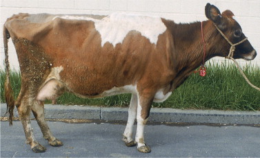
Jersey cow affected with Johne's disease. Poor condition, a dry hair coat, and fecal staining of the hind quarters and tail are apparent.
Figure 6-59.
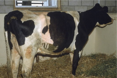
Four-year-old Holstein cow with submandibular edema (bottle jaw) and weight loss caused by Johne's disease. Diarrhea was minimal. The diagnosis was confirmed by right flank laparotomy and ileal lymph node biopsy.
Despite loose manure, loss of body condition, and diminished milk production, cows with Johne's disease do not appear seriously ill until the terminal stages when finally the appetite is markedly reduced. Occasionally cattle have diarrhea intermittently rather than continually, but this is unusual. We have also observed cows with Johne's disease with obvious diarrhea that spontaneously reverted to apparently normal manure after shipment to our hospital for diagnosis. Whether stress associated with shipment or a change in diet is responsible for this temporary improvement in fecal consistency is unknown.
Clinical signs develop only after a prolonged incubation period and usually appear between 2 and 5 years of age. However, signs have been observed in heifers less than 12 months of age and in mature cows up to 8 to 10 years of age (Figure 6-60 ). If several 2-year-old heifers in a herd develop clinical signs of diarrhea, this would suggest a rather heavy dose of MAP at an early age, whereas clinical signs in 5- to 7-year-old cows would suggest a much lower dose of MAP or older age at the time of exposure. Thus age of onset of clinical signs will assist the astute clinician as to the severity of the herd problem. Age of onset is probably affected by many factors, such as dose and duration of exposure to infectious organisms, nutrition, genetics, concurrent diseases or stresses, and other factors. The clinical impression that signs frequently develop following the onset of lactation in the first, second, or third lactations suggests lactational stress may be sufficient to amplify subclinical signs and hypoproteinemia to a clinical state. It also is possible that this observation is simply a reflection of closer monitoring of appetite, production, and body condition in lactating animals as opposed to heifers or dry cows. Lactation stress is not a prerequisite to the development of clinical signs, as proven by bulls and steers having clinical Johne's disease. Interestingly, some severely affected bulls and steers with Johne's disease have developed abomasal displacements during the advanced stages of disease.
Figure 6-60.
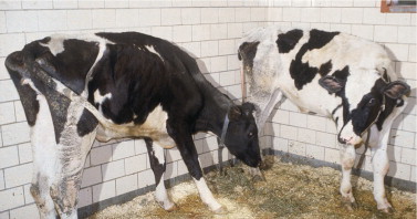
A pair of 18-month-old Holstein heifers with advanced Johne's disease. These heifers were representative of an age-grouped epidemic involving 12- to 24-month-old heifers on a single farm. This would imply extremely heavy environmental contamination with M. paratuberculosis.
Many cattle with signs of Johne's disease are culled because of poor production before a diagnosis of Johne's disease is confirmed or suspected. This is especially common in free stall operations in which an individual cow's manure consistency may not be as obvious as it would be in conventional housing and individual stalls. Cattle with clinical or subclinical infection also may have higher cull rates than uninfected herdmates because of mastitis and reproductive failure. Other studies have found a higher cull rate and reduced production but less mastitis in infected versus noninfected herdmates. Increased mastitis and reproductive failure may be partially explained by hypoproteinemia, negative energy and protein balance, stress, and poor condition.
Diagnosis
Cattle with advanced signs of Johne's disease are easily suspected of having the disease because of diarrhea, hypoproteinemia, production loss, weight loss, and overall deterioration of condition. The only abnormalities detected routinely in serum biochemistry are hypoalbuminemia, hypoproteinemia, and occasionally hyperphosphatemia (.7 mg/dl). Clinical Johne's disease must be differentiated from chronic parasitism, coccidiosis, chronic salmonellosis, toxicities, intestinal neoplasia, heart failure, glomerulonephropathies, renal amyloidosis, eosinophilic enteritis, and chronic BVD infections.
Confirmation of a clinical diagnosis of Johne's disease should focus on the agar gel immunodiffusion-(AGID), ELISA, or PCR test. Typically more than 85% of cattle with clinical Johne's disease will be positive on either the ELISA or AGID test. Nearly 100% should be positive when using a robust PCR test. Each of these tests has a turnaround time of less than 1 week, often 2 to 3 days. Fecal cultures are the most sensitive test but have a much longer turnaround time from submission to final result (12 to 16 weeks for solid media culture and 30 to 42 days for liquid media culture). The gold standard diagnosis is culture of ileum, ileocecal lymph nodes, or other mesenteric lymph nodes for cattle with clinical Johne's disease. This technique has been used to identify Johne's disease-infected cattle at slaughter houses and to gather epidemiologic data regarding prevalence of the disease. Although harvesting ileocecal lymph nodes constitutes an invasive procedure for clinical patients, extremely valuable or individually purchased cows suspected of having Johne's disease may warrant invasive techniques to diagnose the condition definitively, especially when the herd has not been known to have Johne's disease in the past. A right flank exploratory laparotomy is performed to harvest a full-thickness 1.0-cm wedge of ileum and an ileocecal lymph node (Figure 6-61, Figure 6-62 ). The ileal biopsy and half of the lymph node are submitted for culture and histopathology, including a Ziehl-Neelsen stain. The remaining half of the lymph node is used for impression smears that are stained for acid-fast organisms. An absolute diagnosis usually is possible from the impression smears, but if this fails or is questionable, the histopathology generally confirms or denies the diagnosis without the prolonged delay associated with cultures.
Figure 6-61.
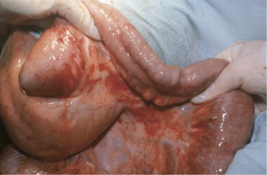
Thickened, edematous ileum and visibly distended lymphatics on the serosal surface in a cow showing early signs of Johne's disease. A rapid definitive diagnosis was established by biopsy of the ileum and ileocecal lymph node.
Figure 6-62.
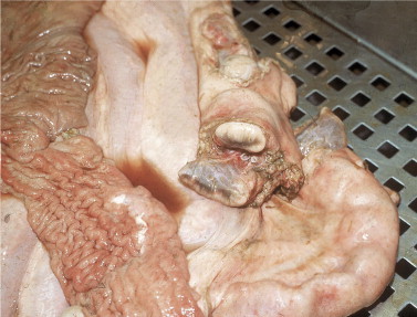
Necropsy view of thickened ileum and edematous (cut) ileocecal lymph node from a cow with advanced Johne's disease.
For asymptomatic infected cows, the sensitivity of all Johne's disease diagnostic tests is much reduced compared with cattle with clinical signs. Specificity of fecal cultures usually has been estimated to be 100%, although recent work confirms that passive excretion of MAP may occur in heavily contaminated environments. Positive fecal cultures in noninfected cows may occur as the result of “pass-through” when super-shedders are present in the herd. Up to 50% of the positive fecal cultures may be the consequence of one or more super-shedders in the herd. Several USDA-licensed ELISA tests are available and commonly used today. The overall ELISA sensitivity for detection of Johne's disease is estimated at 25% as reported by the National Johne's Working Group. The sensitivity of fecal culture was estimated to be 40%. Cattle in the eclipse phase, before onset of fecal shedding are typically negative on all tests for Johne's disease. In an attempt to determine whether such cattle are infected, testing older animals in the herd of origin with pooled fecal cultures or testing environmental manure samples by reverse transcriptase-PCR are good strategies. Because ELISA tests are not 100% specific, fecal samples for all ELISA-positive cattle should be submitted for culture or PCR testing. Overall, approximately one third of fecal samples from ELISA-positive cattle are culture positive. Some herd owners are choosing to do fecal cultures on all adult cattle in the herd, especially those owners who have made the requisite management changes designed to reduce transmission of Johne's disease within the herd. Laboratory testing in the absence of management changes to reduce the risk of transmission is not recommended.
No currently available diagnostic test will detect all infected cattle; thus repeated, usually annually, testing is necessary to detect infected cattle. Thus a negative test for Johne's disease should not be construed as proof of a noninfected animal, but only as data that suggest that if MAP infection was present, it was not detectable at that time. A more sound epidemiological approach would be testing of adult cattle in the herd using pooled or environmental manure samples. Recent reports suggest pooled fecal samples (five samples/pool) and composite environmental manure samples provide excellent tools to detect herd infections, and that if these tests are negative they indicate the herd is at low risk for Johne's disease. Both of these testing scenarios have been incorporated into the National Johne's Herd Status program.
Gross and histological lesions obtained at necropsy or slaughter facilities are extremely helpful to render an absolute diagnosis. Mild clinical cases may have a thickened edematous ileum with distended lymphatics on the serosal surface. Thickening of the mucosal surface and a raised corrugated appearance is typical. Moderate to advanced cases have obvious thickening and edema of the ileum with marked corrugation of the mucosal surface (see Figure 6-62). Lymphatic distention is obvious on the serosal surface of the ileum, and the ileocecal lymph nodes, as well as other mesenteric lymph nodes, are enlarged and edematous on cut sections. Lesions may be present in the cecum and colon of advanced clinical cases and can extend orad from the ileum to more proximal regions of the small intestine. Although MAP may be isolated from other organs such as the liver, uterus, or fetus in some advanced cases, gross lesions consisting of granuloma formation are rare in these organs, and truly disseminated infections having gross lesions are very rare. However, disseminated infections detected by culture of MAP from lymph nodes such as the prescapular, prefemoral, supramammary, or popliteal lymph nodes do occur in cattle with clinical disease. Aortic calcification has been observed in advanced cases.
Histopathology confirms a granulomatous enterocolitis with macrophages and epithelioid cells in the submucosa and lower mucosa. Ziehl-Neelsen staining confirms the presence of MAP in the intestine and lymphatics. However, culture of these same tissues has a much greater sensitivity to detect MAP than does histopathology.
Treatment
Although treatment seldom is attempted, therapeutic options do exist for valuable animals that may justify the expense and the continued exposure risk these cattle may represent for transmission to herdmates.
Clofazimine and isoniazid have both been used to successfully alleviate clinical signs of MAP infection in cattle and small ruminants. Although the drugs reduce fecal shedding, the infection persists despite daily therapy. Typically treated animals will gain weight, have improved manure consistency, and plasma protein levels are restored within a few weeks. Continued daily therapy is necessary to maintain the animal free of clinical signs. Isoniazid (20 mg/kg orally, once daily) has been used either alone or in conjunction with rifampin (20 mg/kg orally, once daily). As with clofazimine, however, the infection is only suppressed, not cured despite an improved clinical picture. Isoniazid is the most economical choice but may require adjunctive therapy with rifampin to achieve clinical improvement. All therapy for Johne's disease involves extralabel drug use, requires prolonged (i.e., months-long) treatment, precludes use of milk or meat for human consumption, and is expensive. Unless isolation of the patient is possible, treated cattle continue to shed MAP and pose a risk to infect herdmates.
Control
Once a diagnosis of Johne's disease has been confirmed, the herd owner must be counseled regarding the economic implications and options for control or eradication of the disease. Economic considerations extend beyond the loss of clinical cases to increased cull rates in subclinical cases; fear of dissemination of disease to noninfected herds through sale of infected but apparently normal calves or cattle; risks inherent to embryo transfer; and decreased productivity. Currently states offer Johne's disease programs to aid control and support testing costs of the herd to detect infected cattle. All federally supported Johne's disease programs require that a Johne's disease–certified veterinarian does a herd risk assessment and a herd management plan before funds are available to support the diagnostic testing.
Although difficult, eradication of the infection from a herd requires intensive and repeated use of fecal cultures on all animals older than 24 months of age for many years. All clinically suspicious cattle should be culled immediately or tested by AGID or ELISA. If positive on either of these tests, the animals should be culled. Calves should be born in disinfected, cleaned maternity areas and removed from the dam immediately. Calves should be raised completely separately—preferably on a separate farm—from the adult cattle. All calves should be fed colostrum from ELISA-negative, or better, fecal culture–negative cows. The purchase of replacement animals from herds of unknown Johne's disease status continues to represent the greatest risk to introduce or reintroduce Johne's disease to the herd. Minimizing fecal contamination of feedstuff, water, pastures, and exposure of calves to adult cow feces is essential and must be evaluated on an individual herd basis. Equipment used for manure removal or that could be contaminated by manure must remain separate from feeding implements and the calf environment.
Although these principles for eradication are straightforward, they may not be practical or affordable in some instances. Compromises in eradication may allow “control” but do not eliminate the disease and continue to compromise sale opportunities for purebred herds.
Vaccines for Johne's disease have been used in Europe, Australia, and several states in the United States. In the United States, a licensed killed vaccine is available, but its use is limited to infected herds, and approval by the state veterinarian is required. The herd must be tuberculin test negative. If approved for use in a specific herd, the herd owner must agree to have all calves vaccinated before 35 days of age. Each calf must have an ear tattoo and a special ear tag that signifies a Johne's disease vaccinate. The vaccine is administered SQ in the brisket and frequently predisposes to a local abscess over the next few months or years. The vaccine should be used in conjunction with management changes to reduce the incidence of infection in herds. However, the vaccine does not prevent infection, but vaccinated cattle shed fewer organisms in their manure. Most importantly, the vaccine prevents clinical signs in nearly all vaccinated cattle. Because premature culling from the herd because of Johne's disease infection is the major economic loss attributable to Johne's disease, the vaccine is considered highly efficacious by herd owners who have years of experience with Johne's disease. Other disadvantages for vaccine use include concern regarding interpretation of tuberculin reactions and accidental self-inoculation of the vaccine by veterinarians.
Foreign Animal Disease
Mucosal diseases such as BVDV, BTV, MCF, and VSV require differentiation from foreign animal diseases that threaten livestock in the United States (Table 6-8 ). Extreme vigilance is necessary to prevent entrance of those diseases to this country, and consultation with regulatory state or federal veterinarians is imperative whenever confusion exists. Because a great deal of overlap is possible for the clinical signs present in domestic and foreign mucosal diseases, positive diagnosis including appropriate serologic and virologic confirmation is essential.
TABLE 6-8.
Foreign or Exotic Animal Diseases Affecting the Gastrointestinal Tract
| Disease | Cause | Clinical Signs | Major Differential Diagnosis | Diagnosis | Reference |
|---|---|---|---|---|---|
| Foot and mouth disease (Aftosa) | FMDV = genus Aphtovirus, family | Fever, salivation, lipsmacking, lameness, teat lesions | Vesicular stomatitis | Call regulatory veterinarians | Kahrs RF: Viral diseases of cattle, Ames, IA, 1981, Iowa State University Press. |
| Picorniviridae | |||||
| (Aphthous fever) | BVDV | Fluid from vesicles | |||
| 7 distinct serotypes with multiple subtypes |
|
|
|
Sutmoller, P: Vesicular diseases. In Committee on Foreign Animal Diseases, editors: Foreign animal diseases, Richmond, VA, 1992, United States Animal Health Association. | |
| Rinderpest (cattle plague) | RV = genus | Peracute— high fever, death | BVDV | Call regulatory veterinarians | Kahrs RF: Viral diseases of cattle, Ames, IA, 1981, Iowa State University Press. |
| Morbillivirus, family | |||||
| (Peste bovine) | |||||
| Paramyxoviridae | |||||
| 1 major serotype with field strains possessing variable pathogenicity | Classic fever, mucous membrane congestion, necrosis, and subsequent erosion | Malignant catarrhal fever | Samples best obtained from febrile animals with mucosal lesions (early cases) | ||
| Vesicular stomatitis | |||||
| Foot and mouth disease | |||||
| Mucous membrane lesions cause salivation, ocular discharge | Salmonellosis | Serologic testing | Seek B, Cook R: Rinderpest. In Committee on Foreign Animal Diseases, editors: Foreign animal diseases, Richmond, VA, 1992, United States Animal Health Association. | ||
| Bluetongue | Viral isolation | ||||
| Severe hemorrhagic diarrhea and tenesmus start several days after mucosal lesions | Arsenic poisoning | ||||
| Dehydration, death | |||||
| Subacute or atypical-lower mortality, greater difficulty in distinguishing from differential diagnosis |
Liver Abscess
Etiology
Abscesses of the liver occur at all ages in cattle. In calves, liver abscesses are often the result of omphalophlebitis, whereas in older cattle they most often are secondary to reticulorumenitis. In feed lot cattle, it is well recognized that the change from pasture to a high concentrate ration causes a rapid increase in rumen fermentation and organic acid production, which may result in erosion and inflammation of the rumen epithelium. Metastasis of bacteria from the inflamed and necrotic rumen wall to the liver occurs via the portal vein. In dairy cattle, similar failure of adaptation of rumen fermentation may occur at the onset of lactation when there is an abrupt increase in the energy content of the diet. Liver abscesses also may occur as a result of traumatic reticulitis. The most common organisms isolated from hepatic abscesses are Fusobacterium necrophorum and Arcanobacterium pyogenes. Streptococci and staphylococci also may be isolated from mixed cultures.
Clinical Signs
Local, circumscribed liver abscesses are characteristically silent clinically and are not associated with systemic abnormalities or with hepatic dysfunction. Such abscesses are found incidentally during the postmortem examination of slaughtered cattle and are of importance economically because of the condemnation of affected livers.
Liver abscess, when located adjacent to the vena cava, may distort the vessel wall and cause phlebitis and thrombosis. Septic thromboembolism from the vena cava may cause a respiratory syndrome characterized by cough, dyspnea, and/or pulmonary hemorrhage that is described in Chapter 4. In a postmortem series of 6337 slaughtered cattle, liver abscesses were found in 368 (5.8%), and of these, 24% were located in the craniodorsal aspect of the liver with the potential for causing vena caval thrombosis.
Liver abscesses may be associated with constitutional abnormalities that include fever, anorexia, weight loss, and reduced milk production. Neutrophilic leukocytosis and significant increases in serum globulin and fibrinogen are characteristic. Growth of a liver abscess near the common bile duct may obstruct bile flow and may result in clinical signs and laboratory abnormalities associated with impeded flow of bile (see below). Liver abscess also has been recognized as a cause of vagal indigestion.
Ultrasonographic examination of the liver is a valuable diagnostic procedure for determining the location of the abscess(es) and for evaluating prognosis and response to therapy (Figure 6-63, Figure 6-64 ). The lesions may vary in diameter from a few centimeters to more than 20 cm. Characteristically they may be visualized in three or four adjacent intercostal spaces, and needle aspiration may not be necessary for diagnosis. On rare occasion a large liver abscess can be seen on radiographs displacing the diaphragm (Figure 6-65, A to C ).
Figure 6-63.
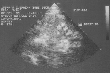
Transabdominal sonogram of the liver in a mature cow with multiple hyperechoic abscesses. The hyperechoic appearance suggests dense purulent exudate, decreasing the chances of successful treatment.
Figure 6-64.
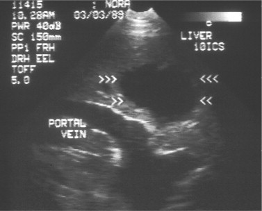
Transabdominal sonogram of the liver in a 3-year-old Holstein cow with weight loss and diminished production. A single large hypoechoic abscess can be seen, and the cow recovered following 1 month of systemically administered penicillin treatment.
Figure 6-65.
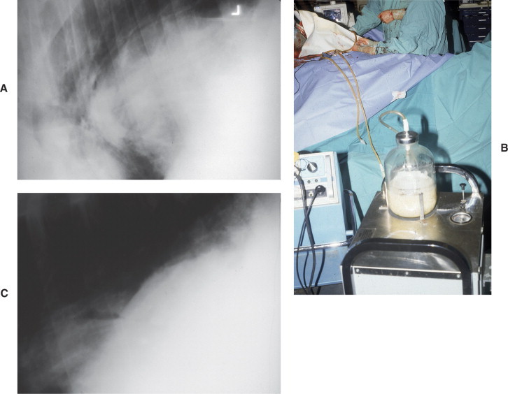
A, Thoracic radiographs of a 14-month-old Holstein heifer showing a very large mass (liver abscess) displacing the diaphragm. B, The liver abscess was drained. C, Radiographs repeated after drainage.
Treatment
When liver abscesses are recognized clinically and their location identified, it is possible to consider antibiotic therapy and/or surgical drainage. The decision regarding drainage of liver abscesses depends on the size, location, and the condition of the cow. Penicillin treatment can be successful in some cows with smaller, hypoechoic abscesses, but relapses often occur unless treatment is for 4 or more weeks. Even with surgical drainage, relapses may occur. Prognosis for treatment of liver abscesses that have caused clinical signs is guarded and is least favorable for large and hyperechoic abscesses. Successful surgical treatment of a liver abscess that caused vagal indigestion has been described.
Bile Duct Obstruction and Cholangitis
Intrahepatic cholestasis is observed in lactating cattle with severe fatty liver during the periparturient period and is described in Chapter 14. Extrahepatic cholestasis is caused by obstruction of bile flow from choleliths within the common bile duct or by obstruction of flow in the common bile duct as a result of external mechanical pressure exerted on the common bile duct by liver abscesses, by extensive adhesions in the area of the cystic and common bile ducts, or by smaller inflammatory lesions of the common duct near the hilus or the duodenal papilla (see video clip 13).
The characteristic sterility of the biliary tract is maintained by the continued production and flow of bile into the intestine. Partial or complete obstruction of bile flow predisposes to ascending infection of the biliary tract by intestinal microorganisms. Infection of the biliary tree causes cholangitis and may result in significant alterations in the physical characteristics of bile, including the accumulation of inspissated products of inflammation and of precipitated bile constituents (bile acids, cholesterol, and even stone formation) (Figure 6-66 ), which further impedes the flow of bile.
Figure 6-66.
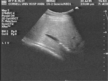
Abdominal sonogram of an adult Holstein hospitalized because of anorexia and mild colic. The cows' GGT was 1500 IU/L. Stones are observed in the intrahepatic ducts. There was a marked clinical improvement within 3 days of initiating therapy with penicillin, IV fluids, flunixin meglumine, and forced feeding.
The clinical signs of extrahepatic bile duct obstruction and cholangitis include malaise, colic, fever, icterus with orange-colored urine (Figure 6-67 ), and, in some cases photodermatitis (Figure 6-68 ) secondary to retention of phylloerythrin. Abnormal laboratory findings consist of leukocytosis, hyperbilirubinemia, bilirubinuria, and elevations in serum globulin and fibrinogen, bilirubin, and in the serum activities of SDH, AST, AP, and GGT. Ultrasonographic findings in cows with extrahepatic cholestasis include severe dilatation of the gallbladder, the cystic and common duct, and other major intrahepatic bile ducts. Dilatation of the gallbladder is not specific because in all anorectic cows, the gallbladder may be distended. The diagnostic findings for extrahepatic cholestasis, however, were dilatation of the cystic and common bile ducts and of the major intrahepatic bile ducts (see video clip 14).
Figure 6-67.
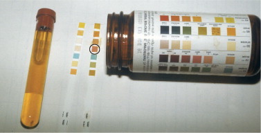
A sample of urine collected from an adult cow with icteric membranes, fever, anorexia, depression, and hepatogenous photosensitization of the muzzle. The cow responded well to symptomatic treatment similar to Figure 6-66. The urine is orange and positive on Multi-strip examination for bilirubin. Circled square is positive test. Untested strip to the left.
Figure 6-68.
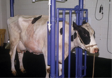
The cow in Figure 6-67 with pronounced hyperemia (photosensitive dermatitis) of the muzzle.
A case of cholelithiasis with cholestasis has been reported by Drs. Rebhun and Cable that was clinically similar to those in which bile duct obstruction was caused by external mechanical pressure on the common bile. A laparotomy was performed, and concretions 1 to 3 cm in diameter were palpated in the gallbladder. The choleliths in the gallbladder were crushed manually, and the material was massaged through the distended cystic and common ducts, into the duodenum, and on into the jejunum. Following the procedure, there was significant improvement in clinical condition and in liver function tests, although the improvement was transient.
A clinical syndrome of unknown etiology has been observed that is clinically similar to that described above but in which there is no laparotomy evidence of extrahepatic cholestasis. The clinical signs and laboratory test results are similar. When force-fed for a few days and treated with penicillin for at least 1 month, there has been gradual improvement in clinical signs and laboratory abnormalities return to normal (see Figure 6-67, Figure 6-68).
Primary hepatic neoplasms are unusual in cattle but could cause obstruction of bile flow and should be considered in cows with both icterus and photosensitization. In a necropsy series of 66 primary bovine hepatic neoplasms, 40 were classified as hepatocellular carcinomas, 10 as hepatocellular adenomas, and 10 as cholangiocellular tumors. Less frequently observed primary tumors of the liver in this series included hemangiosarcoma, hemangioma, fibroma, and Schwannoma. In the postmortem examination of the livers of 24,169 slaughtered cattle, primary liver tumors of hepatocellular origin were identified in 22 (0.09%). In a third series of 1.3 million livers of cattle examined at slaughter, 36 had primary liver tumors of which 13 were classified as primary hepatocellular neoplasms and 21 as cholangiocarcinomas.
The clinical signs of cattle associated with primary hepatic neoplasms have not been extensively described. The expected clinical signs would be those associated with the growth of an expanding hepatic mass or with metastasis to the lung or to the spleen, both of which have been observed at necropsy. If the tumor obstructs bile flow, then icterus and photosensitization would be expected (Figure 6-69, A and B ). Dermatitis caused by photosensitization is frequently most severe on the teats and muzzle, although it may be more generalized (Figure 6-70 ). Ultrasonography should be of value in locating and otherwise assessing the location and prognosis of primary liver tumors.
Figure 6-69.

A, An 8-year-old Holstein cow with weight loss, inappetence, and photosensitization, which is best seen on the teats and udder. B, The cow had a cholangiocarcinoma.
Figure 6-70.
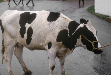
Hepatogenous photosensitization caused by a suspected hepatotoxin.
Hepatic Insufficiency Associated with Sepsis
A syndrome of hepatic insufficiency has been described in lactating cattle following acute septic mastitis or metritis in which the initial clinical signs were compatible with endotoxemia. Subsequent clinical signs included anorexia, weight loss, reduced milk production, and, in one case, photodermatitis. In addition to increased serum activities of liver enzymes, the cows had remarkable delays in the sulfobromophthalein (BSP) plasma clearance test. Liver biopsies showed hepatocellular vacuolization or necrosis that was attributed to the effects of endotoxemia associated with acute systemic infection. Similar hepatic injury has been reported in humans following endotoxic shock. Five such cases were treated by force-feeding and with other symptomatic support. Three of the cows responded satisfactorily to therapy, one failed to respond, and the fifth cow was lost to follow-up evaluation. Based on these observations, it is important to consider the possibility of hepatic injury in the initial management of cows with postpartum sepsis and in the longer term management when there is a sluggish response to therapy of the acute disease. If there is a history of prolonged antimicrobial therapy, intestinal and hepatic mycosis must also be considered (Figure 6-71 ).
Figure 6-71.
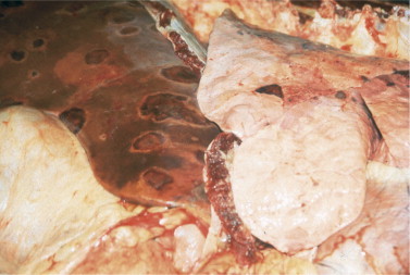
The liver from a 6-year-old Holstein that had intestinal (forestomach and abomasum) and hepatic aspergillosis secondary to generalized sepsis and treatment with broad-spectrum antibiotics.
RECOMMENDED READINGS
- Abutarbush SM, Carmalt JL, Wilson DG. Jejunal hemorrhage syndrome in 2 Canadian beef cows. Can Vet J. 2004;45:48–50. [PMC free article] [PubMed] [Google Scholar]
- Abutarbush SM, Radostits OM. Jejunal hemorrhage syndrome in dairy and beef cattle: 11 cases (2001 to 2003) Can Vet J. 2005;46:711–715. [PMC free article] [PubMed] [Google Scholar]
- Acres SD. Enterotoxigenic Escherichia coli infections in newborn calves: a review. J Dairy Sci. 1985;68:229–256. doi: 10.3168/jds.S0022-0302(85)80814-6. [DOI] [PMC free article] [PubMed] [Google Scholar]
- Ahmed AF, Constable PD, Misk NA. Effect of orally administered cimetidine and ranitidine on abomasal luminal pH in clinically normal milk-fed calves. Am J Vet Res. 2001;62:1531–1538. doi: 10.2460/ajvr.2001.62.1531. [DOI] [PubMed] [Google Scholar]
- Alcaine SD, Sukhnanand SS, Warnick LD. Ceftiofur-resistant Salmonella strains isolated from dairy farms represent multiple widely distributed subtypes that evolved by independent horizontal gene transfer. Antimicrob Agents Chemother. 2005;49:4061–4067. doi: 10.1128/AAC.49.10.4061-4067.2005. [DOI] [PMC free article] [PubMed] [Google Scholar]
- Allison MJ, Robinson IM, Doughtery RW. Grain overload in cattle and sheep: changes in microbial populations in the cecum and rumen. Am J Vet Res. 1975;39:181–185. [PubMed] [Google Scholar]
- Al-Mashat RR, Taylor DR. Production of diarrhoea and dysentery in experimental cakes by feeding pure cultures of Campylobacter fetu s subspecies jejuni. Vet Rec. 1980;107:459–464. doi: 10.1136/vr.107.20.459. [DOI] [PubMed] [Google Scholar]
- Anderson BC. Gastric cryptosporidiosis of feeder cattle beef cows and dairy cows. Bov Pract. 1988;23:99–101. [Google Scholar]
- Ares-Mazas E, Lorenzo MJ, Casal JA. Effect of a commercial disinfectant (‘Virkon’) on mouse experimental infection by Cryptosporidium parvum. J Hosp Infect. 1997;36:141–145. doi: 10.1016/s0195-6701(97)90120-1. [DOI] [PubMed] [Google Scholar]
- Arthington JD, Jaynes CA, Tyler HD. The use of bovine serum protein as an oral support therapy following coronavirus challenge in calves. J Dairy Sci. 2002;85:1249–1254. doi: 10.3168/jds.S0022-0302(02)74189-1. [DOI] [PMC free article] [PubMed] [Google Scholar]
- Audet S, Crim R, Beeler J. Evaluation of vaccines, interferons, and cell substrates for pestivirus contamination. Biologicals. 2000;28:41–46. doi: 10.1006/biol.1999.0240. [DOI] [PubMed] [Google Scholar]
- Bellamy J, Acres SD. A comparison of histopathological changes in calves associated with K99- and K991 strains of enterotoxigenic E. coli. Can J Comp Med. 1983;47:143–149. [PMC free article] [PubMed] [Google Scholar]
- Berchtold JF, Constable PD, Smith GW. Effects of intravenous hyperosmotic sodium bicarbonate on arterial and cerebrospinal fluid acid-base status and cardiovascular function in calves with experimentally induced respiratory and strong ion acidosis. J Vet Intern Med. 2005;19:240–251. doi: 10.1892/0891-6640(2005)19<240:eoihsb>2.0.co;2. [DOI] [PubMed] [Google Scholar]
- Berghaus RD, McCluskey BJ, Callan RJ. Risk factors associated with hemorrhagic bowel syndrome in dairy cattle. J Am Vet Med Assoc. 2005;226:1700–1706. doi: 10.2460/javma.2005.226.1700. [DOI] [PubMed] [Google Scholar]
- Bertone A, Rebhun WC. Tracheal actinomycosis in a cow. J Am Vet Med Assoc. 1984;185:221–222. [PubMed] [Google Scholar]
- Besser TE, Gay CC. Septicemic colibacillosis and failure of passive transfer of colostral immunoglobulin in calves. Vet Clin North Am: Food Anim Pract. 1985;1:445–459. doi: 10.1016/s0749-0720(15)31295-0. [DOI] [PubMed] [Google Scholar]
- Besser TE, Gay CC, Pritchett L. Comparison of three methods of feeding colostrum to dairy calves. J Am Vet Med Assoc. 1991;198:419–422. [PubMed] [Google Scholar]
- Besser TE, Lejeune JT, Rice DH. Increasing prevalence of Campylobacter jejuni in feedlot cattle through the feeding period. Appl Environ Microbiol. 2005;71:5752–5758. doi: 10.1128/AEM.71.10.5752-5758.2005. [DOI] [PMC free article] [PubMed] [Google Scholar]
- Besser TE, McGuire TC, Gay CC. Transfer of functional immunoglobulin G (IgG) antibody into the gastrointestinal tract accounts for IgG clearance in calves. J Virol. 1988;62:2234–2237. doi: 10.1128/jvi.62.7.2234-2237.1988. [DOI] [PMC free article] [PubMed] [Google Scholar]
- Bettini G, Marcato PS. Primary hepatic tumours in cattle. A classification of 66 cases. J Comp Pathol. 1992;107:19–34. doi: 10.1016/0021-9975(92)90092-9. [DOI] [PubMed] [Google Scholar]
- Bezek DM, Mechor GD. Identification and eradication of bovine viral diarrhea virus in a persistently infected dairy herd. J Am Vet Med Assoc. 1992;201:580–586. [PubMed] [Google Scholar]
- Bistner SI, Rubin LF, Saunders LZ. The ocular lesions of bovine viral diarrhea-mucosal disease. Pathol Vet. 1970;7:275–285. doi: 10.1177/030098587000700306. [DOI] [PubMed] [Google Scholar]
- Blanchard PC. Sampling techniques for the diagnosis of digestive disease. Vet Clin N Am Food Anim Pract. 2000;16:23–36. doi: 10.1016/s0749-0720(15)30135-3. [DOI] [PubMed] [Google Scholar]
- Bolin SR, Grooms DL. Origination and consequences of bovine viral diarrhea virus diversity. Vet Clin N Am: Food Anim Pract. 2004;20:51–68. doi: 10.1016/j.cvfa.2003.11.009. [DOI] [PMC free article] [PubMed] [Google Scholar]
- Bolin SR, McClurkin AW, Cutlip RC. Severe clinical disease induced in cattle persistently infected with noncytopathic bovine viral diarrhea virus by superinfection with cytopathic bovine viral diarrhea virus. Am J Vet Res. 1985;46:573–576. [PubMed] [Google Scholar]
- Bonneau KR, DeMaula CD, Mullens BA. Duration of viraemia infectious to Culicoides sonorensis in bluetongue virus-infected cattle and sheep. Vet Microbiol. 2002;88:115–125. doi: 10.1016/s0378-1135(02)00106-2. [DOI] [PubMed] [Google Scholar]
- Booth AJ, Naylor JM. Correction of metabolic acidosis in diarrheal calves by oral administration of electrolyte solutions with or without bicarbonate. J Am Vet Med Assoc. 1987;191:62–68. [PubMed] [Google Scholar]
- Borriello SP, Carman RJ. Clostridial diseases of the gastrointestinal tract in animals. In: Borriello SP, editor. Clostridia in gastrointestinal disease. CRC Press; Boca Raton, FL: 1990. pp. 195–221. [Google Scholar]
- Borzacchiello G, Iovane G, Marcante ML. Presence of bovine papillomavirus type 2 DNA and expression of the viral oncoprotein E5 in naturally occurring urinary bladder tumours in cows. J Gen Virol. 2003;84(Pt 11):2921–2926. doi: 10.1099/vir.0.19412-0. [DOI] [PubMed] [Google Scholar]
- Bowne JG, Luedke AJ, Jochim MM. Bluetongue disease in cattle. J Am Vet Med Assoc. 1968;153:662–668. [PubMed] [Google Scholar]
- Braun U. Ultrasound as a decision-making tool in abdominal surgery in cows. Vet Clin North Am Food Anim Pract. 2005;21:33–35. doi: 10.1016/j.cvfa.2004.11.001. [DOI] [PubMed] [Google Scholar]
- Braun U, Gotz M, Guscetti F. Ultrasonographic findings in a cow with extrahepatic cholestasis and cholangitis. Schweiz Arch Tierheilkd. 1994;136:275–279. [PubMed] [Google Scholar]
- Braun U, Pospischil A, Pusterla N. Ultrasonographic findings in cows with cholestasis. Vet Rec. 1995;137:537–543. doi: 10.1136/vr.137.21.537. [DOI] [PubMed] [Google Scholar]
- Braun U, Pusterla N, Wild K. Ultrasonographic findings in 11 cows with a hepatic abscess. Vet Rec. 1995;137:284–290. doi: 10.1136/vr.137.12.284. [DOI] [PubMed] [Google Scholar]
- Brownlie J: The pathogenesis of mucosal disease. A dual role for bovine viral diarrhea virus. In: Proceedings 2nd University of Nebraska Mini-Symposium on Veterinary Infectious Diseases—Bovine Viral Diarrhea Virus: New Challenges for the New Decade Lincoln, NE, May 19, 1990.
- Brownlie J, Clarke MC, Howard CJ. Experimental production of fatal mucosal disease in cattle. Vet Rec. 1984;114:535–536. doi: 10.1136/vr.114.22.535. [DOI] [PubMed] [Google Scholar]
- Bruer AN. Actinomycosis of the digestive tract in cattle. Vet J. 1955;3:121–122. [Google Scholar]
- Bruner DW, Gillespie JH. Hagan's infectious diseases of domestic animals. ed 8. Comstock Publishing Associates; Ithaca, NY: 1988. [Google Scholar]
- Bueschel DM, Jost BH, Billington SJ. Prevalence of cpb2, encoding beta2 toxin, in Clostridium perfringens field isolates: correlation of genotypee with phenotype. Vet Microbiol. 2003;94:121–129. doi: 10.1016/s0378-1135(03)00081-6. [DOI] [PubMed] [Google Scholar]
- Cable CS, Rebhun WC, Fortier LA. Cholelithiasis and cholecystitis in a dairy cow. J Am Vet Med Assoc. 1997;211:899–900. [PubMed] [Google Scholar]
- Callan RJ. Malignant catarrhal fever: recent findings. Proceedings 19th Annual Forum. Amer Coll Vet Int Med. 2001;19:336–338. [Google Scholar]
- Callan RJ: Unpublished data, Fort Collins, CO, 2006.
- Campbell SG, Cookingham CA. The enigma of winter dysentery. Cornell Vet. 1978;68:423–441. [PubMed] [Google Scholar]
- Campbell SG, Whitlock RH, Timoney JF. An unusual epizootic of actinobacillosis in dairy heifers. J Am Vet Med Assoc. 1975;166:604–606. [PubMed] [Google Scholar]
- Carlson SA, Stoffregen WC, Bolin SR. Abomasitis associated with multiple antibiotic resistant Salmonella enteritica serotype typhimurium phagetype DT104. Vet Microbiol. 2002;85:233–240. doi: 10.1016/s0378-1135(01)00508-9. [DOI] [PubMed] [Google Scholar]
- Carter G, Chengappa M, Roberts AW. Clostridium. Essentials of veterinary microbiology. Williams and Wilkins; Media, PA: 1995. pp. 134–137. [Google Scholar]
- Castrucci G, Frigeri F, Ferrari M. The efficacy of colostrum from cows vaccinated with rotavirus in protecting calves to experimentally induced rotavirus infection. Comp Immun Microbiol Infect Dis. 1984;7:11–18. doi: 10.1016/0147-9571(84)90011-0. [DOI] [PubMed] [Google Scholar]
- Chigerwe M, Dawes ME, Tyler JW. Evaluation of a cow-side immunoassay kit for assessing IgG concentration in colostrum. J Am Vet Med Assoc. 2005;227:129–131. doi: 10.2460/javma.2005.227.129. [DOI] [PubMed] [Google Scholar]
- Cho KO, Halbur PG, Bruna JD. Detection and isolation of coronavirus from feces of three herds of feedlot cattle during outbreaks of winter dysentery-like disease. J Am Vet Med Assoc. 2000;217:1191–1194. doi: 10.2460/javma.2000.217.1191. [DOI] [PubMed] [Google Scholar]
- Cho KO, Hasoksuz M, Nielsen PR. Cross-protection studies between resporatory and calf diarrhea and winter dysentery coronavirus strains in calves and RT-CR and nested PCR for their detection. Arch Virol. 2001;146:2401–2419. doi: 10.1007/s007050170011. [DOI] [PMC free article] [PubMed] [Google Scholar]
- Clark MA. Bovine coronavirus. Br Vet J. 1993;149:51–70. doi: 10.1016/S0007-1935(05)80210-6. [DOI] [PMC free article] [PubMed] [Google Scholar]
- Cobbold RN, Rice DH, Davis MA. Long-term persistence of multi-drug-resistant Salmonella enterica serovar Newport in two dairy herds. J Am Vet Med Assoc. 2006;228:686–692. doi: 10.2460/javma.228.4.585. [DOI] [PubMed] [Google Scholar]
- Constable PD. Antimicrobial use in the treatment of calf diarrhea. J Vet Intern Med. 2004;18:8–17. doi: 10.1111/j.1939-1676.2004.tb00129.x. [DOI] [PMC free article] [PubMed] [Google Scholar]
- Corapi WC, French TW, Dubovi EJ. Severe thrombocytopenia in young calves experimentally infected with noncytopathic bovine viral diarrhea virus. J Virol. 1989;63:3934–3943. doi: 10.1128/jvi.63.9.3934-3943.1989. [DOI] [PMC free article] [PubMed] [Google Scholar]
- Coretese V. Bovine vaccines and herd vaccination programs. In: Smith BP, editor. Large animal internal medicine. ed 3. Mosby; St. Louis: 2002. [Google Scholar]
- Coria MF, McClurkin AW. Specific immune tolerance in an apparently healthy bull persistently infected with bovine viral diarrhea virus. J Am Vet Med Assoc. 1978;172:449–451. [PubMed] [Google Scholar]
- Craig TM. Treatment of external and internal parasites of cattle. Vet Clin N Am: Food Anim Pract. 2003;19:661–678. doi: 10.1016/s0749-0720(03)00053-7. [DOI] [PubMed] [Google Scholar]
- Current WL. Cryptosporidiosis. J Am Vet Med Assoc. 1985;187:1334–1338. [PubMed] [Google Scholar]
- Davidson HP, Rebhun WC, Habel RE. Pharyngeal trauma in cattle. Cornell Vet. 1981;71:15–25. [PubMed] [Google Scholar]
- de Graaf DC, Vanopdenbosch E, Ortega-Mora LM. A review of the importance of cryptosporidiosis in farm animals. Int J Parasitol. 1999;29:1269–1287. doi: 10.1016/S0020-7519(99)00076-4. [DOI] [PMC free article] [PubMed] [Google Scholar]
- de la Rosa C, Hogue DE, Thonney ML. Vaccination schedules to raise antibody concentrations against epsilon toxin of Clostridium perfringens in ewes and their triplet lambs. J Anim Sci. 1997;75:2328–2334. doi: 10.2527/1997.7592328x. [DOI] [PubMed] [Google Scholar]
- Dennison AC, Van Metre DC, Callan RJ. Hemorrhagic bowel syndrome in adult dairy cattle: 22 cases (1997-2000) J Am Vet Med Assoc. 2002;221:686–689. doi: 10.2460/javma.2002.221.686. erratum 221:1149. [DOI] [PubMed] [Google Scholar]
- Dennison AC, Van Metre DC, Morley PS. Comparison of the odds of isolation, genotypes, and in vivo production of major toxins by Clostridium perfringens obtained from the gastrointestinal tract of dairy cows with hemorrhagic bowel syndrome or left displaced abomasum. J Am Vet Med Assoc. 2005;227:132–138. doi: 10.2460/javma.2005.227.132. [DOI] [PubMed] [Google Scholar]
- de Verdier KK. Enhancement of clinical signs in experimentally rotavirus infected calves by combined viral infections. Vet Rec. 2000;147:717–719. [PubMed] [Google Scholar]
- Donis RO. Bovine viral diarrhea: the unraveling of a complex of clinical presentations. Bov Proc. 1988;20:16–22. [Google Scholar]
- Dougherty RW, Habel RE, Bone HE. Esophageal innervation and eructation reflex in sheep. Am J Vet Res. 1958;19:115–118. [PubMed] [Google Scholar]
- Dougherty RW, Hill KJ, Campeti FL. Studies of the pharyngeal and laryngeal activity during eructation in ruminants. Am J Vet Res. 1962;23:213–219. [PubMed] [Google Scholar]
- Drolet BS, Campbell CL, Stuart MA. Vector competence of Culicoides sonorensis (Diptera: Ceratopogonidae) for vesicular stomatitis virus. J Med Entomol. 2005;42:409–418. doi: 10.1603/0022-2585(2005)042[0409:vcocsd]2.0.co;2. [DOI] [PubMed] [Google Scholar]
- Dubovi EJ: Personal communication [to W.C. Rebhun], Ithaca, NY, 1993.
- Elitok B, Elitok OM, Pulat H. Efficacy of azithromycin dihydrate in treatment of cryptosporidiosis in naturally infected dairy calves. J Vet Intern Med. 2005;19:590–593. doi: 10.1892/0891-6640(2005)19[590:eoadit]2.0.co;2. [DOI] [PubMed] [Google Scholar]
- Embury-Hyatt CK, Wobeser G, Simko E. Investigation of a syndrome of sudden death, splenomegaly, and small intestinal hemorrhage in farmed deer. Can Vet J. 2005;46:702–708. [PMC free article] [PubMed] [Google Scholar]
- Ernst JV, Benz GW. Intestinal coccidiosis in cattle. Vet Clin North Am: Food Anim Pract. 1986;2:283–291. doi: 10.1016/s0749-0720(15)31238-x. [DOI] [PubMed] [Google Scholar]
- Espinasse J, Viso M, Laval A. Winter dysentery: a coronavirus-like agent in the faeces of beef and dairy cattle with diarrhoea. Vet Rec. 1982;11:385. doi: 10.1136/vr.110.16.385. [DOI] [PubMed] [Google Scholar]
- Ewaschuk JB, Naylor JM, Palmer R. D-lactate production and excretion in diarrheic calves. J Vet Intern Med. 2004;18:744–747. doi: 10.1892/0891-6640(2004)18<744:dpaeid>2.0.co;2. [DOI] [PubMed] [Google Scholar]
- Ewoldt JM, Anderson DE. Determination of the effect of single abomasal or jejunal inoculation of Clostridium perfringens type A in dairy cows. Can Vet J. 2005;46:821–824. [PMC free article] [PubMed] [Google Scholar]
- Farrell CJ, Shen DT, Wescott RB. An enzyme-linked immunosorbent assay for diagnosis of Fasciola hepatica infection in cattle. Am J Vet Res. 1981;42:237–240. [PubMed] [Google Scholar]
- Fayer R, Morgan U, Upton SJ. Epidemiology of Cryptosporidium: transmission, detection, and identification. Int J Parasitol. 2000;30:1305–1322. doi: 10.1016/s0020-7519(00)00135-1. [DOI] [PubMed] [Google Scholar]
- Fayer R, Santin M, Xiao L. Cryptosporidium bovis n. sp. (Apicomplexa: Cryptosporidiiae) in cattle (Bos taurus) J Parasitol. 2005;91:624–629. doi: 10.1645/GE-3435. [DOI] [PubMed] [Google Scholar]
- Firehammer BD, Myers LL. Campylobacter featus subspecies jejuni: its possible significance in enteric disease of calves and lambs. Am J Vet Res. 1981;42:918–922. [PubMed] [Google Scholar]
- Fleenor WA, Stott GH. Hydrometer test for estimation of immunoglobulin concentration in bovine colostrum. J Dairy Sci. 1980;63:973–977. doi: 10.3168/jds.S0022-0302(80)83034-7. [DOI] [PubMed] [Google Scholar]
- Fossler CP, Wells SJ, Kaneene JB. Herd-level factors associated with isolation of Salmonella in a multi-state study of conventional and organic dairy farms I. Salmonella shedding in cows. Prev Vet Med. 2005;70:257–277. doi: 10.1016/j.prevetmed.2005.04.003. [DOI] [PubMed] [Google Scholar]
- Fowler ME: Recent calf milk replacer research update. In Proceedings: American Association Bovine Practitioners Convention, vol 2, 1992, pp. 168-175.
- Fubini SL, Ducharme NG, Murphy JP. Vagus indigestion syndrome resulting from a liver abscess in dairy cows. J Am Vet Med Assoc. 1985;186:1297–1300. [PubMed] [Google Scholar]
- Garmory HS, Chanter N, French NP. Occurrence of Clostridium perfringens beta2-toxin amongst animals, determined by using genotyping and subtyping PCR assays. Epidemiol Infect. 2000;124:61–67. doi: 10.1017/s0950268899003295. [DOI] [PMC free article] [PubMed] [Google Scholar]
- Georgi JR. Parasitology for veterinarians. ed 4. WB Saunders; Philadelphia: 1985. pp. 62–72. [Google Scholar]
- Gibbs EPJ. Bluetongue—an analysis of current problems, with particular reference to importation of ruminants to the United States. J Am Vet Med Assoc. 1983;182:1190–1194. [PubMed] [Google Scholar]
- Gibbs EPJ. Bluetongue disease. Agri-Practice. 1983;4:31–38. [Google Scholar]
- Gibbs HC, Herd RP. Nematodiasis in cattle: importance, species involved immunity, and resistance. Vet Clin North Am: Food Anim Pract. 1986;2:211–224. [PubMed] [Google Scholar]
- Gibert M, Jolivet–Reynaud C, Popoff M. Beta2 toxin, a novel toxin produced by Clostridium perfringens. Gene. 1997;203:65–73. doi: 10.1016/s0378-1119(97)00493-9. [DOI] [PubMed] [Google Scholar]
- Giles N, Hopper SA, Wray C. Persistence of salmonella-typhimurium in a large dairy herd. Epidemiol Infect. 1989;103:235–242. doi: 10.1017/s0950268800030582. [DOI] [PMC free article] [PubMed] [Google Scholar]
- Gillespie JH, Bartholomew PT, Thomson RG. The isolation of non-cytopathic virus diarrhea from two aborted fetuses. Cornell Vet. 1967;57:564–571. [Google Scholar]
- Givens MD, Waldrop JG. Bovine viral diarrhea virus in embryo and semen production systems. Vet Clin N Am: Food Anim Pract. 2004;20:21–38. doi: 10.1016/j.cvfa.2003.11.002. [DOI] [PubMed] [Google Scholar]
- Godden S, Frank R, Ames T. Survey of Minnesota veterinarians on the occurrence of and potential risk factors for jejunal hemorrhage syndrome in adult dairy cows. Bov Pract. 2001;35:97–103. [Google Scholar]
- Goldman L, Ausiello D. Campylobacter enteritis. In: Wyngaarden JB, Smith LH Jr, editors. Cecil textbook of medicine. ed 22. WB Saunders; Philadelphia: 2004. [Google Scholar]
- Goodger WJ, Thurmond M, Nehay J. Economic impact of an epizootic of bovine vesicular stomatitis in California. J Am Vet Med Assoc. 1985;186:370–373. [PubMed] [Google Scholar]
- Grahn TC, Fahning ML, Zemjanis R. Nature of early reproductive failure caused by bovine viral diarrhea virus. J Am Vet Med Assoc. 1984;185:429–432. [PubMed] [Google Scholar]
- Griesemer RA, Cole CR. Bovine papular stomatitis. J Am Vet Med Assoc. 1960;137:404–410. [PubMed] [Google Scholar]
- Grooms D, Baker JC, Ames TR. Diseases caused by bovine virus diarrhea virus. In: Smith BP, editor. Large animal internal medicine. ed 3. Mosby; St. Louis: 2002. pp. 707–714. [Google Scholar]
- Grooms DL. Reproductive consequences of infection with bovine viral diarrhea virus. Vet Clin N Am: Food Anim Pract. 2004;20:5–19. doi: 10.1016/j.cvfa.2003.11.006. [DOI] [PubMed] [Google Scholar]
- Grooms DL, Brock KV, Ward LA. Detection of bovine viral diarrhea virus in the ovaries of cattle acutely infected with bovine viral diarrhea virus. J Vet Diagn Invest. 1998;10:125–129. doi: 10.1177/104063879801000201. [DOI] [PubMed] [Google Scholar]
- Haggard DL. Bovine enteric colibacillosis. Vet Clin North Am: Food Anim Pract. 1985;1:495–508. doi: 10.1016/S0749-0720(15)31298-6. [DOI] [PMC free article] [PubMed] [Google Scholar]
- Haines DM, Chelck BJ, Naylor JM. Immunoglobulin concentrations in commercially available colostrum supplements for calves. Can Vet J. 1990;31:36–37. [PMC free article] [PubMed] [Google Scholar]
- Hamdy FM, Dardiri AH, Mebus C, et al: Etiology of malignant catarrhal fever outbreak in Minnesota. In Proceedings: 82nd US Animal Health Association, 1978, p. 248. [PubMed]
- Hammerberg B. Pathophysiology of nematodiasis in cattle. Vet Clin North Am: Food Anim Pract. 1986;2:225–234. [PubMed] [Google Scholar]
- Hand MS, Hunt E, Phillips RW. Milk replacers for the neonatal calf. Vet Clin North Am: Food Anim Pract. 1985;1:589–609. doi: 10.1016/s0749-0720(15)31305-0. [DOI] [PubMed] [Google Scholar]
- Hansen DE, Thurmond MC, Thorburn M. Factors associated with the spread of clinical vesicular stomatitis in California dairy cattle. Am J Vet Res. 1985;46:789–795. [PubMed] [Google Scholar]
- Hasoksuz M, Hoet AE, Loersch SC. Detection of respiratory and enteric shedding of bovine coronaviruses in cattle in an Ohio feedlot. J Vet Diagn Invest. 2002;14:308–313. doi: 10.1177/104063870201400406. [DOI] [PubMed] [Google Scholar]
- Hebeller HF, Linton AH, Osborne AD. Atypical actinobacillosis in a dairy herd. Vet Rec. 1961;73:517–521. [Google Scholar]
- Hendrick SH, Kelton DF, Leslie KE. Efficacy of monensin sodium for the reduction of fecal shedding of Mycobacterium avium subsp. paratuberculosis in infected dairy cattle. Prev Vet Med. 2006;75:206–220. doi: 10.1016/j.prevetmed.2006.03.001. [DOI] [PubMed] [Google Scholar]
- Herd RP, Heider LE. Control of internal parasites in dairy replacement heifers by two treatments in the spring. J Am Vet Med Assoc. 1980;177:51–54. [PubMed] [Google Scholar]
- Herd KP, Heider LE. Control of nematodes in dairy heifers by prophylactic treatments with albendazole in the spring. J Am Vet Med Assoc. 1985;186:1071–1074. [PubMed] [Google Scholar]
- Heuschele WP. Foreign animal diseases. United States Animal Health Association; Richmond, VA: 1992. Malignant catarrhal fever. In Committee on Foreign Animal Diseases editors. [Google Scholar]
- Holland RE. Some infectious causes of diarrhea in young farm animals. Clin Microbiol Rev. 1990;3:345–375. doi: 10.1128/cmr.3.4.345. [DOI] [PMC free article] [PubMed] [Google Scholar]
- Holloway NM, Tyler JW, Lakritz J. Serum immunoglobulin G concentrations in calves fed fresh colostrum or a colostrum supplement. J Vet Intern Med. 2002;16:187–191. doi: 10.1892/0891-6640(2002)016<0187:sigcic>2.3.co;2. [DOI] [PubMed] [Google Scholar]
- Holmberg SD, Osterholm MT, Senger KA. Drug-resistant Salmonella from animals fed antimicrobials. N Engl J Med. 1984;311:617–622. doi: 10.1056/NEJM198409063111001. [DOI] [PubMed] [Google Scholar]
- Hornick RB. Salmonella infections other than typhoid lever. In: Wyngaarden JB, Smith LH Jr, editors. Cecil's textbook of medicine. ed 18. WB Saunders; Philadelphia: 1988. [Google Scholar]
- Howard TH, Bean B, Hillman R. Surveillance for persistent bovine viral diarrhea virus infection in four artificial insemination centers. J Am Vet Med Assoc. 1990;196:1951–1955. [PubMed] [Google Scholar]
- Jamaluddin AA, Carpenter TE, Hird DW. Economics of feeding pasteurized colostrum and pasteurized waste milk to dairy calves. J Am Vet Med Assoc. 1996;209:751–756. [PubMed] [Google Scholar]
- Jarrett WFH, Campo MS, Blaxter ML. Alimentary fibropapilloma in cattle. A spontaneous tumor, nonpermissive for papillomavirus replication. J Nat Cancer Inst. 1984;73:499–504. doi: 10.1093/jnci/73.2.499. [DOI] [PubMed] [Google Scholar]
- Jarrett WFH, et al: Papilloma viruses in benign and malignant tumors of cattle. Cold Spring Harbor Conference on Cell Proliferation, 1980, pp. 215-222.
- Jeong WI, Do SH, Sohn MH. Hepatocellular carcinoma with metastasis to the spleen in a Holstein cow. Vet Pathol. 2005;42:230–232. doi: 10.1354/vp.42-2-230. [DOI] [PubMed] [Google Scholar]
- Kahrs R. Effect of bovine viral diarrhea on the developing fetus. J Am Vet Med Assoc. 1973;163:877–878. [Google Scholar]
- Kahrs R, Atkinson G, Baker JA. Serological studies on the incidence of bovine virus diarrhea, infectious bovine rhinotracheitis, bovine myxovirus, parainfluenza-3, and Leptospira pomona in New York State. Cornell Vet. 1964;54:360–369. [PubMed] [Google Scholar]
- Kahrs RF. Rotavirus associated with neonatal diarrhea. In: Kahrs RF, editor. Viral diseases of cattle. ed 2. Iowa State University Press; Ames, IA: 2001. pp. 239–246. [Google Scholar]
- Kahrs RF. Viral diseases of cattle. ed 1. Iowa State University Press; Ames, IA: 1981. [Google Scholar]
- Kahrs RF, Scott FW, de Lahunte A. Congenital cerebella hypoplasia and ocular defects in calves following bovine viral diarrhea-mucosal disease infection in pregnant cattle. J Am Vet Med Assoc. 1970;156:1443–1450. [PubMed] [Google Scholar]
- Katayama SI, Matsushita O, Minami J. Comparison of the alpha-toxin genes of Clostridium perfringens type A and C strains: evidence for extragenic regulation of transcription. Infect Immun. 1993;61:457–463. doi: 10.1128/iai.61.2.457-463.1993. [DOI] [PMC free article] [PubMed] [Google Scholar]
- Kelling CL, Steffen DJ, Cooper VL. Effect of infection with bovine viral diarrhea virus alone, bovine rotavirus alone, or concurrent infection with both on enteric disease in gnotobiotic calves. Am J Vet Res. 2002;63:1179–1186. doi: 10.2460/ajvr.2002.63.1179. [DOI] [PubMed] [Google Scholar]
- Kelling CW. Evolution of bovine viral diarrhea virus vaccines. Vet Clin N Am: Food Anim Pract. 2004;20:115–129. doi: 10.1016/j.cvfa.2003.11.001. [DOI] [PubMed] [Google Scholar]
- Kendrick JW, Franti CE. Bovine viral diarrhea: decay of colostrum-conferred antibody in the calf. Am J Vet Res. 1974;35:589–591. [Google Scholar]
- Kenney DG, Weldon AD, Rebhun WC. Oropharyngeal abscessation in two cows secondary to administration of an oral calcium preparation. Cornell Vet. 1993;83:61–65. [PubMed] [Google Scholar]
- Ketelsen AT, Johnson DW, Muscoplat CC. Depression of bovine monocyte chemotactic responses by bovine viral diarrhea virus. Infect Immun. 1979;25:565–568. doi: 10.1128/iai.25.2.565-568.1979. [DOI] [PMC free article] [PubMed] [Google Scholar]
- Kimball A, Twiehaus MJ, Frank ER. Actinomyces bovis isolated from six cases of bovine orchids: a preliminary report. Am J Vet Res. 1954;15:551–553. [PubMed] [Google Scholar]
- Kingman HE, Paven JS. Streptomycin in the treatment of actinomycosis. J Am Vet Med Assoc. 1951;118:28–30. [PubMed] [Google Scholar]
- Kirkpatrick MA, Kersting KW, Kinyon JM. Case report—Jejunal hemorrhage syndrome of dairy cattle. Bov Pract. 2001;35:104–116. [Google Scholar]
- Knight AP, Messer NT. Vesicular stomatitis. Compend Contin Educ Pract Vet. 1983;5:S517–S534. [Google Scholar]
- Koopmans M, van Wuijckhuise-Sjouke L, Schukken YM. Association of diarrhea in cattle with torovirus infections on farms. Am J Vet Res. 1991;52:1769–1773. [PubMed] [Google Scholar]
- Kumper H. A new treatment for abomasal bloat in calves. Bov Pract. 1995;29:80–82. [Google Scholar]
- Lechtenberg KF, Nagaraja TG. Hepatic ultrasonography and blood changes in cattle with experimentally induced hepatic abscesses. Am J Vet Res. 1991;52:803–809. [PubMed] [Google Scholar]
- Lee KM, Gillespie JH. Propagation of virus diarrhea virus of cattle in tissue culture. Am J Vet Res. 1957;18:952–953. [PubMed] [Google Scholar]
- Lerner PI. Susceptibility of pathogenic actinomycetes to antimicrobial compounds. Antimicrob Agents Chemother. 1974;5:302–309. doi: 10.1128/aac.5.3.302. [DOI] [PMC free article] [PubMed] [Google Scholar]
- Li H, Shen DT, O'Toole D. Investigation of sheep-associated malignant catarrhal fever virus infection in ruminants by PCR and competitive inhibition enzyme-linked immunosorbent assay. J Clin Microbiol. 1995;33:2048–2053. doi: 10.1128/jcm.33.8.2048-2053.1995. [DOI] [PMC free article] [PubMed] [Google Scholar]
- Lilley CW, Hamar DW, Gerlach M. Linking copper deficiency with abomasal ulcers in beef calves. Vet Med. 1985;80:85–88. [Google Scholar]
- Lofstedt J, Dohoo IR, Duizer G. Model to predict septicemia in diarrheic calves. J Vet Intern Med. 1999;13:81–88. doi: 10.1111/j.1939-1676.1999.tb01134.x. [DOI] [PMC free article] [PubMed] [Google Scholar]
- Lorenz I. Influence of D-lactate on metabolic acidosis and on prognosis in neonatal calves with diarrhoea. J Vet Med A Physiol Pathol Clin Med. 2004;51:425–428. doi: 10.1111/j.1439-0442.2004.00662.x. [DOI] [PubMed] [Google Scholar]
- Lucchelli A, Lance SE, Bartlett PB. Prevalence of bovine group A rotavirus shedding among dairy calves in Ohio. Am J Vet Res. 1992;53:169–174. [PubMed] [Google Scholar]
- Luedke AJ, Jones RH, Jochim MM. Transmission of bluetongue between sheep and cattle by Culicoides variipennis. Am J Vet Res. 1967;28:457–460. [PubMed] [Google Scholar]
- MacPherson LW. Bovine virus enteritis (winter dysentery) Can J Comp Med. 1957;21:184–192. [PMC free article] [PubMed] [Google Scholar]
- Maddox C, Hattel A, Drake T, et al: Clostridium perfringens type A strains recovered from acute hemorrhagic enteritis of adult lactating dairy cattle (abstract). Proceedings of the 42nd Annual Meeting, American Association of Veterinary Laboratory Diagnosticians, San Diego, 1999, p. 51.
- Malmquist WA. Bovine viral diarrhea-mucosal disease: etiology, pathogenesis, and applied immunity. J Am Vet Med Assoc. 1968;152:763–768. [Google Scholar]
- Malone JB., Jr Fascioliasis and cestodiasis in cattle. Vet Clin North Am: Food Anim Pract. 1986;2:261–275. [PubMed] [Google Scholar]
- Manteca C, Daube G, Pirson V. Bacterial intestinal flora associated with enterotoxaemia in Belgian Blue calves. Vet Microbiol. 2001;81:21–32. doi: 10.1016/s0378-1135(01)00329-7. [DOI] [PubMed] [Google Scholar]
- Manteca C, Jauniaux T, Daube G. Isolation of Clostridium perfringens from three calves with hemorrhagic abomasitis. Rev Med Vet. 2001;152:637–639. [Google Scholar]
- Markham RJF, Ramnaraine ML. Release of immunosuppressive substances from tissue culture cells infected with bovine viral diarrhea virus. Am J Vet Res. 1985;46:879–883. [PubMed] [Google Scholar]
- McClurkin AW, Bolin SR, Coria MF. Isolation of cytopathic and noncytopathic bovine viral diarrhea virus from the spleen of cattle acutely and chronically affected with bovine viral diarrhea. J Am Vet Med Assoc. 1985;186:568–569. [PubMed] [Google Scholar]
- McClurkin AW, Coria MF, Cutlip RC. Reproductive performance of apparently healthy cattle persistently infected with bovine viral diarrhea virus. J Am Vet Med Assoc. 1979;174:1116–1119. [PubMed] [Google Scholar]
- McClurkin AW, Littledike ET, Cutlip RC. Production of cattle immunotolerant to bovine viral diarrhea virus. Can J Comp Med. 1984;48:156–161. [PMC free article] [PubMed] [Google Scholar]
- McDonough PL: Epidemiology of bovine salmonellosis. In Proceedings of the 18th Annual Convention of American Association Bovine Practitioners, 1985, pp. 169-173.
- McGuirk SM: Colostrum: quality and quantity. In Proceedings of the XVII World Buiatrics Congress vol 2, 1992, pp. 162-167.
- McGuirk SM, Collins M. Managing the production, storage, and delivery of colostrum. Vet Clin N Am: Food Anim Pract. 2004;20:593–603. doi: 10.1016/j.cvfa.2004.06.005. [DOI] [PubMed] [Google Scholar]
- McLauchlin J, Amar C, Pedraza-Diaz S. Molecular epidemiological analysis of Cryptosporidium spp. in the United Kingdom: results of genotyping Cryptosporidium spp. in 1,705 fecal samples from humans and 105 fecal samples from livestock animals. J Clin Microbiol. 2000;38:3984–3990. doi: 10.1128/jcm.38.11.3984-3990.2000. [DOI] [PMC free article] [PubMed] [Google Scholar]
- Mebus CA, Newman LE, Stair EL. Scanning, electron, light and immunofluorescent microscopy of intestine of gnotobiotic calf infected with calf diarrhea coronavirus. Am J Vet Res. 1975;36:1719–1725. [PubMed] [Google Scholar]
- Mebus CA, Underdahl NR, Rhodes MB. Calf diarrhea (scours): reproduced with a virus from a field outbreak. Univ Nebr Res Bull. 1969;233:2–15. [Google Scholar]
- Mechor GD, Gröhn YT, VanSaun RJ. Effect of temperature on colostrometer readings for estimation of immunoglobulin concentration in bovine colostrum. J Dairy Sci. 1991;74:3940–3943. doi: 10.3168/jds.S0022-0302(91)78587-1. [DOI] [PubMed] [Google Scholar]
- Meer R, Songer JG. Multiplex polymerase chain reaction assay for genotyping Clostridium perfringens. Am J Vet Res. 1997;58:702–705. [PubMed] [Google Scholar]
- Meyling A, Jensen AM. Transmission of bovine virus diarrhoea virus (BVDV) by artificial insemination (AI) with semen from a persistently infected bull. Vet Microbiol. 1988;17:97–105. doi: 10.1016/0378-1135(88)90001-6. [DOI] [PubMed] [Google Scholar]
- Miller HV, Drost M. Failure to cause abortion in cows with intravenous sodium iodide treatment. J Am Vet Med Assoc. 1978;172:466–467. [PubMed] [Google Scholar]
- Milne MH, Barrett DC, Mellor DJ. Clinical recognition and treatment of bovine cutaneous actinobacillosis. Vet Rec. 2001;148:273–274. doi: 10.1136/vr.148.9.273. [DOI] [PubMed] [Google Scholar]
- Moon HW, McClurkin AW, Isaacson RE. Pathogenic relationship of rotavirus. Escherichia coli, and other agents in mixed infections in calves. J Am Vet Med Assoc. 1978;173:577–583. [PubMed] [Google Scholar]
- Morley PS, Morris N, Hyatt DR. Evaluation of the efficacy of disinfectant footbaths as used in veterinary hospitals. J Am Vet Med Assoc. 2005;226:2053–2058. doi: 10.2460/javma.2005.226.2053. [DOI] [PubMed] [Google Scholar]
- Motiwala AS, Li L, Kapur V. Current understanding of the genetic diversity of Mycobacterium avium subsp. Paratuberculosis. Microbes Infect. 2006;8:1406–1418. doi: 10.1016/j.micinf.2005.12.003. [DOI] [PubMed] [Google Scholar]
- Muller-Doblies UU, Li H, Hauser B. Field validation of laboratory tests for clinical diagnosis of sheep-associated malignant catarrhal fever. J Clin Microbiol. 1998;36:2970–2972. doi: 10.1128/jcm.36.10.2970-2972.1998. [DOI] [PMC free article] [PubMed] [Google Scholar]
- Nagarja TG, Chengappa MM. Liver abscesses in feedlot cattle: a review. Anim Sci. 1998;76:287–298. doi: 10.2527/1998.761287x. [DOI] [PubMed] [Google Scholar]
- Naylor JM. Neonatal ruminant diarrhea. In: Smith BP, editor. Large animal internal medicine. ed 3. Mosby, Inc.; St. Louis: 2002. pp. 352–381. [Google Scholar]
- Naylor JM. Severity and nature of acidosis in diarrheic calves over and under one week of age. Can Vet J. 1987;28:168–173. [PMC free article] [PubMed] [Google Scholar]
- Neitz WO, Riemerschmid G. The influence of solar radiation on the course of bluetongue. Onderstepoort J Vet Res. 1944;20:29–55. [Google Scholar]
- Niilo L. Clostridium perfringens in animal disease: a review of current knowledge. Can Vet J. 1980;21:141–148. [PMC free article] [PubMed] [Google Scholar]
- Niilo L. Clostridium perfringens type C enterotoxemia. Can Vet J. 1988;29:658–664. [PMC free article] [PubMed] [Google Scholar]
- Norman LM, Hohenboken WD, Kelley KW. Genetic differences in concentration of immunoglobulin G1 and M in serum and colostrum of cows in serum of neonatal calves. J Anim Sci. 1981;53:1465–1472. doi: 10.2527/jas1982.5361465x. [DOI] [PubMed] [Google Scholar]
- Nunamaker RA, Lockwood JA, Stith CE. Grasshoppers (Orthoptera: Acrididea) could serve as reservoirs and vectors of vesicular stomatitis virus. J Med Entomol. 2003;40:957–963. doi: 10.1603/0022-2585-40.6.957. [DOI] [PubMed] [Google Scholar]
- Nydam DV, Mohammed HO. Quantitative risk assessment of Cryptosporidium species infection in dairy calves. J Dairy Sci. 2005;88:3932–3943. doi: 10.3168/jds.S0022-0302(05)73079-4. [DOI] [PubMed] [Google Scholar]
- Ogilvie TH. The persistent isolation of Salmonella typhimurium from the mammary gland of a dairy cow. Can Vet J. 1986;27:329–331. [PMC free article] [PubMed] [Google Scholar]
- Olafson P, MacCallum AD, Fox FH. An apparently new transmissible disease of cattle. Cornell Vet. 1946;36:205–213. [PubMed] [Google Scholar]
- O'Sullivan EN. Two-year study of bovine hepatic abscessation in 10 abattoirs in County Cork, Ireland. Vet Rec. 1999;145:389–393. doi: 10.1136/vr.145.14.389. [DOI] [PubMed] [Google Scholar]
- Palotay JL. Actinobacillosis in cattle. Vet Med. Feb 1951:52–54. [PubMed] [Google Scholar]
- Panciera RJ, Thomas RW, Garner FM. Cryptosporidial infection in a calf. Vet Pathol. 1971;8:479–484. [Google Scholar]
- Parish SM, Evermann JF, Olcott B. A bluetongue epizootic in northwestern United States. J Am Vet Med Assoc. 1982;181:589–591. [PubMed] [Google Scholar]
- Parreno V, Bejar C, Vagnozzi A. Modulation by colostrum-acquired antibodies of systemic and mucosal antibody responses to rotavirus in calves experimentally challenged with bovine rotavirus. Vet Immunol Immunopathol. 2004;100:7–24. doi: 10.1016/j.vetimm.2004.02.007. [DOI] [PMC free article] [PubMed] [Google Scholar]
- Peek SE, Hartmann FA, Thomas CB. Isolation of Salmonella spp from the environment of dairies without any history of clinical salmonellosis. J Am Vet Med Assoc. 2004;225:574–577. doi: 10.2460/javma.2004.225.574. [DOI] [PubMed] [Google Scholar]
- Pellerin C, van den Hurk J, Lecompte J. Identification of a new group of bovine viral diarrhea virus strains associated with severe outbreaks and high mortalities. Virology. 1994;203:260–268. doi: 10.1006/viro.1994.1483. [DOI] [PubMed] [Google Scholar]
- Perdrizet JA, Rebhun WC, Dubovi EJ. Bovine virus diarrhea—clinical syndromes in dairy herds. Cornell Vet. 1987;77:46–74. [PubMed] [Google Scholar]
- Perry GH, Vivanco H, Holmes I. No evidence of Mycobacterium avium subsp. paratuberculosis in in vitro produced cryopreserved embryos derived from subclinically infected cows. Theriogenology. 2006;66:1267–1273. doi: 10.1016/j.theriogenology.2006.02.052. [DOI] [PubMed] [Google Scholar]
- Perryman LE, Kapil SJ, Jones ML. Protection of calves against cryptosporidiosis with immune bovine colostrum induced by a Cryptosporidium parvum recombinant protein. Vaccine. 1999;17:2142–2149. doi: 10.1016/s0264-410x(98)00477-0. [DOI] [PubMed] [Google Scholar]
- Petit L, Gibert M, Popoff M. Clostridium perfringens: toxinotype and genotype. Trend Microbiol. 1999;7:104–110. doi: 10.1016/s0966-842x(98)01430-9. [DOI] [PubMed] [Google Scholar]
- Pettit HV, Ivan M, Brisson GJ. Digestibility and blood parameters in the preruminant calf fed a clotting or nonclotting milk replacer. J Anim Sci. 1988;66:986–991. doi: 10.2527/jas1988.664986x. [DOI] [PubMed] [Google Scholar]
- Phillips RW. Fluid therapy for diarrheic calves: what, how, and how much. Vet Clin North Am: Food Anim Pract. 1985;1:541–562. doi: 10.1016/S0749-0720(15)31302-5. [DOI] [PMC free article] [PubMed] [Google Scholar]
- Plowright W. Malignant catarrhal fever. J Am Vet Med Assoc. 1968;152:795–806. [Google Scholar]
- Pohlenz J, Moon HW, Cheville NF. Cryptosporidiosis as a probable factor in neonatal diarrhea in calves. J Am Vet Med Assoc. 1978;172:452–457. [PubMed] [Google Scholar]
- Popísil Z. Decline in the phytohaemagglutinin responsiveness of lymphocytes from calves infected experimentally with bovine viral diarrhoea-mucosal disease virus and parainfluenza 3 virus. Acta Vet Brno. 1975;44:360–375. [Google Scholar]
- Potgieter LND, McCracken MD, Hopkins FM. Comparison of the pneumopathogenicity of two strains of bovine viral diarrhea virus. Am J Vet Res. 1985;46:151–153. [PubMed] [Google Scholar]
- Powers JG, Van Metre DC, Collins JK. Evaluation of ovine herpesvirus-2 infections, as detected by competitive inhibition ELISA and polymerase chain reaction assay, in dairy cattle without signs of malignant catarrhal fever. J Am Vet Med Assoc. 2005;227:606–611. doi: 10.2460/javma.2005.227.606. [DOI] [PubMed] [Google Scholar]
- Pritchett LC, Gay CC, Besser TE. Management and production factors influencing immunoglobulin G1 concentration in colostrum from Holstein cows. J Dairy Sci. 1991;74:2336–2341. doi: 10.3168/jds.S0022-0302(91)78406-3. [DOI] [PubMed] [Google Scholar]
- Pritchett LC, Gay CC, Hancock DD. Evaluation of the hydrometer for testing immunoglobulin G1 in Holstein colostrum. J Dairy Sci. 1994;77:1761–1767. doi: 10.3168/jds.S0022-0302(94)77117-4. [DOI] [PubMed] [Google Scholar]
- Puntenney SB, Wang Y, Forsberg NE: Mycotic infections in livestock: recent insights and studies on etiology, diagnostics, and prevention of hemorrhagic bowel syndrome. Proceedings, Southwest Animal Nutrition Conference, University of Arizona, Department of Animal Science, Tucson, AZ, 2003, pp. 49-63.
- Pyorala S, Laurila T, Lehtonen S. Local tissue damage in cows after administration of preparations containing phenylbutazone, flunixin, ketoprofen, and metamizole. Acta Vet Scand. 1999;40:145–150. doi: 10.1186/BF03547031. [DOI] [PMC free article] [PubMed] [Google Scholar]
- Qi F, Ridpath JF, Berry ES. Insertion of a bovine SMT3B gene in NS4B and duplication of NS3 in a bovine viral diarrhea virus genome correlate with the cytopathogenicity of the virus. Virus Res. 1998;57:1–9. doi: 10.1016/s0168-1702(98)00073-2. [DOI] [PubMed] [Google Scholar]
- Quilez J, Sanchez-Acedo C, Avendano C. Efficacy of two peroxygen-based disinfectants for inactivation of Cryptosporidium parvum oocysts. Appl Environ Microbiol. 2005;71:2479–2483. doi: 10.1128/AEM.71.5.2479-2483.2005. [DOI] [PMC free article] [PubMed] [Google Scholar]
- Radostits OM. Clinical examination of the alimentary system—ruminants. In: Radostits OM, Mayhew IG, Houston DM, editors. Veterinary clinical examination and diagnosis. WB Saunders; London: 2000. pp. 409–468. [Google Scholar]
- Radostits OM, Gay C, Blood DC. Veterinary medicine. A textbook of the diseases of cattle, sheep, pigs, goats and horses. ed 7. Bailliere Tindall; Philadelphia: 1989. [with contributions by Arundel JH, Gay CC]. (The latest edition in the 9th edition.) [Google Scholar]
- Radostits OM, Stockdale PH. A brief review of bovine coccidiosis in Western Canada. Can Vet J. 1980;21:227–230. [PMC free article] [PubMed] [Google Scholar]
- Ramsey HA: Non-milk protein in milk replacers with special emphasis on soy products, Presented at the Annual Meeting American Dairy Science Association 1982.
- Rebhun WC, French TW, Perdrizet JA. Thrombocytopenia associated with acute bovine virus diarrhea infection in cattle. J Vet Intern Med. 1989;3:42–46. doi: 10.1111/j.1939-1676.1989.tb00327.x. [DOI] [PubMed] [Google Scholar]
- Rebhun WC, King JM, Hillman RB. Atypical actinobacillosis granulomas in cattle. Cornell Vet. 1988;78:125–130. [PubMed] [Google Scholar]
- Reggiardo C, Kaeberle ML. Detection of bacteremia in cattle inoculated with bovine viral diarrhea virus. Am J Vet Res. 1981;42:218–221. [PubMed] [Google Scholar]
- Ridpath J, Bolin S. Differentiation of types Ia, Ib, and 2 bovine viral diarrhea virus (BVDV) by PCR. Mol Cell Probes. 1998;12:101–106. doi: 10.1006/mcpr.1998.0158. [DOI] [PubMed] [Google Scholar]
- Roberts SJ. Winter dysentery in dairy cattle. Cornell Vet. 1957;47:372–388. [PubMed] [Google Scholar]
- Rodak L, Babiuk LA, Acres SD. Detection by radioimmunoassay and enzyme-linked immunosorbent assay of coronavirus antibodies in bovine serum and lacteal secretions. J Clin Microbiol. 1982;16:34–40. doi: 10.1128/jcm.16.1.34-40.1982. [DOI] [PMC free article] [PubMed] [Google Scholar]
- Roden LD, Smith BP, Spier SJ. Effect of calf age and Salmonella bacterin type on ability to produce immunoglobulins directed against Salmonella whole cells or lipopolysaccaride. Am J Vet Res. 1992;53:1895–1899. [PubMed] [Google Scholar]
- Roeder BL, Chengappa MM, Nagaraja TG. Experimental induction of abomasal tympany and abomasal ulceration by intraruminal inoculation of Clostridium perfringens type A in neonatal calves. Am J Vet Res. 1988;49:201–207. [PubMed] [Google Scholar]
- Roeder BL, Chengappa MM, Nagaraja TG. Isolation of Clostridium perfringens type A from neonatal calves with ruminal and abomasal tympany, abomasitis, and abomasal ulceration. J Am Vet Med Assoc. 1987;190:1550–1555. [PubMed] [Google Scholar]
- Rood J, McClane B. The Clostridia, molecular biology and pathogenesis. Academic Press; San Diego: 1997. pp. 153–160. [Google Scholar]
- Ross CE, Dubovi EJ, Donis RO. Herd problems of abortions and malformed calves attributed to bovine viral diarrhea. J Am Vet Med Assoc. 1986;185:429–432. [PubMed] [Google Scholar]
- Ross CE, Rebhun WC. Megaesophagus in a cow. J Am Vet Med Assoc. 1986;188:623–624. [PubMed] [Google Scholar]
- Roth JA, Bolin SR, Frank DE. Lymphocyte blastogenesis and neutrophil function in cattle persistently infected with bovine viral diarrhea virus. Am J Vet Res. 1986;47:1139–1141. [PubMed] [Google Scholar]
- Runnels PL, Moon HW, Matthews PJ. Effects of microbial and host variables on the interaction of rotavirus and Escherichia coli infections in gnotobiotic calves. Am J Vet Res. 1986;47:1542–1550. [PubMed] [Google Scholar]
- Sahal M, Karaer Z, Yasa Duru S. Cryptosporidiosis in newborn calves in Ankara region: clinical, haematological findings and treatment with Lasalocia-NA (article in German) Dtsch Tierarztl Wochenschr. 2005;112:203–208. 210. [PubMed] [Google Scholar]
- Saif LJ, Redman DR, Smith KL. Passive immunity to bovine rotavirus in newborn calves fed colostrum supplements from immunized or nonimmunized cows. Infect Immun. 1983;41:1118–1131. doi: 10.1128/iai.41.3.1118-1131.1983. [DOI] [PMC free article] [PubMed] [Google Scholar]
- Saif LJ, Smith L. Enteric viral infections of calves and passive immunity. J Dairy Sci. 1985;68:206–228. doi: 10.3168/jds.S0022-0302(85)80813-4. [DOI] [PMC free article] [PubMed] [Google Scholar]
- Saliki JT, Dubovi EJ. Laboratory diagnosis of bovine viral diarrhea virus infections. Vet Clin N Am: Food Anim Pract. 2004;20:69–83. doi: 10.1016/j.cvfa.2003.11.005. [DOI] [PubMed] [Google Scholar]
- Sanchez J, Dohoo I, Leslie K. The use of an indirect Ostertagia ostertagi ELISA to predict milk production response after anthelmintic treatment in confined and semi-confined dairy herds. Vet Parasitol. 2005;130:115–124. doi: 10.1016/j.vetpar.2005.03.013. [DOI] [PubMed] [Google Scholar]
- Sattar SA, Jacobsen H, Rahman H. Interruption of rotavirus spread through chemical disinfection. Infect Control Hosp Epidemiol. 1994;15:751–756. doi: 10.1086/646852. [DOI] [PubMed] [Google Scholar]
- Schlafer DH, Scott FW. Prevalence of neutralizing antibody to the calf rotavirus in New York cattle. Cornell Vet. 1979;69:262–271. [PubMed] [Google Scholar]
- Scott FW, Kahrs RF, De Lahunte A. Virus induced congenital anomalies of the bovine fetus: I. cerebellar degeneration (hypoplasia), ocular lesions and fetal mummification following experimental infection with bovine viral diarrhea-mucosal disease virus. Cornell Vet. 1973;63:536–560. [PubMed] [Google Scholar]
- Selim SA, Cullor JS, Smith BP. The effect of Escherichia coli J5 and modified live Salmonella dublin vaccines in artificially reared neonatal calves. Vaccine. 1995;13:381–390. doi: 10.1016/0264-410x(95)98262-9. [DOI] [PubMed] [Google Scholar]
- Sinks GD, Quigley JD, III, Reinemeyer CR. Effects of lasalocid on coccidial infection and growth in young dairy calves. J Am Vet Med Assoc. 1992;200:1947–1951. [PubMed] [Google Scholar]
- Smith BP: Personal communication, 2006.
- Smith BP. Large animal internal medicine. ed 3. Mosby; St. Louis: 2002. Salmonellosis in ruminants; pp. 775–779. [Google Scholar]
- Smith BP. Understanding the role of endotoxins in gram-negative septicemia. Vet Med. 1986;81:1148–1161. [Google Scholar]
- Smith BP, Dilling GW, Roden LD. Aromatic-dependent Salmonella dublin as a parenteral modified live vaccine for calves. Am J Vet Res. 1984;45:2231–2235. [PubMed] [Google Scholar]
- Smith BP, Oliver DG, Singh P. Detection of Salmonella dublin mammary gland infection in carrier cows using an ELISA for antibody in milk or serum. Am J Vet Res. 1989;50:1352–1360. [PubMed] [Google Scholar]
- Smith BP, Reina-Guerra M, Hoiseth SK. Aromatic-dependent Salmonella typhimurium as modified live vaccines for calves. Am J Vet Res. 1984;45:59–66. [PubMed] [Google Scholar]
- Smith HW. A laboratory consideration of the treatment of Actinobacillus lignieresii infection. Vet Rec. 1951;63:674–675. [Google Scholar]
- Smith RH, Wynn CF. Effects of feeding soya products to preruminant calves. Proc Nutr Soc (London) 1971;30:75A. [Google Scholar]
- Smithlie LK, Modderman E: BVD virus in commercial fetal calf serum and normal and aborted fetuses, In Proceedings: 18th Annual Meeting American Association Veterinary Laboratory Diagnosis, 1975, pp. 113-119.
- Snodgrass DR, Fahey KJ, Wells PW. Passive immunity in calf rotavirus infections: maternal vaccination increases and prolongs immunoglobulin G1 antibody secretion in milk. Infect Immun. 1980;28:344–349. doi: 10.1128/iai.28.2.344-349.1980. [DOI] [PMC free article] [PubMed] [Google Scholar]
- Snodgrass DR, Stewart J, Taylor J. Diarrhea in dairy calves reduced by feeding colostrum from cows vaccinated with rotavirus. Res Vet Sci. 1982;32:70–73. doi: 10.1016/S0034-5288(18)32440-8. [DOI] [PMC free article] [PubMed] [Google Scholar]
- Snodgrass DR, Terzolo HR, Sherwood D. Aetiology of diarrhea in young calves. Vet Rec. 1986;119:31–34. doi: 10.1136/vr.119.2.31. [DOI] [PubMed] [Google Scholar]
- Snyder DE, Floyd JG, DiPietro JA. Use of anthelmintics and anticoccidial compounds in cattle. Compend Contin Educ Pract Vet. 1991;13:1847–1860. [Google Scholar]
- Sockett DC, Brower AI, Porter RE, et al: Hemorrhagic bowel syndrome in dairy cattle: preliminary results from a case control study. In Proceedings, 47th Annual Conference, American Association of Veterinary Laboratory Diagnosticians Greensboro, NC, 2004, p. 37.
- Songer JG. Clostridial diseases of small ruminants. Vet Res. 1998;29:219–232. [PubMed] [Google Scholar]
- Songer JG. Clostridial enteric diseases of domestic animals. Clin Microbiol Rev. 1996;9:216–234. doi: 10.1128/cmr.9.2.216. [DOI] [PMC free article] [PubMed] [Google Scholar]
- Songer JG: Clostridium perfringens type A infection in cattle. In Proceedings, 32nd Annual Convention, American Association of Bovine Practitioners, 1999, pp. 40-44.
- Speer CA, Scott MC, Bannantine JP. A novel enzyme-linked immunosorbent assay for diagnosis of Mycobactyerium avium subsp. paratuberculosis infections (Johne's disease) in cattle. Clin Vaccine Immunol. 2006;13:535–540. doi: 10.1128/CVI.13.5.535-540.2006. [DOI] [PMC free article] [PubMed] [Google Scholar]
- Spier SJ, Smith BP, Cullor JS. Persistent experimental Salmonella dublin intramammary infection in dairy cows. J Vet Intern Med. 1991;5:341–350. doi: 10.1111/j.1939-1676.1991.tb03148.x. [DOI] [PubMed] [Google Scholar]
- Stockdale PH, Niilo L. Production of bovine coccidiosis with Eimeria zuernii. Can Vet J. 1976;17:35–37. [PMC free article] [PubMed] [Google Scholar]
- Stott JL. Foreign animal diseases. United States Animal Health Association; Richmond, VA: 1992. Bluetongue and epizootic hemorrhagic disease. In Committee on Foreign Animal Diseases editors. [Google Scholar]
- Sulaiman IM, Xiao L, Yang C. Differentiating human from animal isolates of Cryptosporidium parvum. Emerg Inf Dis. 1998;4:681–685. doi: 10.3201/eid0404.980424. [DOI] [PMC free article] [PubMed] [Google Scholar]
- Swarbrick O. Atypical actinobacillosis in three cows. Br Vet J. 1967;123:70–75. [Google Scholar]
- Sweeney RW, Divers TJ, Whitlock RH. Hepatic failure in dairy cattle following mastitis or metritis. J Vet Intern Med. 1988;2:80–84. doi: 10.1111/j.1939-1676.1988.tb02798.x. [DOI] [PubMed] [Google Scholar]
- Sweeney RW, Uzonna J, Whitlock RH. Tissue predilection sites and effect of dose on Mycobacterium avium subs. paratuberculosis organism recovery in a short-term bovine experimental oral infection model. Res Vet Sci. 2006;80:253–259. doi: 10.1016/j.rvsc.2005.07.007. [DOI] [PubMed] [Google Scholar]
- Sweeney RW, Whitlock RH, McAdams S. Londitudinal study of ELISA seroreactivity to Mycobacterium avium subsp. paratuberculosis in infected cattle and culture-negative herd mates. J Vet Diagn Invest. 2006;18:2–6. doi: 10.1177/104063870601800102. [DOI] [PubMed] [Google Scholar]
- Takahashi E. Epizootic diarrhoea of adult cattle associated with a corona-like agent. Vet Microbiol. 1980;5:151–154. [Google Scholar]
- Tennant B, Ward DE, Braun RK. Clinical management and control of neonatal enteric infections of calves. J Am Vet Med Assoc. 1978;173:654–660. [PubMed] [Google Scholar]
- Terzolo HR, Lawson GHK, Angus KW. Enteric campylobacter infection in gnotobiotic calves and lambs. Res Vet Sci. 1987;43:72–77. [PubMed] [Google Scholar]
- Timoney JF, Gillespie JH, Scott FW. Hagan and Bruner's microbiology and infectious diseases of domestic animals. ed 8. Cornell University Press; New York: 1988. [Google Scholar]
- Tompkins T. Milk replacer options. Large Anim Vet. January 1993:24–29. [Google Scholar]
- Tompkins T, Drackley JK: Clotting factor in bovine preruminant nutrition. In Proceedings: XVII World Buiatrics Congress, vol 2, 1992, pp. 176-181.
- Tompkins T, Jaster EH. Preruminant calf nutrition. Vet Clin North Am: Food Anim Pract. 1991;7:557–576. [PubMed] [Google Scholar]
- Torres-Medina A, Schlafer DH, Mebus CA. Rotaviral and coronaviral diarrhea. Vet Clin North Am: Food Anim Pract. 1985;1:471–493. doi: 10.1016/S0749-0720(15)31297-4. [DOI] [PMC free article] [PubMed] [Google Scholar]
- Trainin Z, et al: Oral immunization of young calves against enteropathogenic E. coli. In Proceedings: XI International Congress on Diseases of Cattle, 1980, p. 1313.
- Traven M, Naslund K, Linde N. Experimental reproduction of winter dysentery in lactating cows using BCV—comparison with BCV infection in milk-fed calves. Vet Microbiol. 2001;81:127–151. doi: 10.1016/S0378-1135(01)00337-6. [DOI] [PMC free article] [PubMed] [Google Scholar]
- Troxel TR, Gadberry MS, Wallace WT. Clostridial antibody response from injection-site lesions in beef cattle, long-term antibody response to single or multiple doses, and response in newborn beef calves. J Anim Sci. 2001;79:2558–2564. doi: 10.2527/2001.79102558x. [DOI] [PubMed] [Google Scholar]
- Tsunemitsu H, Smith DR, Saif LJ. Experimental inoculation of adult dairy cows with bovine coronavirus and detection of coronavirus by RT-PCR. Arch Virol. 1999;144:167–175. doi: 10.1007/s007050050493. [DOI] [PMC free article] [PubMed] [Google Scholar]
- Tyler JW, Hancock DD, Wilson L. Effect of passive transfer status and vaccination with Escherichia coli (J5) on mortality in commingled dairy calves. J Vet Intern Med. 1999;13:36–39. [PubMed] [Google Scholar]
- Tyler JW, Parish SM, Besser TE. Detection of low serum immunoglobulin concentrations in clinically ill calves. J Vet Intern Med. 1999;12:40–43. [PubMed] [Google Scholar]
- Tyler JW, Steevens BJ, Hostetler DE. Colostral IgG concentrations in Holstein and Guernsey cows. Am J Vet Res. 1999;60:1136–1139. [PubMed] [Google Scholar]
- Udall DH. The practice of veterinary medicine. ed 6. published by the author; Ithaca, NY: 1954. p. 624. [Google Scholar]
- Underdahl NR, Grace OD, Hoerlein AB. Cultivation in tissue culture of a cytopathogenic agent from bovine mucosal disease. Proc Soc Exp Biol Med. 1957;94:795–797. doi: 10.3181/00379727-94-23091. [DOI] [PubMed] [Google Scholar]
- Uzal FA, Blanchard P, Songer G, et al: Studies on the so-called “clostridial enteritis” of cattle (abstract). In Proceedings, 43rd Annual Meeting, American Association of Veterinary Laboratory Diagnosticians Birmingham, AL, 2000, p. 15.
- Van Campen H: personal communication, 2006, Fort Collins, CO.
- van Schaik G, Stehman SM, Jacobson RH. Cow-level evaluation of a kinetics ELISA with multiple cutoff values to detect fecal shedding of Mycobacterium avium subspecies paratuberculosis in New York State dairy cows. Prev Vet Med. 2005;73:221–236. doi: 10.1016/j.prevetmed.2005.01.019. [DOI] [PubMed] [Google Scholar]
- Vance HN. A survey of the alimentary tract of cattle for Clostridium perfringens. Can J Comp Med Vet Sci. 1967;31:260–264. [PMC free article] [PubMed] [Google Scholar]
- Voges J, Horner GW, Rowe S. Persistent bovine pestivirus infection localized in the testes of an immunocompetent, non-viraemic bull. Vet Microbiol. 1998;61:165–175. doi: 10.1016/s0378-1135(98)00177-1. [DOI] [PubMed] [Google Scholar]
- Walz PH, Bell TG, Steficek BA. Experimental model of type II bovine viral diarrhea virus-induced thrombocytopenia in neonatal calves. J Vet Diagn Invest. 1999;11:505–514. doi: 10.1177/104063879901100604. [DOI] [PubMed] [Google Scholar]
- Walz PH, Steficek BA, Baker JC. Effect of experimentally induced type II bovine viral diarrhea virus infection on platelet function in calves. Am J Vet Res. 1999;60:1396–1401. [PubMed] [Google Scholar]
- Wang YQ, Puntenney SB, Forsberg NE: Identification of the mechanisms by which OmniGen-AF, a nutritional supplement, augments immune function in ruminant livestock. In Proceedings, Western Section, American Association of Animal Science, 2004, p. 55.
- Ward GM, Roberts SJ, McEntee K. A study of experimentally induced bovine viral diarrhea-mucosal disease in pregnant cows and their progeny. Cornell Vet. 1969;59:525–539. [PubMed] [Google Scholar]
- Warnick LD, Kanistanon K, McDonough PL. Effect of previous antimicrobial treatment on fecal shedding of Salmonella enterica subsp. Enterica serogroup B in New York dairy herds with recent clinical salmonellosis. Prev Vet Med. 2003;56:285–297. doi: 10.1016/s0167-5877(02)00210-6. [DOI] [PubMed] [Google Scholar]
- Washburn KE, Step DL, Kirkpatrick JF. Bluetongue and persistent bovine viral diarrhea virus infection causing generalized edema in an adult bull. J Vet Intern Med. 2000;14:468–469. [PubMed] [Google Scholar]
- Watts TC, Olsoh SM, Rhodes CS. Treatment of bovine actinomycosis with isoniazid. Can Vet J. 1973;14:223–224. [PMC free article] [PubMed] [Google Scholar]
- Weaver DM, Tyler JW, Barrington GM. Passive transfer of colostral immunoglobulins in calves. J Vet Intern Med. 2000;14:569–577. doi: 10.1892/0891-6640(2000)014<0569:ptocii>2.3.co;2. [DOI] [PubMed] [Google Scholar]
- Weaver LD. Malignant catarrhal fever in two California dairy herds. Bov Pract. 1979;14:121–127. [Google Scholar]
- Weldon AD, Moise NS, Rebhun WC. Hyperkalemic atrial standstill in neonatal calf diarrhea. J Vet Intern Med. 1992;6:294–297. doi: 10.1111/j.1939-1676.1992.tb00354.x. [DOI] [PMC free article] [PubMed] [Google Scholar]
- White DM, Wilson WC, Blair CD. Studies on overwintering of bluetongue viruses in insects. J Gen Virol. 2005;2:453–462. doi: 10.1099/vir.0.80290-0. [DOI] [PubMed] [Google Scholar]
- Whitmore HL, Zemjanis R, Olson J. Effect of bovine viral diarrhea virus on conception in cattle. J Am Vet Med Assoc. 1981;178:1065–1067. [PubMed] [Google Scholar]
- Williams JC, Corwin RM, Craig TM. Control strategies for nematodiasis in cattle. Vet Clin North Am: Food Anim Pract. 1986;2:247–260. doi: 10.1016/s0749-0720(15)31235-4. [DOI] [PubMed] [Google Scholar]
- Williams JC, Knox JW, Marbury KS. Osterlagia ostertagi: a continuing problem of recognition and control. Anim Nutr Health. March 1985:42–45. [Google Scholar]


