The coronavirus disease 2019 (COVID-19) from Wuhan, China has become a global challenge since the late December 2019 (1-3). The clinical characteristics of patients infected with severe acute respiratory syndrome coronavirus 2 (SARS-CoV-2) have been defined in recent studies (4-6). In addition, CT imaging characteristics have been described as an important diagnostic tool of COVID-19 (7-9). On February 4, 2020, National Health Commission of the People’s Republic of China released the 5th edition of “Diagnosis and management plan of novel coronavirus pneumonia”, and highlighted the role of CT imaging in Hubei Province to promote the early detection and early isolation.
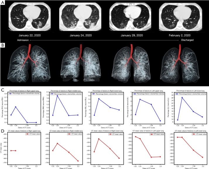
We report a 30-year-old man presented to the hospital with a 3-day history of fever and cough of unknown cause. He indicated that he had a travel history of Wuhan, China [a potential origin of SARS-CoV-2 (1,2)]. Real-time fluorescence polymerase-chain-reaction of his respiratory specimens was positive for SARS-CoV-2 nucleic acid. At admission, unenhanced chest CT showed patchy ground-glass opacity and a few subplueral consolidations in his left lower lobe (Figure 1A). After receiving 2 days of supportive treatment, he was clinically worse and repeat chest CT showed that pneumonia progressed with distribution in bilateral lower lobes, predominantly manifesting as consolidation (Figure 1A). Therefore, therapeutic strategy was adjusted with additional treatment including interferon and antibiotic. The patient showed a good response and recovered well. Chest CT at the follow-up after 7 and 11 days showed that the pneumonia absorbed gradually (Figure 1A). During the hospitalization, radiologists provided dynamic and visible information of pulmonary lesions with three-dimensional volume-rendered reconstruction (Figure 1B). On the basis of intelligent image technology, radiologists could help the physician to monitor the progression and regression of disease using quantitative imaging information including the lesion percentage (Figure 1C) and the CT mean density values (Figure 1D) in each lung lobe. After his respiratory samples checked twice to confirm the negative of SARS-CoV-2 nucleic acid, the patient was discharged home.
Figure 1.
CT imaging of coronavirus disease 2019 (COVID-19). (A) Chest CT shows patchy ground-glass opacity and a few subpleural consolidations in the left lower lobe on admission; Image obtained 2 days after follow-up shows the pneumonia progressed with distribution in bilateral lower lobes, predominantly manifesting as consolidation; images obtained 7 and 11 days after follow-up show the pneumonia absorbed gradually. (B) Three-dimensional volume-rendered reconstruction shows the visible progression and regression of pulmonary lesions. (C) The lesion percentage in each lung lobe shows obvious progression in the second CT scan, and indicates marked improvement in the following CT images obtained 7 and 11 days after follow-up. (D) The CT mean density values in each lung lobe shows a similar change of the lesion percentage at time of CT examination.
In conclusion, CT imaging could play more important role on the basis of intelligent technology in managing patients of COVID-19 from diagnosis to monitoring, and from the qualitative to quantitative.
Acknowledgments
Funding: Supported by Gansu Provincial COVID-19 Science and Technology Major Project, China.
Ethical Statement: The authors are accountable for all aspects of the work in ensuring that questions related to the accuracy or integrity of any part of the work are appropriately investigated and resolved.
Footnotes
Conflicts of Interest: The authors have no conflicts of interest to declare.
References
- 1.Mattiuzzi C, Lippi G. Which lessons shall we learn from the 2019 novel coronavirus outbreak? Ann Transl Med 2020;8:48. 10.21037/atm.2020.02.06 [DOI] [PMC free article] [PubMed] [Google Scholar]
- 2.Zhu N, Zhang D, Wang W, et al. A Novel Coronavirus from Patients with Pneumonia in China, 2019. N Engl J Med 2020;382:727-33. 10.1056/NEJMoa2001017 [DOI] [PMC free article] [PubMed] [Google Scholar]
- 3.Li Q, Guan X, Wu P, et al. Early Transmission Dynamics in Wuhan, China, of Novel Coronavirus-Infected Pneumonia. N Engl J Med 2020. [Epub ahead of print]. 10.1056/NEJMoa2001316 [DOI] [PMC free article] [PubMed] [Google Scholar]
- 4.Wang D, Hu B, Hu C, et al. Clinical Characteristics of 138 Hospitalized Patients With 2019 Novel Coronavirus-Infected Pneumonia in Wuhan, China. JAMA 2020. [Epub ahead of print]. [DOI] [PMC free article] [PubMed] [Google Scholar]
- 5.Chen N, Zhou M, Dong X, et al. Epidemiological and clinical characteristics of 99 cases of 2019 novel coronavirus pneumonia in Wuhan, China: a descriptive study. Lancet 2020;395:507-13. 10.1016/S0140-6736(20)30211-7 [DOI] [PMC free article] [PubMed] [Google Scholar]
- 6.Huang C, Wang Y, Li X, et al. Clinical features of patients infected with 2019 novel coronavirus in Wuhan, China. Lancet 2020;395:497-506. 10.1016/S0140-6736(20)30183-5 [DOI] [PMC free article] [PubMed] [Google Scholar]
- 7.Lei J, Li J, Li X, et al. CT Imaging of the 2019 Novel Coronavirus (2019-nCoV) Pneumonia. Radiology 2020. [Epub ahead of print]. 10.1148/radiol.2020200236 [DOI] [PMC free article] [PubMed] [Google Scholar]
- 8.Chung M, Bernheim A, Mei X, et al. CT Imaging Features of 2019 Novel Coronavirus (2019-nCoV). Radiology 2020. [Epub ahead of print]. 10.1148/radiol.2020200230 [DOI] [PMC free article] [PubMed] [Google Scholar]
- 9.Xie X, Zhong Z, Zhao W, et al. Chest CT for Typical 2019-nCoV Pneumonia: Relationship to Negative RT-PCR Testing. Radiology 2020. [Epub ahead of print]. 10.1148/radiol.2020200343 [DOI] [PMC free article] [PubMed] [Google Scholar]


