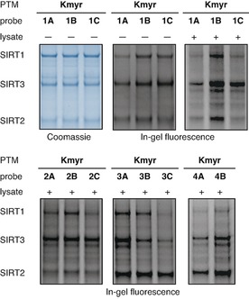
Figure 3.

In‐gel fluorescence showing efficiency of SIRT1–3 labeling using myristoylated probes. Coomassie‐stained gel is shown in blue‐scale and in‐gel fluorescence is shown in gray‐scale. Screening of the labeling efficiency of different myristoylated probes (12.5 μM) against recombinant SIRT1‐3 (1 μM) in pure buffer or buffer containing HeLa cell lysate (2 μg μL−1). The concentration of SIRT2 was slightly lower than expected based on the protein concentration provided by the vendor, which we corrected for in later experiments. See the Supporting Information (Figures S6–S21) for full gel images and further discussion.
