

Abstract
Pharyngitis is an acute inflammatory infection of pharynx and/or tonsils, most cases are caused by viruses and occur as part of common cold syndromes. As there is marked overlap between clinical findings of viral and bacterial pharyngitis and it is difficult to distinguish GABHS from viral causes clinicians tend to prescribe antibiotics for any sore throat and therefore pharyngitis has become the raison d'être for antibiotic misuses. Although clinical findings may not specifically differentiate the various pharyngitis pathogens yet many of these pathogens have clinical markers which could give us some clues to clinical suspicion.
A good picture is equal to thousand words, believing in this axiom article presents few simple clinico-pictorial clues which could help suspecting pharyngitis pathogens, this in turn may boost clinical confidence to avoid unnecessary antibiotic prescriptions.
Keywords: Viral pharyngitis, GABHS, Pharyngoconjunctival fever, Herpangina, Gingivostomatitis
An acute inflammatory syndrome of the pharynx and/or tonsils, pharyngitis (sore throat) is caused by several different groups of microorganisms. It could be a specific infection localized in the pharynx and/or tonsils or can be part of a generalized upper respiratory tract infection (Nasopharyngitis)1; most cases are caused by viruses (isolated in approximately 40% of cases)2 and occur as part of common cold syndromes. Disease is ubiquitous, the entire range of pharyngitis-causing pathogens is observed throughout the world. It is a foremost cause of pediatric ambulatory care visits & is one of the most common causes of absence from school or work.
Primary bacterial pharyngitis accounts for less than 30% of cases in children3; among the bacterial pharyngitis GABHS is the principal bacteria of concern due primarily for its predilection to cause rheumatic fever. As the GABHS is sensitive to plethora of antibiotics, they are liberally used under the pretext of preventing rheumatic fever. As there is marked overlap between clinical findings of viral and bacterial pharyngitis and it is difficult to distinguish GABHS from viral causes clinicians tend to prescribe antibiotics for any sore throat and therefore pharyngitis has become the raison d'être for antibiotic misuses; studies conducted in America, Asia, and Europe clearly document a very high percentage of children receiving antibiotics for the common cold, and other upper respiratory tract infections (URIs).4 Although clinical findings may not specifically differentiate the various pharyngitis pathogens yet many of these pathogens have clinical markers which could give us some clues to clinical suspicion.
A good picture is equal to thousand words, believing in this axiom article presents few simple clinico-pictorial clues which could help suspecting pharyngitis pathogens, this in turn may boost clinical confidence to avoid unnecessary antibiotic prescriptions.
1. Viral vs bacterial pharyngitis
While no single finding or combination of physical findings distinguishes bacterial from a viral etiology certain findings do suggest a particular possibility.
2. Viral pharyngitis
In viral infections pharyngitis is invariably a part of generalized common cold syndrome and therefore involvement of other mucous membranes e.g., conjunctivitis, coryza (Fig. 1, Fig. 2 ) is a common occurrence. Presence of sneezing, rhinorrhea, cough, conjunctivitis, and hoarseness of voice is likely indicators of viral pharyngitis. Throat is usually erythematous and swollen which may create irritation and soreness. Dry hacking cough is usual accompaniment. Erythematous nasal mucosa with reddened nares from nose blowing is generally notable findings.2, 3 A watery nasal discharge turning thick and yellow (Fig. 3 ) over the time is the normal course of the disease; unfortunately for patients clinicians consider this as sign of bacterial super infection and unnecessarily tend to prescribe antibiotics.
Fig. 1.
Nasal, conjunctival watering in viral pharyngitis.
Fig. 2.

Watering eyes in viral pharyngitis.
Fig. 3.

Thick yellow green discharge.
3. Bacterial pharyngitis
Compared to viral infections bacterial pharyngitis tends to be a localized phenomenon.
Marked pharyngeal erythema, enlargement of tonsils, significant pharyngeal and tonsillar exudates (beefy appearance) (Fig. 4 ), soft palate petechiae (Fig. 5 ), and tender anterior cervical adenopathy (Fig. 6 ) are prominent characteristics of bacterial pharyngitis. A sandpapery scarlatina rash (punctate erythematous macules and fine papules) is highly suggestive of GABHS (Fig. 7 ).3, 5 A purulent rhinitis with excoriations of nares and satellite impetiginous lesions may accompany many cases (Fig. 8 ).
Fig. 4.

Tonsillar exudates bacterial pharyngitis.
Fig. 5.
Palatal petechiae bacterial pharyngitis.
Fig. 6.

Anterior cervical adenitis.
Fig. 7.

Sand paper rash-Scarlet rash GABHS.
Fig. 8.

Purulent discharge with excoriations satellite impetiginous lesions.
4. Diphtheria: a forgotten entity
Believing that malady no more exists younger generation of clinicians have forgotten this entity. Thick gray membrane of diphtheria is mistaken for other exudative pharyngitis such as GABHS consequently many a times patients are diagnosed late and they lend into all kinds of serious complications. Diphtheria presents with a friable thick gray membrane which easily bleeds on manipulation (Fig. 9 ). Marked anterior cervical adenopathy lends a bull neck appearance (Fig. 10 ).
Fig. 9.

Membrane Rt tonsil diphtheria.
Fig. 10.

Bull neck diphtheria.
5. Pharyngitis: categories
Realizing the importance of clinical differentiation for a common clinician, the Infectious Diseases Society of America (IDSA) has decided to sort out this issue; based on some of the clinical differentiators. The IDSA categorized pharyngitis as follows6:
Category 1 (probable viral pharyngitis) – conjunctivitis, coryza, cough, diarrhea, viral-like exanthems.
Category 2 (suggestive of possible bacterial pharyngitis) – fever of more than 38.5 °C, tender anterior cervical nodes, headache, petechiae of the palate, abdominal pains, or sudden onset (<12 h).
6. Markers for certain viral etiological agents
Multitude of viruses may cause acute viral pharyngitis, prominent ones are: Rhinovirus, Adenovirus, Echovirus, Coxsackievirus, EBV (mononucleosis), Cytomegalovirus (CMV), Parainfluenza virus, Corona virus. Due to overlapping symptomatology it is difficult to make a specific etiological diagnosis however many of these viruses present with certain symptom complex which may be helpful in distinguishing them clinically.
7. Adenoviral pharyngitis (pharyngoconjunctival fever)
Adenoviral infection generally presents as a severe disease in young children; disease is more intense for a common cold. Child suffers from high grade fever, bad sore throat, dysphagia, and most importantly red eye. Concurrent conjunctivitis is responsible for red eyes (Fig. 11 ). Almost half of the patients of adenoviral infection present with this combination of pharyngitis & conjunctivitis which clinically distinguishes as “pharyngoconjunctival fever”.7 Eyes may pain but are devoid of purulent discharge; both eyes are involved although onset may be monocular; bulbar as well palpebral conjunctivae are affected. Conjunctivitis is often prolonged and may persist beyond resolution of fever and other symptoms. Follicular exudates (Fig. 12 ) and cervical lymphadenopathy may create confusion with bacterial pharyngitis; however presence of concurrent conjunctivitis is a strong differentiator.7
Fig. 11.

Monocular conjunctivitis adenoviral infection.
Fig. 12.

Follicular tonsillitis adenoviral infection.
8. HSV infection: pharyngogingivostomatitis
Eruption of shallow painful ulcers with erythematous margins on the hard and soft palates, posterior pharynx, and tonsillar pillars could occur in herpes. Presence of similar lesions on the tongue, gingiva, lips, or buccal mucosa (associated gingivostomatitis) (Fig. 13 ) almost characterizes herpetic infection.7 High grade fever and tender cervical lymphadenopathy are common. Both herpes simplex virus (HSV) types 1 and 2 can cause pharyngogingivostomatitis.
Fig. 13.

Ulcerative gingivostomatitis herpetic pharyngitis.
9. Coxsackieviral infection: (herpangina & hand foot mouth disease)6
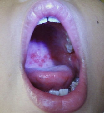
Herpangina presents as multiple small vesicles (1–2 mm) on the tonsils, tonsillar pillars, uvula, and soft palate, vesicles sit on a striking erythematous base7 (Fig. 14 ). Vesicles turn into shallow ulcers over the next 3 days and then heal. As against herpetic infection reminder of oral cavity is normal. Abrupt onset high fever, coryza, and anorexia are common presenting symptoms in infants and young children. Severe odynophagia and vomiting may compromise child's oral intake forcing hospitalization for IV hydration. Neck pain, headache, and back pain are common complaints among older children. Unlike herpetic pharyngitis herpangina is not associated with gingivitis.
Fig. 14.

Ulcers on palate, tonsils, uvula herpangina.
Many cases of herpangina are associated with papulovesicular rash around mouth, and on hands, feet, and buttocks. Entity is called as hand-foot-and-mouth disease. Children have low-grade fever (temperature, 100–102 °F/38–39 °C), sore throat, sore mouth, anorexia, malaise, along with characteristic rash.
10. EBV infection: pharyngitis with infective mononucleosis and lymphoid hyperplasia
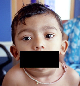
EBV infection produces an intense erythematous inflammation of pharynx and tonsils, an easily detachable membranous exudate may cover both the tonsils. A marked lymphoid hyperplasia manifesting as generalized lymphadenopathy and splenohepatomegaly is a common occurrence with EBV infection, lymphadenopathy is more marked in the posterior and anterior cervical regions (Fig. 15 ), but axillary and inguinal nodes may also be enlarged. 50% of cases have splenomegaly while liver could be enlarged in 10–15% cases. A high grade fever, palatal petechiae, periorbital edema, violaceous ring around eyes (Fig. 16 ), and maculopapular rash (Fig. 17, Fig. 18 ) are usual incident.7 EBV can also produce a subclinical hepatitis presenting as slightly elevated aminotransferases. Presence of atypical lymphocytes is the hallmark of EBV infection.
Fig. 15.

Marked lymphadenopathy EBV infection.
Fig. 16.

Periorbital edema, violaceous ring around eyes EBV infection.
Fig. 17.

Macular rash EBV infection.
Fig. 18.

Petechial rash EBV infection.
11. Pharyngitis caused by CMV
Cytomegalovirus can produce a syndrome which is clinically similar to EBV infection; the differentiating point is much lesser pharyngeal involvement. Atypical lymphocytosis, rash and elevated transaminases are common to both infections.7
12. Importance of pictorial approach
Though multitude of agents could cause pharyngitis a clinico-pictorial approach can help us rightly suspect an organism. Final diagnosis needs viral isolation or identification which in the want of specific treatment is futile most of the times. However a correct suspicion would infuse enough confidence in a clinician to avoid unnecessary antibiotic indulgence.
Conflicts of interest
The author has none to declare.
References
- 1.Perkins A. An approach to diagnosing the acute sore throat. Am Fam Physician. Jan 1997;55(1):131–138. 141-2. [PubMed] [Google Scholar]
- 2.Paradise J.L. Etiology and management of pharyngitis and pharyngotonsillitis in children: a current review. Ann Otol Rhinol Laryngol Suppl. Jan 1992;155:51–57. doi: 10.1177/00034894921010s111. [DOI] [PubMed] [Google Scholar]
- 3.Gerber M.A. Diagnosis and treatment of pharyngitis in children. Pediatr Clin North Am. Jun 2005;52(3):729–747. doi: 10.1016/j.pcl.2005.02.004. [DOI] [PMC free article] [PubMed] [Google Scholar]
- 4.Del Mar C.B., Glasziou P.P., Spinks A.B. Antibiotics for sore throat (Review) Cochrane Collab. 2007;(1):1–41. [Google Scholar]
- 5.Shaikh N., Swaminathan N., Hooper E.G. Accuracy and precision of the signs and symptoms of streptococcal pharyngitis in children: a systematic review. J Pediatr. Mar 2012;160(3):487–493. doi: 10.1016/j.jpeds.2011.09.011. [DOI] [PubMed] [Google Scholar]
- 6.Shulman S.T., Bisno A.L., Clegg H.W. Clinical practice guideline for the diagnosis and management of group A streptococcal pharyngitis: 2012 update by the Infectious Diseases Society of America. Clin Infect Dis. Nov 15 2012;55(10):1279–1282. doi: 10.1093/cid/cis847. [DOI] [PubMed] [Google Scholar]
- 7.Fisher RG, Boyce TG. Nose and Throat Syndromes; Moffet’s Pediatric Infectious Diseases. 4th ed. Lippincott Williams and Wilkins:14–39.