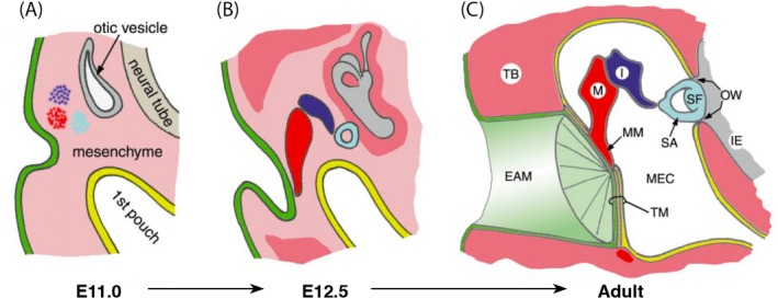
Figure 5.

Development of the tympanic membrane. (a) and (b) Cells from the region of the first cleft invaginate toward the first pouch meanwhile the ossicles form within the neural crest cell derived mesenchyme. (c) The developing ectoderm‐derived ear canal and endoderm‐derived middle ear cavity sandwich a layer of mesenchyme between them forming a three‐layered tympanic membrane. (Modified and reprinted from Mallo, 2001 with permission from Elsevier). EAM, external auditory meatus; I, incus; IE, inner ear; M, malleus; MEC, middle ear cavity; MM, malleus manubrium; OW, oval window; SA, stapedius arm; SF, stapedius footplate; TB, temporal bone; TM, tympanic membrane
