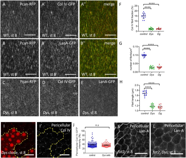
Fig. 2.
The DAPC is important for BM fibril deposition. (A-B″) Basal view of WT (w1118) follicle ECM at stage 8 visualized with Pcan-RFP and ColIV-GFP (A) or Pcan-RFP and LanA-GFP (B). (C-E) Basal view of DysE17/Exel6184 follicles expressing Pcan-RFP (C), ColIV-GFP (D) and LanA-GFP (E). (F-H) Quantification of the BM fibril fraction (F), fibril numbers (G) and fibril length (H) at stage 8 in WT, DysE17/Exel6184 and DgO86/O43 mutant follicles (n>10 follicles). (I,I′) Mutant clones for DysExel6184 marked by the absence of RFP and stained to detect pericellular ColIV. Dashed line indicates the border between WT and mutant cells. (J) Quantification of pericellular ColIV intensity around WT and Dys mutant cells (n>20 cells). (K,L) Accumulation of pericellular LanA in fat2 (K) and fat2, Dys (L) mutant follicles. For all panels, error bars represent s.d.; ****P<0.0001 (unpaired t-test). n.s., not significant. Scale bars: 10 µm.

