

The Upper Respiratory Tract
Jessica Quimby, Michael R. Lappin
Clinical signs of upper respiratory tract disease, including sneezing and nasal discharge, are common in cats (Box 30-1 ). Some diseases are associated with sneezing, and others are more commonly associated with stertorous breathing, with or without gagging. Coughing can sometimes be present, as well as epiphora, halitosis, dysphagia, and nonspecific signs such as lethargy, inappetence, and weight loss.1, 12, 33 Laryngeal disease is rare in the cat but may present as acute or chronic dyspnea, stridor, dysphagia, and signs of upper airway obstruction.84 Common causes of upper respiratory disorders in cats include trauma, foreign bodies, infectious agents, brachycephalic syndrome, inflammatory polyps, tooth root infections or other oral disease, nasopharyngeal stenosis, chronic rhinosinusitis, and neoplasia.1, 12, 33 The most common causes of laryngeal disease in the cat are laryngeal paralysis and laryngeal neoplasia.92 A complete diagnostic workup is important to determine the etiology so that the treatment regimen can be appropriately directed and maximal response to therapy is obtained.76, 77
BOX 30-1. Differential Diagnoses for Nasal Discharge in Cats.
Serous
Viral infection
Precursor to disease with mucopurulent discharge
Normal
Mucopurulent
- Associated with oral disease
-
•Tooth root abscess
-
•Oronasal fistula
-
•
Nasopharyngeal polyp
Foreign body
- Bacterial infection
-
•Primary or secondary
-
•
- Viral infection
-
•Feline herpesvirus-1
-
•Feline calicivirus
-
•
- Fungal infection
-
•Cryptococcus spp.
-
•Aspergillus spp.
-
•
Chronic rhinosinusitis
- Neoplasia
-
•Lymphoma
-
•Squamous cell carcinoma
-
•Adenocarcinoma
-
•
Hemorrhagic
- Nasal disease
-
•Trauma
-
•Foreign body
-
•Chronic rhinosinusitis
-
•Neoplasia
-
•Fungal disease
-
•
- Systemic disease
-
•Hypertension
-
•Polycythemia
-
•Coagulopathy
-
•Hyperviscosity syndrome
-
•
Clinical Signs
Nasal Disease
Nasal discharge is the most common clinical sign associated with nasal disease and can be serous, mucopurulent, or hemorrhagic1, 12, 33 (see Box 30-1). Serous nasal discharge is characteristic of most acute diseases of the nasal cavity and may precede mucopurulent nasal discharge. If the serous nasal discharge is chronic, viral and allergic etiologies are most common. Mucopurulent nasal discharge implies inflammation and occurs in association with fungal disease, primary bacterial disease, or overgrowth of normal bacterial flora secondary to any chronic nasal disease, including neoplasia, chronic rhinosinusitis, oronasal fistula, foreign body, inflammatory polyp, fungal disease, and viral disease. In addition, cats with vomiting or regurgitation can develop sneezing or nasal discharge by aspirating gastrointestinal content into the nose through the nasopharynx.
Epistaxis alone is most common with trauma, acute foreign body, hypertension, and coagulopathy. Epistaxis that develops in conjunction with or after mucopurulent discharge is most common with fungal disease, neoplasia, oronasal fistula, and occasionally chronic foreign bodies. Vasculitis occurs in dogs with diseases such as ehrlichiosis and bartonellosis but is rare in cats. Unilateral nasal discharge is more likely with foreign bodies, oronasal fistula, and neoplasia, although the latter can become bilateral as it progresses. Bilateral discharge is nonspecific and can be found with almost any etiology.33
Sneezing is a superficial reflex that originates in the mucous membranes lining the nasal cavity and is easily induced by chemical or mechanical stimuli. The sneeze results in forceful expulsion of air that passes through the airways with great velocity to clear the respiratory passageways. Sneezing is a common manifestation of nasal disease but is relatively nonspecific.
Stertor is a harsh, audible snoring sound associated with inspiratory breathing. Cats that experience stertor while awake are also likely to snore when sleeping. Stertor indicates airway obstruction and is most common with such conditions as nasopharyngeal polyps, nasopharyngeal stenosis, and neoplastic masses that occlude the airway. It may also occur as a result of airway occlusion caused by turbinate inflammation. Facial deformity is relatively uncommon but is usually associated with neoplastic processes and fungal infections, particularly Cryptococcus spp.1
Laryngeal Disease
Laryngeal disease is rare in the cat but may present as acute or chronic dyspnea, signs of upper airway obstruction, stridor (a harsh, high-pitched sound heard on inspiration), and dysphagia. Coughing or gagging may also be appreciated, and aphonia (loss of voice) or a change in voice has been reported.84
General Diagnostics
Signalment and lifestyle will often help refine the differential list and direct a diagnostic workup. Brachycephalic breeds may be predisposed to nasal disorders because of their physical conformation.85, 112 Neoplasia is more likely in older cats,33 and nasopharyngeal polyps are more common in younger cats.40 Cats with outdoor access are more likely to develop foreign bodies, trauma, or infectious etiologies.33 Cats in crowded housing conditions such as catteries, shelters, and multicat households are more likely to develop acute or chronic viral or bacterial rhinitis.32 Obtaining a complete history is important for determining the duration of the clinical signs. Acute onset of clinical signs is common with viral agents, foreign bodies, and trauma. The diagnostic workup of sneezing and nasal discharge is commonly completed in three phases (Box 30-2 ).
BOX 30-2. Staged Workup of Upper Respiratory Disease.
Phase 1 (Noninvasive)
History
Physical exam
CBC, chemistry, urinalysis
Thoracic +/− cervical radiographs
Cytology of nasal discharge
Cryptococcus antigen titer
Feline leukemia/feline immunodeficiency virus serology
Phase 2 (Sedation or Anesthesia Required)
Oral, pharyngeal, and laryngeal exam
Computed tomography
Nasal radiography
Nasal biopsy and histopathology
Tissue fungal and bacterial culture
Phase 3
Exploratory rhinotomy
Repetition of earlier phases
Phase 1: Noninvasive Tests
Most cats with acute disease are generally evaluated with noninvasive tests and therapeutic trials. A complete physical examination with careful attention to the head and neck should be performed, including ocular retropulsion. Firm resistance to retropulsion of the orbit or a painful reaction could be indicative of a retrobulbar lesion. Otic examination should be completed to evaluate for bulging or discoloration of the tympanum; these changes commonly occur with nasopharyngeal polyps. Deformation of the nose or face, exophthalmia, or pain on palpation of the nasal or facial bones is most consistent with fungal disease or neoplasia.1 Oral examination should be performed to assess for dental disease that could be causing an oronasal fistula, gingivostomatitis that could be consistent with feline herpesvirus (FHV-1) or feline calicivirus (FCV) infections, and defects in the hard or soft palate. External ocular examination may reveal conjunctivitis that could indicate FHV-1, FCV, Mycoplasma spp., or Chlamydophila felis infections. Fundic examination is performed to evaluate for lesions consistent with lymphoma or Cryptococcus neoformans infection. A cold microscope slide can be placed in front of the nose to assess airflow and may aid in determining if disease is unilateral or bilateral, although this should not limit diagnostic investigation to the obstructive side of the nose because bilateral disease may be present.
Although fungal organisms are uncommonly identified, cytology of nasal discharge should be performed on all cats with mucoid to mucopurulent nasal discharge to evaluate for the presence of C. neoformans, Sporothrix schenckii, or hyphae consistent with Aspergillus spp. or Penicillium spp. Neutrophils and bacteria are commonly detected if mucopurulent disease is present but do not prove primary bacterial disease. Hyphae also do not confirm primary fungal disease; they may represent contamination or infection secondary to another underlying cause. Secondary infections result in the same discharge as primary infections.
If lymph nodes draining the head are enlarged, they should be aspirated to evaluate for the presence of lymphoma, metastatic neoplasia, and fungal agents. Bacterial culture and antimicrobial susceptibility testing on nasal discharges are generally not recommended because results are difficult to interpret in that they typically yield normal intranasal bacterial flora.43 However, in respiratory outbreaks in catteries, pet stores, shelters, and multicat households, culture may be indicated to determine whether a pathogenic Bordetella bronchiseptica isolate is present.
Molecular diagnostic assays are now available for many respiratory agents, including FHV-1, FCV, C. felis, Mycoplasma spp., and B. bronchiseptica. However, cats can be asymptomatic carriers of these agents, and the FHV-1, FCV, B. bronchiseptica, and C. felis assays also amplify vaccine strains of the organisms, which means that positive results do not prove a disease association. This is especially true for FHV-1 and FCV, which may have a relatively high prevalence in the healthy cat population.52, 78, 98 Recently, a study failed to link Bartonella spp. infection to rhinitis in cats; therefore at this time recommendations to perform Bartonella spp. serology, culture, or polymerase chain reaction (PCR) assays in cats with upper respiratory tract signs are controversial.6 If a clinician chooses to test for evidence of Bartonella spp. infection, the cat should be evaluated by serology and PCR or culture because serology alone has been shown to yield false-negative results in up to 15% of infected cats.8 In addition, because only approximately 40% of seropositive cats are currently infected, a positive serologic test result does not prove bartonellosis.8 See Chapter 33 and the subsequent sections in this chapter about individual agents for a further discussion of molecular assays.
A complete blood cell count (CBC), serum biochemical panel, and urinalysis is recommended to rule out other systemic disease processes in cats with chronic disease. In general, results of the CBC are of low yield but may reveal eosinophilia in some cats with fungal or allergic disease, thrombocytopenia in some cats with epistaxis, or other cytopenias that might accompany feline leukemia (FeLV) or feline immunodeficiency virus (FIV) infections. FeLV and FIV do not cause sneezing and nasal discharge primarily, but they have been associated with lymphoma and may induce immunodeficiency that predisposes to other infections; therefore testing for these agents is indicated. A Cryptococcus antigen test is also recommended as a preliminary test for any cat with chronic nasal discharge, but particularly for those with nasal deformation, lymphadenopathy, or retinal lesions.59 Although thoracic radiographs are generally normal, they are still indicated to rule out pulmonary involvement of fungal disease and metastatic neoplasia. In cats with epistaxis, blood pressure measurement, coagulation profile, and buccal mucosal bleeding test are recommended, and thromboelastography may also be useful.
During phase 1, therapeutic trials are commonly attempted in cats with mild disease and usually consist of antibiotics, antiviral drugs, immunomodulators, or antihistamines (see the discussions of specific diseases that follow).
Phase 2: Imaging, Biopsy, Deep Cultures
If the physical examination indicates further diagnostic workup, a definitive diagnosis is not made during phase 1, or routine therapeutic trials fail, more aggressive diagnostic testing is indicated (typically requiring general anesthesia). Phase 2 diagnostics usually consist of pharyngeal and laryngeal examination, computed tomography (CT) scan or skull and dental radiographs, rhinoscopy, bacterial and fungal cultures, and biopsy to obtain samples for histology. In preparation for biopsies, a platelet estimate and an activated clotting time or other coagulation function test should be performed before anesthesia.
General anesthesia is induced by administering approximately one third of an induction dose of propofol (4 to 6 mg/kg intravenously), a short-acting thiobarbiturate, or ketamine combined with diazepam (ketamine 5 mg/kg intravenously and valium 0.3 mg/kg intravenously). The arytenoids are examined before intubation to make sure both are abducting normally on inspiration. Dopram can be used to stimulate respiration and increase intrinsic laryngeal motion at a dose of 2.2 mg/kg, administered intravenously. Oropharyngeal examination is performed to evaluate thoroughly for masses, foreign bodies, or palate defects. A spay hook and dental mirror can be used to help manipulate the soft palate to a position allowing visualization of the nasopharynx so that polyps, other masses, foreign material, or nasopharyngeal stenosis can be checked (Figure 30-1 ). A thorough dental examination should be performed and all teeth probed for evidence of oronasal fistula.
FIGURE 30-1.

A spay hook and mirror can be used to assess the nasopharyngeal area.
If a definitive diagnosis is not made, a CT scan or nasal, sinus, and dental radiographs are performed. If radiographs are performed, anesthesia is required for accurate positioning and should include a lateral view, ventrodorsal view, and intraoral and open-mouth bullae views. Nasal imaging can reveal increased density in the nasal cavity or bony lysis that could be consistent with a mass, turbinate destruction consistent with chronic rhinosinusitis or fungal disease, as well as radio-opaque foreign objects or tooth root abscessation.44, 69 Although more expensive and not widely available, a CT scan has the added advantage of better visualization of the sinuses and tympanic bullae and better assessment of bony lysis; it also allows assessment of the cribriform plate and brain so that the extent of a lesion can be evaluated86 (Figure 30-2 ). It is also faster to perform than a full series of skull radiographs and allows for radiotherapy treatment planning if indicated. It is the preferred imaging modality, especially if a mass is suspected. Imaging should be performed before rhinoscopy and biopsy to prevent hemorrhage from obscuring details in the nasal passages.
FIGURE 30-2.


CT appearance of chronic rhinitis (A) versus nasal tumor (B). A mass effect and bony lysis is noted in the nasal cavity.
Depending on imaging findings, the nasopharynx is examined with a flexible rhinoscope, and rigid rhinoscopy of the anterior nasal cavity is then performed (Figure 30-3 ). Rhinoscopy allows direct visualization of the nasal cavity, detection and removal of foreign objects, detection and débridement of fungal plaques, as well as assessment for inflammation, turbinate destruction, and masses. However, should a mass be present, rhinoscopy does not allow assessment of the extent of bony lysis (hence the importance of additional imaging). In addition, because gross appearance of the nasal mucosa on rhinoscopy does not always correlate with histopathologic diagnosis, biopsies should always be performed.37
FIGURE 30-3.


A, Rhinoscopy allows visualization of the nasal mucosa as well as sample collection. B, Erythematous and irregular nasal mucosa.
If no foreign material is visualized on rhinoscopy, the nasal cavity is flushed with sterile saline to evaluate for the presence of hidden material. The cuff of the endotracheal tube should be checked for full inflation before performing nasal lavage with saline administered under pressure. In cats lavaging should be performed from the anterior nares caudally. Gauze should be placed in the oropharyngeal area and then a 20-, 35-, or 60-mL syringe can be used to forcefully flush saline through the nose while the nares are being pinched off to create pressure (Figure 30-4 ). Material flushed from the nose (or oropharynx) should be caught on the gauze and examined for foreign objects. If no foreign material is located, biopsies are then made using a bone curette or the largest biopsy instrument that can be passed through the nares. Most rigid endoscopes are too large for the biopsy sleeve to be used in many cats; a gastroscopic biopsy instrument can often be passed next to the camera of a rigid scope to perform directed biopsies. Alternatively, the biopsy site can be directed by the results of diagnostic imaging or by rhinoscopy. If indicated, bacterial and fungal cultures are made using material from flush or biopsied tissues.39
FIGURE 30-4.

Nasal flush can be performed for diagnostic and therapeutic purposes.
Phase 3: Exploratory Rhinotomy
Exploratory rhinotomy allows for direct visualization of the nasal cavity to identify foreign objects, masses, or fungal plaques and is occasionally performed to aid in the diagnostic workup and the treatment of some diseases. However, in cats it is rarely performed, except for cases requiring removal of chronically embedded foreign bodies or cases of Aspergillus spp. or other infections in the sinus in which endoscopic débridement was not sufficient or the condition was refractory to treatment. Surgical debulking is rarely required for cats with nasal cryptococcosis. In general, there is also no added benefit to debulking nasal tumors before chemotherapy (e.g., lymphoma) or radiation therapy. Although turbinate tissue can be removed to increase airflow through the nasal cavities, bacterial osteomyelitis is often present as well as nasal discharge, so this procedure is generally not recommended for cats with chronic inflammatory rhinitis.
Disease-Specific Recommendations: The Nasal Cavity
Anatomic and Functional Disorders
Nasopharyngeal Polyps
Nasopharyngeal polyps are non-neoplastic, inflammatory nodules that occur most commonly in young cats. They originate in the middle ear or auditory canal and can grow out through the nasopharynx or, alternatively, the tympanum.34, 40 Why the growths occur is unknown, but because they tend to occur when the cat is young, a congenital etiology has been postulated.2 The possible association of polyps with infectious agents has also been explored, including FHV-1, FCV, C. felis, Mycoplasma spp., and Bartonella spp.,41, 108 but to date no organism has definitely been proven to be a cause. Large polyps can be detected by palpation through the soft palate, and otic examination may reveal discoloration or bulging of the tympanum. When extending into the nasopharynx, polyps disrupt the normal flow of secretions, resulting in secondary bacterial infections, mucopurulent nasal discharge, stertorous breathing, and gagging. Signs of middle ear involvement, such as Horner's syndrome and head tilt, can also be seen. Diagnosis can be confirmed with examination of the nasopharynx under sedation with a dental mirror and spay hook or rhinoscope, as previously described. A bulla radiography series or CT scan should be performed to determine whether there is bulla involvement. However, if there is no evidence of middle ear–associated clinical disease and the polyp can be removed by way of the mouth, many clinicians will perform removal using traction and wait for a recurrence before performing a bulla osteotomy on account of the high incidence of morbidity associated with bulla osteotomy40, 108 (Figure 30-5 ). Complications of this procedure include Horner's syndrome, facial nerve paralysis, and discomfort, and the recovery period is similar to that for a relatively invasive surgery. Without bulla osteotomy approximately 30% will be recurrent.108 However, combining removal by traction with a tapering course of glucocorticoids (1 to 2 mg/kg per day, by mouth, for 14 days followed by a taper dosage over the next 2 weeks) may improve the success rate.64 Bulla osteotomy is an effective surgical treatment, and when it is performed at initial presentation or at recurrence, most cases generally experience complete resolution.40, 103, 108
FIGURE 30-5.

Manual removal of nasopharyngeal polyp.
Brachycephalic Syndrome
Cats with brachycephalic conformation may experience difficulty with airflow due to the severe malformation of their nasal passages and nares, and potentially could be predisposed to nasal disease. A recent CT study of brachycephalic cats documented some of the abnormalities associated with this condition. It was found that the greater the degree of brachycephalia, measured by the amount of dorsal rotation of the maxillary canine tooth, the narrower the nasal cavity, nasal passages and nares.85 Stenotic nares also serve to limit inspired airflow. This condition may be improved by alar fold excision, performed with a laser or scalpel technique or alternatively a punch resection alaplasty.104 Nasopharyngeal turbinates have also been documented in brachycephalic cats and may serve to further reduce airflow through the nasopharyngeal area.25 Little information is available regarding surgical options for nasopharyngeal turbinates.
Nasopharyngeal Stenosis
Nasopharyngeal stenosis is a rare condition that involves narrowing of the choanae to the extent that little air is able to pass. This can occur as a result of chronic infections, aspiration rhinitis, or congenital defect.33, 96 Clinical signs typically include stertorous, labored breathing and, less typically, nasal discharge. Diagnosis is determined by retroflex rhinoscopic assessment of the nasopharynx. In the past manual dilation and advanced surgical procedures combined with steroid therapy were the only therapeutic options, and recurrence was common.33 More recently, stenting of the nasopharynx has been described as a successful palliative measure.5
Infectious Disorders
Bacterial Agents
Diagnosis
Almost all cats with mucopurulent or purulent nasal discharge have a bacterial component to their disease. The bacterial agents that have been described as primary respiratory pathogens in cats include B. bronchiseptica, C. felis, Streptococcus canis, and Mycoplasma spp. However, Corynebacterium spp., Escherichia coli, Pasteurella multocida, Pseudomonas aeruginosa, Streptococcus viridans, and Staphylococcus intermedius are also commonly detected but generally thought to be secondary invaders.* Culture of either nasal flush samples or tissue biopsy samples yields similar species results, but aerobic and anaerobic cultures of nasal flushes were positive significantly more often in one study.39 Culture of nasal biopsies may be more representative for deep mucosal infections,38 but this has not been definitively shown. In another study different organisms were isolated from each collection technique, so it may be most complete to culture both nasal flush and biopsy samples.38 However, it should be remembered that positive culture results may not correlate with the cause of the disease on account of the presence of normal flora and other superficial bacteria.
Although B. bronchiseptica is a well-defined primary pathogen in dogs, the organism can be isolated from many clinically normal cats.32 Thus the positive predictive value (PPV) of serologic test results, culture, and PCR assay is low in cats. Many cats have antibodies against B. bronchiseptica, the organism is commonly cultured from cats in crowded environments, and there are sporadic reports of severe lower respiratory disease caused by bordetellosis in kittens and cats in crowded environments or other stressful situations.7, 110 The organism was cultured on necropsy from the lower airways of several cats from shelters in Colorado, and in one shelter the organism was cultured from 19 of 40 cats (47.5%) with upper respiratory disease.91 However, the significance of infection in otherwise healthy pet cats appears to be minimal. For example, in client-owned cats in north central Colorado, the organism was rarely cultured from cats with rhinitis or lower respiratory disease (approximately 3%).109 B. bronchiseptica is easily grown, and culture is superior to PCR for this agent because antimicrobial susceptibility testing can be performed on isolates. Because the organism is not usually eliminated by treatment, follow-up culture or PCR assay after treatment has minimal benefit.9
C. felis is a common differential diagnosis for cats with clinical evidence of conjunctivitis and rhinitis; it is not a common cause of lower airway disease. The organism is difficult to culture, so PCR detection of microbial DNA from conjunctival swabs can be useful clinically. Because of the intracellular nature of the organism, adequate cellular material must be obtained from the conjunctival swab for analysis.28 PCR assay results can be used to prove a cattery has been cleared of the infection after treatment.95 Most, but not all, PCR-positive cats are clinically ill (e.g., 3.3% if healthy cats were positive in one study).13
Mycoplasma spp. are normal commensal organisms of the mucous membranes of multiple species, including cats. M. felis has been associated primarily with conjunctivitis but is suspected as a primary cause of rhinitis in cats as well.31, 38, 39 There are multiple Mycoplasma spp. of cats, and the pathogenic potential for most is unknown. If other primary diseases are present, even nonpathogenic Mycoplasma spp. may be associated with the disease process. Mycoplasma spp. culture can be difficult and takes longer than routine culture, and antimicrobial susceptibility is not provided by most laboratories. Culture of nasal biopsy samples rather than nasal flush samples may increase yield.39 Mycoplasma spp. PCR assays have at least some clinical utility, with some assays allowing for speciation, which is helpful in assessing the pathogenic potential of the organism. However, because Mycoplasma spp. are common flora, the PPV of the assays is likely to be low. Because the organism is not usually eliminated by treatment, follow-up culture or PCR assay after treatment has minimal benefit.
Treatment
If primary bacterial infections are suspected, doxycycline 10 mg/kg, administered orally once daily for cats with rhinitis with or without conjunctivitis, is usually effective (Table 30-1 ). Doxycycline is the treatment of choice for B. bronchiseptica, Mycoplasma spp., and C. felis infections,19, 28 and in the last has been shown to be superior to topical administration of tetracycline.90 Side-effects in young kittens are less of a concern with doxycycline than tetracycline but should still be taken into consideration. Amoxicillin–clavulanate is a good choice in young animals and is effective for most organisms, with the exception of Mycoplasma spp. because these organisms lack a cell wall. Pradofloxacin has been shown to have efficacy against Mycoplasma spp.16, 31 Enrofloxacin has also been shown to be effective for C. felis 23 but should be used with caution in young cats because of possible adverse effects on cartilage. Although the drug has not been shown to damage chondrocytes in cats, this does occur in several other species. Clindamycin penetrates bone and tissues well and has an excellent anaerobic spectrum. Administering the liquid form of this drug is generally well tolerated if given cold. Azithromycin therapy (15 mg/kg, administered orally once daily) can be tried for cats with suspected resistant bacterial infections.*
TABLE 30-1.
Pharmacologic Treatment of Upper Respiratory Tract Disease
| Class | Drug | Dosage |
|---|---|---|
| Antibiotics |
Amoxicillin | 10-22 mg/kg, PO, every 12 hours |
| Amoxicillin-clavulanate | 13.75 mg/kg, PO, every 12 hours | |
| Azithromycin | 15 mg/kg, PO, every 24 hours | |
| Cefadroxil | 22 mg/kg, PO, every 12 hours | |
| Cephalexin | 22 mg/kg, PO, every 8 hours | |
| Chloramphenicol | 10-15 mg/kg, PO, every 12 hours | |
| Clindamycin | 10-12 mg/kg, PO, every 24 hours | |
| Doxycycline | 10 mg/kg, PO, every 24 hours | |
| Enrofloxacin | 2.5-5 mg/kg, PO, every 24 hours | |
| Marbofloxacin | 2.5-5 mg/kg, PO, every 24 hours | |
| Metronidazole | 10-15 mg/kg, PO, every 12 hours | |
| Orbifloxacin | 2.5-5 mg/kg, PO, every 24 hours | |
| Pradofloxacin | 5-10 mg/kg, PO, every 24 hours | |
| Trimethoprim-sulfonamide | 15 mg/kg, PO, every 12 hours | |
| Antihistamines |
Cetirizine | 2.5-5 mg/cat, PO, every 24 hours |
| Chlorpheniramine | 2 mg/cat, PO, every 12 hours | |
| Clemastine | 0.68 mg/cat, PO, every 12 hours | |
| Fexofenadine | 5-10 mg/cat, PO, every 12 to 24 hours | |
| Hydroxyzine | 5-10 mg/cat, PO, every 8 to 12 hours | |
| Loratadine | 5 mg/cat, PO, every 24 hours | |
| Antifungals |
Deoxycholate amphotericin B | 1) IV: 0.1-0.5 mg/kg: M, W, F; to 16 mg/kg total cumulative dose 2) SC: 0.5-0.8 mg/kg in 400 mL of 0.45% saline/2.5% dextrose; M, W, F; to 16 mg/kg total cumulative dose |
| Fluconazole | 50 mg/cat, PO, every 12 to 24 hours | |
| Itraconazole | 10 mg/kg, PO, every 24 hours | |
| Liposomal amphotericin B | 1 mg/kg IV; Mon, Wed, Fri; to 12 mg/kg total cumulative dose | |
| Antivirals |
Cidofovir topical (0.5%) | 1 drop OU, every 12 hours |
| Famciclovir | 62.5 mg/cat, PO, every 12 hours, 14 days | |
| Interferon-alpha | 10 U PO, every 24 hours (chronic); 10,000 U SC, every 24 hours, 21 days (acute) | |
| Lysine | 500 mg/cat, PO, every 12 hours | |
| NSAIDs |
Meloxicam | 0.025-0.1 mg/kg, PO, every 2 to 3 days |
| Piroxicam | 0.3 mg/kg, PO, every 2 days | |
| Glucocorticoids | Beclomethasone (inhaled) | 1-2 puffs, every 12 to 24 hours |
| Fluticasone (inhaled) | 1-2 puffs, every 12 to 24 hours | |
| Methylprednisolone acetate | 5-15 mg IM, every 3 to 4 weeks, as needed | |
| Prednisolone | 2.5-5 mg/cat, PO, every 1 to 2 days | |
PO, By mouth; IV, intravenously; SC, subcutaneously; OU, each eye; NSAIDs, nonsteroidal antiinflammatory drugs; IM, intramuscularly.
Doxycycline and clindamycin have been associated with esophagitis and esophageal strictures in cats4, 24, 61 because of the poor secondary esophageal contractions in this species. The authors recommend never administering dry pills or capsules to cats. Drugs should be compounded into a liquid, administered, and then followed with a 3- to 6-mL liquid bolus or food, administered coated with butter or a product such as Nutri-Cal, or administered in a pill-delivery treat.27, 50, 111 Cats with acute disease are treated for 7 to 10 days, except for C. felis, in which 28 days of therapy is needed to eliminate infection.31, 95 Chronic bacterial disease may require treatment for 6 to 8 weeks to adequately clear the infection if osteomyelitis exists. Pulse therapy may help some chronically affected cats but may induce antimicrobial resistant bacteria in other cats. Most cases of bacterial rhinitis are secondary to other diseases, including trauma, neoplasia, inflammation induced by viral infection, foreign bodies, inflammatory polyps, chronic rhinosinusitis, and tooth root abscessation. Thus if routine antibiotic therapy fails, a diagnostic workup should be performed.
Prevention
The currently available B. bronchiseptica vaccine for intranasal administration can be administered as early as 4 weeks of age, has an onset of immunity as early as 72 hours, and has a minimum duration of immunity of 1 year.81 The American Association of Feline Practitioners (AAFP) Feline Vaccine Advisory Panel, and the European Advisory Board on Cat Diseases (ABCD) recommendations suggest that Bordetella vaccination should be considered primarily for use in cats at high risk for exposure and disease, such as those with a history of respiratory problems and living in humane shelters with culture-proven outbreaks.19, 81 However, because the vaccine is administered by the intranasal route, mild sneezing and coughing can result, which may influence case management of kittens housed in shelters or humane societies. Because the disease is apparently not life-threatening in adult cats, is uncommon in pet cats, responds to a variety of antibiotics, and is considered minimally zoonotic,18 routine use of this vaccine in the majority of client-owned cats seems unnecessary.
Killed and modified live C. felis–containing vaccines are available. In recent studies C. felis was amplified from conjunctival swabs of 3.2% of cats with conjunctivitis49 but 0% of nasal discharges from cats housed in a humane society.91 FVRCP vaccines that also contained C. felis were associated with more vaccine reactions in cats when compared to other products.63 Because infection of cats by C. felis generally results only in mild conjunctivitis, is easily treated with antibiotics, has variable prevalence rates, and is of minimal zoonotic risk to humans, some researchers have questioned whether C. felis vaccination is ever necessary in the United States.14 Duration of immunity for Chlamydophila vaccines may be short-lived, so high-risk cats, such as those in multicat environments or where there is a history of chlamydial infection, should be immunized before a potential exposure.
Viral Agents
Diagnosis
The most common viruses associated with feline respiratory disease are FCV and FHV-1. Both viruses are extremely common in cats, particularly those from crowded environments such as pet stores, catteries, and shelters.14, 32 There are many strains of FCV, and mutations resulting in new strains are common. This organism is a common differential diagnosis for cats with clinical evidence of rhinitis, stomatitis, oral ulceration, and conjunctivitis (Figure 30-6 ). Less commonly, FCV is associated with polyarthritis, lower airway disease in kittens, and virulent systemic disease.78 Some variants of FCV are thought to induce systemic vasculitis in cats (virulent systemic calicivirus [VS-FCV]), and clinical signs can be severe even in cats previously vaccinated with FVRCP vaccines.10, 36, 72, 87, 105 VS-FCV strains arise spontaneously from endogenous FCV strains, and outbreaks have resolved quickly after the initial cases were recognized. Currently, it is unknown how often these outbreaks occur and whether the number of outbreaks is increasing. The VS-FCV strains evaluated to date have been genetically and antigenically diverse.42, 71
FIGURE 30-6.

Clinical appearance of a kitten with herpesvirus infection.
Virus isolation can be used to document current infection but takes at least several days for results and is not performed by all laboratories. Because of widespread exposure and vaccination, the PPV of serologic tests is poor. Reverse transcriptase (RT) PCR assays can be used to amplify the RNA of calicivirus, and results can be made available quickly. However, these assays also amplify vaccine strains of FCV (Lappin MR: unpublished data, 2010). FCV RNA can be amplified from samples collected from normal carrier cats as well as clinically ill cats, and PCR assays therefore have poor PPV. In addition, amplification of FCV RNA cannot be used to prove virulent systemic calicivirus infection.78 False-negative results of FCV RT-PCR can also occur if inadequate RNA is present on the submitted swab or if the organism has been cleared to levels below the sensitivity limits of the assay by specific immune responses. Because treatment does not eliminate FCV infection, there is no benefit to follow-up culture or RT-PCR testing.
FHV-1 is a common differential diagnosis for cats with clinical evidence of rhinitis, stomatitis, conjunctivitis, keratitis, and facial dermatitis. Because of widespread exposure and vaccination, the PPV of serologic tests is poor. FHV-1 infection can be documented by direct fluorescence staining of conjunctival scrapings, virus isolation, or PCR.98 FHV-1 DNA can be amplified from conjunctival cells of approximately 20% of healthy cats; therefore the PPV of PCR assays for this agent is low.79 Currently used PCR assays also detect vaccine strains of FHV-1, further lessening the PPV.52 Quantitative PCR may ultimately prove to correlate with the presence or absence of disease but failed to correlate to presence of conjunctivitis in one small study in the authors’ laboratory.49 The negative predictive value of the FHV-1 PCR assays is also in question because many cats that are likely to have FHV-1–associated disease are PCR negative. This may relate to clearance of FHV-1 DNA from tissues by the immune reaction. Tissue biopsies have greater sensitivity than conjunctival swabs but do not necessarily have greater predictive value.93 FHV-1 DNA can be amplified from the aqueous humor of some cats, but whether this indicates FHV-1–associated uveitis is unknown.54 Because treatment does not eliminate FHV-1 infection, there is no benefit to follow-up culture or PCR testing.
Treatment
Therapy for FCV consists mainly of supportive care, which is often needed for cats with VS-FCV infections, and may consist of intravenous fluids, antibiotics for concurrent bacterial infections, and interferon. Interferon may augment immune responses to viral infections by upregulating key cytokines.101 Feline interferon omega (1 to 2.5 million IU/kg, intravenously or subcutaneously, every 24 to 48 hours for up to 5 doses, then reduce to twice weekly and eventually to once weekly, depending on clinical response) (Virbagen Omega, Virbac Animal Health) inhibits FCV replication in vitro, but results of controlled studies evaluating efficacy in clinically affected cats with respiratory disease are not available. If human alpha interferon is used systemically for cats with life-threatening FCV or FHV-1 infections, 10,000 U/kg, administered subcutaneously, can be administered safely once daily, but controlled data concerning efficacy are not available. Recently, use of feline interferon therapy has been used to improve quality of life in cats with FeLV and FIV infections.11 In one study low-dose oral interferon therapy (10 U/kg, orally, daily alternating 7 days on, 7 days off, for 6 months) improved quality of life in cats with FIV infections.73 The effect of oral interferon is thought to be from mediation of inflammatory cytokines. There may also be effects against chronic FHV-1 or FCV infections, but controlled data are not available. Topical administration of alpha interferon in saline to the eyes of cats with conjunctivitis or the nose has been recommended by some veterinarians as an aid in the management of some cats with acute or chronic FHV-1 or FCV infections.
Recently, antiviral drugs have become more popular in the management of cats with acute or chronic FHV-1 infections. Currently available antiviral medications are efficacious only for DNA viral infections such as FHV-1, and not RNA viruses such as FCV, because they interfere with viral DNA synthesis and thus viral replication. Acyclovir and valacyclovir have been administered to some cats but can induce bone marrow suppression and are minimally effective for FHV-1; therefore they should no longer be used.51, 66 Famciclovir is safe and effective and is used for both acute and long-term therapy for cats with FHV-1 infections. One dose that has been used with apparent clinical efficacy is 62.5 mg, orally, every 12 hours for 14 days.58 However, recent pharmacokinetic studies indicate that higher doses may be needed for activity against FHV-1.99
Idoxuridine and trifluridine have been used topically in cats with conjunctivitis or keratitis resulting from FHV-1 infection, but these must be administered multiple times per day and are irritating. Recently, cidofovir was used in a small experimental FHV-1 conjunctivitis study and was shown to lessen clinical signs and FHV-1 shedding.21 Lysine at 250 to 500 mg, orally, every 12 hours may be helpful in some cats with acute or chronic rhinosinusitis caused by FHV-1 infection (not FCV).53 However, in several controlled studies of cats fed a lysine-fortified diet, a significant positive effect was not noted.17, 55, 80
Intranasal administration of modified live FHV-1 and FCV vaccines may lessen disease in some chronically infected cats, but controlled data are lacking. If there is a positive response to intranasal vaccination in a cat with chronic disease, this form of immunotherapy can be administered up to three times per year. Intranasal vaccination has been shown to potentiate cell-mediated immunity to FHV-1 better than parenteral vaccination.48 Chronic administration of one commercially available probiotic (FortiFlora, Purina Veterinary Diets) was shown to enhance T-helper lymphocyte numbers in cats.106 When this probiotic was administered to cats with chronic FHV-1 infections that were then subjected to mild stress, improved conjunctivitis scores were noted in some of the cats in the treatment group.47 See the section on chronic rhinosinusitis for a discussion of other nonspecific therapies.
Prevention
Specific pathogen-free (SPF) cats inoculated with one dose of intranasal modified live FVRCP vaccine had significantly less clinical signs than control cats as soon as 4 days when challenged with virulent FHV-1 in one study.45 Administration of the intranasal FVRCP vaccine was also shown to induce FCV antibody responses in SPF kittens more quickly than a modified live FVRCP vaccine administered parenterally.46 Thus the intranasal route of administration may be preferred for the primary or booster immunization of kittens housed in environments at high risk for exposure to FHV-1 or FCV, such as shelters, humane societies, catteries, boarding facilities, and multicat households. However, because administration of intranasal FHV-1 and FCV vaccines can induce transient mild sneezing or coughing, the owners should be informed of these potential side effects. Additionally, these vaccine side effects may influence case management of kittens housed in shelters or humane societies. Subcutaneous vaccines are recommended if concerns about the respiratory side effects of intranasal vaccines exist. Currently, yearly revaccination for cats in high-risk environments (e.g., shelters, catteries, multicat household) and a 3-year revaccination interval for cats in low-risk environments (indoor-only with no contact with other cats) are recommended.78, 81, 98
Inactivated vaccines containing VS-FCV are now available (Calicivax, Boehringer Ingelheim Vetmedica). The product contains a traditional FCV vaccine strain as well as a VS-FCV strain. Cross-neutralization studies show that cats inoculated with more than one FCV strain inactivate more FCV strains in vitro than cats inoculated with one FCV strain.74, 75 In addition, a recent challenge study illustrated that kittens vaccinated with the dual-strain product were protected from clinical signs of VS-FCV.35
Fungal Agents
C. neoformans and Aspergillus spp. are the most common causes of respiratory tract fungal infection in cats.3, 57, 68 Cryptococcosis is most common and should be considered a differential diagnosis for cats with respiratory tract disease, subcutaneous nodules, lymphadenopathy, intraocular inflammation, fever, and central nervous system disease.57 Infected cats range from 6 months to 16 years of age, and male cats are overrepresented in some studies.67 Infection of the nasal cavity is reported most frequently and commonly results in sneezing and nasal discharge. The nasal discharge can be unilateral or bilateral, ranges from serous to mucopurulent, and often contains blood. Granulomatous lesions extruding from the external nares, facial deformity over the bridge of the nose, and ulcerative lesions on the nasal planum are common (Figure 30-7 ). Submandibular lymphadenopathy is detected in most cats with rhinitis. Definitive diagnosis of cryptococcosis is based on antigen testing or cytologic, histopathologic, or culture demonstration of the organism.
FIGURE 30-7.

Although Cryptococcus and Aspergillus are the most common upper respiratory fungal infections in cats, nasal deformity may also be associated with Sporothrix spp. infection.
Cats with cryptococcosis have been treated with amphotericin B, ketoconazole, itraconazole, fluconazole, and 5-flucytosine alone and in varying combinations. Responses have varied among studies, but good to excellent treatment responses are often achieved in cats administered fluconazole or itraconazole.60, 67 Because of toxicity and the availability of more efficacious drugs, ketoconazole is no longer recommended by these authors. Fluconazole at 50 mg per cat, orally, once or twice daily is recommended because it results in the fewest side effects, has the best penetration across the blood–brain and blood–ocular barriers of the azoles, and has been shown to have good efficacy.60, 67 If life-threatening infection is occurring or the cat is failing to respond to an azole drug, amphotericin B should be used.67 A typical amphotericin protocol involves intravenous infusions on a Monday–Wednesday–Friday schedule until a cumulative dose of 16 mg/kg has been reached. Nephrotoxicity is the most serious side effect, and an initial infusion dose of 0.1 mg/kg is used as a test dose. The dose can be slowly increased to 0.5 mg/kg if it is well tolerated clinically and if renal values remain stable.26, 56 A successful subcutaneous protocol has also been described in which 0.5 to 0.8 mg/kg is added to 0.45% saline/2.5% dextrose, and the total volume is given two to three times weekly to a cumulative dose of 8 to 26 mg/kg.56, 67
In addition to the deoxycholate form of amphotericin B, a liposomal formulation is also available. It is thought that less nephrotoxicity is seen with the liposomal product, and a recommended dose regimen is 1 mg/kg, intravenously, on a Monday–Wednesday–Friday schedule until a cumulative dose of 12 mg/kg has been reached.26 Focal nasal and cutaneous cryptococcosis generally resolves with treatment; central nervous system, ocular, and disseminated diseases are less likely to respond to treatment.60, 67 Treatment should be continued for at least 1 to 2 months past resolution of clinical disease, or until antigen titers are negative.20, 67 People and animals can have the same environmental exposure to Cryptococcus spp., but zoonotic transfer from contact with infected animals is unlikely.
Aspergillosis is less common than cryptococcosis but can be equally devastating.3, 102, 112 Clinical signs of mild disease are similar to those of nasal cryptococcosis. Sino-orbital aspergillosis was recently described in cats and appears to be more aggressive than canine aspergillosis, involving invasion into surrounding structures.3 Ocular involvement, such as exophthalmos and ocular discharge, can be seen in addition to nasal signs. Diagnosis of aspergillosis is based on visualization of fungal plaques on rhinoscopy or fungal hyphae on cytology or biopsy. Infection can be caused by either Aspergillus spp. or Penicillium spp., which can be difficult to differentiate cytologically. Fungal culture seems less sensitive and specific than visualization on rhinoscopy or biopsy.112 Therapy with oral itraconazole and fluconazole has been documented as 50% to 60% efficacious,102, 112 and better efficacy with nasal clotrimazole therapy has been reported in a few cases.102
Chronic Inflammatory Disorders
Chronic Rhinosinusitis
Lymphocytic–plasmacytic, eosinophilic, and idiopathic rhinosinusitis are a constellation of diagnoses obtained by way of histopathology that are collectively referred to as chronic rhinosinusitis. In many cases this is a diagnosis of exclusion when other etiologies have been ruled out. This syndrome is one of the most significant causes of sneezing and nasal discharge in the cat.33 The nasal discharge is generally serous, but secondary bacterial infections can result in the development of mucopurulent nasal discharge (Figure 30-8 ), and inflammation can be severe enough to cause intermittent hemorrhage.33 The clinical signs can persist for years. Cats with relatively stable disease that encounter a sudden change in severity should be re-evaluated for the presence of a more severe secondary disease, such as fungal rhinitis or neoplasia.
FIGURE 30-8.

Mucopurulent nasal discharge is common with chronic rhinosinusitis.
There is a subset of chronic rhinosinusitis cats that have a history of acute FHV-1 or FCV upper respiratory infections at a younger age, and it is postulated that an early severe viral infection may trigger chronic disease.38 In addition, it is estimated that approximately 80% of cats are latently infected with FHV-1,22 and so another possible scenario for chronic viral rhinosinusitis is the presence of latent FHV-1 viral infections that are triggered into recrudescence by stressful events. In such cats with a prior history of viral infection, therapies such as lysine, antivirals, and immunomodulators are often tried, as previously discussed. Subjective improvement in clinical signs has been noted in response to cationic liposome DNA complexes (CLDC) immunomodulatory therapy in a small pilot study currently under way at Colorado State University, as well as in a previously published study.107 Stress is thought to play a role in the clinical severity and potential for recurrence of chronic viral rhinosinusitis, particularly if FHV-1 latency or chronic FCV infections are involved. Environmental measures to decrease stress, allocate resources in multicat households, and provide antianxiety therapies such as feline facial pheromone use (Feliway, Ceva Animal Health) may also provide some benefit. Controversy surrounds the use of immunosuppressive therapy in these patients: It may not be beneficial and runs the risk of exacerbating viral and bacterial components of the disease syndrome.
In many cases there is no history of viral infection or any other predisposing cause. Generally, idiopathic chronic rhinosinusitis is somewhat refractory to treatment, and palliation of clinical signs, rather than cure, is the goal of medical management. Broad-spectrum antibiotics are often prescribed to manage secondary infections. Administration of antihistamines such as chlorpheniramine at 1 to 2 mg, orally, every 12 hours may lessen clinical signs of disease in some cats. Several newer antihistamines are now available (see Table 30-1), and because response to therapy is variable from patient to patient, an alternative drug should be tried if no improvement is seen with the initial choice. Moistening therapies such as nebulization and saline nasal drops can help loosen secretions and soothe mucosa, particularly in drier climates.
The role of immunosuppressive drugs as therapeutic agents in the treatment of chronic idiopathic rhinosinusitis is poorly understood, likely because of the multifactorial nature of this condition. Individual patients will respond variably to this approach. Prednisolone may be used at a maximum dose of 1 to 2 mg/kg, orally, every 12 hours. If a positive response is noted, the lowest dose and the longest dosing interval that is effective should be determined by adjusting the dose over time. Inhaled glucocorticoids can be used as an alternative to decrease the systemic side effects of oral glucocorticoid use and have the benefit of directly affecting the nasal mucosa. Beclomethasone or fluticasone can be administered by metered dose inhaler (MDI) with an inhalation chamber at 1 to 2 puffs once to twice daily. Resistant cases may respond to administration of cyclosporine at up to 7.5 mg/kg, orally, daily or every other day, but controlled data are lacking. Trough blood levels should be checked 2 weeks after initiation of therapy to ensure that excessive blood levels are not achieved; this may activate latent infectious diseases.
Neoplasia
Neoplasia of the nasal cavity is relatively rare in the cat compared with the dog; lymphoma is the most common tumor type, followed by adenocarcinoma and squamous cell carcinoma.12, 33, 65 Lymphoma is treated with chemotherapy, often in conjunction with radiation therapy, and has potential for a good long-term prognosis.89 Palliative radiation therapy is indicated for other nasal neoplasms, and surgical debulking is generally not required.62 Prognosis depends on the aggressiveness and extent of the tumor, which is best determined by CT scan. Piroxicam administered at 0.3 mg/kg, orally, every 48 to 72 hours can control inflammation and clinical signs of disease in some cats with nonlymphoproliferative nasal neoplasia. Meloxicam (0.1 mg/kg, orally, every other day) may be another efficacious alternative. However, neither drug may result in antitumor effects against squamous cell carcinoma because it has been shown that there is minimal expression of cyclooxygenase-2 in this cancer type in the cat.15 If nonsteroidal antiinflammatory therapy is used, the cat should be monitored for renal and gastrointestinal side effects (e.g., PCV to assess for gastrointestinal hemorrhage and renal values) because these are potential side effects of this drug family.
Spontaneous Disorders
Trauma
Trauma to the nasal cavity most commonly results in massive hemorrhage, and thus initial evaluation should include assessment and treatment for hypovolemic shock. Nasal tissue may be easily damaged because of its fragile structure. Placement of a nasal cannula may aid in airflow and allowing healing that maintains a nasal passage. It may take 2 to 3 weeks for healing to occur. Generally, surgical correction of fractures in the nasal cavity is not necessary, although solitary bone fragments should be removed to prevent the formation of sequestra. Trauma may also lead to chronic complications as a result of damage to the nasal passage.33
Foreign Bodies
Nasal foreign bodies are more common in cats than generally realized.33, 82 In dogs the foreign material is usually inspired into the anterior nares and is found in the ventral meatus just caudal to the nares. Most nasal foreign bodies in cats are plant material that lodges above the soft palate after coughing or vomiting. Clinical signs may include sneezing, reverse sneezing, gagging, and repeated attempts at swallowing. Retroflex rhinoscopic examination of the nasopharynx can sometimes confirm diagnosis and aid in removal of the object. Nasal lavage under general anesthesia is often more effective. The cuff of the endotracheal tube should be checked for full inflation before performing nasal lavage with saline administered under pressure. In cats lavaging from the anterior nares caudally is recommended. Gauze should be placed in the oropharyngeal area, and then a 20-, 35-, or 60-mL syringe can be used to forcefully flush saline through the nose while the nares are being pinched off to create pressure. Material flushed from the nose (or oropharynx) should be caught on the gauze and examined for foreign material.
Disease-Specific Recommendations: The Larynx
Laryngeal Paralysis
Laryngeal paralysis is a relatively rare condition in the cat compared with the dog, but it may occur as a result of a variety of etiologies.84 Clinical signs include respiratory distress, inspiratory stridor, change in vocalization, coughing, dysphagia, and nonspecific signs such as weight loss and anorexia. Continuous positive airway pressure may be helpful in managing acute cases when intubation is not possible.100 When possible, depending on the stability of the patient, cervical and thoracic radiographs are helpful for ruling out other causes of dyspnea. In a recent study 60% of cats with laryngeal paralysis had evidence of upper airway obstruction on radiographs. Findings included lung hyperinflation; caudal displacement of the larynx; and air in the larynx, pharynx, esophagus, and stomach.84 Insofar as laryngeal paralysis is a functional disease, laryngeal examination provides a definitive diagnosis. In one study 75% of cats examined had bilateral disease.84 In this study medical management was instituted for cats with unilateral disease with good outcome. Surgical management with arytenoid lateralization is reported to be variably successful (50% to 70%) for cats with laryngeal paralysis and is more commonly recommended in cats with bilateral disease. Aspiration pneumonia (with bilateral lateralization procedures), temporary tracheotomy necessitated by laryngeal swelling, and repetition of the surgery have been reported as possible sequelae of surgical management.30
Inflammatory Laryngitis
A few cases of inflammatory laryngeal disease have been reported.97 These are clinically and grossly similar in presentation to other causes of laryngeal disease but are apparently non-neoplastic and noninfectious in etiology. Thus a biopsy should always be performed to distinguish inflammatory disease from a neoplastic process. Histopathology of this condition appears to be either granulomatous or lymphocytic-plasmacytic and neutrophilic. A temporary tracheostomy may be necessary while treatment is initiated, and surgical resection of inflamed tissue may be necessary. Favorable response to glucocorticoids has been reported, with good long-term prognosis in some cases.97
Laryngeal Neoplasia
Laryngeal neoplasia most commonly presents with clinical signs similar to those of other laryngeal disorders. A physical examination with special attention to the cervical area may identify a mass originating from tissues adjacent to the larynx. Laryngeal examination may reveal a mass, swelling, or irregularity in the appearance of the larynx. A biopsy is necessary to confirm the diagnosis and differentiate the condition from inflammatory laryngeal disease. The most common laryngeal neoplasms are lymphoma, squamous cell carcinoma, and adenocarcinoma.70 Lymphoma may respond to chemotherapy. Complete surgical excision is usually not possible in this anatomic area, but it can be performed as a palliative measure. The long-term prognosis is poor. Permanent tracheostomy can be performed, but complications are common.29, 92
References
- 1.Allen HS, Broussard J, Noone K. Nasopharyngeal diseases in cats: a retrospective study of 53 cases (1991-1998) J Am Anim Hosp Assoc. 1999;35:457. doi: 10.5326/15473317-35-6-457. [DOI] [PubMed] [Google Scholar]
- 2.Baker G. Nasopharyngeal polyps in cats. Vet Rec. 1982;111:43. doi: 10.1136/vr.111.2.43. [DOI] [PubMed] [Google Scholar]
- 3.Barrs VR, Beatty JA, Lingard AE. Feline sino-orbital aspergillosis:an emerging clinical syndrome. Aust Vet J. 2007;85:N23. [PubMed] [Google Scholar]
- 4.Beatty JA, Swift N, Foster DJ. Suspected clindamycin-associated oesophageal injury in cats: five cases. J Feline Med Surg. 2006;8:412. doi: 10.1016/j.jfms.2006.04.006. [DOI] [PMC free article] [PubMed] [Google Scholar]
- 5.Berent AC, Weisse C, Todd K. Use of a balloon-expandable metallic stent for treatment of nasopharyngeal stenosis in dogs and cats: six cases (2005-2007) J Am Vet Med Assoc. 2008;233:1432. doi: 10.2460/javma.233.9.1432. [DOI] [PubMed] [Google Scholar]
- 6.Berryessa NA, Johnson LR, Kasten RW. Microbial culture of blood samples and serologic testing for bartonellosis in cats with chronic rhinosinusitis. J Am Vet Med Assoc. 2008;233:1084. doi: 10.2460/javma.233.7.1084. [DOI] [PubMed] [Google Scholar]
- 7.Binns SH, Dawson S, Speakman AJ. Prevalence and risk factors for feline Bordetella bronchiseptica infection. Vet Rec. 1999;144:575. doi: 10.1136/vr.144.21.575. [DOI] [PubMed] [Google Scholar]
- 8.Brunt J, Guptill L, Kordick DL. American Association of Feline Practitioners 2006 Panel report on diagnosis, treatment, and prevention of Bartonella spp. infections. J Feline Med Surg. 2006;8:213. doi: 10.1016/j.jfms.2006.05.006. [DOI] [PMC free article] [PubMed] [Google Scholar]
- 9.Coutts AJ, Dawson S, Binns S. Studies on natural transmission of Bordetella bronchiseptica in cats. Vet Microbiol. 1996;48:19. doi: 10.1016/0378-1135(95)00128-x. [DOI] [PubMed] [Google Scholar]
- 10.Coyne KP, Jones BR, Kipar A. Lethal outbreak of disease associated with feline calicivirus infection in cats. Vet Rec. 2006;158:544. doi: 10.1136/vr.158.16.544. [DOI] [PubMed] [Google Scholar]
- 11.de Mari K, Maynard L, Sanquer A. Therapeutic effects of recombinant feline interferon-omega on feline leukemia virus (FeLV)-infected and FeLV/feline immunodeficiency virus (FIV)-coinfected symptomatic cats. J Vet Intern Med. 2004;18:477. doi: 10.1892/0891-6640(2004)18<477:teorfi>2.0.co;2. [DOI] [PubMed] [Google Scholar]
- 12.Demko JL, Cohn LA. Chronic nasal discharge in cats: 75 cases (1993-2004) J Am Vet Med Assoc. 2007;230:1032. doi: 10.2460/javma.230.7.1032. [DOI] [PubMed] [Google Scholar]
- 13.Di Francesco A, Piva S, Baldelli R. Prevalence of Chlamydophila felis by PCR among healthy pet cats in Italy. New Microbiol. 2004;27:199. [PubMed] [Google Scholar]
- 14.Di Martino B, Di Francesco CE, Meridiani I. Etiological investigation of multiple respiratory infections in cats. New Microbiol. 2007;30:455. [PubMed] [Google Scholar]
- 15.DiBernardi L, Dore M, Davis JA. Study of feline oral squamous cell carcinoma: potential target for cyclooxygenase inhibitor treatment. Prostaglandins Leukot Essent Fatty Acids. 2007;76:245. doi: 10.1016/j.plefa.2007.01.006. [DOI] [PubMed] [Google Scholar]
- 16.Dossin O, Gruet P, Thomas E. Comparative field evaluation of marbofloxacin tablets in the treatment of feline upper respiratory infections. J Small Anim Pract. 1998;39:286. doi: 10.1111/j.1748-5827.1998.tb03652.x. [DOI] [PubMed] [Google Scholar]
- 17.Drazenovich TL, Fascetti AJ, Westermeyer HD. Effects of dietary lysine supplementation on upper respiratory and ocular disease and detection of infectious organisms in cats within an animal shelter. Am J Vet Res. 2009;70:1391. doi: 10.2460/ajvr.70.11.1391. [DOI] [PubMed] [Google Scholar]
- 18.Dworkin MS, Sullivan PS, Buskin SE. Bordetella bronchiseptica infection in human immunodeficiency virus–infected patients. Clin Infect Dis. 1999;28:1095. doi: 10.1086/514761. [DOI] [PubMed] [Google Scholar]
- 19.Egberink H, Addie D, Belak S. Bordetella bronchiseptica infection in cats. ABCD guidelines on prevention and management. J Feline Med Surg. 2009;11:610. doi: 10.1016/j.jfms.2009.05.010. [DOI] [PMC free article] [PubMed] [Google Scholar]
- 20.Flatland B, Greene RT, Lappin MR. Clinical and serologic evaluation of cats with cryptococcosis. J Am Vet Med Assoc. 1996;209:1110. [PubMed] [Google Scholar]
- 21.Fontenelle JP, Powell CC, Veir JK. Effect of topical ophthalmic application of cidofovir on experimentally induced primary ocular feline herpesvirus-1 infection in cats. Am J Vet Res. 2008;69:289. doi: 10.2460/ajvr.69.2.289. [DOI] [PubMed] [Google Scholar]
- 22.Gaskell RM, Povey RC. Experimental induction of feline viral rhinotracheitis virus re-excretion in FVR-recovered cats. Vet Rec. 1977;100:128. doi: 10.1136/vr.100.7.128. [DOI] [PubMed] [Google Scholar]
- 23.Gerhardt N, Schulz BS, Werckenthin C. Pharmacokinetics of enrofloxacin and its efficacy in comparison with doxycycline in the treatment of Chlamydophila felis infection in cats with conjunctivitis. Vet Rec. 2006;159:591. doi: 10.1136/vr.159.18.591. [DOI] [PubMed] [Google Scholar]
- 24.German AJ, Cannon MJ, Dye C. Oesophageal strictures in cats associated with doxycycline therapy. J Feline Med Surg. 2005;7:33. doi: 10.1016/j.jfms.2004.04.001. [DOI] [PMC free article] [PubMed] [Google Scholar]
- 25.Ginn JA, Kumar MS, McKiernan BC. Nasopharyngeal turbinates in brachycephalic dogs and cats. J Am Anim Hosp Assoc. 2008;44:243. doi: 10.5326/0440243. [DOI] [PubMed] [Google Scholar]
- 26.Greene CE, Hartmann K, Calpin J. Antimicrobial Drug Formulary. In: Greene CE, editor. Infectious diseases of the dog and cat. ed 3. Elsevier; St Louis: 2006. p. 1200. [Google Scholar]
- 27.Griffin B, Beard DM, Klopfenstein KA. Use of butter to facilitate the passage of tablets through the esophagus of cats. J Vet Intern Med. 2003;17:445. [Google Scholar]
- 28.Gruffydd-Jones T, Addie D, Belak S. Chlamydophila felis infection. ABCD guidelines on prevention and management. J Feline Med Surg. 2009;11:605. doi: 10.1016/j.jfms.2009.05.009. [DOI] [PMC free article] [PubMed] [Google Scholar]
- 29.Guenther-Yenke CL, Rozanski EA. Tracheostomy in cats: 23 cases (1998-2006) J Feline Med Surg. 2007;9:451. doi: 10.1016/j.jfms.2007.06.002. [DOI] [PMC free article] [PubMed] [Google Scholar]
- 30.Hardie RJ, Gunby J, Bjorling DE. Arytenoid lateralization for treatment of laryngeal paralysis in 10 cats. Vet Surg. 2009;38:445. doi: 10.1111/j.1532-950X.2009.00547.x. [DOI] [PubMed] [Google Scholar]
- 31.Hartmann AD, Helps CR, Lappin MR. Efficacy of pradofloxacin in cats with feline upper respiratory tract disease due to Chlamydophila felis or Mycoplasma infections. J Vet Intern Med. 2008;22:44. doi: 10.1111/j.1939-1676.2007.0012.x. [DOI] [PubMed] [Google Scholar]
- 32.Helps CR, Lait P, Damhuis A. Factors associated with upper respiratory tract disease caused by feline herpesvirus, feline calicivirus, Chlamydophila felis and Bordetella bronchiseptica in cats: experience from 218 European catteries. Vet Rec. 2005;156:669. doi: 10.1136/vr.156.21.669. [DOI] [PubMed] [Google Scholar]
- 33.Henderson SM, Bradley K, Day MJ. Investigation of nasal disease in the cat—a retrospective study of 77 cases. J Feline Med Surg. 2004;6:245. doi: 10.1016/j.jfms.2003.08.005. [DOI] [PMC free article] [PubMed] [Google Scholar]
- 34.Holt DE. Nasopharyngeal polyps. In: King L, editor. Textbook of respiratory disease in dogs and cats. Saunders; Philadelphia: 2004. p. 328. [Google Scholar]
- 35.Huang C, Hess J, Gill M. A dual-strain feline calicivirus vaccine stimulates broader cross-neutralization antibodies than a single-strain vaccine and lessens clinical signs in vaccinated cats when challenged with a homologous feline calicivirus strain associated with virulent systemic disease. J Feline Med Surg. 2010;12:129. doi: 10.1016/j.jfms.2009.08.006. [DOI] [PMC free article] [PubMed] [Google Scholar]
- 36.Hurley KE, Pesavento PA, Pedersen NC. An outbreak of virulent systemic feline calicivirus disease. J Am Vet Med Assoc. 2004;224:241. doi: 10.2460/javma.2004.224.241. [DOI] [PubMed] [Google Scholar]
- 37.Johnson LR, Clarke HE, Bannasch MJ. Correlation of rhinoscopic signs of inflammation with histologic findings in nasal biopsy specimens of cats with or without upper respiratory tract disease. J Am Vet Med Assoc. 2004;225:395. doi: 10.2460/javma.2004.225.395. [DOI] [PubMed] [Google Scholar]
- 38.Johnson LR, Foley JE, De Cock HE. Assessment of infectious organisms associated with chronic rhinosinusitis in cats. J Am Vet Med Assoc. 2005;227:579. doi: 10.2460/javma.2005.227.579. [DOI] [PubMed] [Google Scholar]
- 39.Johnson LR, Kass PH. Effect of sample collection methodology on nasal culture results in cats. J Feline Med Surg. 2009;11:645. doi: 10.1016/j.jfms.2008.12.004. [DOI] [PMC free article] [PubMed] [Google Scholar]
- 40.Kapatkin AS, Matthiesen DT, Noone KE. Results of surgery and long-term follow-up in 31 cats with nasopharyngeal polyps. J Am Anim Hosp Assoc. 1990;26:387. [Google Scholar]
- 41.Klose TC, Rosychuk RA, MacPhail CM. Association between upper respiratory tract infections and inflammatory aural and nasopharyngeal polyps in cats. J Vet Intern Med. 2007;21:628. [Google Scholar]
- 42.Kreutz LC, Johnson RP, Seal BS. Phenotypic and genotypic variation of feline calicivirus during persistent infection of cats. Vet Microbiol. 1998;59:229. doi: 10.1016/s0378-1135(97)00158-2. [DOI] [PubMed] [Google Scholar]
- 43.Kuehn NF. Chronic rhinitis in cats. Clin Tech Small Anim Pract. 2006;21:69. doi: 10.1053/j.ctsap.2005.12.013. [DOI] [PubMed] [Google Scholar]
- 44.Lamb CR, Richbell S, Mantis P. Radiographic signs in cats with nasal disease. J Feline Med Surg. 2003;5:227. doi: 10.1016/S1098-612X(03)00023-8. [DOI] [PMC free article] [PubMed] [Google Scholar]
- 45.Lappin MR, Sebring RW, Porter M. Effects of a single dose of an intranasal feline herpesvirus 1, calicivirus, and panleukopenia vaccine on clinical signs and virus shedding after challenge with virulent feline herpesvirus 1. J Feline Med Surg. 2006;8:158. doi: 10.1016/j.jfms.2005.12.001. [DOI] [PMC free article] [PubMed] [Google Scholar]
- 46.Lappin MR, Veir J, Hawley J. Feline panleukopenia virus, feline herpesvirus-1, and feline calicivirus antibody responses in seronegative specific pathogen-free cats after a single administration of two different modified live FVRCP vaccines. J Feline Med Surg. 2009;11:159. doi: 10.1016/j.jfms.2008.05.004. [DOI] [PMC free article] [PubMed] [Google Scholar]
- 47.Lappin MR, Veir JK, Satyaraj E. Pilot study to evaluate the effect of oral supplementation of Enterococcus faecium SF68 on cats with latent feline herpesvirus 1. J Feline Med Surg. 2009;11:650. doi: 10.1016/j.jfms.2008.12.006. [DOI] [PMC free article] [PubMed] [Google Scholar]
- 48.Lappin MR, Veir JK, Sebring RW. Feline lymphocyte blastogenesis in response to feline herpesvirus-1 antigens and concanavalin A after vaccination with five FVRCP vaccines. J Vet Intern Med. 2005;19:467. [Google Scholar]
- 49.Low HC, Powell CC, Veir JK. Prevalence of feline herpesvirus 1, Chlamydophila felis, and Mycoplasma spp DNA in conjunctival cells collected from cats with and without conjunctivitis. Am J Vet Res. 2007;68:643. doi: 10.2460/ajvr.68.6.643. [DOI] [PubMed] [Google Scholar]
- 50.MacPhail CM, Bennet AD, Gibbons DS. The esophageal transit time of tablets or capsules following administration with FlavorRx pill glide or pill delivery treats. J Vet Intern Med. 2009;23:736. [Google Scholar]
- 51.Maggs DJ, Clarke HE. In vitro efficacy of ganciclovir, cidofovir, penciclovir, foscarnet, idoxuridine, and acyclovir against feline herpesvirus type-1. Am J Vet Res. 2004;65:399. doi: 10.2460/ajvr.2004.65.399. [DOI] [PubMed] [Google Scholar]
- 52.Maggs DJ, Clarke HE. Relative sensitivity of polymerase chain reaction assays used for detection of feline herpesvirus type 1 DNA in clinical samples and commercial vaccines. Am J Vet Res. 2005;66:1550. doi: 10.2460/ajvr.2005.66.1550. [DOI] [PubMed] [Google Scholar]
- 53.Maggs DJ, Collins BK, Thorne JG. Effects of L-lysine and L-arginine on in vitro replication of feline herpesvirus type-1. Am J Vet Res. 2000;61:1474. doi: 10.2460/ajvr.2000.61.1474. [DOI] [PubMed] [Google Scholar]
- 54.Maggs DJ, Lappin MR, Nasisse MP. Detection of feline herpesvirus–specific antibodies and DNA in aqueous humor from cats with or without uveitis. Am J Vet Res. 1999;60:932. [PubMed] [Google Scholar]
- 55.Maggs DJ, Nasisse MP, Kass PH. Efficacy of oral supplementation with L-lysine in cats latently infected with feline herpesvirus. Am J Vet Res. 2003;64:37. doi: 10.2460/ajvr.2003.64.37. [DOI] [PubMed] [Google Scholar]
- 56.Malik R, Craig AJ, Wigney DI. Combination chemotherapy of canine and feline cryptococcosis using subcutaneously administered amphotericin B. Aust Vet J. 1996;73:124. doi: 10.1111/j.1751-0813.1996.tb10003.x. [DOI] [PubMed] [Google Scholar]
- 57.Malik R, Krockenberger MB, O’Brien CR. Cryptococcus. In: Greene CE, editor. Infectious diseases of the dog and cat. ed 3. Elsevier; St Louis: 2006. p. 584. [Google Scholar]
- 58.Malik R, Lessels NS, Webb S. Treatment of feline herpesvirus-1 associated disease in cats with famciclovir and related drugs. J Feline Med Surg. 2009;11:40. doi: 10.1016/j.jfms.2008.11.012. [DOI] [PMC free article] [PubMed] [Google Scholar]
- 59.Malik R, McPetrie R, Wigney DI. A latex cryptococcal antigen agglutination test for diagnosis and monitoring of therapy for cryptococcosis. Aust Vet J. 1996;74:358. doi: 10.1111/j.1751-0813.1996.tb15445.x. [DOI] [PubMed] [Google Scholar]
- 60.Malik R, Wigney DI, Muir DB. Cryptococcosis in cats: clinical and mycological assessment of 29 cases and evaluation of treatment using orally administered fluconazole. J Med Vet Mycol. 1992;30:133. doi: 10.1080/02681219280000181. [DOI] [PubMed] [Google Scholar]
- 61.Melendez LD, Twedt DC, Wright M. Suspected doxycycline-induced esophagitis and esophageal stricture formation in three cats. Feline Pract. 2000;28:10. [Google Scholar]
- 62.Mellanby RJ, Herrtage ME, Dobson JM. Long-term outcome of eight cats with non-lymphoproliferative nasal tumours treated by megavoltage radiotherapy. J Feline Med Surg. 2002;4:77. doi: 10.1053/jfms.2001.0159. [DOI] [PMC free article] [PubMed] [Google Scholar]
- 63.Moore GE, DeSantis-Kerr AC, Guptill LF. Adverse events after vaccine administration in cats: 2,560 cases (2002-2005) J Am Vet Med Assoc. 2007;231:94. doi: 10.2460/javma.231.1.94. [DOI] [PubMed] [Google Scholar]
- 64.Muilenburg RK, Fry TR. Feline nasopharyngeal polyps. Vet Clin North Am Small Anim Pract. 2002;32:839. doi: 10.1016/s0195-5616(02)00024-4. [DOI] [PubMed] [Google Scholar]
- 65.Mukaratirwa S, van der Linde-Sipman JS, Gruys E. Feline nasal and paranasal sinus tumours: clinicopathological study, histomorphological description and diagnostic immunohistochemistry of 123 cases. J Feline Med Surg. 2001;3:235. doi: 10.1053/jfms.2001.0141. [DOI] [PMC free article] [PubMed] [Google Scholar]
- 66.Nasisse MP, Dorman DC, Jamison KC. Effects of valacyclovir in cats infected with feline herpesvirus 1. Am J Vet Res. 1997;58:1141. [PubMed] [Google Scholar]
- 67.O’Brien CR, Krockenberger MB, Martin P. Long-term outcome of therapy for 59 cats and 11 dogs with cryptococcosis. Aust Vet J. 2006;84:384. doi: 10.1111/j.1751-0813.2006.00040.x. [DOI] [PubMed] [Google Scholar]
- 68.O’Brien CR, Krockenberger MB, Wigney DI. Retrospective study of feline and canine cryptococcosis in Australia from 1981 to 2001: 195 cases. Med Mycol. 2004;42:449. doi: 10.1080/13693780310001624547. [DOI] [PubMed] [Google Scholar]
- 69.O’Brien RT, Evans SM, Wortman JA. Radiographic findings in cats with intranasal neoplasia or chronic rhinitis: 29 cases (1982-1988) J Am Vet Med Assoc. 1996;208:385. [PubMed] [Google Scholar]
- 70.Ogilvie GK, Moore AS. Laryngeal tumors. In: Ogilvie GK, Moore AS, editors. Feline oncology. Veterinary Learning Systems; Trenton, NJ: 2000. p. 375. [Google Scholar]
- 71.Ossiboff RJ, Sheh A, Shotton J. Feline caliciviruses (FCVs) isolated from cats with virulent systemic disease possess in vitro phenotypes distinct from those of other FCV isolates. J Gen Virol. 2007;88:506. doi: 10.1099/vir.0.82488-0. [DOI] [PubMed] [Google Scholar]
- 72.Pedersen NC, Elliott JB, Glasgow A. An isolated epizootic of hemorrhagic-like fever in cats caused by a novel and highly virulent strain of feline calicivirus. Vet Microbiol. 2000;73:281. doi: 10.1016/S0378-1135(00)00183-8. [DOI] [PMC free article] [PubMed] [Google Scholar]
- 73.Pedretti E, Passeri B, Amadori M. Low-dose interferon-alpha treatment for feline immunodeficiency virus infection. Vet Immunol Immunopathol. 2006;109:245. doi: 10.1016/j.vetimm.2005.08.020. [DOI] [PubMed] [Google Scholar]
- 74.Porter CJ, Radford AD, Gaskell RM. Comparison of the ability of feline calicivirus (FCV) vaccines to neutralise a panel of current UK FCV isolates. J Feline Med Surg. 2008;10:32. doi: 10.1016/j.jfms.2007.06.011. [DOI] [PMC free article] [PubMed] [Google Scholar]
- 75.Poulet H, Brunet S, Leroy V. Immunisation with a combination of two complementary feline calicivirus strains induces a broad cross-protection against heterologous challenges. Vet Microbiol. 2005;106:17. doi: 10.1016/j.vetmic.2004.12.010. [DOI] [PubMed] [Google Scholar]
- 76.Quimby JM, Lappin MR. Update on feline upper respiratory diseases: introduction and diagnostics. Compend Cont Ed Pract Vet. 2009;31 [PubMed] [Google Scholar]
- 77.Quimby JM, Lappin MR. Update on feline upper respiratory diseases: condition-specific recommendations. Compend Cont Ed Pract Vet. 2010;32 [PubMed] [Google Scholar]
- 78.Radford AD, Addie D, Belak S. Feline calicivirus infection. ABCD guidelines on prevention and management. J Feline Med Surg. 2009;11:556. doi: 10.1016/j.jfms.2009.05.004. [DOI] [PMC free article] [PubMed] [Google Scholar]
- 79.Rampazzo A, Appino S, Pregel P. Prevalence of Chlamydophila felis and feline herpesvirus 1 in cats with conjunctivitis in northern Italy. J Vet Intern Med. 2003;17:799. doi: 10.1111/j.1939-1676.2003.tb02517.x. [DOI] [PubMed] [Google Scholar]
- 80.Rees TM, Lubinski JL. Oral supplementation with L-lysine did not prevent upper respiratory infection in a shelter population of cats. J Feline Med Surg. 2008;10:510. doi: 10.1016/j.jfms.2008.03.004. [DOI] [PMC free article] [PubMed] [Google Scholar]
- 81.Richards JR, Elston TH, Ford RB. The 2006 American Association of Feline Practitioners Feline Vaccine Advisory Panel report. J Am Vet Med Assoc. 2006;229:1405. doi: 10.2460/javma.229.9.1405. [DOI] [PubMed] [Google Scholar]
- 82.Riley P. Nasopharyngeal grass foreign body in eight cats. J Am Vet Med Assoc. 1993;202:299. [PubMed] [Google Scholar]
- 83.Ruch-Gallie RA, Veir JK, Spindel ME. Efficacy of amoxycillin and azithromycin for the empirical treatment of shelter cats with suspected bacterial upper respiratory infections. J Feline Med Surg. 2008;10:542. doi: 10.1016/j.jfms.2008.04.001. [DOI] [PMC free article] [PubMed] [Google Scholar]
- 84.Schachter S, Norris CR. Laryngeal paralysis in cats: 16 cases (1990-1999) J Am Vet Med Assoc. 2000;216:1100. doi: 10.2460/javma.2000.216.1100. [DOI] [PubMed] [Google Scholar]
- 85.Schlueter C, Budras KD, Ludewig E. Brachycephalic feline noses: CT and anatomical study of the relationship between head conformation and the nasolacrimal drainage system. J Feline Med Surg. 2009;11:891. doi: 10.1016/j.jfms.2009.09.010. [DOI] [PMC free article] [PubMed] [Google Scholar]
- 86.Schoenborn WC, Wisner ER, Kass PP. Retrospective assessment of computed tomographic imaging of feline sinonasal disease in 62 cats. Vet Radiol Ultrasound. 2003;44:185. doi: 10.1111/j.1740-8261.2003.tb01269.x. [DOI] [PubMed] [Google Scholar]
- 87.Schorr-Evans EM, Poland A, Johnson WE. An epizootic of highly virulent feline calicivirus disease in a hospital setting in New England. J Feline Med Surg. 2003;5:217. doi: 10.1016/S1098-612X(03)00008-1. [DOI] [PMC free article] [PubMed] [Google Scholar]
- 88.Schulz BS, Wolf G, Hartmann K. Bacteriological and antibiotic sensitivity test results in 271 cats with respiratory tract infections. Vet Rec. 2006;158:269. doi: 10.1136/vr.158.8.269. [DOI] [PubMed] [Google Scholar]
- 89.Sfiligoi G, Theon AP, Kent MS. Response of nineteen cats with nasal lymphoma to radiation therapy and chemotherapy. Vet Radiol Ultrasound. 2007;48:388. doi: 10.1111/j.1740-8261.2007.00262.x. [DOI] [PubMed] [Google Scholar]
- 90.Sparkes AH, Caney SM, Sturgess CP. The clinical efficacy of topical and systemic therapy for the treatment of feline ocular chlamydiosis. J Feline Med Surg. 1999;1:31. doi: 10.1016/S1098-612X(99)90007-4. [DOI] [PubMed] [Google Scholar]
- 91.Spindel ME, Veir JK, Radecki SV. Evaluation of pradofloxacin for the treatment of feline rhinitis. J Feline Med Surg. 2008;10:472. doi: 10.1016/j.jfms.2008.04.003. [DOI] [PMC free article] [PubMed] [Google Scholar]
- 92.Stepnik MW, Mehl ML, Hardie EM. Outcome of permanent tracheostomy for treatment of upper airway obstruction in cats: 21 cases (1990-2007) J Am Vet Med Assoc. 2009;234:638. doi: 10.2460/javma.234.5.638. [DOI] [PubMed] [Google Scholar]
- 93.Stiles J, McDermott M, Bigsby D. Use of nested polymerase chain reaction to identify feline herpesvirus in ocular tissue from clinically normal cats and cats with corneal sequestra or conjunctivitis. Am J Vet Res. 1997;58:338. [PubMed] [Google Scholar]
- 94.Sturgess CP, Gruffydd-Jones TJ, Harbour DA. Controlled study of the efficacy of clavulanic acid–potentiated amoxycillin in the treatment of Chlamydia psittaci in cats. Vet Rec. 2001;149:73. doi: 10.1136/vr.149.3.73. [DOI] [PubMed] [Google Scholar]
- 95.Sykes JE, Studdert VP, Browning GF. Comparison of the polymerase chain reaction and culture for the detection of feline Chlamydia psittaci in untreated and doxycycline-treated experimentally infected cats. J Vet Intern Med. 1999;13:146. doi: 10.1892/0891-6640(1999)013<0146:cotpcr>2.3.co;2. [DOI] [PubMed] [Google Scholar]
- 96.Talavera Lopez J, Josefa Fernandez Del Palacio MA, Cano FG. Nasopharyngeal stenosis secondary to soft palate dysgenesis in a cat. Vet J. 2009;181:200. doi: 10.1016/j.tvjl.2008.02.026. [DOI] [PubMed] [Google Scholar]
- 97.Tasker S, Foster DJ, Corcoran BM. Obstructive inflammatory laryngeal disease in three cats. J Feline Med Surg. 1999;1:53. doi: 10.1016/S1098-612X(99)90010-4. [DOI] [PubMed] [Google Scholar]
- 98.Thiry E, Addie D, Belak S. Feline herpesvirus infection. ABCD guidelines on prevention and management. J Feline Med Surg. 2009;11:547. doi: 10.1016/j.jfms.2009.05.003. [DOI] [PMC free article] [PubMed] [Google Scholar]
- 99.Thomasy SM, Lim CC, Reilly CM. Safety and efficacy of orally administered famciclovir in cats experimentally infected with feline herpesvirus-1. Am J Vet Res. 2011;72:85. doi: 10.2460/ajvr.72.1.85. [DOI] [PubMed] [Google Scholar]
- 100.Ticehurst K, Zaki S, Hunt GB. Use of continuous positive airway pressure in the acute management of laryngeal paralysis in a cat. Aust Vet J. 2008;86:395. doi: 10.1111/j.1751-0813.2008.00350.x. [DOI] [PubMed] [Google Scholar]
- 101.Tompkins WA. Immunomodulation and therapeutic effects of the oral use of interferon-alpha: mechanism of action. J Interferon Cytokine Res. 1999;19:817. doi: 10.1089/107999099313325. [DOI] [PubMed] [Google Scholar]
- 102.Tomsa K, Glaus TM, Zimmer C. Fungal rhinitis and sinusitis in three cats. J Am Vet Med Assoc. 2003;222:1380. doi: 10.2460/javma.2003.222.1380. [DOI] [PubMed] [Google Scholar]
- 103.Trevor PB, Martin RA. Tympanic bulla osteotomy for treatment of middle-ear disease in cats: 19 cases (1984-1991) J Am Vet Med Assoc. 1993;202:123. [PubMed] [Google Scholar]
- 104.Trostel CT, Frankel DJ. Punch resection alaplasty technique in dogs and cats with stenotic nares: 14 cases. J Am Anim Hosp Assoc. 2009;46:5. doi: 10.5326/0460005. [DOI] [PubMed] [Google Scholar]
- 105.Turnquist SE, Ostlund E. Calicivirus outbreak with high mortality in a Missouri feline colony. J Vet Diagn Invest. 1997;9:195. doi: 10.1177/104063879700900215. [DOI] [PubMed] [Google Scholar]
- 106.Veir JK, Knorr R, Cavadini C. Effect of supplementation with Enterococcus faecium (SF68) on immune functions in cats. Vet Ther. 2007;8:229. [PubMed] [Google Scholar]
- 107.Veir JK, Lappin MR, Dow SW. Evaluation of a novel immunotherapy for treatment of chronic rhinitis in cats. J Feline Med Surg. 2006;8:400. doi: 10.1016/j.jfms.2006.03.005. [DOI] [PMC free article] [PubMed] [Google Scholar]
- 108.Veir JK, Lappin MR, Foley JE. Feline inflammatory polyps: historical, clinical, and PCR findings for feline calici virus and feline herpes virus-1 in 28 cases. J Feline Med Surg. 2002;4:195. doi: 10.1053/jfms.2002.0172. [DOI] [PMC free article] [PubMed] [Google Scholar]
- 109.Veir JK, Ruch-Gallie R, Spindel ME. Prevalence of selected infectious organisms and comparison of two anatomic sampling sites in shelter cats with upper respiratory tract disease. J Feline Med Surg. 2008;10:551. doi: 10.1016/j.jfms.2008.04.002. [DOI] [PMC free article] [PubMed] [Google Scholar]
- 110.Welsh RD. Bordetella bronchiseptica infections in cats. J Am Anim Hosp Assoc. 1996;32:153. doi: 10.5326/15473317-32-2-153. [DOI] [PubMed] [Google Scholar]
- 111.Westfall DS, Twedt DC, Steyn PF. Evaluation of esophageal transit of tablets and capsules in 30 cats. J Vet Intern Med. 2001;15:467. doi: 10.1892/0891-6640(2001)015<0467:eoetot>2.3.co;2. [DOI] [PubMed] [Google Scholar]
- 112.Whitney BL, Broussard J, Stefanacci JD. Four cats with fungal rhinitis. J Feline Med Surg. 2005;7:53. doi: 10.1016/j.jfms.2004.02.004. [DOI] [PMC free article] [PubMed] [Google Scholar]
Lower Respiratory Tract Diseases
Lower respiratory tract (LRT) diseases include the following:
-
1
Bronchial disease (i.e., pathology affecting the airways distal to the tracheal bifurcation)
-
2
Pulmonary interstitial disease, which is predominantly pneumonia or neoplasia (but also pulmonary edema and pulmonary fibrosis)
These distinctions are important to make for management purposes, but the clinician should not lose sight of the fact that combinations of these can occur, such as in bronchopulmonary pneumonia. Severe cases of LRT disease usually present for dyspnea or tachypnea. In milder cases cats may present for coughing, wheezing, or “loud breathing.”
The diagnostic processes must not only distinguish bronchial from interstitial disease but also LRT diseases from upper respiratory tract (URT) or cardiac disease and other intrathoracic diseases (e.g., those causing pleural effusion or intrathoracic masses). LRT diseases can be classified as infectious or noninfectious, and noninfectious causes can be considered as neoplastic or non-neoplastic. The most common LRT conditions encountered are idiopathic bronchial diseases, commonly described as asthma or chronic bronchitis.
Clinical Signs
The most common presenting signs for cats with LRT disorders are dyspnea, coughing, or other abnormal respiratory sounds or patterns; some cats can present for general malaise, and respiratory signs are first recognized by the clinician rather than the owner. Signs can be intermittent or constant. The clinician must, in the first instance, distinguish LRT signs from those caused by URT, cardiac, or pleural disorders. A recent study of 90 dyspneic cats divided the underlying causes (and proportions) as cardiac (38%), non-neoplastic URT and LRT disease (32%), and neoplasia of the URT, LRT, or pleural cavity (20%).263
Typically, louder (or harsher) expiratory sounds are auscultated with LRT disease compared with the harsher inspiratory sounds auscultated with URT disorders. Disease confined to the pleural space or pulmonary parenchyma does not result in auscultatory change when the airways are not affected. Rapid breathing with decreased depth indicates restrictive disease which can result from alveolar or interstitial infiltrates or masses (LRT) or pleural diseases such as effusion, mediastinal masses, diaphragmatic herniation, or pneumothorax. Auscultation of a murmur usually points toward cardiac disease, but cardiomyopathies do not necessarily result in a murmur.
Coughing and wheezing are considered typical signs of LRT disorders such as bronchial diseases or pneumonia and do not typically occur with congestive heart failure (CHF) in cats.
Palpably decreased chest wall compliance is considered typical of pleural diseases such as effusion or masses but is certainly recognized with bronchopulmonary disease as well.
Diagnostics
Radiology
Radiology is the hallmark modality to diagnose LRT diseases and distinguish them from pleural and cardiac diseases. In acute cases screening images to determine the anatomic location of pathology should be taken in the most comfortable position for the cat; many cats are comfortable sitting sternally, with their elbows stabilizing themselves, which can allow a dorsoventral image. Multiple views of good-quality images are important for precise diagnostics but only after respiratory distress has been relieved. When radiographs are taken for more precise diagnostics, three views should be taken: (1) left lateral, (2) right lateral, and (3) dorsoventral or ventrodorsal.
Typically, bronchopulmonary disease is interpreted radiographically by a pattern-based approach of assessment28 (Table 30-2 ).
TABLE 30-2.
Radiographic Features of Pulmonary Patterns in Dyspneic Cats
| Pattern Name | Radiographic Features | Comments | Disease Examples (Not All Inclusive) |
|---|---|---|---|
| Alveolar | Lobar sign; uniform soft tissue opacity; air bronchograms; will not see pulmonary vessels or airways; border effacement of heart or diaphragm | Location is important for formulating a differential list; is the easiest pulmonary pattern to recognize | Aspiration pneumonia; bronchopneumonia; cardiogenic and noncardiogenic pulmonary edema; neoplasia; hemorrhage; smoke inhalation; etc. |
| Bronchial | Rings and lines are noted within the pulmonary parenchyma; visible in the periphery and away from the pulmonary hilum | Usually generalized; be sure to evaluate in the peripheral lung fields and in the thin areas of lung | Chronic bronchitis; pulmonary infiltrates with eosinophilia; heartworm disease; allergic lung disease; feline asthma |
| Vascular | Increased in size of the pulmonary arteries, veins or both (left-to-right shunting lesions) | Added lung opacity secondary to enlargement of the pulmonary vessels | Pulmonary arteries—heartworm disease or cor pulmonale; pulmonary veins—left-sided heart failure; both—left-to-right PDA, VSD, ASD, or overcirculation secondary to volume overload |
| Interstitial—nodules or miliary pattern (structured) | Multiple “millet seeds” or small miliary nodules noted throughout the lung fields; variably sized pulmonary nodules | Usually needs to be at least 5 mm in size to be seen as a distinct nodule; “fake-outs” include nipples, end-on vessels, and pulmonary osteomas | Lymphoma, disseminated neoplasia (carcinoma), and fungal disease; parasitic, eosinophilic, or pyogranulomatous pneumonias; nodules can be cavitated |
| Unstructured interstitial | Increased opacity to the lung fields with decreased visualization of the pulmonary vessels, aorta, and caudal vena cava | Typically generalized and never mild! | Check exposure factors, expiration, lymphoma, fibrosis, fungal infection, edema, hemorrhage, infectious etiologies |
PDA, Patent ductus arteriosus; VSD, ventricular septal defect; ASD, atrial septal defect.
From Berry CR: Small animal thoracic radiology: the dyspneic cat, in Proceedings, Western Veterinary Conference, 2010.
Bronchial patterns result from fluid or cellular infiltrate within the bronchial walls and peribronchial and perivascular connective tissue of the lung. This leads to increased radiodensity of bronchial walls. When these prominent bronchi are viewed longitudinally, they appear as paired, parallel lines that branch and have been compared to train tracks. In cross-section they appear as tissue-dense circles with radiolucent centers that have been described as resembling “doughnuts” (Figure 30-9 ).
FIGURE 30-9.

Thoracic radiograph (right lateral projection) demonstrating bronchial pattern. Note the increased radiodensity of bronchial walls resulting in “tram tracks” (two, marked with green arrows) and “doughnuts” (one, marked with a red arrow).
Vascular patterns result from an increase (or decrease) in prominence (i.e., size, shape, number) of pulmonary arteries and veins. Vessels appear as radiodense linear structures running parallel to the main lobar airways that taper. Vascular patterns are more common in cardiovascular disease than LRT disease.
Alveolar patterns result from alveolar collapse or flooding of pulmonary acini with fluid. The acini are those parts of the lung distal to each terminal bronchiole. As each acinus becomes flooded, the fluid spreads to the adjoining acinus, resulting in a “fluffy” density. As these “fluffy” densities spread, they coalesce. Any bronchial structures within the density appear as radiolucent lines (known as “air bronchograms”). Alveolar patterns indicate that the disease process is within the end-air spaces and not the lung interstitium (or pleural space or mediastinum) (Figure 30-10 ).
FIGURE 30-10.

Thoracic radiograph (right lateral projection) demonstrating alveolar pattern. This cat has pulmonary edema subsequent to congestive heart failure. Note the bronchial structure (“air bronchogram”) within the density caused by flooded alveoli appearing as a radiolucent line (marked with green arrow). There is also cardiomegaly, and the stomach is filled with air, as is often the case with dyspneic cats.
Interstitial patterns result from fluid or cellular accumulation within the lung interstitium (i.e., not airways). The consequence is generally a haze or “veil” over the lung fields that obscures the vascular outlines. Distinct linear densities or nodules are also possible (Figure 30-11 ).
FIGURE 30-11.

Thoracic radiograph (right lateral projection) demonstrating interstitial pattern. Note that the radiodensity of the lung fields is increased with a “veil-like” quality. The radiodensity of normal lung fields should approach that of the air surrounding the cat.
In most cases a combination of patterns (mixed pattern) will result. For example, bronchopneumonia may result in alveolar and bronchial patterns. In all cases the clinician must determine the dominant pattern to help decide the most appropriate next diagnostic step.
Although radiography narrows down the possible diagnoses, a precise diagnosis cannot be made without a cytologic assessment. The radiographic pattern can help decide which sampling technique is likely to produce a higher cellular yield. Sampling from airways (bronchoalveolar washing) is more likely to be successful when bronchial or alveolar patterns are present. Aspiration directly from lung tissue is more likely to aid diagnosis when an interstitial pattern predominates or discrete nodules are present.
Computed Tomography
CT is being used more frequently in referral practice. CT eliminates superimposition of overlying structures and offers superior contrast resolution compared with conventional radiography. Most reports of CT, often with CT-guided fine-needle aspiration (FNA), for diagnosis of feline LRT disorders have been for neoplastic disease.106, 292 CT is also useful for assessment of airway wall thickening, mucus obstruction, emphysematous changes, secondary bronchiectasis, mucus accumulation in small airways, and air trapping.120, 121
Bronchoscopy
Bronchoscopy is not yet widely available in general practice but is becoming more so. Bronchoscopy allows examination of patency, color, and character of mucosa; presence and character of secretions; and presence and location of masses or foreign bodies. A major limitation of feline bronchoscopy is the requirement for very narrow bronchoscopes (2.5 to 3.8 mm outer diameter). Even with such narrow scopes, the airways are mostly occluded, requiring intravenous anesthesia for maintenance as well as induction. Oxygen can be supplied through jet ventilation or an oxygen cannula passed adjacent to the scope. In one study of bronchoscopy in 68 cats, complications resulted in approximately one third of cases (26 of 68 cats). In most cases the complications were mild, but in six cases they were serious, with death resulting in four cats. The deaths were considered to be associated with the severity of the underlying pulmonary disease. Complications were predominantly associated with oxygen desaturation of hemoglobin. Pretreatment with terbutaline (0.01 mg/kg subcutaneously) and preoxygenation appear to reduce complication rates.122, 132
When bronchial disease is recognized radiographically or suspected despite no radiographic evidence, airway fluid should be collected for analysis by blind or transoral bronchial lavage or by bronchoscopy. Pretreatment with terbutaline (0.01 mg/kg subcutaneously) is recommended for blind sampling as well as with bronchoscopic evaluation.
Bronchoalveolar Lavage
Bronchoscopic bronchoalveolar lavage (BAL) samples are taken after visual inspection. The bronchoscope is removed from the airway and the biopsy channel rinsed and the outer surface wiped. The bronchoscope is then reintroduced and wedged in the smallest bronchus that accommodates it. Three 10-mL aliquots of warmed, sterile saline are introduced and then retrieved.121 The technique for blind bronchial lavage170, 222 is described in Box 30-3 (Figure 30-12 ). Samples collected should be assessed cytologically and submitted for culture and sensitivity testing. Samples should be evaluated promptly because storage for 48 hours may alter cytologic evaluation.193 Table 30-3 summarizes normal bronchial lavage cytologic findings from several studies.
BOX 30-3. Blind Bronchoalveolar Lavage Technique.
Equipment
-
1
Two sterile 10-mL red-top blood collection tubes to submit samples for cytology and culture
-
2
Sterile endotracheal (ET) tube (usually 4 mm)
-
3
Two or three sterile 10-mL syringes filled with 5-mL aliquots of warmed sterile 0.9% saline
-
4
Sterile red rubber feeding tube or polypropylene catheter (usually 8 French). The end of the red rubber feeding tube should be cut off and smoothed so that the sterile saline will flow more easily than through the side holes. The proximal end of the feeding tube may also need to be trimmed so that the syringe will fit.
-
5
Laryngoscope and topical lidocaine
Procedure
-
1
Premedicate with an injectable bronchodilator such as terbutaline.
-
2
Administer a short-acting intravenous anesthetic agent (e.g., propofol, ketamine, midazolam). The entire procedure will only take a few minutes if all the equipment is organized in advance.
-
3
Desensitize the larynx with topical lidocaine, and use the laryngoscope to insert the sterile endotracheal tube, trying to avoid contamination of the tip of the tube in the oral cavity.
-
4
Provide a few minutes of supplemental oxygen before the procedure.
-
5
With the cat in lateral recumbency, the sterile red rubber feeding tube or polypropylene catheter is inserted into the lumen of the endotracheal tube and advanced until resistance is felt when the tip is lodged in a small airway. Do not force the catheter too far into the airway, or trauma may result.
-
6
A syringe with warmed saline is attached to the end of the red rubber feeding tube or catheter and a 5-mL aliquot is instilled. With gentle suction the fluid is aspirated back from the airways. When negative pressure is felt on the syringe, suction should be decreased, the catheter backed out slightly, and aspiration continued. The hindquarters of the cat can be elevated to facilitate aspiration, and gentle coupage of the chest wall may also be helpful. The presence of foam in the aspirated fluid indicates surfactant and a successful deep alveolar lavage. The volume of saline retrieved is typically less than half of what was instilled. If an inadequate volume is retrieved, a second aliquot can be instilled and the procedure repeated. Typically a total volume of 5 to 10 mL sterile saline is required to retrieve adequate samples, although total volumes of up to 60 mL have been described in cats.
-
7
The red rubber feeding tube or catheter is removed from the endotracheal tube, and supplemental oxygen is provided until the cat is ready for extubation. Elevating the hindquarters and providing gentle coupage can assist in clearance of remaining fluid from the airways. Once the endotracheal tube is removed, the cat should be closely observed for respiratory distress for the next 15 to 30 minutes.
-
8
Samples collected should be divided into sterile red-top blood collection tubes and submitted for cytology as well as culture.
Adapted from McCarthy G, Quinn PJ: The development of lavage procedures for the upper and lower respiratory tract of the cat, Irish Vet J 40:6, 1986; Reinero CR: Bronchoalveolar lavage fluid collection using a blind technique, Clinician's Brief 8:58, 2010.
FIGURE 30-12.

Blind bronchoalveolar lavage. Note that the 8F feeding tube is within the endotracheal tube.
TABLE 30-3.
Bronchial Lavage Cytologic Findings of Normal Cats: Cell Counts and Percentage of Major Cell Types Found
| Authors (year) | Total Nucleated Cell Counts (/µL ± SEM) | Alveolar Macrophages (%) | Eosinophils (%) | Neutrophils (%) |
|---|---|---|---|---|
| McCarthy, Quinn (1986)170 | 300 | 90.5 | 7.5 | 0.7 |
| Hawkins et al (1990)102 | 241 ± 101 | 70.6 | 16.1 | 6.7 |
| Padrid et al (1991)211 | 301 ± 126 | * | 25 | * |
| Lecuyer et al (1995)144 | 280 ± 270 | 60 | 11 | 24 |
| Mills et al (2006)184 | 185 | 71 | 7 | 6 |
| Dehard et al (2008)54 | 567 ± 74 | 89 | 3.1 | 7.4 |
SEM, Standard error of the mean.
Unreported. Alveolar macrophages are noted as “cell type most frequently encountered.”
BAL findings are dependent on cell recovery and yield, which is influenced by the distribution and location of lesions in the lung. One study found poor correlation between BAL cytology findings and histology199; however, this is in direct contrast to a prior study that found good correlation.91 A retrospective of 88 BAL specimens from 80 cats with broad-ranging lower respiratory diseases found inconclusive results in 28 cases.79 Series of cats with bronchial disease have mostly shown consistent BAL cytologic findings. Moise and colleagues188 recognized exudates in 58 of 65 bronchial washes. Dye and colleagues63 showed that severely affected cats with bronchial disease had significantly higher percentages of eosinophils or neutrophils (or both) than moderately affected cats compared with normal cats. Corcoran and coworkers45 found that 16 of 24 had BAL cytologic findings consistent with bronchial disease, and the other eight cats had milder radiographic changes. Foster and coworkers75 found no association between BAL cytology or radiographic severity and disease severity, but no direct comparison of cytology to radiologic changes were made. These findings can be summarized as follows: BAL will give helpful clinical information in most (but not all) cases with diffuse bronchial disease. Generalized interstitial disease or focal lesions are less likely to lend themselves to cytologic diagnosis from BAL.
Fine-Needle Aspiration
Focal lesions can be assessed by FNA. Although aspiration is the commonly used term for this technique, it is a misnomer because, when tissue samples are obtained by ultrasound guidance, the needle tip (with attached syringe) should be moved back and forth gently within the lesion 5 to 10 times, instead of using negative pressure to obtain lung tissue. This technique has been described as “woodpeckering”; it decreases sample volume but improves the cellularity of the sample because there is less hemodilution.180 The reported accuracy of samples obtained by FNA compared with histopathology ranges from 83% to 89%.52, 266, 290
Lung Parenchymal Biopsy
Lung parenchymal biopsy may be warranted when a diagnosis cannot be reached and empiric treatment has not been successful. A surgical technique of obtaining lung biopsies by a “keyhole” approach has been described. Smaller incisions result in less tissue trauma.200 Nevertheless, the decision to obtain lung biopsy samples should be weighed against the potential additional morbidity that may be created by thoracotomy in a cat with respiratory disease.
Lower Respiratory Tract Diseases
LRT diseases may be defined as noninfectious or infectious. Noninfectious LRT disease can be considered as non-neoplastic or neoplastic. By far the most common LRT disorders encountered are idiopathic bronchopulmonary diseases,79 commonly referred to as feline asthma and chronic bronchitis.
Asthma and Chronic Bronchitis
Definition
There are no formal definitions of feline idiopathic, inflammatory airway diseases, highlighting the fact that these diseases are still poorly understood. The terms asthma, bronchial asthma, allergic bronchitis, acute bronchitis, and chronic bronchitis have all been borrowed from human medical literature. The cat is the only animal species that commonly develops a syndrome of asthma similar to that experienced by humans, with eosinophilic airway inflammation, spontaneous bronchoconstriction, and airway remodeling.208, 227 Despite these similarities, direct comparison between human and feline lower airway disease is difficult because pulmonary function testing is used to help classify bronchial diseases in humans, and this testing is not readily available for cats.188, 227
Essentially, asthma is defined as reversible bronchoconstriction, and chronic bronchitis results from airway remodeling, leading to fixed airway obstruction. Clinically, reversible bronchoconstriction (the defining feature of asthma) can be recognized by the rapid response to treatment with a bronchodilator such as terbutaline, and chronic bronchitis requires the presence of a daily cough (which may be intermittent with asthma).207 Asthma is characterized by predominantly eosinophilic airway inflammation and chronic bronchitis by nondegenerate neutrophilic inflammation.135, 192, 278
Because the diagnosis, prognosis, and management overlap considerably, these conditions will be considered concurrently.
Epidemiology
One study recognized 128 cases of feline bronchial disease compared to 13,831 hospital admissions over the same period, giving a disease prevalence of approximately 1%.188 Cats with idiopathic bronchial disease can present between 4 months and 15 years of age, with a mean age of 4.9 years in one study63 and medians of 5, 5.5, 6, and 9 years reported in others.2, 45, 75, 260 Sex predisposition findings have been inconsistent, with two studies finding two thirds of cases to be female,2, 188 another two studies finding two thirds to be male,63, 260 and two others finding no sex predispositions.45, 75 Two North American studies found Siamese to be overrepresented, accounting for between 16% and 17% of cases, with one of these studies compared to a hospital population of 0.6% Siamese cats over the duration of the study63, 188; one European study reported 12 of 22 cats (55%) to be Siamese, compared with a hospital prevalence of 30%.2
Etiopathogenesis
The clinical signs of feline bronchial disease can result from bronchoconstriction caused by increased airway reactivity or increased mucus production (or both) and smooth muscle hypertrophy arising from inflammation of the bronchial wall.121, 207, 208, 278 The underlying cause of airway inflammation is believed to be instigated by antigenic or allergic stimulation causing activation of a T-Helper 2 (Th2) response. A Th2 response instigates secretion of interleukins (IL) 4, 5, and 13, and this cascade results in recruitment of and subsequent degranulation of eosinophils. Degranulation of eosinophils results in damage and destruction of the epithelial lining of the airways.73, 201 Cats with predominantly neutrophilic inflammation may have similar toxic damage with subsequent repair processes.121
The consequences of these responses are metaplasia and proliferation of airway epithelium, hyperplasia of mucous glands with production of excess mucus, hypertrophy and hyperplasia of airway smooth muscle, and distal emphysematous changes in the pulmonary parenchyma. Hyperresponsiveness of airway smooth muscle results in airway constriction in response to nonspecific stimuli such as airway irritants, allergens, parasites, and viral particles.121
Experimental models in which cats are first sensitized and then challenged with Bermuda grass antigen, house dust mite antigen, or pig roundworm (Ascaris suum) have been developed. These models have resulted in not only the clinical signs but also the airway hyperreactivity, typical airway cytology, and histologic lesions that are recognized in cats with naturally occurring bronchial disease.133, 201
Clinical Signs
Cats with bronchial diseases typically present for coughing, wheezing, loud breathing, and rapid or labored respiration. Exercise intolerance can be seen, and some cats present for general lethargy without the owner realizing that the signs are attributable to respiratory disease. Because coughing can be such an active process involving considerable abdominal effort, owners sometimes confuse it with regurgitation or vomiting and may mention gastrointestinal signs to the veterinarian at presentation. The frequency of clinical signs can vary from intermittent (with cats apparently entirely normal between episodes) to daily.
Physical examination findings vary markedly. Cats presenting with severe respiratory distress should have only a cursory initial examination, during which the clinician's aim is to determine if the clinical signs are from LRT disease or pleural effusion. Radiographs in the position most comfortable for the cat may be required to help make this distinction. Oxygen therapy should be instituted as soon as practicable, and a bronchodilator such as terbutaline (0.01 mg/kg subcutaneously) administered if bronchial disease is suspected. At the other extreme, some cats have no specific abnormalities present. In all but emergency situations, it is ideal to observe the cat before handling to watch for tachypnea and any signs of increased or prolonged expiratory effort. Auscultation often reveals increased expiratory sounds, which can sound harsh or wheezy. Chronic disease can result in a barrel-chested appearance and decreased thoracic compliance.
Diagnosis
Idiopathic, inflammatory bronchial disease is the most common cause of coughing and wheezing in cats. There is no single diagnostic test that is pathognomonic for this diagnosis. Diagnosis is made on the basis of collection of diagnostic clues, the results of which can sometimes be inconsistent, and the exclusion of other known causes of lower respiratory disease, mainly parasitic (e.g., lungworm, heartworm) or other infectious causes. Other causes of respiratory signs, such as pleural effusion, cardiomyopathies, and neoplasia, are mostly distinguished on the basis of radiographic findings.
In most cases radiography and bronchial wash analysis, together with clinical history and physical examination findings, will give enough information for a working diagnosis. Response to therapy is also a useful indication; most cats with asthma will respond to corticosteroid therapy within 1 week.207 Lungworm and heartworm can be difficult to rule out definitively; these parasites are considered in subsequent sections.
Radiography
Radiography is a vital aspect in diagnosis of lower airway disease but cannot provide a definitive diagnosis of the cause of bronchial disease. The radiographic finding of a bronchial pattern helps guide the clinician to determine the cause; however, the absence of such a pattern does not rule out bronchial diseases. Cats with bronchial diseases can have a variety of radiographic findings, including no abnormalities.45
One recent study assessed the radiographic findings of 40 cats with bronchial disease; 37 of 40 cats (92.5%) had a bronchial pattern, but a large number of these cats also had an unstructured interstitial pattern (30 of 40 cats). Nonspecific respiratory signs were also prominent, with lung hyperinflation (31 of 40), hyperlucency (21 of 40), and aerophagia (19 of 40) recognized. Further, lung soft tissue opacities were seen in 11 of 40 cats. This study also found variation in interobserver interpretation; however, there was disagreement in only 2 of 24 cases with severe bronchial disease.83
These findings are similar to one other study in which Foster and coworkers75 found a bronchial or mixed bronchial pattern in 20 of 22 cats (91%). Adamama-Moraitou and coworkers2 recognized a bronchial pattern in 16 of 22 cats (73%), whereas Corcoran and coworkers45 found a bronchial or mixed bronchial–interstitial pattern to be less consistent, occurring in only 17 of 29 cats (59%). Moise and coworkers188 used a bronchial pattern as part of the inclusion criteria but found that 46% also had an interstitial pattern and 37% also had a patchy alveolar pattern. The other consistent radiographic finding in cats with bronchial disease is collapsed lung lobes, occurring in 4% to 11% of cases*; the right middle lung lobe is most frequently collapsed because this lobe's main bronchus has a dorsal-ventral orientation within the bronchial tree, so the accumulated mucus is subject to the effects of gravity.208 With chronicity, miliary broncholithiasis can develop that appears radiographically as a generalized nodular pattern with multiple mineral opacities.264
FIGURE 30-13, FIGURE 30-14 show the progression of radiographic appearance of a cat with bronchial disease over a 9-year period.
FIGURE 30-13.


Progression of radiographic appearance (right lateral views) of a cat with bronchial disease over 9 years. A, A bronchointerstitial pattern, with the interstitial change focused about the bronchial tree. Hyperinflation of the lungs is evident. Initially, the cat was diagnosed with Mycoplasma infection. B, Six weeks after the prior radiograph, after treatment with doxycyline during that time. Note improvement in the interstitial pattern, with a bronchial pattern predominating. Bronchoalveolar lavage at this stage showed eosinophils predominating. C, Three years after the initial radiographs. Although the bronchial pattern is less intense, there is evidence of early stage mineralization about the airways. D, Five years after the initial radiographs. Mineralization is more obvious and beginning to organize into nodules. E, Seven years after the initial diagnosis. Note an organized nodular pattern with multiple mineral opacities, typical of broncholithiasis. A collapsed lung lobe is also visible caudoventrally; this is more evident on the dorsoventral view (see Figure 30-14). F, Nine years after the initial diagnosis. Note further broncholithiasias; where lung is visible caudally, a bronchial pattern can be seen. After the initial course of doxycyline, this cat remained on prednisolone and terbutaline, which (for the most part) managed this cat's clinical signs for the duration of this series.
FIGURE 30-14.

Dorsoventral view of cat with chronic bronchial disease in prior figures. Note that the collapsed lung lobes (marked with green arrows) are dependent lobes. There is also marked broncholithiasis.
In summary, a bronchial or bronchointerstitial pattern has a high correlation with idiopathic bronchial diseases, but other findings, including normal radiographs, still allow the possibility of this diagnosis.
Airway Cytology and Culture
Airway cytology and culture are important in the diagnosis of idiopathic bronchial disease, as well as to rule out specific causes of LRT disease, such as infection. As noted previously, samples can be obtained bronchoscopically (after bronchoscopic examination) or by blind BAL. There are no definitive guidelines for assessment of tracheobronchial wash cytology; contamination of samples is hard to avoid, and up to 75% of cases will result in light growth of bacteria that are not clinically relevant.63, 75 Studies of healthy cats have found highly variable nucleated cell counts in BAL fluid, as well as a variation in the proportion of cell types; eosinophils have been reported to make up as much as 25% of the cells retrieved in normal cats (see Table 30-3).184, 211 However, comparisons of bronchial wash cytology of cats with bronchial diseases, when directly compared with that of healthy cats, have shown higher cell counts in those cats with airway inflammation.54, 63, 187 Cell counts from normal cats are typically in the order of 200 to 300 nucleated cells/µL; however, counts as a high as 600/µL can occur. In contrast, cell counts can exceed 1500/µL with airway inflammation.54, 102 The predominant cell type in bronchial wash fluid is alveolar macrophages, which can make up as much as 90% of the population of cells retrieved from healthy cats.54, 170, 171 For asthma and chronic bronchitis, the predominant cell type can be eosinophils or neutrophils.45, 63, 75, 188 It has been suggested that asthma (reversible bronchoconstriction) can be characterized by eosinophilic inflammation, and chronic bronchitis (permanently remodeled airways) may have neutrophils as the predominant inflammatory cell present.278 Although this has not been confirmed clinically, experimental models in which cats are sensitized to an antigen such as Bermuda grass or house dust mites have shown substantial increases in BAL eosinophil proportions from less than 10% to 35% to 45%.145, 201 The proportions of inflammatory cells found in wash cytology are not always mentioned in clinical reports, but in one study all eosinophilic exudates comprised more than 60% eosinophils,75 and another noted that mixed cell exudates contained 30% to 50% eosinophils.188 Although a mean of 25% eosinophils was found in one population of healthy cats,211 it could be expected that 25% eosinophils from a wash of 1500 to 3000 nucleated cells/µL would have greater significance than the same proportion found in a wash of only 200 to 300 cells/µL. A large proportion of eosinophils may also reflect other conditions, such as parasitism, which must be ruled out. When the neutrophil population is elevated, the distinction of inflammatory disease from infection must be made. As well as the expectation of a positive culture result (from bacterial infection), the neutrophils associated with infection can be expected to show toxic, degenerative changes, whereas nondegenerate neutrophils resembling those of normal peripheral blood would be expected with inflammatory, noninfectious disease.
Not only is culture of Mycoplasma spp. important to rule out pneumonia, but this organism may have an interaction with idiopathic bronchial diseases.35, 76, 78, 273 Mycoplasma spp. infections will be considered with infectious bronchial diseases.
Less Routine Diagnostic Interventions
Bronchoscopy
Bronchoscopy requires specific training and experience because the procedure carries the risks of bronchospasm and pneumothorax. The consequences of these risks are increased because the patient cannot be intubated. In competent hands bronchoscopy allows direct visualization of airways and a means of guided BAL. However, in most cases, bronchoscopy is not required to make an accurate diagnosis of asthma or bronchitis.
Computed Tomography
CT provides more precise imaging information than radiography. Specifically for lower airway diseases, CT can provide precise information about airway thickening, mucus accumulation, and lung lobe consolidation.
Exhaled Breath Condensate Analysis
Exhaled breath condensate (EBC) analysis has been assessed experimentally in cats.136, 253 Essentially, increased concentrations of particular biomarkers are recognized in the exhaled air of people, and this technique has been applied to cats to measure exhaled hydrogen peroxide (H2O2). This noninvasive technique involves cats being placed in a chamber similar to an oxygen chamber; the exhaled air is passed through a steel tube that runs through an ice bath for condensation of the exhaled air. The steel tube is disconnected and shaken vigorously to collect the condensate droplets, which are then assessed to measure H2O2, considered to be a marker of lower airway inflammation.136, 253
Pulmonary Function Testing
Pulmonary function testing is commonly used in people to determine airway resistance associated with reduced airway diameter caused by bronchoconstriction and mucus accumulation. There are descriptions of pulmonary function testing in cats, but this testing is not widely available.63, 111, 134 Plethysmography allows pulmonary function testing in conscious cats by calculating expiratory time and peak inspiratory and expiratory flows from a cat in a sealed (and calibrated) Plexiglas box.111, 134
Identification of Allergens
Identification of allergens has been performed for cats with bronchial diseases in both clinical190 and experimental145, 198 settings with both intradermal skin testing(IDT)145, 190 and IgE serology.145, 190, 198 These studies show promise, and if specific allergens can be identified, immunotherapy is conceivable as a treatment option. The remaining obstacles for allergy testing of cats with lower respiratory disease are that positive allergen reactions reflect only exposure and may not be clinically relevant. Further, IDT in cats is regarded as problematic to interpret because subtle positive reactions occur and repeatability of both serum IgE and IDT testing cannot always be demonstrated.190 Interestingly, one study found case recruitment difficult because numerous cats had concurrent allergic skin disease (an exclusion criteria for this study); concurrent allergic skin disease and lower airway disease had not previously been reported.190
Treatment
Aims
Treatment aims are as follows:
-
1
To reduce airway smooth muscle contraction (bronchodilators)
-
2
To decrease underlying inflammation (corticosteroids)
Use of bronchodilators is the primary method of treatment when acute signs develop but are not appropriate as sole therapy. It is important to recognize that human (and most likely feline) airways show evidence of chronic, ongoing inflammation whether the patient is showing clinical signs or not; thus many cats require long-term treatment with corticosteroids.208 Asthma is a chronic disorder without a cure, and even well-controlled patients may experience occasional exacerbations of clinical signs. Although the prognosis for most patients receiving appropriate diagnostic evaluation and treatment is good for control of disease, owners should have realistic expectations about the need for long-term therapy.
Emergency Treatment
Cats presenting with acute respiratory distress such as open-mouth breathing or abdominal breathing should be handled minimally (to reduce further stress). The clinician's primary aim should be to distinguish LRT disease from pleural effusion or pulmonary edema; to this end, survey radiographs should be taken in the position most comfortable for the cat (usually dorsoventrally, with the cat supported by its elbows) after the patient has been stabilized as much as possible. It is appropriate to provide the cat with an oxygen-rich environment; details of supplementary oxygen–delivery techniques can be found in Box 30-5 later in the chapter. A bronchodilator such as terbutaline should be administered at 0.01 mg/kg parenterally. A response is usually seen in 10 to 30 minutes. The response to terbutaline alone can aid in the diagnosis of bronchoconstriction as the cause of respiratory distress. Terbutaline can be readministered 30 minutes later at the same dose if minimal effect has been noted. An alternative is to administer salbutamol–albuterol using an MDI (discussed later). The typical dose is 2 actuations every 30 minutes as needed. The drug takes effect rapidly with improvement in clinical signs seen in less than 5 minutes. If oxygen therapy and bronchodilation do not substantially reduce respiratory rate and effort, corticosteroids such as dexamethasone sodium phosphate (0.2 to 1 mg/kg, intravenously or intramuscularly) can be administered. Failure to respond to these therapeutic measures should alert the clinician to other causes of respiratory distress.
Long-Term Treatment Corticosteroids
Corticosteroids are the mainstay for treatment of inflammatory airway disease, having demonstrated success in several clinical studies.45, 75 Corticosteroids reduce inflammation and migration of inflammatory cells into the airway. However, there have been few studies to determine efficacy or optimal dosing routines for systemic corticosteroids in cats with asthma. One study has shown that prednisolone administered at 5 mg/cat every 12 hours orally for 2 weeks resulted in a significantly lower percentage of eosinophils (mean = 5%) in BAL cytology compared with a control substance (mean eosinophils = 33.7%).226 Typical recommendations are for prednisolone to be administered at 1 to 2 mg/kg every 12 hours for 5 to 10 days before tapering the dose over at least 2 months (e.g., reducing at weekly intervals until 0.5 mg/kg every second day).121, 208
Treatment with injectable long-acting corticosteroids (e.g., methylprednisolone acetate, 10 to 20 mg/cat, intramuscularly every 4 to 8 weeks) is the least desirable approach and should be reserved for patients for which no other method of drug administration is possible. Chronic use of these formulations often results in serious side effects, such as weight gain, diabetes mellitus, and an impaired immune response. Owners should be fully informed of the significant risks of this form of therapy.
Inhaled corticosteroids have been used to manage feline bronchial diseases for over a decade.204 In veterinary medicine human MDIs containing a propellant rather than dry powder inhalers (e.g., Diskus inhalers) are used. Inhalational delivery of corticosteroids by use of MDI allows for local antiinflammatory activity while minimizing systemic absorption and the potential adverse effects that may arise. Systemic absorption of inhaled medications still occurs with some drugs; one study demonstrated hypothalamic–pituitary–adrenal axis (HPAA) suppression in healthy cats receiving 250 µg flunisolide every 12 hours,223 but another study of cats receiving 220 µg fluticasone every 12 hours showed no suppression.40 Research using nebulized radiopharmaceutical agents has demonstrated that inhaled medications delivered using a face mask and spacer can be distributed throughout the lung fields of cats.242 The most commonly used inhaled corticosteroid is fluticasone, although others are sometimes available (e.g., beclomethasone) and may be less expensive (but potentially less effective). Fluticasone has the longest half-life of the available inhalant corticosteroids, is the most potent, and is the least likely to be absorbed systemically.205 The drug is available in three strengths per actuation, with the labeling varying by country: 44 or 50 µg, 110 or 125 µg, and 220 or 250 µg. In the United States, MDIs are labeled by the amount of drug delivered at the mouthpiece, whereas in other countries, they are labeled by the amount of drug delivered from the valve, which accounts for what appears to be different strengths of the same product. Inhaled fluticasone at doses of 44 µg every 12 hours and 110 µg every 12 hours was found to be effective in ameliorating signs and reducing inflammation in experimentally induced eosinophilic airway inflammation.40 Further, 220 µg of inhaled fluticasone every 24 hours reduced the inflammatory response in cats with neutrophilic airway inflammation (but no clinical signs).135
Each MDI delivers a set dose per actuation (a “puff”), and each container has a fixed number of doses. For MDIs that do not include a dose-counting mechanism, the owner must keep track of the doses used so that an empty container is not used. MDIs require slow, deep inhalation on the part of the patient. This type of inspiration is not possible for animals and infants, so a spacer and face mask must be used. Spacers decrease the amount of drug deposited in the oropharynx. A spacer and face mask of appropriate size for cats should be used (e.g., AeroKat, Trudell Medical International). To administer the medication, the MDI is shaken to open an internal valve in the canister and is then attached to the spacer. If the spacer does not contain a valve, the spacer and mask should be applied to the cat's face before the MDI is actuated. The MDI is then actuated to deliver the drug into the spacer and the owner should observe the cat take 7 to 10 breaths to ensure that the drug has been inhaled. The AeroKat system has an indicator valve that makes it easier for the owner to tell when the cat takes a breath. If a patient is receiving both an inhaled bronchodilator and corticosteroid, the bronchodilator should be administered first and the corticosteroid 5 to 10 minutes later. The owner should follow the manufacturer's instructions for cleaning and maintenance of the mask and spacer system.
It has been stated that the most effective long-term treatment of asthma is systemically administered corticosteroids,208 and it is prudent to start severely affected cats with oral prednisolone with a view to maintenance with inhaled fluticasone when the severity of signs has decreased. It is appropriate to start therapy with inhaled fluticasone in mildly to moderately affected cases. Anecdotal responses indicate that 44/50 µg fluticasone is not always effective clinically but that 110/125 µg every 12 hours is effective in managing most cases of mild to moderate disease and cats with more serious disease require 220/250 µg every 12 hours; it has been suggested that once-daily dosing is occasionally effective and dosing less often is not helpful.208 Inhaled corticosteroid therapy is considerably more expensive than oral therapy, and cost will be a barrier for some owners.
The author's approach to corticosteroid use in severe cases has been to use oral prednisolone, starting at 10 mg/cat every 24 hours or 5 mg/cat every 12 hours and tapering weekly until 5 mg/cat every second day is reached. At this point inhaled therapy with fluticasone can be started on the days prednisolone is not given; it should be given daily when the cat has been weaned from prednisolone entirely. It may take 1 to 2 weeks for fluticasone to take effect, so weaning from oral prednisolone should take place over a 2- to 3-week period. Mild to moderately affected cats can be started with inhaled fluticasone (without use of systemic corticosteroids). The author uses an MDI product containing 250 µg fluticasone combined with 25 µg salmeterol and has found that most cats can be managed at dosages between one puff every second day and two puffs twice a day. MDIs containing fluticasone combined with salmeterol are not available in all countries, although they may be available as separate products. Salmeterol is available as 25 µg/actuation in some countries and as 21 µg/actuation in others. Management appears to be most successful when the dose is started higher and reduced at 1- to 2-week intervals to the lowest effective dose.
Avoidance of oral corticosteroids may be important for certain patients with concurrent diseases, such as diabetes mellitus or herpesvirus infection. In one series of 300 cases,206 80% of 246 cats with mild to moderate disease were weaned from oral corticosteroids and maintained on inhaled fluticasone alone. Of 54 cats with severe disease, 63% were weaned from oral corticosteroids and maintained on inhaled fluticasone. The remaining cats were maintained on a combination of oral and inhaled corticosteroids, but in most cases with a lower oral dose than if inhaled medication was not used. About 85% of owners were able to use a mask and spacer system effectively, and the rate of adverse effects was low, such as 5% of cats with ocular irritation.
Box 30-4 provides details of guidelines for the use of inhaled medications in cats (FIGURE 30-15, FIGURE 30-16 ).
BOX 30-4. Guidelines for Use of Inhaled Medications in Cats.
Inhaled medications have been successfully used to manage bronchial diseases in cats for more than a decade. Successful therapy depends on habituating the cat (and the owner) to receiving (and giving) medication in this way. There are several commercially available spacers designed for both veterinary and human pediatric use. The clinician should explore available options and become familiar with the equipment.
Choices of chamber and mask should be made based on the basis of the following factors:
-
1
Size of chamber: Ideally, the size of the chamber should be appropriate for a cat's tidal volume (10 to 20 mL/kg). Veterinary brands and those designed for human neonates that are approximately 10 to 12 cm long and 3 to 4 cm in diameter are appropriate.
-
2
Is there a valve present? Presence of a valve between the chamber and the mask allows preloading of the chamber. This reduces the potential stress of the noise of actuating the metered dose inhaler. When using a chamber without such a valve, the owner may need to habituate the cat to the sound of the actuation.
-
3
Does the mask fit snugly over the cat's muzzle? Commercial spacers usually have a specially shaped opening that allows only their brand of mask to be fitted. Veterinary systems often include more than one size of mask. Sometimes masks can be cut down to fit better.
Habituation:
-
1
Some cats dislike the mask being placed over their muzzle. These cats can be introduced to the mask slowly by offering food from the mask (just a few kibbles) without the chamber attached for approximately 1 week and then allowing the cat to breathe air through the mask before attaching the chamber. After a few weeks, medications can be introduced to the chamber. Cats may need oral medication during the period of habituation.
-
2
When using a chamber without a valve, the owner may need to habituate the cat to the sound of the actuation.
Dosing:
-
1
Anecdotal information indicates that 44 to 50 µg fluticasone is not effective clinically but that 110 to 125 µg every 12 hours is effective in managing most cases of mild to moderate disease and that cats with more serious disease require 220 to 250 µg every 12 hours; it has been suggested that once-daily dosing is occasionally effective and dosing less often is not helpful.
-
2
The author uses an MDI with 250 µg fluticasone/25 µg salmeterol and has found that most cats can be managed between one puff every second day and two puffs twice day. Management appears to be most successful when the dose is started higher and reduced at 1- to 2-week intervals to the lowest effective dose.
-
3
Albuterol/salbutamol 90 to 100 µg per actuation can be used before administering fluticasone or, in emergencies, every 30 minutes for up to 4 to 6 hours. Chronic use of albuterol/salbutamol is not recommended.
-
4
It may take 1 to 2 weeks for fluticasone to take full effect, so some patients may benefit from concurrent administration of oral prednisolone in tapering doses over 2 to 3 weeks.
FIGURE 30-15.

Metered dose inhaler connected to chamber and mask ready for delivery of inhaled medication. The mask should fit snugly and comfortably over the cat's muzzle. The ideal chamber size should approximate the cat's tidal volume.
FIGURE 30-16.

Administration of inhaled medications should be in a relaxed setting where the cat feels comfortable.
Bronchodilators
Because bronchoconstriction is considered a major aspect of inflammatory airway disease, bronchodilators can be expected to be beneficial. Reductions in airway obstruction have been demonstrated with the beta2-receptor agonists, terbutaline (intravenously)63 and a combination of inhaled salbutamol (albuterol) and ipratropium.132 The bronchodilatory effects of inhaled salbutamol, salmeterol, and ipratropium have also been demonstrated against induced bronchoconstriction in healthy cats.148
There are three major classes of bronchodilators: beta2-receptor agonists, methylxanthines, and anticholinergics.
Beta2-Receptor Agonists
The most commonly used beta2-receptor agonists are terbutaline (mostly used through injectable routes or orally) and salbutamol/albuterol (mostly used as an inhaled medication but available in oral preparations). Beta2-receptor specificity should reduce cardiovascular side effects, but tachycardia has been recognized with terbutaline.63 Terbutaline can be given parenterally (subcutaneously, intramuscularly, or intravenously) at 0.01 mg/kg63, 64, 175 for emergency care and is recommended to be given before bronchoscopy or blind BAL. Oral dosing has been determined to be 0.625 mg every 12 hours.64, 175 Terbutaline should be used with care in patients with preexisting cardiac disease, hyperthyroidism, hypertension, or seizures.
Salbutamol (International Nonproprietary Name) and albuterol (United States Adopted Name) are different names for the same drug. The drug is available in an MDI as salbutamol (100 µg/actuation) in most of the world and as albuterol (90 µg/actuation) in the United States. In humans salbutamol or albuterol results in significant bronchodilation within 5 to 15 minutes and lasts for 3 to 4 hours.65 Similar results have been found in cats; the longer acting beta2-receptor agonist salmeterol had less effect but lasted for 24 hours.148 There are two forms (enantiomers) of salbutamol or albuterol, the pharmacologically active R-salbutamol or albuterol and inactive S-salbutamol or albuterol; most formulations available are combinations of the two forms, with the proportion of active form varying from 16% to 50%.4, 100, 117, 202, 228 Chronic use of the inactive form can result in worsening airway inflammation in humans100, 117, 202 and in cats.228 Where available, single enantiomer R-salbutamol or albuterol should be used. It is used once daily when needed and is administered before fluticasone. Salbutamol or albuterol should be used with care in patients with preexisting cardiac disease, hyperthyroidism, hypertension, or seizures.
The addition of inhaled long acting beta2-agonists to corticosteroids is believed to increase the efficacy of inhaled corticosteroid effects in moderate to severe asthma and chronic obstructive pulmonary disease in humans.36 The author uses 100 µg salbutamol or albuterol as emergency treatment and 25 µg salmeterol (combined with fluticasone) as maintenance therapy for cats with stable disease.
Methylxanthines
Theophylline and aminophylline are not recommended for routine use; however, propentofylline may show promise. Drugs of this class are generally considered less potent bronchodilators than beta2-agonists209 and remain controversial in human respiratory medicine, with some authors considering these drugs obsolete for airway disease.141 The recommended dose of standard-preparation theophylline is 4 mg/kg orally every 8 to 12 hours; the sustained release preparation dose has been reported at 10 mg/kg orally every 24 hours121 and 25 mg/kg orally every 24 hours.208 However, sustained-release formulations known to have acceptable pharmacokinetics in cats, such as Theo-Dur, were discontinued more than 10 years ago. Extended-release formulations from various manufacturers do not have the same pharmacokinetics, preventing extrapolation of doses from brand to brand and making routine use of these products impractical. In addition, there are several known drug interactions with theophylline that the clinician must consider. The dose rate for aminophylline is reported to be 5 to 6 mg/kg orally every 12 hours.208 A recent clinical study indicated that cats treated with propentofylline (a methylxanthine derivative marketed for dementia in dogs) improved more on owner and clinician scoring as well as radiographic features than those treated with prednisolone alone.260 This drug's usefulness could be assessed further by a crossover study (which is not usually practical for a clinical study as was undertaken).
Anticholinergics
Anticholinergics are not widely used in veterinary medicine. Several recent papers have indicated synergistic benefit of inhaled ipratropium bromide when used with salbutamol in healthy cats148 and experimentally allergen-sensitized cats.132 These drugs may prove useful in cats with severe disease in which more routinely used bronchodilators are not sufficiently helpful.
Other Therapies
Antibiotics
Antibiotic use is not warranted in a great majority of cases of feline idiopathic airway disease. Antimicrobial therapy should be instituted only on the basis of culture and sensitivity findings from bronchial wash fluid. However, a positive culture result does not always indicate a lower respiratory infection and must be interpreted on the basis of cytologic findings, whether there is pure or mixed growth, and how heavy the growth is. Generally, greater than 105 organisms/mL is consistent with infection.208 LRT infections with Mycoplasma spp. have the strongest association with idiopathic airway disease. Mycoplasma spp. have not been isolated from airways of healthy cats,221 and associations have been made with human asthma and mycoplasma infection.76, 78 Treatment with doxycycline 5 mg/kg orally every 12 hours for 3 weeks is effective to clear infection in most cases. More information about mycoplasma infections appears later in this chapter in the section about LRT infections.
Cyclosporin-A
An initial study indicated that cyclosporin-A (CsA) inhibits late phase asthmatic responses in experimentally induced allergic airway disease in cats,210 but a subsequent study by the same investigators showed that CsA treatment does not inhibit the early phase asthmatic response or mast cell degranulation.185 No further work appears to have been done, and no recommendations to use CsA for idiopathic lower airway disease can be made as of this writing.
Antihistamines
One study has demonstrated that cyproheptadine blocks airway smooth muscle contraction in vitro,212 but the author of this paper has stated that clinical observations do not support these in vitro findings.208 A subsequent study showed minimal reduction in BAL eosinophil percentage compared with a control substance and far less reduction than inhaled or orally administered corticosteroids; however, two of six cats did indicate reduced airway resistance.226 A more recent paper confirmed minimal reduction in BAL eosinophil percentage for both cyproheptadine and another antihistamine, ceterizine, compared with a placebo in experimentally induced allergic airway disease.240 No recommendations to use antihistamines for idiopathic lower airway disease can be made as of this writing.
Antileukotrienes
Antileukotrienes such as zafirlukast or montelukast have been advocated for the treatment of feline bronchial diseases.165 However, one study failed to show any significant reduction in BAL eosinophil percentage for zafirlukast compared with a placebo in experimentally induced allergic airway disease.226 There is no evidence to support the use of this class of drug for feline bronchial diseases.
Omega-3 Polyunsaturated Fatty Acids
A recent study has indicated a significant modulation in the development of allergen-induced airway disease in cats administered a combination of lipid extract of New Zealand green-lipped mussel (providing omega-3 polyunsaturated fatty acids) and lutolein.147 This treatment shows promise as a therapeutic option for feline allergic airway disease.
Immunotherapy
The identification of allergens responsible for lower airway disease allows the possibility of allergen-specific immune therapy. Several studies from the same investigative group have demonstrated the success of immune therapy in experimentally induced allergic lower airway disease.146, 224, 225 Because disease was experimentally induced in these cases, the inciting allergen (Bermuda grass allergen) was known; the challenge remains to demonstrate efficacy in natural cases where the inciting allergen (or allergens) must be determined from intradermal skin testing (notoriously difficult in cats) or IgE serology. Nevertheless, immunotherapy holds great promise not only to manage allergic airway disease (as other treatments do) but actually to cure the underlying disease so that ongoing therapy is no longer required.
Long-Term Management
In the great majority of cases, cats with lower airway disease require chronic, mostly lifelong, treatment. Cats undergoing long-term therapy (for any condition) should be assessed at regular intervals. Follow-up visits should be scheduled approximately 2 weeks after instituting therapy, 1 month later, and subsequently every 3 to 6 months. The schedules are not fixed and cats should be seen ahead of schedule if the cat fails to respond to treatment. The initial visits are important to assess not only the cat's response to therapy but also the owner's ability to administer medication, whether inhaled or oral. As well as the cat's clinical signs at home at examination, doses and frequency of medication should be assessed and confirmed. Home control measures should be discussed with the clients, such as avoidance of aerosol triggers such as cigarette smoke, fireplace smoke, and dusty cat litters; use of air filters can also help control signs. Cats on long-term systemic glucocorticoid treatment should also have periodic blood glucose assessment. Repeat thoracic radiographs or other investigations may be warranted if a cat's clinical signs persist, recur, or are not controlled entirely.
Other Noninfectious Lower Respiratory Tract Disease
The diagnosis of noninfectious, non-neoplastic causes of LRT disease in cats is often aided by clinical history. For example, a history of blunt trauma, exposure to smoke inhalation, lipid aspiration from owner administration of mineral oil, or electric shock is often known at the time of admission; likewise, aspiration pneumonia is usually associated with an esophageal disorder and chronic vomiting if not associated with anesthesia. The major exception to this generalization is idiopathic pulmonary fibrosis.
Idiopathic Pulmonary Fibrosis
Despite few reports, idiopathic pulmonary fibrosis has been well characterized in one case series of 23 cats41 that included 16 cats from an earlier study,286 which detailed the histologic findings and compared the condition to that in people. Most cats in this study were middle-aged or older, with a median age of 8.3 years (range 1.9 to 15 years). Most cases present for respiratory signs—predominantly labored or rapid respiration but also coughing. Lethargy, anorexia, and weight loss are also prominent (but not always present). Respiratory distress is often recognized at presentation and is often inspiratory or mixed inspiratory and expiratory compared with bronchial disease, in which expiratory signs predominate. Respiratory sounds auscultated were described as harsh or loud in numerous cases and wheezes or crackles, or both were recognized in approximately half of the cases.41
Radiographic changes affecting the parenchyma resulting in an interstitial or bronchointerstitial pattern are usually marked with diffuse involvement (Figure 30-17 ), but patchy distribution with greater severity in some regions (particularly caudal lung lobes) was recognized in 10 of 18 cats. BAL findings may show a mild increase in nondegenerate neutrophils or be normal. FNA, when performed, is either nondiagnostic or misleading. Diagnosis depends on histopathologic examination of affected lung tissue; either performed ante mortem by thoracotomy or thorascopic biopsy but usually at necropsy. The characteristic histopathologic finding is interstitial fibrosis with fibroblast or myofibroblast foci, honeycombing with alveolar interstitial smooth muscle metaplasia. Interstitial inflammation is not a prominent feature. As in people, coincident neoplasia may be present.41, 286 CT has been used to diagnose idiopathic pulmonary fibrosis presumptively in people with an accuracy of approximately 90%; the key feature is cystic dilation of air spaces leading to peripheral honeycombing. Although this has not been described in cats, it may serve as a useful diagnostic aid.130
FIGURE 30-17.


Left lateral (A), right lateral (B), and dorsoventral (C) radiographic views of cat with idiopathic pulmonary fibrosis. Note the diffuse reticular interstitial pattern present throughout all lung lobes. The left caudal lung lobe has consolidation caudally.
There is no known effective treatment. Corticosteroids (e.g., prednisolone at 10 mg/cat, orally every 24 hours) and bronchodilators (e.g., terbutaline) appear to help some cats but have no beneficial effect in many. Cyclophosphamide (12.5 mg orally, on 4 days out of 7) was used for several weeks in the only cat that was alive at the time the report was written. With no definitive cause or known pathogenesis, there can be no definitive treatment. Therapeutic approaches in human patients are aimed at interactions between fibroblasts and other pulmonary cells.41
Aspiration Pneumonia
Aspiration pneumonia results from the aspiration of endogenous secretions or exogenous substances into the LRT. The extent of damage depends on the frequency, volume, and character of the aspirated material, as well as the effectiveness of host defense mechanisms. Defense mechanisms include airway closure during swallowing; cough reflex; mucociliary transport apparatus; and pulmonary cellular defenses, which minimize the degree of damage when minimal amounts of material are aspirated.265
Aspiration pneumonia is a potential consequence in cats with esophageal or swallowing disorders, including those that are anesthetized, comatose, or otherwise debilitated. Gastric contents are acidic and therefore result in chemical pneumonitis that can cause necrosis of bronchial and alveolar epithelium, as well as pulmonary edema. Supportive care with oxygen therapy is appropriate, and treatment for pulmonary edema may be required.265
Aspiration of small amounts of inert substances such as water or barium (or less acidic gastric material) usually results in transient, self-limiting hypoxia. Larger volumes aspirated can result in asphyxia, and solid material aspirated may result in a more severe reaction than gastric fluid alone. Solid material, such as food particles, should be removed by suction or bronchoscopy and must be removed as soon as possible to prevent death from asphyxiation. Oxygen therapy and other supportive care should also be instituted.265 Bacterial pneumonia is a longer-term consequence, and preemptive antibiotics are appropriate.
Overnight fasting before anesthesia reduces the risk of aspiration pneumonia as an anesthetic complication.
Lipid aspiration pneumonia may result if mineral or paraffin oil is inhaled during oral administration for management of hairballs.34 Because such oils are nonirritating, they do not cause reflex inhibition of aspiration. The resultant pneumonia elicits an extensive macrophage response with fibrosis. A mixed alveolar–interstitial infiltrate is often seen in the middle lung lobe, but diffuse nodular densities that may be confused with metastatic lesions can also be seen.34, 265 This condition can be fatal; bronchodilators and corticosteroids may help some cats, but effective treatment may require lobectomy.
Endogenous lipid pneumonia can result when underlying obstructive pulmonary disease damages pneumocytes, allowing release of lipid from degenerating cells, which acts as a direct irritant to the lung and triggers an inflammatory response. This condition does not normally result in death, and the underlying condition should be managed.125
Trauma
Thoracic trauma subsequent to automobile accidents or high-rise falls can result in pulmonary contusion (accumulation of blood and other fluids) or bullae (acutely formed pockets of air). Contused lung appears in a patchy alveolar pattern, similar to pulmonary edema; lung cysts appear as isolated air-fluid levels with poorly delineated margins within lung parenchyma and, if they rupture, can result in pneumothorax. Lung cysts and contusions are more obvious radiographically 24 to 48 hours after trauma. These lesions usually resolve with cage rest as other traumatic injuries are managed. Lung cysts sometimes require lobectomy of the affected lobe.6
Smoke Inhalation
Both carbon monoxide intoxication and direct bronchopulmonary injury result from smoke inhalation. Compromise to surfactant activity causes atelectasis, resulting in necrotizing bronchiolitis and intraalveolar hemorrhage. Radiographic changes include diffuse peribronchial densities or patchy, interstitial infiltration and, as with trauma, may not be apparent for 24 hours. Cats should be placed in an oxygen chambers (see Box 30-5 ), and bronchodilators are indicated. Prognosis depends on the degree of damage caused, which is associated with the amount of smoke inhaled.265
BOX 30-5. Supplemental Oxygen Delivery.
The fraction of inspired oxygen (FiO2) in normal room air is 0.21 (21%). Providing dyspneic cats with supplemental oxygen (O2) aims to increase FiO2 and thus help increase the patient's arterial oxygen pressure; this, in turn, enables an increase in the oxygen saturation of hemoglobin—that is, a shift to the right of the oxygen–hemoglobin dissociation curve (see Figure 30-25). Different modes of O2 supplementation provide differing FiO2s and require differing O2 flow rates.
Flow-by oxygen (Figure 30-26)
-
•
O2 line held 1 to 3 cm in front of the cat's nose and mouth
-
•
O2 flow rate of 6 to 8 L/min used
-
•
FiO2 of 0.25 to 0.45 typically achieved
-
•
Minimal stress to patient
-
•
Requires physical presence of caregiver to hold O2 line in place
-
•
High O2 flow rates
-
•
Appropriate at initial presentation while carrying out other procedures (e.g., thoracocentesis)
Face mask (Figure 30-27)
-
•
Well-fitted mask held over cat's muzzle
-
•
O2 flow rate of 6 to 8 L/min used
-
•
FiO2 of 0.35 to 0.55 typically achieved (reservoir bag may increase to as high as 0.8)
-
•
Poor elimination of carbon dioxide
-
•
Stressful for many cats (stress further increases O2 requirements)
-
•
Only to be used if tolerated
Elizabethan collar canopy (Figure 30-28)
-
•
Elizabethan collar placed snugly around the neck
-
•
O2 line placed inside the collar from caudal direction (tip may be taped inside collar)
-
•
Front of the collar then covered in plastic wrap with vent holes created or portion left uncovered (to eliminate expired air)
-
•
FiO2 of 0.30 to 0.40
-
•
O2 flow rate of 2 to 5 L/min
-
•
Mostly well tolerated
-
•
Stable patient can be left in cage, affording full accessibility for examination, treatment
-
•
Potential heat and humidity, CO2 retention within canopy if vents inadequate
-
•
Variability in O2 concentration within the hood, depending on the size of the vent, minute ventilation, exact placement of the oxygen hose within hood
Nasal catheter (Figure 30-29)
-
•
Topical anesthetic (e.g., 2% lidocaine, proparacaine) instilled into one nostril
-
•
5 Fr lubricated, soft rubber catheter (with multiple fenestrations distally) introduced through nostril to the level of the medial canthus then secured to the skin at the nostril with adhesive glue or suture (usually least irritant long term)
-
•
Remainder of catheter attached to dorsolateral aspect of nose and head with further sutures or adhesive, then attached to the O2 source
-
•
Ideal for in-line bubble humidifier
-
•
FiO2 of 0.3 to 0.5
-
•
O2 flow rates of 100 to 150 mL/kg/min
-
•
Stable patient can be left in cage, affording full accessibility for examination, treatment
-
•
Can be stressful for the patient, so severe respiratory distress may preclude safe placement
Oxygen chamber (Figures 30-30 and 30-31)
-
•
Most commercially available oxygen chambers/cages inappropriate
-
•
Must be able to regulate O2 concentration, eliminate expired CO2, as well as ambient temperature and humidity
-
•
Without temperature and humidity regulation, overheating of patient almost inevitable (leading to increased O2 demand)
-
•
Human neonatal incubators (humidicribs) most appropriate (see Figure 30-31)
-
•
FiO2 of 0.4 to 0.5
-
•
Flow rates variable, determined by unit
-
•
Temperature to be maintained ~22°C (~70°F), humidity 40-50%
-
•
Inability to conduct physical examination while animal in O2-rich environment
-
•
Loss of O2-rich environment when cage door opened, several minutes to refill with O2
Adapted from Camps-Palau MA, Marks SL, Cornick JL: Small animal oxygen therapy, Compend Contin Educ Pract Vet 21:587, 1999; Drobatz KJ, Hackner S, Powell S: Oxygen supplementation. In Bonagura JD, Kirk RW, editors: Kirk's current veterinary therapy XII, Philadelphia, 1995, Saunders, p 175; Manning AM: Oxygen therapy and toxicity, Vet Clin North Am Small Anim Pract 32:1005, 2002; Tseng LW, Drobatz KJ: Oxygen supplementation and humidification. In King LG, editor: Textbook of respiratory disease in dogs and cats, St Louis, 2004, Saunders, p 205.
Neoplasia
Primary pulmonary neoplasia is rare in cats179, 182, 267; no recent prevalence data are available, but the annual incidence was estimated at 2.2 per 100,000 cats approximately 30 years ago.267 Older cats are more likely to be affected, with a mean age of 10 to 14 years reported.13, 179 Most tumors are adenocarcinomas of bronchial or bronchoalveolar origin.94 Presenting signs are most commonly referable to the respiratory tract and can include coughing, exercise intolerance, tachypnea, and dyspnea94; however, cats can present for nonrespiratory signs such as lethargy, inappetence, and lameness associated with metastasis to digits; this is also known as “lung–digit syndrome” and carries an extremely poor prognosis.87, 189 Metastasis to other locations such as skin,217 skeletal muscle,142, 181 and the eye32 has also been reported. Hypercalcemia of malignancy is possible,5, 22, 238 and hypertrophic osteodystrophy has been reported in 5% to 25% of cases.13, 94
In many cases all lung lobes are affected when assessed radiographically, and pleural effusion is present in 35% to 65% of cases. Typical radiographic findings are a mixed bronchoalveolar pattern, an ill-defined alveolar mass, or a mass with cavitation. Some form of bronchial disease is often present and may represent local airway metastasis (Figure 30-18 ).10, 13, 179, 182
FIGURE 30-18.

Right lateral radiographic view of a cat with primary pulmonary adenocarcinoma. As well as the overt lung lobe consolidation and pleural effusion, local osteolysis of the ninth rib (red arrow) is evident and the two crurae of the diaphragm (green arrows) are not aligned. The tumor was adhered to the rib and one diaphragmatic crus.
In comparison, pulmonary metastasis from other locations appears radiographically as interstitial nodules (either well or ill defined) or a diffuse pulmonary pattern. The latter often consists of an alveolar pattern with or without ill-defined pulmonary nodules or pleural effusion. The most commonly represented primary tumor is mammary gland adenocarcinoma.74
Definitive diagnosis depends on cytologic or histologic assessment. Ultrasound-guided FNA of discrete masses or pleural fluid can provide a cytologic diagnosis in many cases but can lead to missed diagnoses because of poor cell recovery as a result of small sample size, poor exfoliation of certain cell types and minimal exfoliation of certain cell types (e.g., mesenchymal cells), necrosis, or failure to obtain a representative sample.52 Histology of lung biopsy samples may be required for definitive diagnosis.52, 199, 200
The prognosis for long-term survival with pulmonary adenocarcinoma has been reported to be poor.13 The degree of differentiation of the tumor is the only recognized prognostic factor associated with survival in cats with primary pulmonary neoplasia; cats with moderately differentiated tumors survived a median 698 days (19 to 1526 days) compared with cats that had poorly differentiated tumors at 75 days (13 to 634 days), although all cats eventually died of metastatic disease. The overall median survival time is 115 days.95 Long-term individual treatment success is possible: A recent paper reported that a cat remained well, with no radiographic evidence of disease at 34 months after left-sided pneumonectomy and adjuvant chemotherapy with mitoxantrone, administered every 3 to 5 weeks, for 10 doses after surgery.38
Lower Respiratory Tract Infections
In most cases LRT infection results in pneumonia (inflammation of the lung parenchyma) in cats, although occasionally pathology is limited to the airways.17, 78, 161 Pneumonia is uncommon in cats, which is demonstrated by the scarcity of case series that have been published, including only 39 cases over 10 years161 and 21 cases over 5 years,78 each from teaching hospitals. The potentially serious nature of infectious pneumonia is highlighted by two17, 161 of only three case series being postmortem studies; however, early recognition and intervention can lead to successful treatment, as noted in 18 of 20 cats in the only clinical cases series.78 Successful treatment has also been noted in numerous case reports.76, 80
Isolation of organisms from BAL fluid or tissue obtained from focal lesions by way of FNA or fuller biopsy forms the basis for diagnosis of infectious pneumonia.78, 161 Isolation of bacteria may represent contamination or commensal organisms, although culture of a single organism (“pure” culture) implies infection.207 Diagnosis of LRT infection, therefore, is based on identification of an organism with supportive historical, clinical, radiographic, and cytologic findings; further, diagnosis is confirmed only with an unambiguous response to appropriate therapy.78
Clinical signs may not relate directly to the respiratory tract. In one study 14 of 39 cats (36%) did not have respiratory signs,161 although in another, 18 of 20 cats (90%) had coughing (15 of 20) or dyspnea (3 of 20).78 Anorexia, lethargy, and fever were absent in 14 of 39 cats (41%) in one study,161 and pyrexia was noted in only 25% in another.78 Hematologic indication of infection (neutrophilia with or without a left shift) is helpful diagnostically if present but can also be absent.161 Infectious pneumonia is most often associated with an alveolar radiographic pattern, but any pattern is possible; one study found predominantly interstitial patterns (with normal radiographs in 3 of 13 cats),161 and another, although finding 67% with an alveolar pattern, also found 81% with a bronchial pattern.78 Neutrophils with toxic changes can be expected to predominate in bronchial wash fluid.
Treatment requires appropriate antibiosis, ideally based on culture and sensitivity findings, as well as supportive care to maintain hydration and electrolyte balance. In some cases of focal pneumonia, pulmonary lobectomy may be required to effect a cure by removing the nidus of infection.191
Viral
Until recently, most, if not all, viral causes of LRT infection were diagnosed with lung histopathology and specific viral detection techniques, such as virus isolation by culture, immunofluorescent antibody testing, electron microscopy, or PCR of pulmonary parenchyma obtained, in most cases, post mortem* but in one case ante mortem.124 In one recent paper, influenza A (H1N1), commonly known as “swine flu,” was identified by PCR of BAL fluid.255 Perhaps the increasing availability of PCR testing will allow more ante mortem diagnoses of viral pneumonia in such a manner. Most reported cases of viral pneumonia have been in kittens, but virulent strain calicivirus115, 216 and influenza virus† have also been regularly described in adult cats. Additionally, a case series of nine cats with herpesvirus pneumonia included two adult cats.37
Herpesvirus
Herpesvirus infections most often result in URT and ocular clinical signs. Necrotizing pneumonia arises in rare cases in kittens or otherwise immune-debilitated cats.* In a recent study, three of nine kittens were also FeLV positive.37 In all but one124 reported case, herpesvirus pneumonia has been fatal. The airways, as well as lung parenchyma, are affected either diffusely or in multifocal distribution with fibrinonecrotic pneumonia. Concurrent infections such as B. bronchiseptica or E. coli may be recognized, and these most likely represent secondary infection. In many cases typical signs of herpesvirus URT infections may be recognized before lower respiratory signs. There may be an overrepresentation of male cats for herpesvirus pneumonia37 (although not for herpesvirus causing only URT signs), but with so few cases recognized, this may be a statistical anomaly. The one reported case of survival from herpesvirus pneumonia also had orthopoxvirus demonstrated by immunohistochemistry and electron microscopy on a sample of lung tissue obtained by Tru-Cut biopsy. The cat was 5 years old, and herpesvirus infection was thought to be as a result of recrudescence. The cat required approximately 2 weeks of hospitalization with supportive care of intravenous fluids and parenteral broad-spectrum antibiotics.124
Calicivirus
Pneumonia as a result of calicivirus infection was first described in the 1940s.9 Then, as now, most cases of calicivirus infection resulted in URT signs, often with glossal ulceration. Pneumonia usually cannot be demonstrated ante mortem, but necropsies often reveal grayish, densely consolidated areas in the anterior lobes. The disease is rarely fatal. These findings were confirmed with experimental infection studies in the 1970s that indicated pneumonia was transient and resolved with an appropriate immune response.112, 220 Occasional reports of kittens dying with calicivirus pneumonia after natural infection160 were most likely associated with either an inability to mount an appropriate immune response or the pathogenicity of the strain of virus.
Outbreaks of virulent strains of calicivirus (named FCV-Ari and FCV-Kaos) in the late 1990s and early 2000s led to new cases of calicivirus pneumonia.115, 215, 216 Signs referable to pneumonia were not a clinical feature of cats affected with these virulent strains of calicivirus, but severe pneumonia, often with secondary infection, such as with Aspergillus spp., was demonstrated post-mortem. Distinctive clinical signs included facial and limb edema in febrile, anorexic, dehydrated cats, with hair loss and ulcerative dermatitis of the face and feet, and sudden death.115, 215 In these outbreaks 33% to 50% of infected cats died. Cats need intensive supportive care of intravenous fluids and parenteral antibiotics. Phosphorodiamidate morpholino oligomers (PMOs) are compounds that enter cells to target viral-specific sequences and block viral replication. Use of an anticalicivirus-specific PMO appeared to improve survival in cats affected with virulent calicivirus.249 The virus transmits readily, including by way of fomite dissemination, and sodium hypochlorite solution should be used for disinfection whenever contamination is suspected.115 These outbreaks of virulent calicivirus appeared to be self-limiting, but others may occur in the future given the nature of these organisms to mutate readily. More information on virulent calicivirus infections is found in Chapter 33.
Influenza
Until this century, clinical signs resulting from influenza viruses were not recognized in cats despite the recognition that cats mount an immunologic response and develop antibodies against experimental infection.110, 213, 232
The H5N1 avian influenza pandemic in southeast Asia from the early 2000s resulted in sporadic deaths from natural infection in domestic cats. Infection results from ingestion of the carcass of infected birds,139, 252 and horizontal transmission to other cats by respiratory and gastrointestinal routes has been demonstrated after experimental infections.139 Infected cats can develop clinical signs, including significant pyrexia, in 1 to 5 days after infection, and virus attachment in the LRT can result in pneumonic signs in only 1 more day.139, 230, 274 The disease can be fatal, but some cats are asymptomatic, and this wide range of severity of clinical signs is thought to be dose dependent.149 Supportive care with intravenous fluids and parenteral, broad-spectrum antibiotics to treat secondary infection is warranted. There have been no reports of humans contracting the infection from diseased cats.101
The pandemic (H1N1) 2009 influenza virus (swine flu) was first reported in pigs in Canada in May of 2009281 and has subsequently been recognized in multiple species, including humans, on multiple continents. Natural infection has been reported in three domestic cats between the ages of 8 and 13 years; in each case the cats are believed to have been infected by aerosol transmission from their owners.157, 255 Pneumonia was recognized radiographically between 1 and 4 days after the onset of clinical signs of inappetence, lethargy, and dyspnea. In one case four other cats in the household had less severe respiratory signs that resolved without any intervention. Two cats died, and H1N1 was diagnosed by PCR of postmortem samples (one of lung tissue and the other of a nasal swab).157 The other cat was diagnosed by PCR of BAL fluid. The BAL fluid comprised 65% macrophages, 25% nondegenerate neutrophils, and 10% small lymphocytes, and radiographs demonstrated opacity of right and left caudal lung lobes. The cat was essentially managed as an outpatient with at-home administration of subcutaneous fluids and oral, broad-spectrum antibiotics.255
Experimental infection of cats with H1N1 influenza resulted in lesions confined to the lungs compared with experimental infection with H5N1 influenza, which also results in extrarespiratory lesions.275
A seroprevalence survey across several states in the United States during the 2009 to 2010 influenza season found that 17 of 78 (21.8%) cats sampled had appreciable antibody titers against the novel H1N1 strain. The high seroprevalence and infrequent reporting of confirmed disease suggest that most infections are clinically inapparent, because there was no increase in pneumonia of undiagnosed cause over the same duration.174
Poxvirus
Cowpox, of the genus Orthopoxvirus, is found only in Eurasia, and the reservoir hosts are voles or wood mice.19 Poxvirus-associated pneumonia is rare in cats. This virus typically causes focal cutaneous lesions, with few to no systemic signs.18, 218 Experimental infection of domestic cats has been shown to induce fatal pulmonary disease,293 and occasional fatal pneumonia has been reported in natural cases.109, 239, 243 A recent report described the diagnosis and successful treatment of a 5-year-old cat with cowpox virus and herpesvirus pneumonia. Radiographs demonstrated a consolidated caudal right lung lobe. Neutrophils predominated on BAL cytology. Bacterial culture of the BAL fluid yielded a negative result. Tru-Cut biopsy of the consolidated lung lobe demonstrated severe, acute, necrotizing bronchopneumonia with necrosis affected both the bronchiolar and the alveolar epithelium. Cowpox virus and herpesvirus were demonstrated by immunohistochemistry and electron microscopy. Interestingly, the cat had skin lesions additional to the respiratory signs. The cat improved after nearly 2 weeks of hospitalization with intravenous fluid therapy and parenteral antibiotics. The reason for the prolonged hospitalization was that the owner was concerned about the zoonotic potential.124 The potential of poxvirus infection to veterinary staff and owners should be considered when treating affected cats because cat-to-human transmission has been reported.103, 243
Coronavirus
In one study feline infectious peritonitis (FIP) was the cause of pneumonia in 9 of 11 cats with viral pneumonia.161 FIP is unlikely to cause pneumonia in cats without pleural effusion, and there are usually other indicators of infection, such as fever. FIP is covered in detail in Chapter 33.
Bacterial
Bacteria represent the most common cause of LRT infections, with postmortem studies recognizing bacterial pneumonia in approximately 50% of infectious pneumonia cases17, 161 and a clinical study recognizing 18 of 21 (86%) LRT infections to be bacterial.78 A postmortem study noted 12 of 20 cases (60%) of pneumonia to be due to hematogenous spread.161 A clinical study used BAL as the major mode to retrieve samples, also finding a bronchial radiographic pattern in 81% of cases78; these findings indicate airway involvement and may suggest inhalation as the primary route of infection. These disparate findings may represent the difference between an antemortem and a postmortem study.
Important causes of bacterial pneumonia in cats include Mycoplasma spp.,* Neisseria animaloris (previously described as Eugonic Fermenter 4a or EF-4a),† Pasteurella spp.,17, 78, 161, 186, 257 B. bronchiseptica,‡ Streptococcus spp.,17, 161, 261 Mycobacterium spp.,12, 46, 80, 93, 126 E. coli,17, 37, 108, 262 Salmonella spp.,16, 72, 78, 231, 258 and Yersinia pestis.67, 127
Mycoplasma
Mycoplasma spp. deserve special mention in relation to feline LRT infections. In case series, two postmortem studies reported Mycoplasma pneumonia prevalence of 0%161 and 15%,17 respectively, yet a clinical paper found 13 of 17 (76%) of bacterial LRT infections to have mycoplasma infection (and 11 of 13 had purely mycoplasma infection).78 Further, Mycoplasma spp. feature prominently in other reports of feline LRT infections.35, 48, 76, 163, 273 This discrepancy between antemortem and postmortem findings may be because Mycoplasma spp. lends itself to more ready diagnosis and successful treatment can be achieved in most cases. Mycoplasma spp. have not been isolated from airways of healthy cats,221 yet mycoplasma LRT infections are often considered to be a consequence of preexisting pulmonary disease, mostly asthma or chronic bronchitis. In human asthma it is recognized that mycoplasma infection can exacerbate asthma and that asthma is induced subsequent to mycoplasma infection; similar associations may apply in feline medicine.76, 78 In most cases infection is associated with the airways, but pulmonary parenchymal involvement also may be present (see Figure 30-13).35, 76, 78, 221, 273 In some cases pulmonary abscessation may be seen (Figure 30-19 ).48, 78
FIGURE 30-19.

Right lateral radiographic view of pulmonary abscess (green arrow) caused by Mycoplasma infection. Note that because the abscess is over the cardiac silhouette, the abscess could not be seen in a left lateral projection.
In most cases treatment with doxycycline 5 mg/kg orally every 12 hours for 3 weeks is often effective to clear infection, but if a cat has underlying airway disease (asthma or chronic bronchitis), management of the concurrent disease is required.78
Neisseria animaloris
One of the most frequently reported causes of feline pneumonia is Neisseria animaloris, † which was not formally classified until 2006276 despite reports in cats dating back to 1973.118 N. animaloris is considered to be part of the normal oral flora of the dog and cat and has been recovered from the oral cavity of 30% to 82% of normal dogs8, 234; no specific studies appear to have been done in cats, but they are thought to be normal flora in this species also.279 The organism causes fatal necrotizing pneumonia with a multifocal distribution, which suggests hematogenous dissemination. The precise pathogenesis of N. animaloris infection in cats is not well understood. All previous cases have appeared acute clinically, with death occurring within 1 week of onset of clinical signs and often within 2 days, although necropsy and histologic findings were indicative of chronicity. N. animaloris has been shown to have inherently low virulence because experimental inoculation of guinea pigs with low numbers of bacteria did not result in any discernible change, but inoculation with larger numbers resulted in death within 18 hours.156 It has been postulated that prolonged infection overwhelms the host's defenses, resulting in periodic subclinical bacteremia with hematogenous dissemination to the lungs and an eventual acute on chronic terminal exacerbation.70 Thus the eventual outcome for the patient most likely depends on whether the organism spreads to locations that favor its survival and replication. Because of its small size and Gram stain reaction, the organism is difficult to visualize in cytologic and histologic preparations, necessitating culture of specimens to recognize infection. The organism is susceptible to a broad range of antibiotics, but no successful treatment has been recorded on account of the severity of signs at time of presentation.11
Pasteurella spp.
Pasteurella spp. represent approximately one quarter of all LRT infections in two studies17, 161 and 32 of 68 (47%) of positive bacterial lower airway cultures in another.257 The organism is recognized as indigenous microflora of the nasopharynx and large airways of dogs and cats.89 Prior respiratory disease such as viral infection89 or potassium bromide–induced airway disease78 contributes to proliferation and then migration to lower airways. Reduced defense mechanisms lead to impaired bacterial clearance from the lung and resultant pneumonia. Pasteurella spp. are usually susceptible to a broad range of antibiotics; however, the pneumonia may be slow to resolve, and abscesses or pleuritis may develop.89
Bordetella
Pneumonia as a result of B. bronchiseptica infection has been recognized both experimentally47, 116 and as a result of natural infection,282, 287 although in most cases disease will be confined to the URT. It is likely that in cases of natural infection other factors may be involved in disease, including environmental factors such as stress or overcrowding or, in some cases, preexisting viral infection.37, 66 Multiple cases of LRT disease associated with Bordetella have been reported in breeding catteries, boarding catteries, and in veterinary clinics with boarding facilities. Dogs with Bordetella may spread the infection to cats.21, 105, 154 Most cases of Bordetella pneumonia are in kittens; a postmortem study of pneumonia in which 65% of cases were kittens younger than 12 weeks of age found 30% of confirmed bacterial infections to be B. bronchiseptica, 17 whereas only 1 of 68 (1.5%) of positive lower airway cultures from cats of unspecified age were recognized in another study.257
Antibiotic choice should be based on sensitivity findings; however, almost all isolates of B. bronchiseptica from cats are sensitive to tetracyclines, and doxycycline dosed at 5 mg/kg orally every 12 hours for 21 days is the antimicrobial of choice. Feline isolates of B. bronchiseptica are less susceptible to clavulanate-potentiated amoxicillin, and a high level of resistance has been detected to ampicillin and trimethoprim.254 Supportive care is almost always required initially. Feline vaccines against B. bronchiseptica are available in many countries. Although pneumonia is a severe consequence of this organism, in most cases bordetellosis is a mild disease of low prevalence in the small populations typical of most pet cats, so routine vaccinations are not recommended. Use of this vaccine should be limited to those cats living in or moving into high-density populations of cats with a history of bordetellosis.66 B. bronchiseptica is a zoonosis, and disease has been reported in immune-compromised humans in association with living with cats.283
Others
Other bacteria have been recognized as causative agents of pneumonia in cats. Some, such as E. coli, 17 Salmonella spp.,72, 258 or Streptococcus spp.261 may be recognized in cases of disseminated septicemia. However, these same organisms have been recognized in cases of apparent primary respiratory infection17, 108, 231, 262 or in association with viral pneumonia.37 Interestingly, Salmonella pneumonia has been recognized on two occasions with the lungworm Aelurostrongylus abstrusus, and it has been suggested that migrating lungworm larvae may act as carriers for intestinal bacteria.16, 78 The pneumonic form of plague, caused by Y. pestis, was found in 10% of cats with this disease in one study.67 The overall mortality of plague in cats is 33%, with pneumonic cases posing the greatest risk.67, 127 Various forms of mycobacterial species have been recognized to cause pneumonia both as part of disseminated12, 93, 126 and localized disease.46, 80 In all cases logical approaches to diagnosis by defining organisms from cultures from BAL fluid or FNA or fuller biopsy samples and treatment by antibiotics determined from sensitivity studies with appropriate supportive care should be used.
Fungal
Fungal pneumonia is rare in cats, representing 0.8%,17 5%,78 and 15%161 of pneumonia cases in case series. The incidence of fungal infections, generally, depends on whether organisms are endemic to particular regions. Even in endemic areas, fungal pneumonia is not a common consequence of infection, as demonstrated by the fact that there was only one case (Cryptococcus neoformans) among 20 cats with pneumonia in Sydney, Australia,78 where this organism is considered endemic. A diagnosis of fungal pneumonia, therefore, should lead to a high degree of suspicion of immune suppression. Reported causes of fungal pneumonia in cats include Cryptococcus neoformans, * Histoplasma capsulatum, † Aspergillus spp.,‡ Sporothrix schenkii,§ Blastomycosis dermatitides,¶ Coccidioides immitis, 90 and Mucor spp.,17, 203 and Candida spp.161, 172, 203 In many cases pneumonia is one manifestation of systemic, disseminated disease.
Parasitic
Parasitic infections of the feline LRT include the metastrongyloid A. abstrusus; the capillarid Eucoleus aerophilus; and, in endemic areas, the trematode Paragonimus kellicotti. Collectively, A. abstrusus and E. aerophilus are known as lungworm; P. kellicotti is known as lung fluke. Additionally, the filarioid, Dirofilaria immitis, or heartworm, although a resident of the pulmonary artery in cats, causes predominantly respiratory disease, and the protozoan Toxoplasma gondii can lead to pulmonary involvement in systemic disease.
Lungworm
A. abstrusus has worldwide distribution.42 Adult A. abstrusus lungworms live in the respiratory bronchioles and alveolar ducts of cats. After mating, the females produce eggs that hatch in this same location. The first stage larvae (L1) then ascend the bronchial tree to the pharynx, from where they are swallowed and subsequently excreted in the feces and into the environment. L1s are then ingested by slugs and snails that act as intermediate hosts, with rodents, frogs, lizards, snakes, or birds acting as paratenic hosts. Cats become infected by ingesting a mollusk or a paratenic host (or both).272
Most infections are subclinical96, 244; however, heavy infections can result in clinical signs as a result of damage to the lung parenchyma induced by eggs and larvae. Severe disease can be fatal. Severe clinical disease was reproduced experimentally after kittens were infected with 800 L3 larvae; coughing developed 6 weeks after exposure.97 A. abstrusus infection can mimic allergic respiratory disease because radiographs often demonstrate a bronchointerstitial pattern (although an alveolar pattern predominates during the period of heaviest larval shedding at 5 to 15 weeks post infection) and BAL fluids comprise predominantly eosinophils; further, cats can show an initial positive response to administration of corticosteroids and bronchodilators.88, 271
Diagnosis depends on recognition of the organism in feces, BAL fluid, or pleural fluid.140, 272 In one study standard fecal examination recognized A. abstrusus in only 21.7% of infected cats.128 Using the Baermann technique to examine feces is considered the most sensitive method for larval detection; however, sensitivity is less than 90%.140, 285 A PCR assay for A. abstrusus has recently been validated for use on Baermann sediment, feces, and pharyngeal swabs. This shows great promise to aid in diagnosis, with a reported specificity of 100% and sensitivity of 96.6%.272
Many parasiticides have proved to be effective in treating cats with A. abstrusus infection. Fenbendazole has been used at 20 mg/kg orally every 24 hours for 5 days or 50 mg/kg orally every 24 hours for 15 days.16, 88, 98 Ivermectin (0.4 mg/kg subcutaneously, repeated 2 weeks later) has been reported to be effective in some reported cases.27, 131 Abamectin (0.3 mg/kg subcutaneously, repeated 2 weeks later) was effective in the treatment of one cat.78 A single topical application of imidacloprid/moxidectin reduced larval counts by 100% in one study,269a and topical application of emodopside/praziquental reduced larval counts by 99.4% in another.272a Two applications of selamectin (6 mg/kg, topical) were effective in the treatment of only one of three cats.88
Eucoleus aerophilus (formerly Capillaria aerophilus) has been recognized worldwide.15, 197, 270 The life cycle is considered direct. There is, however, some speculation that earthworms may serve as a paratenic or intermediate host. Infection may not result in clinical signs; alternatively, a chronic cough and weight loss may develop. Infection is rarely fatal.42 Definitive diagnosis is by detection of eggs on sugar and zinc sulfate solution fecal flotation270; eggs may also be detected in BAL samples.15 Radiographs typically show a diffuse interstitial lung pattern, and BAL cytology shows an eosinophilic inflammatory response.42 Abamectin (0.3 mg/kg subcutaneously, repeated 2 weeks later) has been reported as effective treatment in one cat.15 Other anthelmintics, such as ivermectin or milbemycin oxime, may also be effective in cats.42
Lung Fluke
P. kellicotti is a trematode that can infect the lungs of cats and dogs in the Eastern United States, mainly in areas surrounding the Mississippi River. Other species of Paragonimus can affect cats across eastern Asia and Central and South America. The disease in man is endemic in southeast Asia and portions of central Africa.214 Adult flukes live within cysts inside the lung parenchyma and are about 6 mm long. Eggs are deposited into the lung tissue, where they are coughed up, swallowed, and then passed in the feces. If the eggs enter fresh water, they begin to develop and produce a ciliated miracidium, which hatches and seeks out a young snail host. After asexual multiplication in the snail, the cercarial stage is produced, which penetrates the shell of a crayfish and encysts in an area near the heart of the crustacean. Within the crayfish the cercaria forms a cyst wall and becomes a metacercaria. When the crayfish is ingested by a cat (or dog), the excised metacercariae penetrate the intestinal tract and enter the peritoneal cavity, within which they migrate for 7 to 10 days before entering the pleural cavity through the diaphragm. The lungs are entered approximately 2 weeks after infection.259 Dogs and cats typically become infected by eating metacercariae in crayfish, but rats can serve as paratenic hosts and transmammary or transplacental transmission is also thought to be possible.23 Clinical signs such as occasional coughing are usually mild, although pneumothorax can result from migrating flukes. Early lesions appear radiographically as indistinct nodular densities containing small air cavities and having irregular, sharply defined margins; older cysts are usually air-filled pneumatocysts, but ill-defined interstitial nodular densities may also be seen.229, 280 Successful treatment has been reported with praziquantel (25 mg/kg orally every 8 hours for a total dose of 150 mg/kg) and albendazole (25 mg/kg orally every 12 hours for 11 to 24 days).23, 61, 114
Toxoplasma
Toxoplasma gondii may cause self-limiting small bowel diarrhea but typically causes no disease in cats. However, transplacentally or lactationally infected kittens and immune-suppressed older cats can show severe systemic signs.60 In a study of 100 cases with histologically confirmed toxoplasmosis, 76.7% of 86 lung tissue samples assessed had organisms present, and in the 36 cats considered to have generalized toxoplasmosis, 26 had predominantly pulmonary lesions.59 Retrospective studies of feline LRT infection have found T. gondii in 1 of 20 cats (5%)78 and 6 of 245 cases (2.4%).17 Diagnosis is achieved by recognition of Toxoplasma tachyzoites, which can be found in BAL fluid14 or FNA of affected lung tissue.219, 235 Diffuse interstitial to alveolar patterns are typically described,14, 219, 235 but a bronchial influence is also possible14 (Figure 30-20 ). Serology can aid diagnosis.60 Recognition of Toxoplasma in an adult cat should instigate investigations to determine a cause of immune suppression, which is often FIV related50, 78 but can be iatrogenic (e.g., due to administration of corticosteroids or cyclosporin).14, 77 Treatment is covered in detail in Chapter 23 but is typically with clindamycin at 12.5 to 25 mg/kg orally every 12 hours for 2 weeks60; combination therapy with pyrimethamine at 0.25 to 0.5 mg/kg orally every 12 hours may help the prognosis.14 Supportive care such as oxygen therapy maintenance of fluid and electrolyte balance is also important. Successful treatment of clinical feline toxoplasmosis has been described infrequently.14, 59, 143
FIGURE 30-20.

Left lateral radiographic view of cat with Toxoplasma pneumonia. There is a mixed pattern with consolidated ventral lung lobe and pleural effusion.
Heartworm
Despite prevalence studies demonstrating a worldwide distribution and recognizing heartworm infection in up to 18% of cats tested,* the prevalence rate being approximately 5% to 10% of that found in dogs,84, 150, 153 and the prevalence being found similar to that of FIV and FeLV infection,150, 151, 158 feline heartworm disease remains underdiagnosed in general practice.30, 195 This is likely because clinical signs vary dramatically from no signs to sudden death and definitive diagnosis can be difficult.20, 30, 153, 196
Heartworm disease is caused by the filarioid nematode Dirofilaria immitis, which is transmitted by mosquitos and for which dogs are the usual definitive host. Therefore a prerequisite for heartworm infection is a climate with adequate temperature and humidity to support a viable mosquito population and allow maturation of larvae within the intermediate host. The sexually dimorphic adult heartworm mate within an infected dog's pulmonary artery, producing immature forms called microfilariae. Circulating microfilariae are ingested by mosquitoes feeding on infected dogs. Within mosquitoes, microfilariae transform into larval stages. There are five larval stages (L1 to L5); L1 to L3 transformations occur within the mosquito, and L3 is the form that is transmitted from mosquitos to dogs and cats. Maturation from L3 to other larval stages occurs in subcutaneous tissues and peripheral veins of mammalian hosts; immature worms in peripheral veins are carried in the bloodstream to and through the heart, with adult heartworms ultimately residing at the caudal pulmonary arteries.1
There are significant differences between feline and canine heartworm disease because the parasite is only partially adapted to the cat. The cat is susceptible to heartworm infection but more resistant than the dog. The cat represents a dead-end host because it cannot act as a reservoir for infection. The prepatent period in the cat is 7 to 8 months (1 to 2 months longer than in dogs).169 In dogs, most juvenile worms mature into adults that can live for 5 to 7 years, whereas in cats most juvenile worms die shortly after arriving in the pulmonary arteries, instigating a severe vascular and parenchymal inflammatory response. Pulmonary lesions may be long lasting. The clinical response in the cat has been termed heartworm-associated respiratory disease (HARD). In a small percentage of cats, a few worms develop to mature adults that can live for 2 to 3 years.195
Cats with adult heartworms develop pulmonary changes characterized by intimal proliferation, eosinophilic endarteritis, intimal fibrosis, and disruption of the internal elastic lamina. Arterial wall changes can lead to pulmonary hypertension with resultant pulmonary arterial distention.173 Damage can be caused by juvenile heartworms without development to the adult stage, resulting in increase in thickness of the pulmonary arterial wall that in turn causes occlusion.26, 56 There have been suggestions that Wolbachia pipientis, a symbiotic gram-negative intracellular bacterium harbored by D. immitis induces further pulmonary pathology.57 A strong antibody response against Wolbachia surface protein has been demonstrated in heartworm-infected cats. Further research is required to define the exact relationship between Wolbachia and HARD in cats. Upon death of heartworms, it has been hypothesized that degenerating parasites may cause an acute anaphylactic reaction and thromboembolism, which can result in fulminant pulmonary failure and sudden death of the cat.152 Even the death of one adult heartworm can be lethal. Adult heartworms are able to suppress pulmonary intravascular macrophage activity and therefore induce little inflammation until they die.
Clinical signs can vary from no clinical signs, reported in 28% to 79% of cases,7, 84 to acute or chronic respiratory tract signs. Sudden death has been reported in 7% to 47% of naturally infected cats with mature adult heartworm infections.7, 68, 84, 277 Additional signs that may be seen include neurologic signs or vomiting.7, 277
On most occasions cats will present clinically with respiratory tract signs mimicking asthma or chronic bronchitis, such as coughing or dyspnea.7, 84, 277 Physical examination findings are nonspecific. Investigations should follow those for any cats with respiratory signs, but the diagnosis of heartworm disease is sometimes reached only when a high degree of suspicion of this disease is maintained. Cats are rarely microfilaremic, so filtration or immunofluorescent assay for microfilaria is not recommended. No single diagnostic test can detect feline heartworm at all life stages of the worm (Table 30-4 ). Serologic testing, if positive, is the most straightforward for demonstration of infection, but false-negative results occur frequently. According to the American Heartworm Society, the primary reasons for heartworm testing in cats are as follows:
-
1
To establish a diagnosis in cats that, on the basis of other clinical evidence, are suspected of infection
-
2
To monitor the clinical course of cats diagnosed with heartworm disease
-
3
To establish a baseline reference prior to starting prophylaxis
TABLE 30-4.
Interpretation of Heartworm Diagnostic Procedures Tests in the Cat
| Test | Description | Result | Interpretation | Limitation |
|---|---|---|---|---|
| Antibody test |
Detects antibodies produced in response to heartworm larvae; may detect infection as early as 8 weeks after transmission |
Negative | Lowers index of suspicion | Antibodies confirm infection with heartworm larvae but do not confirm disease causality |
| Positive | Increases index of suspicion, confirms cat is at risk of disease; 50% or more of cats will have pulmonary arterial disease | |||
| Antigen test |
Detects antigen produced by adult female heartworms or from >5 dying male or female heartworms |
Negative | Lowers index of suspicion | Immature or male-only heartworm infections are rarely detected |
| Positive | Confirms presence of heartworms | |||
| Thoracic radiography |
Detects vascular enlargement, pulmonary parenchymal inflammation, edema |
Normal | Lowers index of suspicion | Radiographic signs are subjective, affected by clinical interpretation |
| Signs consistent with heartworm disease | Enlarged arteries greatly increases index of suspicion | |||
| Echocardiography | Detects echogenic walls of immature or mature heartworms in the lumen of the pulmonary arterial tree | No heartworms seen | Does not change index of suspicion | Experience of ultrasonographer influences accuracy |
| Heartworms seen | Confirms presence of heartworms | |||
Note: In the cat no single test will detect all heartworm cases. Although the antigen tests are highly specific for detecting adult heartworm antigen, they will not detect infections with only live male worms. The clinician must use a combination of test results to determine the likelihood of heartworm disease as the etiology of the cat's clinical signs.
Adapted and reprinted with permission from the American Heartworm Society (www.heartwormsociety.org/veterinary-resources/feline-guidelines.html).
Heartworm antigen testing detects mature female heartworm genital parts, so sensitivity increases when more female worms are present. Because heartworm antigen testing is regarded as highly specific, false-positive results are very rare. One study found 36% sensitivity with only one female heartworm and increasing to 93% sensitivity with seven females present. Cats with only male heartworms test as negative.177 A more recent study comparing antigen tests detected 79.3% to 86.2% of necropsy confirmed heartworm infections. Most cats with false-negative antigen tests had a single male worm.20 On the basis of this study, it can be assumed that antigen testing will detect at least three quarters of cats infected with adult heartworm. However, as previously noted, clinical signs can result from larval infections that do not progress to adult stage heartworms.
Antibody tests will help determine exposure, but up to 79% cats with exposure do not become symptomatic.84 A negative antibody test does not rule out infection. The different antibody tests available vary in sensitivity and specificity, insofar as each brand may detect a different stage of larval development. Also, up to 30% of cats on heartworm prophylaxis may become antibody positive, although they are not at risk for HARD. Combining antigen and antibody testing achieves higher sensitivity than either test alone but may generate more false-positive results.250
Radiographic findings may add to a clinician's index of suspicion for heartworm disease, but results are inconsistent. Significant enlargement of the central and peripheral caudal lobar pulmonary arteries (greater than 1.6 times the width of the ninth rib) in the ventrodorsal view has been considered a typical radiographic sign of heartworm infection in cats. However, this was recognized in only 53% of cases in one study237 and only 1 of 11 cats in another.277 The latter study found diffuse or focal bronchointerstitial patterns to be most common, with focal changes supporting a better prognosis.277
Echocardiography, in experienced hands, can also aid diagnosis. Adult heartworms typically appear as two echodense parallel lines within the pulmonary artery, one of its branches, or the right ventricle. One study found heartworms in 17 of 43 cats with echocardiography but, it is important to note, allowed a definitive diagnosis to be made in five cats in which antigen test results were negative; four of these five cats had positive antibody test results.53
There is no definitive treatment for heartworm disease in cats. Most cats with infection are asymptomatic, and it is appropriate to start prophylactic treatment in these cats. Heartworm prophylactic treatments have been demonstrated to be slowly adulticidal in dogs,168 and there is no reason to assume that this would not also be the case in cats. Ivermectin has the most potent adulticidal activity, milbemycin the least, with selamectin and moxidectin in between.168 More rapid kill of adult heartworm with arsenical agents such as thiacetarsemide or melarsomine is not recommended. These agents are believed to be less potent in cats and have significant toxicity, and death of heartworms may result in fatal pulmonary embolism.153 Successful surgical removal of heartworms has been reported, although there are substantial risks associated with the procedure. In particular, crushing or transecting the adult heartworms can induce a fatal shock reaction. Trauma to the jugular vein during multiple heartworm retrievals may result in transection of the vein.248
In most cases, in addition to heartworm prophylaxis, it is appropriate to manage the clinical signs of infected cats with corticosteroids and bronchodilators. Prednisolone has been used at 2 mg/kg orally every 24 hours and then tapered at remission of clinical signs.84 If W. pipientis is demonstrated to be relevant to clinical disease, a 3-week course of doxycycline (5 mg/kg orally every 12 hours) would be appropriate.57 Monitoring of heartworm-infected cats with radiographic evidence of disease may include repeat thoracic radiographs at 6- to 12-month intervals. Infected cats can also be monitored with repeat serologic testing. Recovery is indicated by an improvement in radiographic signs and seroconversion of a positive antigen test to negative.
In areas where heartworm is endemic for dogs, all serologic evidence points to the appropriateness of cats (including those confined to indoors) receiving regular prophylactic treatment. There are several macrocytic lactone drugs registered for feline heartworm prophylaxis: ivermectin (Heartguard FX chewables, Merial), milbemycin oxide (Milbemax, Novartis), moxidectin (Advocate/Advantage Multi, Bayer; NB also contains imidacloprid), and selamectin (Revolution or Stronghold, Pfizer).
References
- 1.Abraham D. Biology of Dirofilaria immitis. In: Boreham PFL, Atwell RB, editors. Dirofilariasis. CRC Press; Boca Raton, Fla: 1988. p. 29. [Google Scholar]
- 2.Adamama-Moraitou KK, Patsikas MN, Koutinas AF. Feline lower airway disease: a retrospective study of 22 naturally occurring cases from Greece. J Feline Med Surg. 2004;6:227. doi: 10.1016/j.jfms.2003.09.004. [DOI] [PMC free article] [PubMed] [Google Scholar]
- 3.Alden CL, Mohan R. Ocular blastomycosis in a cat. J Am Vet Med Assoc. 1974;164:527. [PubMed] [Google Scholar]
- 4.Ameredes BT, Calhoun WJ. R)-albuterol for asthma: pro [a.k.a. (s)-albuterol for asthma: con] Am J Resp Crit Care Med. 2006;174:965. doi: 10.1164/rccm.2606001. [DOI] [PubMed] [Google Scholar]
- 5.Anderson T, Legendre A, McEntee M. Probable hypercalcemia of malignancy in a cat with bronchogenic adenocarcinoma. J Am Anim Hosp Assoc. 2000;36:52. doi: 10.5326/15473317-36-1-52. [DOI] [PubMed] [Google Scholar]
- 6.Aron DN, Kornegay JN. The clinical significance of traumatic lung cysts and associated pulmonary abnormalities in the dog and cat. J Am Anim Hosp Assoc. 1983;19:903. [Google Scholar]
- 7.Atkins CE, DeFrancesco TC, Coats JR. Heartworm infection in cats: 50 cases (1985-1997) J Am Vet Med Assoc. 2000;217:355. doi: 10.2460/javma.2000.217.355. [DOI] [PubMed] [Google Scholar]
- 8.Bailie WE, Stowe EC, Schmitt AM. Aerobic bacterial flora of oral and nasal fluids of canines with reference to bacteria associated with bites. J Clin Microbiol. 1978;7:223. doi: 10.1128/jcm.7.2.223-231.1978. [DOI] [PMC free article] [PubMed] [Google Scholar]
- 9.Baker JA. A virus causing pneumonia in cats and producing elementary bodies. J Exp Med. 1944;79:159. doi: 10.1084/jem.79.2.159. [DOI] [PMC free article] [PubMed] [Google Scholar]
- 10.Ballegeer EA, Forrest LJ, Stepien RL. Radiographic appearance of bronchoalveolar carcinoma in nine cats. Vet Radiol Ultrasound. 2002;43:267. doi: 10.1111/j.1740-8261.2002.tb01001.x. [DOI] [PubMed] [Google Scholar]
- 11.Baral RM, Catt MJ, Soon L. Successful treatment of a localised CDC Group EF-4a infection in a cat. J Feline Med Surg. 2007;9:67. doi: 10.1016/j.jfms.2006.05.007. [DOI] [PMC free article] [PubMed] [Google Scholar]
- 12.Baral RM, Metcalfe SS, Krockenberger MB. Disseminated Mycobacterium avium infection in young cats: overrepresentation of Abyssinian cats. J Feline Med Surg. 2006;8:23. doi: 10.1016/j.jfms.2005.06.004. [DOI] [PMC free article] [PubMed] [Google Scholar]
- 13.Barr F, Gruffydd-Jones TJ, Brown PJ. Primary lung tumours in the cat. J Small Anim Pract. 1987;28:1115. [Google Scholar]
- 14.Barrs VR, Martin P, Beatty JA. Antemortem diagnosis and treatment of toxoplasmosis in two cats on cyclosporin therapy. Aust Vet J. 2006;84:30. doi: 10.1111/j.1751-0813.2006.tb13119.x. [DOI] [PubMed] [Google Scholar]
- 15.Barrs VR, Martin P, Nicoll RG. Pulmonary cryptococcosis and Capillaria aerophila infection in an FIV-positive cat. Aust Vet J. 2000;78:154. doi: 10.1111/j.1751-0813.2000.tb10581.x. [DOI] [PubMed] [Google Scholar]
- 16.Barrs VR, Swinney GR, Martin P. Concurrent Aelurostrongylus abstrusus infection and salmonellosis in a kitten. Aust Vet J. 1999;77:229. doi: 10.1111/j.1751-0813.1999.tb11707.x. [DOI] [PubMed] [Google Scholar]
- 17.Bart M, Guscetti F, Zurbriggen A. Feline infectious pneumonia: a short literature review and a retrospective immunohistological study on the involvement of Chlamydia spp. and distemper virus. Vet J. 2000;159:220. doi: 10.1053/tvjl.1999.0451. [DOI] [PubMed] [Google Scholar]
- 18.Bennett M, Gaskell CJ, Baxby D. Feline cowpox virus infection. J Small Anim Pract. 1990;31:167. [Google Scholar]
- 19.Bennett M, Gaskell RF, Baxby D. Poxvirus infection. In: Greene CE, editor. Infectious diseases of the dog and cat. ed 3. Saunders Elsevier; St Louis: 2006. p. 158. [Google Scholar]
- 20.Berdoulay P, Levy JK, Snyder PS. Comparison of serological tests for the detection of natural heartworm infection in cats. J Am Anim Hosp Assoc. 2004;40:376. doi: 10.5326/0400376. [DOI] [PubMed] [Google Scholar]
- 21.Binns S, Dawson S, Speakman A. Prevalence and risk factors for feline Bordetella bronchiseptica infection. Vet Rec. 1999;144:575. doi: 10.1136/vr.144.21.575. [DOI] [PubMed] [Google Scholar]
- 22.Bolliger AP, Graham PA, Richard V. Detection of parathyroid hormone—related protein in cats with humoral hypercalcemia of malignancy. Vet Clin Pathol. 2002;31:3. doi: 10.1111/j.1939-165x.2002.tb00268.x. [DOI] [PubMed] [Google Scholar]
- 23.Bowman DD, Frongillo MK, Johnson RC. Evaluation of praziquantel for treatment of experimentally induced paragonimiasis in dogs and cats. Am J Vet Res. 1991;52:68. [PubMed] [Google Scholar]
- 24.Breider MA, Walker TL, Legendre AM. Blastomycosis in cats: five cases (1979-1986) J Am Vet Med Assoc. 1988;193:570. [PubMed] [Google Scholar]
- 25.Breshears DE. What is your diagnosis? J Am Vet Med Assoc. 1968;152:1555. [PubMed] [Google Scholar]
- 26.Browne LE, Carter TD, Levy JK. Pulmonary arterial disease in cats seropositive for Dirofilaria immitis but lacking adult heartworms in the heart and lungs. Am J Vet Res. 2005;66:1544. doi: 10.2460/ajvr.2005.66.1544. [DOI] [PubMed] [Google Scholar]
- 27.Burgu A, Sarimehmetoglu O. Aelurostrongylus abstrusus infection in two cats. Vet Rec. 2004;154:602. doi: 10.1136/vr.154.19.602. [DOI] [PubMed] [Google Scholar]
- 28.Burk RL, Feeney DA. Saunders Elsevier; St Louis: 2003. The thorax: small animal radiology and ultrasonography: a diagnostic atlas and text. p 25. [Google Scholar]
- 29.Burk RL, Joseph R, Baer K. Systemic aspergillosis in a cat. Vet Radiol. 1990;31:26. [Google Scholar]
- 30.Buzhardt L, Blagburn BL, Cousins M. Feline heartworm disease. Compendium. 2008;30:1. [Google Scholar]
- 31.Carleton RE, Tolbert MK. Prevalence of Dirofilaria immitis and gastrointestinal helminths in cats euthanized at animal control agencies in northwest Georgia. Vet Parasitol. 2004;119:319. doi: 10.1016/j.vetpar.2003.10.019. [DOI] [PubMed] [Google Scholar]
- 32.Cassotis NJ, Dubielzig RR, Gilger BC. Angioinvasive pulmonary carcinoma with posterior segment metastasis in four cats. Vet Ophthalmol. 1999;2:125. doi: 10.1046/j.1463-5224.1999.00068.x. [DOI] [PubMed] [Google Scholar]
- 33.Ceyssens K, Devriese LA, Maenhout T. Necrotizing pneumonia in cats associated with infection by EF-4a bacteria. Zentralbl Veterinarmed B. 1989;36:314. doi: 10.1111/j.1439-0450.1989.tb00607.x. [DOI] [PubMed] [Google Scholar]
- 34.Chalifoux A, Morin M, Lemieux R. Lipid pneumonia and severe pulmonary emphysema in a Persian cat. Feline Pract. 1987;17:6. [Google Scholar]
- 35.Chandler JC, Lappin MR. Mycoplasmal respiratory infections in small animals: 17 Cases (1988-1999) J Am Anim Hosp Assoc. 2002;38:111. doi: 10.5326/0380111. [DOI] [PubMed] [Google Scholar]
- 36.Chung K, Caramori G, Adcock I. Inhaled corticosteroids as combination therapy with β adrenergic agonists in airways disease: present and future. Eur J Clin Pharmacol. 2009;65:853. doi: 10.1007/s00228-009-0682-z. [DOI] [PubMed] [Google Scholar]
- 37.Chvala-Mannsberger S, Bagó Z, Weissenböck H. Occurrence, morphological characterization and antigen localization of felid herpesvirus–induced pneumonia in cats: a retrospective study (2000-2006) J Comp Pathol. 2009;141:163. doi: 10.1016/j.jcpa.2009.04.008. [DOI] [PubMed] [Google Scholar]
- 38.Clements DN, Hogan AM, Cave TA. Treatment of a well differentiated pulmonary adenocarcinoma in a cat by pneumonectomy and adjuvant mitoxantrone chemotherapy. J Feline Med Surg. 2004;6:199. doi: 10.1016/j.jfms.2004.01.007. [DOI] [PMC free article] [PubMed] [Google Scholar]
- 39.Clinkenbeard KD, Cowell RL, Tyler RD. Disseminated histoplasmosis in cats: 12 cases (1981-1986) J Am Vet Med Assoc. 1987;190:1445. [PubMed] [Google Scholar]
- 40.Cohn LA, DeClue AE, Cohen RL. Effects of fluticasone propionate dosage in an experimental model of feline asthma. J Feline Med Surg. 2010;12:91. doi: 10.1016/j.jfms.2009.05.024. [DOI] [PMC free article] [PubMed] [Google Scholar]
- 41.Cohn LA, Norris CR, Hawkins EC. Identification and characterization of an idiopathic pulmonary fibrosis-like condition in cats. J Vet Intern Med. 2004;18:632. doi: 10.1892/0891-6640(2004)18<632:iacoai>2.0.co;2. [DOI] [PubMed] [Google Scholar]
- 42.Conboy G. Helminth parasites of the canine and feline respiratory tract. Vet Clin North Am Small Anim Pract. 2009;39:1109. doi: 10.1016/j.cvsm.2009.06.006. [DOI] [PubMed] [Google Scholar]
- 43.Corboz L, Ossent P, Gruber H. Isolation and characterization of group EF-4 bacteria from various lesions in cat, dog and badger. Zentralbl Bakteriol. 1993;279:140. doi: 10.1016/s0934-8840(11)80500-1. [DOI] [PubMed] [Google Scholar]
- 44.Corboz L, Ossent P, Gruber H. [Local and systemic infections with bacteria of group EF-4 in dogs, cats and in a badger: bacteriologic and pathologico-anatomic results] Schweizer Archiv fur Tierheilkunde. 1993;135:96. [PubMed] [Google Scholar]
- 45.Corcoran BM, Foster DJ, Fuentes VL. Feline asthma syndrome: a retrospective study of the clinical presentation in 29 cats. J Small Anim Pract. 1995;36:481. doi: 10.1111/j.1748-5827.1995.tb02787.x. [DOI] [PubMed] [Google Scholar]
- 46.Couto SS, Artacho CA. Mycobacterium fortuitum pneumonia in a cat and the role of lipid in the pathogenesis of atypical mycobacterial infections. Vet Pathol. 2007;44:543. doi: 10.1354/vp.44-4-543. [DOI] [PubMed] [Google Scholar]
- 47.Coutts AJ, Dawson S, Binns S. Studies on natural transmission of Bordetella bronchiseptica in cats. Vet Microbiol. 1996;48:19. doi: 10.1016/0378-1135(95)00128-x. [DOI] [PubMed] [Google Scholar]
- 48.Crisp MS, Birchard SJ, Lawrence AE. Pulmonary abscess caused by a Mycoplasma sp in a cat. J Am Vet Med Assoc. 1987;191:340. [PubMed] [Google Scholar]
- 49.Crothers SL, White SD, Ihrke PJ. Sporotrichosis: a retrospective evaluation of 23 cases seen in northern California (1987-2007) Vet Dermatol. 2009;20:249. doi: 10.1111/j.1365-3164.2009.00763.x. [DOI] [PubMed] [Google Scholar]
- 50.Davidson MG, Rottman JB, English RV. Feline immunodeficiency virus predisposes cats to acute generalized toxoplasmosis. Am J Pathol. 1993;143:1486. [PMC free article] [PubMed] [Google Scholar]
- 51.Davies C, Troy G. Deep mycotic infections in cats. J Am Anim Hosp Assoc. 1996;32:380. doi: 10.5326/15473317-32-5-380. [DOI] [PubMed] [Google Scholar]
- 52.DeBerry JD, Norris CR, Samii VF. Correlation between fine-needle aspiration cytopathology and histopathology of the lung in dogs and cats. J Am Anim Hosp Assoc. 2002;38:327. doi: 10.5326/0380327. [DOI] [PubMed] [Google Scholar]
- 53.DeFrancesco TC, Atkins CE, Miller MW. Use of echocardiography for the diagnosis of heartworm disease in cats: 43 cases (1985-1997) J Am Vet Med Assoc. 2001;218:66. doi: 10.2460/javma.2001.218.66. [DOI] [PubMed] [Google Scholar]
- 54.Dehard S, Bernaerts F, Peeters D. Comparison of bronchoalveolar lavage cytospins and smears in dogs and cats. J Am Anim Hosp Assoc. 2008;44:285. doi: 10.5326/0440285. [DOI] [PubMed] [Google Scholar]
- 55.Desvaux S, Marx N, Ong S. Highly pathogenic avian influenza virus (H5N1) outbreak in captive wild birds and cats, Cambodia. Emerg Infect Dis. 2009;15:475. doi: 10.3201/eid1503.081410. [DOI] [PMC free article] [PubMed] [Google Scholar]
- 56.Dillon AR, Blagburn BL, Tillson DM. Immature heartworm infection produces pulmonary parenchyma, airway, and vascular disease in cats [abstract] J Vet Intern Med. 2007;21:608. [Google Scholar]
- 57.Dingman P, Levy JK, Kramer LH. Association of Wolbachia with heartworm disease in cats and dogs. Vet Parasitol. 2010;170:50. doi: 10.1016/j.vetpar.2010.01.037. [DOI] [PubMed] [Google Scholar]
- 58.Drolet R, Kenefick KB, Hakomaki MR. Isolation of group eugonic fermenter-4 bacteria from a cat with multifocal suppurative pneumonia. J Am Vet Med Assoc. 1986;189:311. [PubMed] [Google Scholar]
- 59.Dubey JP, Carpenter JL. Histologically confirmed clinical toxoplasmosis in cats: 100 cases (1952-1990) J Am Vet Med Assoc. 1993;203:1556. [PubMed] [Google Scholar]
- 60.Dubey JP, Lappin MR. Toxoplasmosis and neosporosis. In: Greene CE, editor. Infectious diseases of the dog and cat. ed 3. Saunders Elsevier; St Louis: 2006. p. 754. [Google Scholar]
- 61.Dubey JP, Stromberg PC, Toussant MJ. Induced paragonimiasis in cats: clinical signs and diagnosis. J Am Vet Med Assoc. 1978;173:734. [PubMed] [Google Scholar]
- 62.Dunstan RW, Reimann KA, Langham RF. Feline sporotrichosis. J Am Vet Med Assoc. 1986;189:880. [PubMed] [Google Scholar]
- 63.Dye JA, McKiernan BC, Rozanski EA. Bronchopulmonary disease in the cat: historical, physical, radiographic, clinicopathologic, and pulmonary functional evaluation of 24 affected and 15 healthy cats. J Vet Intern Med. 1996;10:385. doi: 10.1111/j.1939-1676.1996.tb02086.x. [DOI] [PubMed] [Google Scholar]
- 64.Dye JA, McKiernan BC, Rozanski EA. Pharmacokinetics of terbutaline in two cats [abstract] J Vet Intern Med. 1990;4:118. [Google Scholar]
- 65.Easton PA, Jadue C, Dhingra S. A Comparison of the bronchodilating effects of a beta-2 adrenergic agent (albuterol) and an anticholinergic agent (ipratropium bromide), given by aerosol alone or in sequence. New Engl J Med. 1986;315:735. doi: 10.1056/NEJM198609183151205. [DOI] [PubMed] [Google Scholar]
- 66.Egberink H, Addie D, Belák S. Bordetella bronchiseptica infection in cats. ABCD guidelines on prevention and management. J Feline Med Surg. 2009;11:610. doi: 10.1016/j.jfms.2009.05.010. [DOI] [PMC free article] [PubMed] [Google Scholar]
- 67.Eidson M, Thilsted JP, Rollag OJ. Clinical, clinicopathologic, and pathologic features of plague in cats: 119 cases (1977-1988) J Am Vet Med Assoc. 1991;199:1191. [PubMed] [Google Scholar]
- 68.Evans EA, Litster AL, Gunew MNM. Forty-five cases of feline heartworm in Australia 1990-1998. Aust Vet Pract. 2000;30:11. [Google Scholar]
- 69.Feinstein L, Miller GF, Penney BE. Diagnostic exercise: lethal pneumonia in neonatal kittens. Lab Anim Sci. 1998;48:190. [PubMed] [Google Scholar]
- 70.Fenwick BW, Jang SS, Gillespie DS. Pneumonia caused by a eugonic fermenting bacterium in an African lion. J Am Vet Med Assoc. 1983;183:1315. [PubMed] [Google Scholar]
- 71.Fernandez C, Chikweto A, Mofya S. A serological study of Dirofilaria immitis in feral cats in Grenada, West Indies. J Helminthol. 2010;84:390. doi: 10.1017/S0022149X10000027. [DOI] [PubMed] [Google Scholar]
- 72.Foley JE, Orgad U, Hirsh DC. Outbreak of fatal salmonellosis in cats following use of a high-titer modified-live panleukopenia virus vaccine. J Am Vet Med Assoc. 1999;214:67. [PubMed] [Google Scholar]
- 73.Fondati A, Carreras E, Fondevila MD. Characterization of biological activities of feline eosinophil granule proteins. Am J Vet Res. 2004;65:957. doi: 10.2460/ajvr.2004.65.957. [DOI] [PubMed] [Google Scholar]
- 74.Forrest LJ, Graybush CA. Radiographic patterns of pulmonary metastasis in 25 cats. Vet Radiol Ultrasound. 1998;39:4. doi: 10.1111/j.1740-8261.1998.tb00317.x. [DOI] [PubMed] [Google Scholar]
- 75.Foster SF, Allan GS, Martin P. Twenty-five cases of feline bronchial disease (1995-2000) J Feline Med Surg. 2004;6:181. doi: 10.1016/j.jfms.2003.12.008. [DOI] [PMC free article] [PubMed] [Google Scholar]
- 76.Foster SF, Barrs VR, Martin P. Pneumonia associated with Mycoplasma spp in three cats. Aust Vet J. 1998;76:460. doi: 10.1111/j.1751-0813.1998.tb10178.x. [DOI] [PubMed] [Google Scholar]
- 77.Foster SF, Charles JA, Canfield PJ. Reactivated toxoplasmosis in a FIV-positive cat. Aust Vet Pract. 1998;28:159. [Google Scholar]
- 78.Foster SF, Martin P, Allan GS. Lower respiratory tract infections in cats: 21 cases (1995-2000) J Feline Med Surg. 2004;6:167. doi: 10.1016/j.jfms.2003.11.006. [DOI] [PMC free article] [PubMed] [Google Scholar]
- 79.Foster SF, Martin P, Braddock JA. A retrospective analysis of feline bronchoalveolar lavage cytology and microbiology (1995-2000) J Feline Med Surg. 2004;6:189. doi: 10.1016/j.jfms.2003.12.001. [DOI] [PMC free article] [PubMed] [Google Scholar]
- 80.Foster SF, Martin P, Davis W. Chronic pneumonia caused by Mycobacterium thermoresistibile in a cat. J Small AnimPract. 1999;40:433. doi: 10.1111/j.1748-5827.1999.tb03118.x. [DOI] [PubMed] [Google Scholar]
- 81.Fox JG, Murphy JC, Shalev M. Systemic fungal infections in cats. J Am Vet Med Assoc. 1978;173:1191. [PubMed] [Google Scholar]
- 82.Fulton RW, Cho DY, Downing M. Isolation of feline herpesvirus 1 from a young kitten. Vet Rec. 1980;106:479. doi: 10.1136/vr.106.23.479. [DOI] [PubMed] [Google Scholar]
- 83.Gadbois J, d’Anjou MA, Dunn M. Radiographic abnormalities in cats with feline bronchial disease and intra- and interobserver variability in radiographic interpretation: 40 cases (1999-2006) J Am Vet Med Assoc. 2009;234:367. doi: 10.2460/javma.234.3.367. [DOI] [PubMed] [Google Scholar]
- 84.Genchi C, Venco L, Ferrari N. Feline heartworm (Dirofilaria immitis) infection: a statistical elaboration of the duration of the infection and life expectancy in asymptomatic cats. Vet Parasitol. 2008;158:177. doi: 10.1016/j.vetpar.2008.09.005. [DOI] [PubMed] [Google Scholar]
- 85.Gerds-Grogan S, Dayrell-Hart B. Feline cryptococcosis: a retrospective evaluation. J Am Anim Hosp Assoc. 1997;33:118. doi: 10.5326/15473317-33-2-118. [DOI] [PubMed] [Google Scholar]
- 86.Gilor C, Graves TK, Barger AM. Clinical aspects of natural infection with Blastomyces dermatitidis in cats: 8 cases (1991-2005) J Am Vet Med Assoc. 2006;229:96. doi: 10.2460/javma.229.1.96. [DOI] [PubMed] [Google Scholar]
- 87.Gottfried S, Popovitch C, Goldschmidt M. Metastatic digital carcinoma in the cat: a retrospective study of 36 cats (1992-1998) J Am Anim Hosp Assoc. 2000;36:501. doi: 10.5326/15473317-36-6-501. [DOI] [PubMed] [Google Scholar]
- 88.Grandi G, Calvi LE, Venco L. Aelurostrongylus abstrusus (cat lungworm) infection in five cats from Italy. Vet Parasitol. 2005;134:177. doi: 10.1016/j.vetpar.2005.06.015. [DOI] [PubMed] [Google Scholar]
- 89.Greene CE, Norris-Reinero C. Bacterial respiratory infections. In: Greene CE, editor. Infectious diseases of the dog and cat. ed 3. Saunders Elsevier; St Louis: 2006. p. 866. [Google Scholar]
- 90.Greene RT, Troy GC. Coccidioidomycosis in 48 cats: a retrospective study (1984-1993) J Vet Intern Med. 1995;9:86. doi: 10.1111/j.1939-1676.1995.tb03277.x. [DOI] [PubMed] [Google Scholar]
- 91.Greenlee PG, Roszel JF. Feline bronchial cytology: histologic/cytologic correlation in 22 cats. Vet Pathol. 1984;21:308. doi: 10.1177/030098588402100308. [DOI] [PubMed] [Google Scholar]
- 92.Guérin-Faublée V, Thollot I, Fournel C. The isolation of Group EF-4 bacteria from a case of septic pleural effusion in a cat. [French] Revue de Medecine Veterinaire. 1995;146:821. [Google Scholar]
- 93.Gunn-Moore DA, Jenkins PA, Lucke VM. Feline tuberculosis: a literature review and discussion of 19 cases caused by an unusual mycobacterial variant. Vet Rec. 1996;138:53. doi: 10.1136/vr.138.3.53. [DOI] [PubMed] [Google Scholar]
- 94.Hahn KA, McEntee MF. Primary lung tumors in cats: 86 cases (1979-1994) J Am Vet Med Assoc. 1997;211:1257. [PubMed] [Google Scholar]
- 95.Hahn KA, McEntee MF. Prognosis factors for survival in cats after removal of a primary lung tumor: 21 cases (1979-1994) Vet Surg. 1998;27:307. doi: 10.1111/j.1532-950x.1998.tb00132.x. [DOI] [PubMed] [Google Scholar]
- 96.Hamilton JM. Aelurostrongylus abstrusus infestation of the cat. Vet Rec. 1963;75:417. [Google Scholar]
- 97.Hamilton JM. The number of Aelurostrongylus abstrusus larvae required to produce pulmonary disease in the cat. J Comp Pathol. 1967;77:343. doi: 10.1016/0021-9975(67)90045-x. [DOI] [PubMed] [Google Scholar]
- 98.Hamilton JM, Weatherley A, Chapman AJ. Treatment of lungworm disease in the cat with fenbendazole. Vet Rec. 1984;114:40. doi: 10.1136/vr.114.2.40. [DOI] [PubMed] [Google Scholar]
- 99.Hamilton TA, Hawkins EC, DeNicola DB. Bronchoalveolar lavage and tracheal wash to determine lung involvement in a cat with cryptococcosis. J Am Vet Med Assoc. 1991;198:655. [PubMed] [Google Scholar]
- 100.Handley D. The asthma-like pharmacology and toxicology of (S)-isomers of beta agonists. J Allergy Clin Immunol. 1999;104:S69. doi: 10.1016/s0091-6749(99)70276-9. [DOI] [PubMed] [Google Scholar]
- 101.Harder TC, Vahlenkamp TW. Influenza virus infections in dogs and cats. Vet Immunol Immunopathol. 2010;134:54. doi: 10.1016/j.vetimm.2009.10.009. [DOI] [PubMed] [Google Scholar]
- 102.Hawkins EC, DeNicola DB, Kuehn NF. Bronchoalveolar lavage in the evaluation of pulmonary disease in the dog and cat. J Vet Intern Med. 1990;4:267. doi: 10.1111/j.1939-1676.1990.tb03120.x. [DOI] [PubMed] [Google Scholar]
- 103.Hawranek T, Tritscher M, Muss WH. Feline orthopoxvirus infection transmitted from cat to human. J Am Acad Dermatol. 2003;49:513. doi: 10.1067/s0190-9622(03)00762-x. [DOI] [PubMed] [Google Scholar]
- 104.Hazell K, Swift I, Sullivan N. Successful treatment of pulmonary aspergillosis in a cat. Aust Vet J. 2011;89:101. doi: 10.1111/j.1751-0813.2010.00664.x. [DOI] [PubMed] [Google Scholar]
- 105.Helps C, Lait P, Damhuis A. Factors associated with URT disease caused by feline herpesvirus, feline calicivirus, Chlamydophila felis and Bordetella bronchiseptica in cats: experience from 218 European catteries. Vet Rec. 2005;156:669. doi: 10.1136/vr.156.21.669. [DOI] [PubMed] [Google Scholar]
- 106.Henninger W. Use of computed tomography in the diseased feline thorax. J Small Anim Pract. 2003;44:56. doi: 10.1111/j.1748-5827.2003.tb00121.x. [DOI] [PubMed] [Google Scholar]
- 107.Hermesmeyer M, Limberg-Child RK, Murphy AJ. Prevalence of Dirofilaria immitis infection among shelter cats. J Am Vet Med Assocn. 2000;217:211. doi: 10.2460/javma.2000.217.211. [DOI] [PubMed] [Google Scholar]
- 108.Highland MA, Byrne BA, DebRoy C. Extraintestinal pathogenic Escherichia coli–induced pneumonia in three kittens and fecal prevalence in a clinically healthy cohort population. J Vet Diagn Invest. 2009;21:609. doi: 10.1177/104063870902100504. [DOI] [PubMed] [Google Scholar]
- 109.Hinrichs U, van de Poel H, van den Ingh TSGAM. Necrotizing pneumonia in a cat caused by an orthopox virus. J Comp Pathol. 1999;121:191. doi: 10.1053/jcpa.1999.0309. [DOI] [PubMed] [Google Scholar]
- 110.Hinshaw VS, Webster RG, Easterday BC. Replication of avian influenza A viruses in mammals. Infect Immun. 1981;34:354. doi: 10.1128/iai.34.2.354-361.1981. [DOI] [PMC free article] [PubMed] [Google Scholar]
- 111.Hoffman AM, Dhupa N, Cimetti L. Airway reactivity measured by barometric whole-body plethysmography in healthy cats. Am J Vet Res. 1999;60:1487. [PubMed] [Google Scholar]
- 112.Hoover EA, Kahn DE. Experimentally induced feline calicivirus infection: clinical signs and lesions. J Am Vet Med Assoc. 1975;166:463. [PubMed] [Google Scholar]
- 113.Hoover EA, Rohovsky MW, Griesemer RA. Experimental feline viral rhinotracheitis in the germfree cat. Am J Pathol. 1970;58:269. [PMC free article] [PubMed] [Google Scholar]
- 114.Hoskins JD, Malone JB, Root CR. Albendazole therapy in naturally-occurring feline paragonimiasis. J Am Anim Hosp Assoc. 1981;17:265. [Google Scholar]
- 115.Hurley KF, Pesavento PA, Pedersen NC. An outbreak of virulent systemic feline calicivirus disease. J Am Vet Med Assoc. 2004;224:241. doi: 10.2460/javma.2004.224.241. [DOI] [PubMed] [Google Scholar]
- 116.Jacobs AA, Chalmers WS, Pasman J. Feline bordetellosis: challenge and vaccine studies. Vet Rec. 1993;133:260. doi: 10.1136/vr.133.11.260. [DOI] [PubMed] [Google Scholar]
- 117.Jacobson GA, Chong FV, Wood-Baker R. (R,S)-salbutamol plasma concentrations in severe asthma. J Clin Pharm Ther. 2003;28:235. doi: 10.1046/j.1365-2710.2003.00483.x. [DOI] [PubMed] [Google Scholar]
- 118.Jang SS, Demartini JC, Henrickson RV. Focal necrotizing pneumonia in cats associated with a gram negative eugonic fermenting bacterium. Cornell Vet. 1973;63:446. [PubMed] [Google Scholar]
- 119.Jasmin AM, Carroll JM, Baucom JN. Systemic blastomycosis in siamese cats. Vet Med Small Anim Clin. 1969;64:33. [PubMed] [Google Scholar]
- 120.Johnson EG, Wisner ER. Advances in respiratory imaging. Vet Clin North Am Small Anim Pract. 2007;37:879. doi: 10.1016/j.cvsm.2007.05.004. [DOI] [PubMed] [Google Scholar]
- 121.Johnson LR. Bronchial disease. In: August JR, editor. Consultations in feline internal medicine. ed 5. Elsevier Saunders; St Louis: 2006. p. 361. [Google Scholar]
- 122.Johnson LR, Drazenovich TL. Flexible bronchoscopy and bronchoalveolar lavage in 68 cats (2001-2006) J Vet Intern Med. 2007;21:219. doi: 10.1892/0891-6640(2007)21[219:fbabli]2.0.co;2. [DOI] [PubMed] [Google Scholar]
- 123.Johnson LR, Fry MM, Anez KL. Histoplasmosis infection in two cats from California. J Am Anim Hosp Assoc. 2004;40:165. doi: 10.5326/0400165. [DOI] [PubMed] [Google Scholar]
- 124.Johnson MS, Martin M, Stone B. Survival of a cat with pneumonia due to cowpox virus and feline herpesvirus infection. J Small Anim Pract. 2009;50:498. doi: 10.1111/j.1748-5827.2009.00784.x. [DOI] [PubMed] [Google Scholar]
- 125.Jones DJ, Norris CR, Samii VF. Endogenous lipid pneumonia in cats: 24 cases (1985-1998) J Am Vet Med Assoc. 2000;216:1437. doi: 10.2460/javma.2000.216.1437. [DOI] [PubMed] [Google Scholar]
- 126.Jordan HL, Cohn LA, Armstrong PJ. Disseminated Mycobacterium avium complex infection in three Siamese cats. J Am Vet Med Assoc. 1994;204:90. [PubMed] [Google Scholar]
- 127.Kaufmann AF, Mann JM, Gardiner TM. Public health implications of plague in domestic cats. J Am Vet Med Assoc. 1981;179:875. [PubMed] [Google Scholar]
- 128.Kelly JD, Ng BKY, Whitlock HV. Helminth parasites of dogs and cats. Aust Vet Pract. 1976;6:89. [Google Scholar]
- 129.Kier AB, Mann PC, Wagner JE. Disseminated sporotrichosis in a cat. J Am Vet Med Assoc. 1979;175:202. [PubMed] [Google Scholar]
- 130.King TE, Costabel U, Cordier J-F. Idiopathic pulmonary fibrosis: diagnosis and treatment. international consensus statement. Am J Resp Crit Care Med. 2000;161:646. doi: 10.1164/ajrccm.161.2.ats3-00. [DOI] [PubMed] [Google Scholar]
- 131.Kirkpatrick CE, Megella C. Use of ivermectin in treatment of Aelurostrongylus abstrusus and Toxocara cati infection in a cat. J Am Vet Med Assoc. 1987;190:1309. [PubMed] [Google Scholar]
- 132.Kirschvink N, Leemans J, Delvaux F. Bronchodilators in bronchoscopy-induced airflow limitation in allergen-sensitized cats. J Vet Intern Med. 2005;19:161. doi: 10.1892/0891-6640(2005)19<161:bibali>2.0.co;2. [DOI] [PubMed] [Google Scholar]
- 133.Kirschvink N, Leemans J, Delvaux F. Functional, inflammatory and morphological characterisation of a cat model of allergic airway inflammation. Vet J. 2007;174:541. doi: 10.1016/j.tvjl.2006.11.004. [DOI] [PubMed] [Google Scholar]
- 134.Kirschvink N, Leemans J, Delvaux F. Non-invasive assessment of airway responsiveness in healthy and allergen-sensitised cats by use of barometric whole body plethysmography. Vet J. 2007;173:343. doi: 10.1016/j.tvjl.2005.10.007. [DOI] [PubMed] [Google Scholar]
- 135.Kirschvink N, Leemans J, Delvaux F. Inhaled fluticasone reduces bronchial responsiveness and airway inflammation in cats with mild chronic bronchitis. J Feline Med Surg. 2006;8:45. doi: 10.1016/j.jfms.2005.07.001. [DOI] [PMC free article] [PubMed] [Google Scholar]
- 136.Kirschvink N, Marlin D, Delvaux F. Collection of exhaled breath condensate and analysis of hydrogen peroxide as a potential marker of lower airway inflammation in cats. Vet J. 2005;169:385. doi: 10.1016/j.tvjl.2004.03.002. [DOI] [PubMed] [Google Scholar]
- 137.Klopfleisch R, Wolf PU, Uhl W. Distribution of lesions and antigen of highly pathogenic avian influenza virus A/Swan/Germany/R65/06 (H5N1) in domestic cats after presumptive infection by wild birds. Vet Pathol. 2007;44:261. doi: 10.1354/vp.44-3-261. [DOI] [PubMed] [Google Scholar]
- 138.Kramer L, Genchi C. Feline heartworm infection: serological survey of asymptomatic cats living in northern Italy. Vet Parasitol. 2002;104:43. doi: 10.1016/s0304-4017(01)00602-1. [DOI] [PubMed] [Google Scholar]
- 139.Kuiken T, Rimmelzwaan G, van Riel D. Avian H5N1 influenza in cats. Science. 2004;306:241. doi: 10.1126/science.1102287. [DOI] [PubMed] [Google Scholar]
- 140.Lacorcia L, Gasser RB, Anderson GA. Comparison of bronchoalveolar lavage fluid examination and other diagnostic techniques with the Baermann technique for detection of naturally occurring Aelurostrongylus abstrusus infection in cats. J Am Vet Med Assoc. 2009;235:43. doi: 10.2460/javma.235.1.43. [DOI] [PubMed] [Google Scholar]
- 141.Lam A, Newhouse MT. Management of asthma and chronic airflow limitation. Are methylxanthines obsolete? Chest. 1990;98:44. doi: 10.1378/chest.98.1.44. [DOI] [PubMed] [Google Scholar]
- 142.Langlais LM, Gibson J, Taylor JA. Pulmonary adenocarcinoma with metastasis to skeletal muscle in a cat. Can Vet J. 2006;47:1122. [PMC free article] [PubMed] [Google Scholar]
- 143.Lappin MR, Greene CE, Winston S. Clinical feline toxoplasmosis. Serologic diagnosis and therapeutic management of 15 cases. J Vet Intern Med. 1989;3:139. doi: 10.1111/j.1939-1676.1989.tb03089.x. [DOI] [PubMed] [Google Scholar]
- 144.Lecuyer M, Dube PG, DiFruscia R. Bronchoalveolar lavage in normal cats. Can Vet J. 1995;36:771. [PMC free article] [PubMed] [Google Scholar]
- 145.Lee-Fowler TM, Cohn LA, DeClue AE. Comparison of intradermal skin testing (IDST) and serum allergen-specific IgE determination in an experimental model of feline asthma. Vet Immunol Immunopathol. 2009;132:46. doi: 10.1016/j.vetimm.2009.09.014. [DOI] [PubMed] [Google Scholar]
- 146.Lee-Fowler TM, Cohn LA, DeClue AE. Evaluation of subcutaneous versus mucosal (intranasal) allergen-specific rush immunotherapy in experimental feline asthma. Vet Immunol Immunopathol. 2009;129:49. doi: 10.1016/j.vetimm.2008.12.008. [DOI] [PubMed] [Google Scholar]
- 147.Leemans J, Cambier C, Chandler T. Prophylactic effects of omega-3 polyunsaturated fatty acids and luteolin on airway hyperresponsiveness and inflammation in cats with experimentally-induced asthma. Vet J. 2010;184:111. doi: 10.1016/j.tvjl.2009.01.008. [DOI] [PubMed] [Google Scholar]
- 148.Leemans J, Kirschvink N, Bernaerts F. A pilot study comparing the antispasmodic effects of inhaled salmeterol, salbutamol and ipratropium bromide using different aerosol devices on muscarinic bronchoconstriction in healthy cats. Vet J. 2009;180:236. doi: 10.1016/j.tvjl.2007.11.008. [DOI] [PubMed] [Google Scholar]
- 149.Leschnik M, Weikel J, Mostl K. Subclinical infection with avian influenza A (H5N1) virus in cats. Emerg Infect Dis. 2007;13:243. doi: 10.3201/eid1302.060608. [DOI] [PMC free article] [PubMed] [Google Scholar]
- 150.Levy JK, Edinboro CH, Glotfelty C-S. Seroprevalence of Dirofilaria immitis, feline leukemia virus, and feline immunodeficiency virus infection among dogs and cats exported from the 2005 Gulf Coast hurricane disaster area. J Am Vet Med Assoc. 2007;231:218. doi: 10.2460/javma.231.2.218. [DOI] [PubMed] [Google Scholar]
- 151.Levy JK, Snyder PS, Taveres LM. Prevalence and risk factors for heartworm infection in cats from northern Florida. J Am Anim Hosp Assoc. 2003;39:533. doi: 10.5326/0390533. [DOI] [PubMed] [Google Scholar]
- 152.Litster A, Atkins C, Atwell R. Acute death in heartworm-infected cats: unraveling the puzzle. Vet Parasitol. 2008;158:196. doi: 10.1016/j.vetpar.2008.09.007. [DOI] [PubMed] [Google Scholar]
- 153.Litster AL, Atwell RB. Feline heartworm disease: a clinical review. J Feline Med Surg. 2008;10:137. doi: 10.1016/j.jfms.2007.09.007. [DOI] [PMC free article] [PubMed] [Google Scholar]
- 154.Little S. Bordetella bronchiseptica infection in a cat. Feline Pract. 2000;28:12. [Google Scholar]
- 155.Liu J, Song KH, Lee SE. Serological and molecular survey of Dirofilaria immitis infection in stray cats in Gyunggi province, South Korea. Vet Parasity. 2005;130:125. doi: 10.1016/j.vetpar.2005.03.026. [DOI] [PubMed] [Google Scholar]
- 156.Lloyd J, Allen JG. The isolation of group EF-4 bacteria from a case of granulomatous pneumonia in a tiger cub. Aust Vet J. 1980;56:399. doi: 10.1111/j.1751-0813.1980.tb09574.x. [DOI] [PubMed] [Google Scholar]
- 157.Lohr CV, DeBess EE, Baker RJ. Pathology and viral antigen distribution of lethal pneumonia in domestic cats due to pandemic (H1N1) 2009 influenza A virus. Vet Pathol. 2010;47:378. doi: 10.1177/0300985810368393. [DOI] [PMC free article] [PubMed] [Google Scholar]
- 158.Lorentzen L, Caola AE. Incidence of positive heartworm antibody and antigen tests at IDEXX Laboratories: trends and potential impact on feline heartworm awareness and prevention. Vet Parasitol. 2008;158:183. doi: 10.1016/j.vetpar.2008.09.006. [DOI] [PubMed] [Google Scholar]
- 159.Love DN. Feline herpesvirus associated with interstitial pneumonia in a kitten. Vet Rec. 1971;89:178. doi: 10.1136/vr.89.7.178. [DOI] [PubMed] [Google Scholar]
- 160.Love DN. Pathogenicity of a strain of feline calicivirus for domestic kittens. Aust Vet J. 1975;51:541. doi: 10.1111/j.1751-0813.1975.tb09376.x. [DOI] [PubMed] [Google Scholar]
- 161.Macdonald ES, Norris CR, Berghaus RB. Clinicopathologic and radiographic features and etiologic agents in cats with histologically confirmed infectious pneumonia: 39 cases (1991-2000) J Am Vet Med Assoc. 2003;223:1142. doi: 10.2460/javma.2003.223.1142. [DOI] [PubMed] [Google Scholar]
- 162.Mahaffey E, Gabbert N, Johnson D. Disseminated histoplasmosis in three cats. J Am Anim Hosp Assoc. 1977;13:46. [Google Scholar]
- 163.Malik R, Love DN, Hunt GB. Pyothorax associated with a Mycoplasma species in a kitten. J Small AnimPract. 1991;32:31. [Google Scholar]
- 164.Malik R, Wigney DI, Muir DB. Cryptococcosis in cats: clinical and mycological assessment of 29 cases and evaluation of treatment using orally administered fluconazole. Med Mycol. 1992;30:133. doi: 10.1080/02681219280000181. [DOI] [PubMed] [Google Scholar]
- 165.Mandelker L. Experimental drug therapy for respiratory disorders in dogs and cats. Vet Clin North Am Small Anim Pract. 2000;30:1357. doi: 10.1016/s0195-5616(00)06011-3. [DOI] [PubMed] [Google Scholar]
- 166.Mavropoulou A, Grandi G, Calvi L. Disseminated histoplasmosis in a cat in Europe. J Small Anim Pract. 2010;51:176. doi: 10.1111/j.1748-5827.2010.00866.x. [DOI] [PMC free article] [PubMed] [Google Scholar]
- 167.McArdle HC, Dawson S, Coutts AJ. Seroprevalence and isolation rate of Bordetella bronchiseptica in cats in the UK. Vet Rec. 1994;135:506. doi: 10.1136/vr.135.21.506. [DOI] [PubMed] [Google Scholar]
- 168.McCall JW. The safety-net story about macrocyclic lactone heartworm preventives: a review, an update, and recommendations. Vet Parasitol. 2005;133:197. doi: 10.1016/j.vetpar.2005.04.005. [DOI] [PubMed] [Google Scholar]
- 169.McCall JW, Calvert CA, Rawlings CA. Heartworm infection in cats: a life-threatening disease. Vet Med. 1994;89:639. [Google Scholar]
- 170.McCarthy G, Quinn PJ. The development of lavage procedures for the upper and lower respiratory tract of the cat. Ir Vet J. 1986;40:6. [Google Scholar]
- 171.McCarthy GM, Quinn PJ. Bronchoalveolar lavage in the cat: cytological findings. Can J Vet Res. 1989;53:259. [PMC free article] [PubMed] [Google Scholar]
- 172.McCausland IP. Systemic mycoses of two cats. N Z Vet J. 1972;20:10. doi: 10.1080/00480169.1972.33988. [DOI] [PubMed] [Google Scholar]
- 173.McCracken MD, Patton S. Pulmonary arterial changes in feline dirofilariasis. Vet Pathol. 1993;30:64. doi: 10.1177/030098589303000108. [DOI] [PubMed] [Google Scholar]
- 174.McCullers JA, Van De Velde LA, Schultz RD. Seroprevalence of seasonal and pandemic influenza A viruses in domestic cats. Arch Virol. 2011;156(1):117. doi: 10.1007/s00705-010-0809-7. [DOI] [PubMed] [Google Scholar]
- 175.McKiernan BC, Dye JA, Powell M. Terbutaline pharmacokinetics in cats [abstract] J Vet Intern Med. 1991;5:122. [Google Scholar]
- 176.McParland PJ, O’Hagan J, Pearson GR. Pathological changes associated with group EF-4 bacteria in the lungs of a dog and a cat. Vet Rec. 1982;111:336. doi: 10.1136/vr.111.15.336. [DOI] [PubMed] [Google Scholar]
- 177.McTier TL, Supakorndej N, McCall JW. Evaluation of ELISA-based adult heartworm antigen test kits using well-defined sera from experimentally and naturally infected cats. Proceedings of the American Association of Veterinary Parasitologists. 1993;38:37. [Google Scholar]
- 178.Medleau L, Jacobs GJ, Marks MA. Itraconazole for the treatment of cryptococcosis in cats. J Vet Intern Med. 1995;9:39. doi: 10.1111/j.1939-1676.1995.tb03270.x. [DOI] [PubMed] [Google Scholar]
- 179.Mehlhaff CJ, Mooney S. Primary pulmonary neoplasia in the dog and cat. Vet Clin North Am Small AnimPract. 1985;15:1061. doi: 10.1016/s0195-5616(85)50110-2. [DOI] [PubMed] [Google Scholar]
- 180.Menard M, Papageorges M. Technique for ultrasound-guided fine needle biopsies. Vet Radiol Ultrasound. 1995;36:137. [Google Scholar]
- 181.Meyer A, Hauser B. [Lung tumor with unusual metastasis in a cat—a case report] Schweizer Archiv fur Tierheilkunde. 1995;137:54. [PubMed] [Google Scholar]
- 182.Miles KG. A review of primary lung tumors in the dog and cat. Vet Radiol. 1988;29:122. [Google Scholar]
- 183.Miller PE, Miller LM, Schoster JV. Feline blastomycosis: a report of three cases and literature review:(1961-1988) J Am Anim Hosp Assoc. 1990;26:417. [Google Scholar]
- 184.Mills PC, Litster A. Using urea dilution to standardise cellular and non-cellular components of pleural and bronchoalveolar lavage (BAL) fluids in the cat. J Feline Med Surg. 2006;8:105. doi: 10.1016/j.jfms.2005.11.001. [DOI] [PMC free article] [PubMed] [Google Scholar]
- 185.Mitchell RW, Cozzi P, Maurice Ndukwu I. Differential effects of cyclosporine A after acute antigen challenge in sensitized cats in vivo and ex vivo. Br J Pharmacol. 1998;123:1198. doi: 10.1038/sj.bjp.0701716. [DOI] [PMC free article] [PubMed] [Google Scholar]
- 186.Mohan K, Kelly PJ, Hill FWG. Phenotype and serotype of Pasteurella multocida isolates from diseases of dogs and cats in Zimbabwe. Comp Immunol Microbiol Infect Dis. 1997;20:29. doi: 10.1016/s0147-9571(96)00029-x. [DOI] [PubMed] [Google Scholar]
- 187.Moise NS, Blue JT. Bronchial washings in the cat: procedure and cytology evaluation. Comp Cont Ed Pract Vet. 1983;5:621. [Google Scholar]
- 188.Moise NS, Wiedenkeller D, Yeager AE. Clinical, radiographic, and bronchial cytologic features of cats with bronchial disease: 65 cases (1980-1986) J Am Vet Med Assoc. 1989;194:1467. [PubMed] [Google Scholar]
- 189.Moore AS, Middleton DJ. Pulmonary adenocarcinoma in three cats with nonrespiratory signs only. J Small Anim Pract. 1982;23:501. [Google Scholar]
- 190.Moriello KA, Stepien RL, Henik RA. Pilot study: prevalence of positive aeroallergen reactions in 10 cats with small-airway disease without concurrent skin disease. Vet Derm. 2007;18:94. doi: 10.1111/j.1365-3164.2007.00573.x. [DOI] [PubMed] [Google Scholar]
- 191.Murphy ST, Mathews KG, Ellison GW. Pulmonary lobectomy in the management of pneumonia in five cats. J Small Anim Pract. 1997;38:159. doi: 10.1111/j.1748-5827.1997.tb03454.x. [DOI] [PubMed] [Google Scholar]
- 192.Nafe LA, DeClue AE, Lee-Fowler TM. Evaluation of biomarkers in bronchoalveolar lavage fluid for discrimination between asthma and chronic bronchitis in cats. Am J Vet Res. 2010;71:583. doi: 10.2460/ajvr.71.5.583. [DOI] [PubMed] [Google Scholar]
- 193.Nafe LA, DeClue AE, Reinero CR. Storage alters feline bronchoalveolar lavage fluid cytological analysis. J Feline Med Surg. 2011;13:94. doi: 10.1016/j.jfms.2010.09.017. [DOI] [PMC free article] [PubMed] [Google Scholar]
- 194.Nasisse MP, van Ee RT, Wright B. Ocular changes in a cat with disseminated blastomycosis. J Am Vet Med Assoc. 1985;187:629. [PubMed] [Google Scholar]
- 195.Nelson CT. Dirofilaria immitis in cats: anatomy of a disease. Compendium. 2008;30:382. [PubMed] [Google Scholar]
- 196.Nelson CT. Dirofilaria immitis in cats: diagnosis and management. Compendium. 2008;30:393. [PubMed] [Google Scholar]
- 197.Nolan TJ, Smith G. Time series analysis of the prevalence of endoparasitic infections in cats and dogs presented to a veterinary teaching hospital. Vet Parasitol. 1995;59:87. doi: 10.1016/0304-4017(94)00742-u. [DOI] [PubMed] [Google Scholar]
- 198.Norris CR, Decile KC, Byerly JR. Production of polyclonal antisera against feline immunoglobulin E and its use in an ELISA in cats with experimentally induced asthma. Vet Immunol Immunopathol. 2003;96:149. doi: 10.1016/s0165-2427(03)00157-0. [DOI] [PubMed] [Google Scholar]
- 199.Norris CR, Griffey SM, Samii VF. Thoracic radiography, bronchoalveolar lavage cytopathology, and pulmonary parenchymal histopathology: a comparison of diagnostic results in 11 cats. J Am Anim Hosp Assoc. 2002;38:337. doi: 10.5326/0380337. [DOI] [PubMed] [Google Scholar]
- 200.Norris CR, Griffey SM, Walsh P. Use of keyhole lung biopsy for diagnosis of interstitial lung diseases in dogs and cats: 13 cases (1998-2001) J Am Vet Med Assoc. 2002;221:1453. doi: 10.2460/javma.2002.221.1453. [DOI] [PubMed] [Google Scholar]
- 201.Norris Reinero CR, Decile KC, Berghaus RD. An experimental model of allergic asthma in cats sensitized to house dust mite or Bermuda grass allergen. Int Arch Allergy Immunol. 2004;135:117. doi: 10.1159/000080654. [DOI] [PubMed] [Google Scholar]
- 202.Nowak R, Emerman C, Hanrahan JP. A comparison of levalbuterol with racemic albuterol in the treatment of acute severe asthma exacerbations in adults. Am J Emerg Med. 2006;24:259. doi: 10.1016/j.ajem.2006.01.027. [DOI] [PubMed] [Google Scholar]
- 203.Ossent P. Systemic aspergillus and mucormycosis in 23 cats. Vet Rec. 1987;120:330. doi: 10.1136/vr.120.14.330. [DOI] [PubMed] [Google Scholar]
- 204.Padrid P. Feline asthma. Diagnosis and treatment. Vet Clin North Am Small Anim Pract. 2000;30:1279. doi: 10.1016/s0195-5616(00)06007-1. [DOI] [PubMed] [Google Scholar]
- 205.Padrid P. Use of inhaled medications to treat respiratory diseases in dogs and cats. J Am Anim Hosp Assoc. 2006;42:165. doi: 10.5326/0420165. [DOI] [PubMed] [Google Scholar]
- 206.Padrid P. Inhaled steroids to treat feline lower airway disease: 300 cases 1995-2007. Proceedings of the American College of Veterinary Internal Medicine Forum. 2008:456. [Google Scholar]
- 207.Padrid P. Chronic bronchitis and asthma in cats. In: Bonagura JD, Twedt DC, editors. Current veterinary therapy XIV. Saunders Elsevier; Philadelphia: 2009. p. 650. [Google Scholar]
- 208.Padrid P. Asthma. In: August JR, editor. Consultations in feline internal medicine. ed 6. Elsevier Saunders; St Louis: 2010. p. 447. [Google Scholar]
- 209.Padrid P, Church DB. Drugs used in the management of respiratory diseases. In: Maddison JE, Page SW, Church DB, editors. Small animal clinical pharmacology. ed 2. Saunders Elsevier; Philadelphia: 2008. p. 458. [Google Scholar]
- 210.Padrid PA, Cozzi P, Leff AR. Cyclosporine A inhibits airway reactivity and remodeling after chronic antigen challenge in cats. Am J Resp Crit Care Med. 1996;154:1812. doi: 10.1164/ajrccm.154.6.8970375. [DOI] [PubMed] [Google Scholar]
- 211.Padrid PA, Feldman BF, Funk K. Cytologic, microbiologic, and biochemical analysis of bronchoalveolar lavage fluid obtained from 24 healthy cats. Am J Vet Res. 1991;52:1300. [PubMed] [Google Scholar]
- 212.Padrid PA, Mitchell RW, Ndukwu IM. Cyproheptadine-induced attenuation of type-I immediate-hypersensitivity reactions of airway smooth muscle from immune-sensitized cats. Am J Vet Res. 1995;56:109. [PubMed] [Google Scholar]
- 213.Paniker CK, Nair CM. Infection with A2 Hong Kong influenza virus in domestic cats. Bull World Health Org. 1970;43:859. [PMC free article] [PubMed] [Google Scholar]
- 214.Pechman RD., Jr Pulmonary paragonimiasis in dogs and cats: a review. J Small AnimPract. 1980;21:87. doi: 10.1111/j.1748-5827.1980.tb01218.x. [DOI] [PubMed] [Google Scholar]
- 215.Pedersen NC, Elliott JB, Glasgow A. An isolated epizootic of hemorrhagic-like fever in cats caused by a novel and highly virulent strain of feline calicivirus. Vet Microbiol. 2000;73:281. doi: 10.1016/S0378-1135(00)00183-8. [DOI] [PMC free article] [PubMed] [Google Scholar]
- 216.Pesavento PA, Maclachlan NJ, Dillard-Telm L. Pathologic, immunohistochemical, and electron microscopic findings in naturally occurring virulent systemic feline calicivirus infection in cats. Vet Pathol. 2004;41:257. doi: 10.1354/vp.41-3-257. [DOI] [PubMed] [Google Scholar]
- 217.Petterino C, Guazzi P, Ferro S. Bronchogenic adenocarcinoma in a cat: an unusual case of metastasis to the skin. Vet Clin Pathol. 2005;34:401. doi: 10.1111/j.1939-165x.2005.tb00069.x. [DOI] [PubMed] [Google Scholar]
- 218.Pfeffer M, Kaaden OR, Pfleghaar S. Retrospective investigation of feline cowpox in Germany. Vet Rec. 2002;150:50. doi: 10.1136/vr.150.2.50. [DOI] [PubMed] [Google Scholar]
- 219.Poitout F, Weiss DJ, Dubey JP. Lung aspirate from a cat with respiratory distress. Vet Clin Pathol. 1998;27:10. doi: 10.1111/j.1939-165x.1998.tb01072.x. [DOI] [PubMed] [Google Scholar]
- 220.Povey RC, Hale CJ. Experimental infections with feline caliciviruses (picornaviruses) in specific-pathogen-free kittens. J Comp Pathol. 1974;84:245. doi: 10.1016/0021-9975(74)90065-6. [DOI] [PubMed] [Google Scholar]
- 221.Randolph JF, Moise NS, Scarlett JM. Prevalence of mycoplasmal and ureaplasmal recovery from tracheobronchial lavages and of mycoplasmal recovery from pharyngeal swab specimens in cats with or without pulmonary disease. Am J Vet Res. 1993;54:897. [PubMed] [Google Scholar]
- 222.Reinero CR. Bronchoalveolar lavage fluid collection using a blind technique. Clinician's Brief. 2010;8:58. [Google Scholar]
- 223.Reinero CR, Brownlee L, Decile KC. Inhaled flunisolide suppresses the hypothalamic-pituitary-adrenocortical axis, but has minimal systemic immune effects in healthy cats. J Vet Intern Med. 2006;20:57. doi: 10.1892/0891-6640(2006)20[57:ifstha]2.0.co;2. [DOI] [PubMed] [Google Scholar]
- 224.Reinero CR, Byerly JR, Berghaus RD. Rush immunotherapy in an experimental model of feline allergic asthma. Vet Immunol Immunopathol. 2006;110:141. doi: 10.1016/j.vetimm.2005.09.013. [DOI] [PubMed] [Google Scholar]
- 225.Reinero CR, Cohn LA, Delgado C. Adjuvanted rush immunotherapy using CpG oligodeoxynucleotides in experimental feline allergic asthma. Vet Immunol Immunopathol. 2008;121:241. doi: 10.1016/j.vetimm.2007.09.013. [DOI] [PubMed] [Google Scholar]
- 226.Reinero CR, Decile KC, Byerly JR. Effects of drug treatment on inflammation and hyperreactivity of airways and on immune variables in cats with experimentally induced asthma. Am J Vet Res. 2005;66:1121. doi: 10.2460/ajvr.2005.66.1121. [DOI] [PubMed] [Google Scholar]
- 227.Reinero CR, DeClue AE, Rabinowitz P. Asthma in humans and cats: Is there a common sensitivity to aeroallegens in shared environments? Environ Res. 2009;109:634. doi: 10.1016/j.envres.2009.02.001. [DOI] [PubMed] [Google Scholar]
- 228.Reinero CR, Delgado C, Spinka C. Enantiomer-specific effects of albuterol on airway inflammation in healthy and asthmatic cats. Int Arch Allergy Immunoly. 2009;150:43. doi: 10.1159/000210379. [DOI] [PubMed] [Google Scholar]
- 229.Rendano VT. Paragonimiasis in the cat: a review of five cases. J Small Anim Pract. 1974;15:637. doi: 10.1111/j.1748-5827.1974.tb06508.x. [DOI] [PubMed] [Google Scholar]
- 230.Rimmelzwaan GF, van Riel D, Baars M. Influenza A virus (H5N1) infection in cats causes systemic disease with potential novel routes of virus spread within and between hosts. Am J Pathol. 2006;168:176. doi: 10.2353/ajpath.2006.050466. [DOI] [PMC free article] [PubMed] [Google Scholar]
- 231.Rodriguez CO, Jr., Moon ML, Leib MS. Salmonella choleraesuis pneumonia in a cat without signs of gastrointestinal tract disease. J Am Vet Med Assoc. 1993;202:953. [PubMed] [Google Scholar]
- 232.Romvary J, Rozsa J, Farkas E. Infection of dogs and cats with the Hong Kong influenza A (H3N2) virus during an epidemic period in Hungary. Acta Vet Acad Sci Hung. 1975;25:255. [PubMed] [Google Scholar]
- 233.Roomiany PL, Axtell RC, Scalarone GM. Comparison of seven Blastomyces dermatitidis antigens for the detection of antibodies in humans with occupationally acquired blastomycosis. Mycoses. 2002;45:282. doi: 10.1046/j.1439-0507.2002.00764.x. [DOI] [PubMed] [Google Scholar]
- 234.Saphir DA, Carter GR. Gingival flora of the dog with special reference to bacteria associated with bites. J Clin Microbiol. 1976;3:344. doi: 10.1128/jcm.3.3.344-349.1976. [DOI] [PMC free article] [PubMed] [Google Scholar]
- 235.Sardinas JC, Chastain CB, Collins BK. Toxoplasma pneumonia in a cat with incongruous serological test results. J Small Anim Pract. 1994;35:104. [Google Scholar]
- 236.Sauter JH, Steelte DS, Henry JE. Aspergillosis in a cat. J Am Vet Med Ass. 1955;127:518. [Google Scholar]
- 237.Schafer M, Berry CR. Cardiac and pulmonary artery mensuration in feline heartworm disease. Vet Radiol Ultrasound. 1995;36:499. [Google Scholar]
- 238.Schoen K, Block G, Newell SM. Hypercalcemia of malignancy in a cat with bronchogenic adenocarcinoma. J Am Anim Hosp Assoc. 2010;46:265. doi: 10.5326/0460265. [DOI] [PubMed] [Google Scholar]
- 239.Schöniger S, Chan DL, Hollinshead M. Cowpox virus pneumonia in a domestic cat in Great Britain. Vet Rec. 2007;160:522. doi: 10.1136/vr.160.15.522. [DOI] [PubMed] [Google Scholar]
- 240.Schooley EK, McGee Turner JB, JiJi RD. Effects of cyproheptadine and cetirizine on eosinophilic airway inflammation in cats with experimentally induced asthma. Am J Vet Res. 2007;68:1265. doi: 10.2460/ajvr.68.11.1265. [DOI] [PubMed] [Google Scholar]
- 241.Schubach TM, Schubach A, Okamoto T. Haematogenous spread of Sporothrix schenckii in cats with naturally acquired sporotrichosis. J Small Anim Pract. 2003;44:395. doi: 10.1111/j.1748-5827.2003.tb00174.x. [DOI] [PubMed] [Google Scholar]
- 242.Schulman RL, Crochik SS, Kneller SK. Investigation of pulmonary deposition of a nebulized radiopharmaceutical agent in awake cats. Am J Vet Res. 2004;65:806. doi: 10.2460/ajvr.2004.65.806. [DOI] [PubMed] [Google Scholar]
- 243.Schulze C, Alex M, Schirrmeier H. Generalized fatal cowpox virus infection in a cat with transmission to a human contact case. Zoonoses Public Health. 2007;54:31. doi: 10.1111/j.1863-2378.2007.00995.x. [DOI] [PubMed] [Google Scholar]
- 244.Scott DW. Current knowledge of aelurostrongylosis in the cat. Cornell Vet. 1973;63:483. [PubMed] [Google Scholar]
- 245.Sekhon AS, Bogorus MS, Sims HV. Blastomycosis: report of three cases from Alberta with a review of Canadian cases. Mycopathologia. 1979;68:53. doi: 10.1007/BF00490391. [DOI] [PubMed] [Google Scholar]
- 246.Sheldon WG. Pulmonary blastomycosis in a cat. Lab Anim Care. 1966;16:280. [PubMed] [Google Scholar]
- 247.Shields RP, Gaskin JM. Fatal generalized feline viral rhinotracheitis in a young adult cat. J Am Vet Med Assoc. 1977;170:439. [PubMed] [Google Scholar]
- 248.Small MT, Atkins CE, Gordon SG. Use of a nitinol gooseneck snare catheter for removal of adult Dirofilaria immitis in two cats. J Am Vet Med Assoc. 2008;233:1441. doi: 10.2460/javma.233.9.1441. [DOI] [PubMed] [Google Scholar]
- 249.Smith AW, Iversen PL, O’Hanley PD. Virus-specific antiviral treatment for controlling severe and fatal outbreaks of feline calicivirus infection. Am J Vet Res. 2008;69:23. doi: 10.2460/ajvr.69.1.23. [DOI] [PubMed] [Google Scholar]
- 250.Snyder PS, Levy JK, Salute ME. Performance of serologic tests used to detect heartworm infection in cats. J Am Vet Med Assoc. 2000;216:693. doi: 10.2460/javma.2000.216.693. [DOI] [PubMed] [Google Scholar]
- 251.Soltys MA, Sumner-Smith G. Systemic mycoses in dogs and cats. Can Vet J. 1971;12:191. [PMC free article] [PubMed] [Google Scholar]
- 252.Songserm T, Amonsin A, Jam-on R. Avian influenza H5N1 in naturally infected domestic cat. Emerg Infect Dis. 2006;12:681. doi: 10.3201/eid1204.051396. [DOI] [PMC free article] [PubMed] [Google Scholar]
- 253.Sparkes AH, Mardell EJ, Deaton C. Exhaled breath condensate (EBC) collection in cats—description of a non-invasive technique to investigate airway disease. J Feline Med Surg. 2004;6:335. doi: 10.1016/j.jfms.2003.12.002. [DOI] [PMC free article] [PubMed] [Google Scholar]
- 254.Speakman AJ, Dawson S, Binns SH. Bordetella bronchiseptica infection in the cat. J Small Anim Pract. 1999;40:252. doi: 10.1111/j.1748-5827.1999.tb03074.x. [DOI] [PubMed] [Google Scholar]
- 255.Sponseller BA, Strait E, Jergens A. Influenza A pandemic (H1N1) 2009 virus infection in domestic cat. Emerg Infect Dis. 2010;16:534. doi: 10.3201/eid1603.091737. [DOI] [PMC free article] [PubMed] [Google Scholar]
- 256.Spradbrow PB, Carlisle C, Watt DA. The association of a herpesvirus with generalised disease in a kitten. Vet Rec. 1971;89:542. doi: 10.1136/vr.89.20.542. [DOI] [PubMed] [Google Scholar]
- 257.Stein JE, Lappin MR. Bacterial culture results in cats with upper and lower airway disease: 255 cases (1995-1999) [abstract] J Vet Intern Med. 2001;15:320. [Google Scholar]
- 258.Stiver SL, Frazier KS, Mauel MJ. Septicemic salmonellosis in two cats fed a raw-meat diet. J Am Anim Hosp Assoc. 2003;39:538. doi: 10.5326/0390538. [DOI] [PubMed] [Google Scholar]
- 259.Stromberg PC, Dubey JP. The life cycle of Paragonimus kellicotti in cats. J Parasitol. 1978;64:998. [PubMed] [Google Scholar]
- 260.Stursberg U, Zenker I, Hecht S. Use of propentofylline in feline bronchial disease: prospective, randomized, positive-controlled study. J Am Anim Hosp Assoc. 2010;46:318. doi: 10.5326/0460318. [DOI] [PubMed] [Google Scholar]
- 261.Sura R, Hinckley LS, Risatti GR. Fatal necrotising fasciitis and myositis in a cat associated with Streptococcus canis. Vet Rec. 2008;162:450. doi: 10.1136/vr.162.14.450. [DOI] [PubMed] [Google Scholar]
- 262.Sura R, Van Kruiningen HJ, DebRoy C. Extraintestinal pathogenic Escherichia coli–induced acute necrotizing pneumonia in cats. Zoonoses Public Health. 2007;54:307. doi: 10.1111/j.1863-2378.2007.01067.x. [DOI] [PubMed] [Google Scholar]
- 263.Swift S, Dukes-McEwan J, Fonfara S. Aetiology and outcome in 90 cats presenting with dyspnoea in a referral population. J Small Anim Pract. 2009;50:466. doi: 10.1111/j.1748-5827.2009.00767.x. [DOI] [PMC free article] [PubMed] [Google Scholar]
- 264.Talavera J, del Palacio MJF, Bayon A. Broncholithiasis in a cat: clinical findings, long-term evolution and histopathological features. J Feline Med Surg. 2008;10:95. doi: 10.1016/j.jfms.2007.06.012. [DOI] [PMC free article] [PubMed] [Google Scholar]
- 265.Tams TR. Aspiration pneumonia and complications of inhalation of smoke and toxic gases. Vet Clin North Am Small Animal Pract. 1985;15:971. doi: 10.1016/s0195-5616(85)50105-9. [DOI] [PubMed] [Google Scholar]
- 266.Teske E, Stokhof A, van den Ingh T. Transthoracic needle aspiration biopsy of the lung in dogs and cats with pulmonic disease. J Am Anim Hosp Assoc. 1991;27:289. [Google Scholar]
- 267.Theilen GH, Madewell BR. Tumours of the respiratory tract and thorax. In: Theilen GH, Madewell BR, editors. Veterinary cancer medicine. ed 1. Lea & Febiger; Philadelphia: 1979. p. 341. [Google Scholar]
- 268.Thiry E, Zicola A, Addie D. Highly pathogenic avian influenza H5N1 virus in cats and other carnivores. Vet Microbiol. 2007;122:25. doi: 10.1016/j.vetmic.2006.12.021. [DOI] [PubMed] [Google Scholar]
- 269.Traversa D, Di Cesare A, Conboy G. Canine and feline cardiopulmonary parasitic nematodes in Europe: emerging and underestimated. Parasit Vectors. 2010;3:62. doi: 10.1186/1756-3305-3-62. [DOI] [PMC free article] [PubMed] [Google Scholar]
- Traversa D, Di Cesare A, Milillo P. Efficacy and safety of imidacloprid 10%/moxidectin 1% spot-on formulation in the treatment of feline aelurostrongylosis. Parasitol Res. 2009;105:S55. doi: 10.1007/s00436-009-1496-8. [DOI] [PubMed] [Google Scholar]
- 270.Traversa D, Di Cesare A, Milillo P. Infection by Eucoleus aerophilus in dogs and cats: is another extra-intestinal parasitic nematode of pets emerging in Italy? Res Vet Sci. 2009;87:270. doi: 10.1016/j.rvsc.2009.02.006. [DOI] [PubMed] [Google Scholar]
- 271.Traversa D, Guglielmini C. Feline aelurostrongylosis and canine angiostrongylosis: a challenging diagnosis for two emerging verminous pneumonia infections. Vet Parasitol. 2008;157:163. doi: 10.1016/j.vetpar.2008.07.020. [DOI] [PubMed] [Google Scholar]
- 272.Traversa D, Iorio R, Otranto D. Diagnostic and clinical implications of a nested PCR specific for the ribosomal DNA of feline lungworm Aelurostrongylus abstrusus (Nematoda, Strongylida) J Clin Microbiol. 2008;46:1811. doi: 10.1128/JCM.01612-07. [DOI] [PMC free article] [PubMed] [Google Scholar]
- Traversa D, Milillo P, Di Cesare A. Efficacy and safety of emodepside 2.1%/praziquantel 8.6% spot-on formulation in the treatment of feline aelurostrongylosis. Parasitol Res. 2009;105:S83. doi: 10.1007/s00436-009-1499-5. [DOI] [PubMed] [Google Scholar]
- 273.Trow AV, Rozanski EA, Tidwell AS. Primary mycoplasma pneumonia associated with reversible respiratory failure in a cat. J Feline Med Surg. 2008;10:398. doi: 10.1016/j.jfms.2008.02.003. [DOI] [PMC free article] [PubMed] [Google Scholar]
- 274.Vahlenkamp TW, Harder TC, Giese M. Protection of cats against lethal influenza H5N1 challenge infection. J Gen Virol. 2008;89:968. doi: 10.1099/vir.0.83552-0. [DOI] [PubMed] [Google Scholar]
- 275.van den Brand JM, Stittelaar KJ, van Amerongen G. Experimental pandemic (H1N1) 2009 virus infection of cats. Emerg Infect Dis. 2010;16:1745. doi: 10.3201/eid1611.100845. [DOI] [PMC free article] [PubMed] [Google Scholar]
- 276.Vandamme P, Holmes B, Bercovier H. Classification of Centers for Disease Control Group Eugonic Fermenter (EF)-4a and EF-4b as Neisseria animaloris sp. nov. and Neisseria zoodegmatis sp. nov., respectively. Int J Syst Evol Microbiol. 2006;56:1801. doi: 10.1099/ijs.0.64142-0. [DOI] [PubMed] [Google Scholar]
- 277.Venco L, Genchi C, Genchi M. Clinical evolution and radiographic findings of feline heartworm infection in asymptomatic cats. Vet Parasitol. 2008;158:232. doi: 10.1016/j.vetpar.2008.09.011. [DOI] [PubMed] [Google Scholar]
- 278.Venema CM, Patterson CC. Feline asthma: What's new and where might clinical practice be heading? J Feline Med Surg. 2010;12:681. doi: 10.1016/j.jfms.2010.07.012. [DOI] [PMC free article] [PubMed] [Google Scholar]
- 279.Weaver RE, Hollis DG, Bottone EJ. Gram-negative fermentative bacteria and Francisella tularensis. In: Lennette EH, Balows A, Hausle WJ Jr, editors. Manual of clinical microbiology. American Society for Microbiology; Washington DC: 1985. p. 309. [Google Scholar]
- 280.Weina PJ, England DM. The American lung fluke, Paragonimus kellicotti, in a cat model. J Parasitol. 1990;76:568. [PubMed] [Google Scholar]
- 281.Weingartl HM, Berhane Y, Hisanaga T. Genetic and pathobiologic characterization of pandemic H1N1 2009 influenza viruses from a naturally infected swine herd. J Virol. 2010;84:2245. doi: 10.1128/JVI.02118-09. [DOI] [PMC free article] [PubMed] [Google Scholar]
- 282.Welsh R. Bordetella bronchiseptica infections in cats. J Am Anim Hosp Assoc. 1996;32:153. doi: 10.5326/15473317-32-2-153. [DOI] [PubMed] [Google Scholar]
- 283.Wernli D, Emonet S, Schrenzel J. Evaluation of eight cases of confirmed Bordetella bronchiseptica infection and colonization over a 15-year period. Clin Microbiol Infect. 2011;17:201. doi: 10.1111/j.1469-0691.2010.03258.x. [DOI] [PubMed] [Google Scholar]
- 284.Weyant RS, Burris JA, Nichols DK. Epizootic feline pneumonia associated with Centers for Disease Control group EF-4a bacteria. Lab Anim Sci. 1994;44:180. [PubMed] [Google Scholar]
- 285.Willard MD, Roberts RE, Allison N. Diagnosis of Aelurostrongylus abstrusus and Dirofilaria immitis infections in cats from a human shelter. J Am Vet Med Assoc. 1988;192:913. [PubMed] [Google Scholar]
- 286.Williams K, Malarkey D, Cohn L. Identification of spontaneous feline idiopathic pulmonary fibrosis. Chest. 2004;125:2278. doi: 10.1378/chest.125.6.2278. [DOI] [PubMed] [Google Scholar]
- 287.Willoughby K, Dawson S, Jones RC. Isolation of B. bronchiseptica from kittens with pneumonia in a breeding cattery. Vet Rec. 1991;129:407. doi: 10.1136/vr.129.18.407. [DOI] [PubMed] [Google Scholar]
- 288.Wolf AM, Belden MN. Feline histoplasmosis: a literature review and retrospective study of 20 new cases. J Am Anim Hosp Assoc. 1984;20:995. [Google Scholar]
- 289.Wolf AM, Green RW. The radiographic appearance of pulmonary histoplasmosis in the cat. Vet Radiol. 1987;28:34. [Google Scholar]
- 290.Wood EF, O’Brien RT, Young KM. Ultrasound-guided fine-needle aspiration of focal parenchymal lesions of the lung in dogs and cats. J Vet Intern Med. 1998;12:338. doi: 10.1111/j.1939-1676.1998.tb02132.x. [DOI] [PubMed] [Google Scholar]
- 291.Yingst SL, Saad MD, Felt SA. Qinghai-like H5N1 from domestic cats, northern Iraq. Emerg Infect Dis. 2006;12:1295. doi: 10.3201/eid1208.060264. [DOI] [PMC free article] [PubMed] [Google Scholar]
- 292.Zekas LJ, Crawford JT, O’Brien RT. Computed tomography-guided fine-needle aspirate and tissue-core biopsy of intrathoracic lesions in thirty dogs and cats. Vet Radiol Ultrasound. 2005;46:200. doi: 10.1111/j.1740-8261.2005.00043.x. [DOI] [PubMed] [Google Scholar]
- 293.Zhukova OA, Tsanava SA, Marennikova SS. Experimental infection of domestic cats by cowpox virus. Acta Virol. 1992;36:329. [PubMed] [Google Scholar]
The Thoracic Cavity
General Considerations
The thoracic cavity is contained within the thoracic wall and the diaphragm caudally. It encloses the lungs and the mediastinum, the potential space located between the right and left pleural cavities. The mediastinum contains numerous vital structures, including the heart, trachea, esophagus, thymus, and great vessels. The pleural space is lined by the visceral and parietal pleura, with visceral pleura covering the surface of the lungs and parietal pleura covering the diaphragm, costal surface, and mediastinum.
Diseases affecting the thoracic cavity most commonly result in the following:
-
1
Fluid accumulation within the pleural space (pleural effusion)
-
2
Air accumulation within the pleural space (pneumothorax)
-
3
Solid tissue within the pleural space; such tissue may arise from within the thoracic cavity, such as neoplastic masses, or be introduced, such as abdominal viscera with diaphragmatic herniation
In most cases cats with pathology affecting the thoracic cavity will present with dyspnea, but in early stages signs may be so mild that no obvious effect on respiration is evident. Recognition of a disorder affecting the thoracic cavity usually requires radiographic recognition of fluid, air, or masses within the chest or disruption of the diaphragm or chest wall.
Clinical Signs
The presence of fluid, air, or masses within the pleural cavity restricts lung expansion, resulting in rapid, shallow breaths with increased inspiratory effort. Lung volume and expansion are also affected with pathology of the pulmonary parenchyma (e.g., pneumonia, pulmonary edema), so a similar breathing pattern may result. In early stages, signs may be subtle, and cats may simply reduce activity levels. Cats may prefer a sitting or crouched sternal posture with elbows abducted from the thorax, perhaps with head or neck extended to maximize air intake. Coughing is not considered a typical sign with pleural space disease but may result from tracheal compression (e.g., from neoplasia) or pulmonary parenchymal involvement or pleuritis.11, 105 Coughing was recognized in 30 of 37 cats with chylothorax in one series.39 Other clinical signs depend on the underlying cause of intrathoracic disease but may include weight loss, anorexia, or fever.
Physical Examination
Cats presenting in severe respiratory distress should have minimal initial handling and only a cursory examination on presentation because further stress can lead to decompensation. Cats in this situation often need supplemental oxygen (see Box 30-5 later in this chapter). In some circumstances, survey radiographs in the position most comfortable for the cat (often dorsoventral with the cat resting on its elbows) should be taken in lieu of a physical examination. Some authors advocate thoracocentesis (see Box 30-6 later in this chapter) ahead of thoracic radiography when pleural effusion is strongly suspected.42, 77
BOX 30-6. Thoracocentesis.
-
•
Thoracocentesis should be considered both a therapeutic and diagnostic procedure.
-
•
Ultrasound guidance is helpful but rarely necessary for large volume effusions.
-
•Equipment required:
-
•19- to 23-gauge butterfly needle; larger bore appropriate for thicker secretions such as pyothorax, does not seem more painful to cats
-
•Alternatively, an IV catheter can be used (up to 18 gauge); in this case, the skin is punctured, but the thorax is not penetrated until after tunneling along 1 rib space. The needle is then withdrawn, leaving the catheter in place. This technique may allow further penetration into the thoracic cavity with minimal increased risk of trauma
-
•Three-way stopcock; so only one placement of needle
-
•10- to 20-mL syringe; 10-mL syringe provides more manual control and can generate more negative pressure but requires more draws
-
•Extension tubing attached for drainage away from cat
-
•
-
•Positioning of cat: Different positions have different advantages and disadvantages.
-
•Sternal recumbency ideal for maximal drainage of fluid, but ventral access can be difficult when the cat is in this position
-
•Cat standing appropriate but too much effort for many cats with respiratory distress
-
•Best access in lateral recumbency, but too much respiratory stress for some cats
-
•
-
•Location for needle insertion:
-
•6th, 7th, or 8th intercostal space
-
•Ventral to the costochondral junction
-
•Care taken to avoid intercostal vessels and nerves near the caudal rib margin
-
•Necessary to clip and surgically prepare the site
-
•
-
•Anesthetics/sedation:
-
•Usually not necessary since tolerated well by most cats
-
•Local anesthetic instillation recommended by some; appears to hurt as much (or more) than placement of butterfly needle
-
•
-
•Samples of pleural fluid (first aliquot) to be collected:
-
•EDTA tube for cell counts and cytology
-
•Plain serum tube for biochemical analysis
-
•Sterile container for aerobic and anaerobic culture (e.g., pediatric BACTEC bottle that will grow both aerobes and anaerobes); must be inoculated without letting air in
-
•Slides with smears should be prepared and subsequently assessed
-
•
-
•Drainage:
-
•After collection of diagnostic specimens
-
•Stay in one location but continue removing fluid
-
•Three-way stopcock adjusted for syringe filling and emptying
-
•Sometimes slight repositioning of needle necessary to keep fluid flowing because the lungs move in relation to the pleura as fluid drains
-
•After no more fluid retrieved at initial site, repeat on contralateral side
-
•
On auscultation, heart and lung sounds may be reduced or absent, particularly ventrally. These signs are obvious in severe cases but may be subtle or absent with milder disease. Thoracic percussion is difficult to perform in cats; tapping against an intercostal space can result in dull, hyporesonant sounds in the case of pleural effusion or pulmonary consolidation; a “drumlike,” resonant sound may be recognized with pneumothorax.
Other physical examination findings can be helpful to determine the underlying cause. For example, the presence of a heart murmur, tachycardia, or jugular distention or pulsation can point toward primary cardiac disease (but absence of these signs does not preclude an underlying cardiopathy); hyperthyroidism can result in cardiac disease, so palpation for a thyroid nodule is important in older cats; signs of neoplasia may be present in other locations around the body; abdominal palpation may reveal displaced organs in cats with diaphragmatic hernia. In many cases the signalment of the cat and clinical history will direct the approach. For example, an older cat with a poor body condition score may have neoplasia or hyperthyroidism-associated CHF; a young cat with a fever may have FIP; a cat with known trauma may have diaphragmatic herniation, pneumothorax, or hemothorax.
Pleural Effusion
In normal cats the pleural space contains a tiny amount of fluid (approximately 0.25 mL/kg in dogs) that enables lubrication of intrathoracic organs during respiration. The control of volume and composition of the pleural liquid is affected by a number of mechanisms, including Starling forces (the balance of interstitial and capillary hydrostatic and oncotic pressures), lymphatic drainage through the parietal pleura stomata, as well as the activity of mesothelial cells.90, 91, 140 Pleural effusions arise when one or more of these factors are altered—that is, fluid formation or accumulation is increased, absorption is decreased, or both. Multiple underlying processes can result in pleural effusion, but in most cases the underlying cause is FIP, CHF, neoplasia, pyothorax, or idiopathic chylothorax. A recent review accumulated results from five prior studies ascertaining that of 265 cats with a definitive diagnosis of pleural effusion, 88% to 100% of cases had one of these first four diagnoses11; idiopathic chylothorax is a diagnosis of exclusion and represents 10% to 15% of cats with pleural effusion.23, 129, 141
Because clinical signs of pleural effusion are essentially the same as for other diseases affecting the pleural space, radiography (or ultrasonography) is required to confirm the presence of pleural fluid.
Radiography
Radiography should only be used to confirm the presence or absence of pleural effusion in a dyspneic cat but cannot determine the nature or etiology of the effusion. Multiple radiographic views for precise diagnostics should be attempted only in stable cats. In severe cases, the stress from handling can result in decompensation and death, so some authors advocate thoracocentesis (discussed later and in Box 30-6) before radiography when pleural effusion is suspected.42, 77 In many circumstances, cats with pleural effusion present very similarly to cats with pulmonary edema or bronchial disease, and radiography is required to confirm the presence of pleural fluid. Handling should be minimal, and initially only a single radiograph in the position most comfortable for the cat should be taken, with no additional handling to improve symmetry, for example. In most cases dyspneic cats are most comfortable sitting sternally and resting on their elbows, so a dorsoventral view is most appropriate. Supplementary oxygen should be provided by mask or flow-by ventilation if this is not stressful to the cat (see Box 30-5). Horizontal beam radiographs can be used to document fluid lines or distinguish masses from free-flowing fluid; in one study, however, this view did not contribute additional information to that gained from plain radiographs in eight of nine cats.23
Small volumes of pleural effusion are not visible radiographically. It has been shown that 50 mL of effusion is required before radiographic signs are visible in 15-kg dogs,73 but no similar studies appear to have been performed in cats. The first radiographic signs in small volume effusions are interlobar fissure lines, rounding of lung margins at costophrenic angles, separation of lung borders from the thoracic wall, scalloping of lung margins dorsal to the sternum, blurring to absence of the cardiac silhouette, and widening of the mediastinum. With larger volume effusions the visibility of the heart and mediastinum reduces, lung lobes may collapse, the trachea can be elevated dorsally, and the diaphragm and liver may be displaced dorsally42, 94, 97 (FIGURE 30-21, FIGURE 30-22, FIGURE 30-23, FIGURE 30-24 ).
FIGURE 30-21.

For dyspneic cats it is appropriate to take a single radiographic view in the position most comfortable for the cat, even if the radiograph is imperfect. In this poorly aligned dorsoventral radiograph, the lung is separated from the thoracic wall (yellow arrow) and the lung margins are rounded (red arrow). The cardiac silhouette is barely visible. Additionally, pulmonary edema can be seen as patchy opacities in lung lobes on the left side. In this case pleural effusion is more obvious in the lateral view (see Figure 30-22). This cat was subsequently diagnosed with congestive heart failure secondary to hypertrophic cardiomyopathy.
FIGURE 30-22.

Right lateral radiographic view of cat from Figure 30-21. Pleural effusion is recognized by interlobar fissure lines, rounding of lung margins at costophrenic angles, separation of lung borders from the thoracic wall, scalloping of lung margins dorsal to the sternum, blurring of the cardiac silhouette, and dorsal elevation of the trachea. This cat was subsequently diagnosed with congestive heart failure secondary to hypertrophic cardiomyopathy.
FIGURE 30-23.
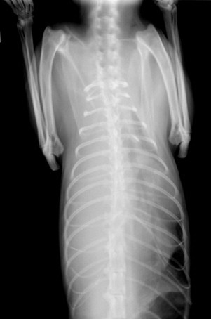
Dorsoventral radiographic view of severe pleural effusion. This cat had pyothorax.
FIGURE 30-24.

Right lateral radiographic view of severe pleural effusion. Note the distention of the thoracic cavity resulting in extreme dorsal displacement of the diaphragm and liver. This cat had pyothorax.
Ultrasonography can be used to detect the presence of small volumes of pleural effusion when radiographic findings are equivocal.72, 101 In one study in humans, pleural fluid was recognized by ultrasonography in 93% of cases, compared with only 83% by radiology.53 Ultrasonography can also provide gross estimations of effusion volume and assess effusion characteristics on the basis of echogenicity.72, 101
Stabilization
Cats (and other animals) with respiratory disease decompensate easily because although the oxygen saturation of hemoglobin is relatively stable, this is only provided the arterial partial pressure of oxygen (PO2) remains above approximately 60 mm Hg. Below this point the amount of oxygen carried by hemoglobin drops dramatically, so for an already respiratory-compromised cat, very small reductions in oxygen intake can result in extreme clinical effects. This is demonstrated by the oxygen–hemoglobin dissociation curve (Figure 30-25 ), where it can be seen that on the flat (right-hand side) part of the curve, decreases in PO2 from 100 to 60 mm Hg result in only small drops in oxygen hemoglobin saturation. As PO2 decreases below approximately 60 mm Hg, the curve drops to the left steeply, demonstrating the large decrease in the amount of oxyhemoglobin that results from only small drops in PO2; PO2 less than 60 mm Hg is defined as hypoxemic respiratory failure. If a cat is underventilating for any reason, the arterial carbon dioxide (PCO2) will rise; therefore, if the patient is inspiring room air, hypoxia will worsen; PCO2 greater than 45 mm Hg is defined as hypercapnic (ventilatory) respiratory failure.55, 77 A patient can have both hypercapnic and hypoxemic respiratory failure occurring to differing degrees simultaneously.
FIGURE 30-25.

Oxygen–hemoglobin dissociation curve demonstrating that as oxygen partial pressure (PO2) decreases below 60 mm Hg, the hemoglobin saturation percentage drops rapidly.
Rights were not granted to include this figure in electronic media. Please refer to the printed book.
(From West JB: Respiratory physiology: the essentials, ed 5, Philadelphia, 1995, Williams & Wilkins.)
© 2012 Williams & Wilkins
Stabilization requires an increase in the patient's arterial oxygen pressure to enable increased oxygen saturation of hemoglobin—that is, a shift to the right of the oxygen–hemoglobin dissociation curve. The two key components of stabilization are as follows:
-
1
Oxygen therapy
-
2
Pleurocentesis
Administration of an oxygen-enriched inspired gas mixture will at least partially correct hypoxemia depending on severity of hypoventilation, atmospheric pressure, flow rates, underlying pathology, hemoglobin concentration, cardiac output, and method of administration. The administration of an oxygen-enriched inspired gas mixture improves oxygenation for most respiratory causes of hypoxia.
Provision of pure oxygen provides higher concentrations of oxygen than normal air, so a greater amount of oxygen is moved to the alveoli with each breath. Oxygen therapy can be provided in the short term by flow-by delivery (Figure 30-26 ) or face mask delivery (Figure 30-27 ). More prolonged oxygen delivery can be achieved with the use of an Elizabethan collar canopy (Figure 30-28 ) or nasal catheter (Figure 30-29 ). Oxygen cages are widely used but can be problematic. Many commercially available cages do not permit manipulation of the temperature and humidity within the cage (Figure 30-30 ); the increasing heat and humidity caused by the presence of the cat in the sealed cage increases the cat's oxygen demands, thus further reducing the oxygen saturation of hemoglobin. Moreover, each time the cage door is opened to access the patient, the oxygen-rich environment not only is lost (which can lead to decompensation) but takes time to reach the desired oxygen concentration again. Human neonatal incubators (humidicribs) are most appropriate because they do allow manipulation of temperature and humidity, as well as having smaller openings (Figure 30-31 ). In severe cases intratracheal catheterization may be necessary, and those cats that cannot sustain PO2 greater than 60 mm Hg through their own efforts despite conventional oxygen supplementation will require mechanical ventilation. Further details about oxygen therapy are contained in Box 30-5.
FIGURE 30-26.

“Flow-by” oxygen delivery.
FIGURE 30-27.

Face mask oxygen delivery.
FIGURE 30-28.

Elizabethan collar canopy.
FIGURE 30-29.

Nasal catheter oxygen delivery to a recumbent cat. The catheter is marked with a yellow arrow and is glued to the cat's zygomatic arch.
(Courtesy Dr. Peter Best, South Tamworth Animal Hospital, Tamworth, Australia.)
FIGURE 30-30.

Cat in an inappropriate oxygen cage with no temperature, humidity, or fraction of inspired oxygen control.
FIGURE 30-31.

Cat in a human neonatal incubator (humidicrib).
(Courtesy Dr. Peter Best, South Tamworth Animal Hospital, Tamworth, Australia.)
Drainage of pleural fluid by pleurocentesis (puncture and aspiration from the pleural cavity) allows improved lung expansion, thereby enabling further oxygen saturation of hemoglobin. Some authors advocate thoracocentesis (puncture and aspiration from the thorax and not necessarily from the pleural space) before imaging when pleural effusion is strongly suspected because it can result in life-saving improvement for the patient.42, 77 In experienced hands thoracocentesis is a safe procedure and very well tolerated by cats. The major complications are pneumothorax, hemothorax, pneumohemothorax, or organ laceration, and these risks may be greater in cases when there is a small volume effusion (including no effusion), uncooperative patients, use of a large needle, and the need for multiple needle passes.103 Thoracocentesis is usually performed at the 6th, 7th, or 8th intercostal space, just ventral to the costochondral junction; this site should be clipped and surgically prepared. The positioning of the cat will vary from case to case. Sternal recumbency is ideal for maximal drainage of fluid, but access ventrally can be difficult when the cat is in this position; having the cat stand is appropriate, but this requires too much effort for many cats with respiratory distress; lateral recumbency results in the best access but may be too stressful for the cat. Butterfly catheter needles of 19 to 23G are appropriate to use and ideally should be mounted to a three-way stopcock and a 10-mL to 20-mL syringe.42, 103 It is important that fluid be collected for assessment as well as drained for therapeutic purposes. Further details about pleurocentesis are contained in Box 30-6 and Figure 30-32 .
FIGURE 30-32.

Thoracocentesis. Note the milky appearance of the effusion. This chylous effusion was subsequent to right-sided congestive heart failure.
Pleural Effusion Analysis
Assessment of pleural effusion fluid is the key step to determining the underlying cause. Assessments are based initially on the gross appearance (color, turbidity, presence of fibrin), then the protein concentration and cytology. The findings should always take into account the signalment of the cat and clinical findings.
Opaque effusion fluid can be milky white or milky pink (chyle), red (hemorrhage, although blood contamination can give a similar appearance); pyothorax often results in green or yellow opaque, malodorous fluid with flocculent material (but this can vary). Translucent effusions can be clear, yellow, or pink tinged and are initially distinguished by protein concentration, with lower protein effusions most typically arising from CHF and higher protein effusions most typically arising from FIP. Neoplasia can result in chylous, hemorrhagic, or translucent effusions of high or low protein concentration; cytology is often, but not always, helpful for diagnosis. More details about pleural fluid analysis can be found in Table 30-5 .
TABLE 30-5.
Guidelines for Categorization of Feline Pleural Effusions
| Translucent Effusions |
Opaque Effusions |
|||||
|---|---|---|---|---|---|---|
| Transudate | Modified Transudate | Nonseptic Exudate | Septic exudate | Chylous Effusion | Hemorrhage | |
| Color | Colorless to pale yellow | Yellow or pink | Yellow or pink | Yellow or pink | Milky white | Red |
| Turbidity | Clear | Clear to cloudy | Clear to cloudy | Cloudy to opaque; flocculent | Opaque | Opaque |
| Protein (g/L) | <25 | 25-50 | 30-60 (FIP: 35-85) | 30-70 | 25-60 | >30 |
| Fibrin | Absent | Absent | Present | Present | Variable | Present |
| Triglyceride | Absent | Absent | Absent | Absent | Present | Absent |
| Bacteria | Absent | Absent | Absent | Present | Absent | Absent |
| Nucleated cells/UL | <1000 | 1000-7,000 (LSA: 1000-100,000) | 5000-20,000 (LSA: 5,000-100,000) | 7000-300,000 | 1000-20,000 | Similar to that of peripheral blood |
| Cytologic features | Mostly mesothelial cells | Mostly macrophages and mesothelial cells; few nondegenerative neutrophils; may be neoplastic cells (LSA, carcinoma) in some cases | Mostly nondegenerate neutrophils and macrophages; neoplastic cells (LSA, carcinoma) in some cases | Mostly degenerate neutrophils, bacteria, also macrophages | Small lymphocytes, neutrophils, and macrophages in variable proportions | Mostly erythrocytes; macrophages with erythrocytosis |
| Disease associations | Hypoalbuminemia (glomerulopathy, hepatopathy, protein-losing enteropathy); early CHF (rare); hyperthyroid has been reported | CHF; neoplasia (LSA, carcinoma); diaphragmatic hernia | FIP; neoplasia; diaphragmatic hernia; lung lobe torsion; pancreatitis | Pyothorax | Chylothorax; obstructed thoracic duct or cranial vena cava (lymphangectasia, central venous thrombosis); ruptured thoracic duct, CHF, heartworm, neoplasia, idiopathic | Hemothorax Trauma Coagulopathies Lung lobe Torsion Neoplasia |
| Further diagnostics to differentiate causes | Blood biochemistry (perhaps T4), urinalysis with UPC; If primary hepatic: abdominal U/S and FNA, or fuller hepatic biopsy. If primary cardiac, echocardiography | Echocardiography NB hernia may not be seen on radiographs |
May need biopsy sample to differentiate neoplasia and FIP (although signalment often helpful); FIP: unresponsive pyrexia, hyperglobulinemia, nonregen erative anemia, lymphopenia (perhaps serology), coronavirus IFA of effusion fluid may help diagnose; neoplasia: LSA may be noted from cytology; pancreatitis may require laparotomy and biopsy | Culture and sensitivity required for appropriate antibiosis | Echocardiography; neoplasia may require FNA or biopsy; idiopathic by rule outs | Must distinguish between blood contamination of effusion; haemothorax fluid as haematocrit of 25-50% of that of peripheral blood History, presentation (mainly for trauma, exposure to rodenticides) Clotting factors. Neoplasia by U/S and FNA or biopsy (clotting factors first!) |
FIP, Feline infectious peritonitis; LSA, lymphosarcoma; CHF, congestive heart failure; U/S, ultrasound; UPC, urine protein:creatinine ratio; FNA, fine-needle aspirate; IFA, immunofluorescent assay; NB, nota bene.
Repeat radiography after pleurocentesis of effusion is often helpful diagnostically.
Causes of Pleural Effusion
Congestive Heart Failure
Effusion Characteristics
Effusion fluid from cats with CHF may be translucent (often clear to pale yellow, but blood contamination during pleurocentesis can result in pink or reddish tinge) or chylous (white or pink if blood contamination). The fluid is often a modified transudate (with protein concentrations of 25 to 50 g/L), but transudative or chylous effusion is also possible. Left-sided CHF typically results in pleural effusion in cats associated with increased ventricular diastolic pressure resulting in increased capillary hydrostatic pressure. Right-sided CHF can also cause pleural effusion, and chylothorax is often seen in this circumstance41, 68 (Figure 30-33 ).
FIGURE 30-33.

Right lateral thoracic radiograph showing pleural effusion and pulmonary edema caused by congestive heart failure. Note the significant cardiomegaly.
Further Diagnostics
The underlying cardiac disease can be determined only by echocardiography; cardiomyopathies such as hypertrophic or unclassified cardiomyopathy are most common. Auscultable changes such as a murmur or gallop rhythm are not necessarily present, and cardiac disease may be secondary to another problem, such as hyperthyroidism. CHF is covered in more detail in Chapter 20.
Feline Infectious Peritonitis
In one survey of 390 cats with effusive FIP, 17% had thoracic effusions, 62% had ascites, and 21% had effusions in both body cavities57 (Figure 30-34 ).
FIGURE 30-34.

Right lateral thoracic radiograph showing pleural effusion caused by feline infectious peritonitis (FIP); there is no distinguishing radiological feature to discern FIP. The color of the fluid and the high protein content in a young, febrile cat were clinical clues of the diagnosis.
Effusion Characteristics
The effusion fluid found with FIP is typically straw colored to golden yellow (although the shade can vary quite a bit), often contains fibrin clots, and has a high protein concentration. The total protein content is greater than 35 g/L, and often greater than 45 g/L, with globulins making up 50% or more.114 One study described an effusion with total protein greater than 80 g/L as 90% specific, 55% sensitive, and having a 0.78 PPV for the diagnosis of FIP.
Further Diagnostics
The Rivalta test recognizes high-protein content, as well as high concentrations of fibrinogen and inflammatory mediators in effusion fluid. It has been found to be very sensitive but only 80% specific; this test is performed by adding one drop of acetic acid (98%) to 5 mL of distilled water. This fluid is mixed thoroughly, and then one drop of effusion is gently placed on the surface of the mixture. If the drop stays at the top of the fluid or slowly floats to the bottom, the test is considered to be positive. This test can give inaccurate results if inappropriate technique is used or if there is a significant temperature difference between the fluid sample and the acetic acid solution. A positive Rivalta test can also result from lymphosarcoma or septic effusions (these can be distinguished from FIP by cytology and culture). Immunofluorescence staining of effusion fluid for coronavirus antigen in macrophages has a PPV of 1.00 but a negative predictive value of 0.57.58
The potential clinical presentations, diagnostics, and management of FIP are covered in greater detail in Chapter 33.
Neoplasia
Effusion Characteristics
Effusion fluid resulting from neoplasia can vary greatly depending on the mechanism responsible for fluid production. For example, exudates are more likely to occur when large numbers of neoplastic cells are exfoliated, and modified transudates are associated with less exfoliation. However, inflammation associated with neoplasia can result in an exudate with poorly exfoliating neoplasia. Chylous effusions can result when there is obstruction of intrathoracic lymph flow. Occasionally, hemorrhagic effusions may result from hemorrhagic neoplasia or vessel rupture from neoplastic invasion.
Further Diagnostics
Cytologic analysis is often useful to recognize neoplasia. One study showed that cytology had a sensitivity of 61% and a specificity of 100% in detecting neoplasia in body cavity effusions.62 Direct smears are often of adequate cellularity for assessment, but samples should be concentrated before examination in cases of low cellularity. In cases when cytology is not diagnostic, repeat radiography after drainage of pleural fluid often reveals a mass, commonly in the mediastinum, cranial to the cardiac silhouette. Care should be taken when interpreting radiographs because atelectatic lung lobes may mimic the appearance of a mass. Ultrasound-guided FNA can usually be used to confirm the diagnosis in these cases; in some cases, thoracotomy (or thoracoscopy) is required for an adequate biopsy sample.70
Specific Causes
Most studies cite mediastinal lymphosarcoma (Figure 30-35 ) as the most common neoplastic cause of pleural effusion in cats, representing approximately two thirds to three quarters of cases.21, 23, 52, 62 One study indicated that pleural effusion occurred in 90% of cats (55 of 61) with mediastinal lymphosarcoma.21 In a more recent study, however, only one of five cases (20%) of cats with pleural effusion associated with neoplasia was diagnosed with lymphosarcoma, with the remaining four cases associated with carcinoma.141 Most cats, particularly young cats, with mediastinal lymphosarcoma seem to respond well to routine lymphosarcoma treatment protocols.76, 119, 124
FIGURE 30-35.

Right lateral thoracic radiograph showing mediastinal mass (marked with yellow arrow). Fine-needle aspiration allowed a cytologic diagnosis of lymphosarcoma.
Primary pulmonary neoplasia is rare in cats.84, 86, 120 In many cases all lung lobes are affected when assessed radiographically, and pleural effusion is present in between 35% and 65% of cases. Other radiographic findings can include a mixed bronchoalveolar pattern, an ill-defined alveolar mass, or a mass with cavitation. Some form of bronchial disease is often present and may represent local airway metastasis.5, 6, 84, 86 Pulmonary metastasis from other locations appears radiographically as interstitial nodules (which may be well or ill defined) or a diffuse alveolar patter with or without ill-defined pulmonary nodules or pleural effusion (or both). Mammary gland adenocarcinoma is the most commonly represented primary tumor.32 Digital metastasis of bronchial carcinomas is also recognized as an uncommon cause of lameness in older cats; it is also known as lung–digit syndrome and carries an extremely poor prognosis.48 The prognosis for long-term survival with pulmonary adenocarcinoma is generally poor, with an overall median survival time of 115 days reported.56
Differential Diagnoses
Other mediastinal pathology can result in mediastinal masses; most cases are thymoma, which does not typically (but can) result in pleural effusion; thymic hyperplasia; idiopathic mediastinal and thymic hemorrhage; cystic thymus24; and thymolipoma.125 Thymic masses can be associated with paraneoplastic processes such as exfoliative dermatitis and myasthenia gravis.* Thymectomy typically results in resolution of all clinical signs (including paraneoplastic signs) and results in prolonged survival,* with one study noting median survival of 1825 days.139 Occasional reports have recognized the development of myasthenia gravis after a thymectomy.47, 107
Pyothorax
Effusion Characteristics
Septic exudates are usually turbid to opaque because of a large cell count and often contain flocculent material. They are usually cream or pale yellow in color but can be pink, green tinged, or red tinged. Greater than 80% of cases of pyothorax are caused by mixed anaerobic infections; consequently the effusion is typically malodorous (like cat-fight abscesses, which are also largely due to anaerobic bacterial infection). A lack of odor does not rule out pyothorax but makes aerobic or yeast infection probable in an effusion predominated by neutrophils. Typical of exudates, the protein content is greater than 30 g/L because of a high nucleated cell count (at least 7,000/µL), which often comprises more than 85% degenerate neutrophils; bacteria may be recognized intracellularly or extracellularly.8
Other Findings
Cats with pyothorax are not necessarily dyspneic on presentation and may present with nonspecific systemic signs of inappetence or lethargy. Pyothorax is typically a disease of young cats, with a mean age of 4 to 6 years reported, but cats of any age can be affected.7, 23, 25, 128, 130 Pyrexia is not necessarily present; hypothermia and bradycardia can be present as indicators of severe sepsis.128 Hematology may reflect sepsis with a left shift neutrophilia, and neutropenia with a degenerative left shift can occur with advanced sepsis. Plasma biochemistry may demonstrate hyperglobulinemia with hypoalbuminemia, and hyperglycemia or hypoglycemia are potential consequences of sepsis.
Treatment
Treatment of pyothorax should be considered in terms of treatment of a cat-fight abscess—that is, drainage, appropriate antibiotic therapy, and any required supportive care.
Drainage and Lavage
Drainage is best achieved by placement of bilateral thoracostomy tubes, which generally remain in place for 5 to 6 days to enable repeated thoracic lavage. Placement of thoracostomy tubes is covered in Box 30-7 .
BOX 30-7. Placement of Thoracostomy Tubes.
-
•
Thoracostomy tubes should be placed after stabilization of respiratory compromise (by thoracocentesis and oxygen therapy) as well as any fluid and electrolyte imbalances.
-
•Tubes should be placed bilaterally:
-
•Helps effective drainage if loculation of fluid or mediastinum intact
-
•Drainage still provided if single side tube failure (obstruction or kinking)
-
•
-
•
Commercially available veterinary thoracic trocar catheters are available; generally 14 to 16 Fr are the most appropriate size.
-
•General anesthesia is required with intubation.
-
•Intermittent positive pressure ventilation (IPPV) required; ~10-12 breaths per minute, with a tidal volume of about 20 mL per kg.
-
•
-
•The chest tube enters the skin two or more intercostal spaces (ICSs) caudal to where the tube enters the thoracic cavity (Figure 30-36 ).
-
•Surgical site prepared
-
•Small stab incision (just large enough to accommodate the size of the thoracic drain but no larger) for entry of catheter at approximately 10th to 12th ICS (dorsally).
-
•Subcutaneous tunnel made cranioventrally with trocar as far as 7th to 8th ICS
-
•Helpful if assistant pulls the thoracic skin approximately 5 cm cranially and ventrally just before driving trocar through thoracic wall; enables the trocar to enter thoracic wall perpendicularly
-
•Trocar advanced through subcutaneous tunnel, and then driven into the pleural space at 7th to 8th ICS
-
•Tube pushed off the trocar and advanced ~12 to 18 cm cranially and ventrally within the thoracic cavity (assistant releases the skin)
-
•End of tube clamped to prevent pneumothorax
FIGURE 30-36.
 Positioning of a thoracostomy tube within the pleural space.Rights were not granted to include this figure in electronic media. Please refer to the printed book.(From Barrs VR, Beatty JA: Feline pyothorax: new insights into an old problem. Part 2: Treatment recommendations and prophylaxis, Vet J 179:171, 2009.)© 2012 Vet J
 -
•
-
•Alternative technique:
-
•Direct dissection of the subcutaneous tunnel using Mayo scissors or hemostat
-
•Red rubber catheter grasped and clamped in the tips of a hemostat, to drive into the pleural space.
-
•
-
•Tube is secured to the thoracic wall with purse-string suture, and a Chinese finger trap suture is placed to prevent the tube from slipping (Figure 30-37 ).
FIGURE 30-37.
 Thoracostomy tube in situ, secured to the thoracic wall with purse-string suture and a Chinese finger trap suture (which cannot easily be seen).
 -
•
Three-way stopcock is placed at the end of the tube, and any remaining exudate or air that entered during the procedure is evacuated
-
•After bilateral tubes are placed, each tube should be used to lavage the thorax on two or three occasions, with about 100 mL of warmed lactated Ringer's solution (Figure 30-38 ).
-
•The thorax should be lavaged multiple times in the first 24 to 48 hours.
-
•Subsequently, two to three times daily lavage is appropriate.
FIGURE 30-38.

 A, Warmed lactated Ringer's solution is used to lavage the pleural space. The author uses multiple 20-mL syringes for this. B, Introduced fluid is removed. Note the discoloration of the fluid compared with that in the prior image.
 -
•
-
•A light two-layer bandage should be applied without excessive pressure (not so tight that it interferes with the cat's breathing) (Figure 30-39 ).
FIGURE 30-39.
 A, Thoracoscopy tubes covered by a light two-layer bandage should be applied without excessive pressure. In this view the three-way stopcocks are exposed (as when lavage and drainage are performed) to show gauze used as padding under the stopcocks. B, The three-way stopcocks should be covered over for when the cat is returned to its hospital cage.
 -
•
Hygienic precautions such as wearing gloves should be adhered to at all times when changing bandage dressing or performing manual aspiration of the tube.
-
•Tubes are generally removed after 4 to 6 days on the basis of the following factors:
-
•Reduction of pleural effusion to approximately 2 mL/kg per day
-
•Resolution of pleural effusion on thoracic radiographs
-
•Cytologically:
-
○Absence of microorganisms
-
○Reduction of neutrophil numbers with loss of degenerative appearance
-
○
-
•
-
•
Appearance of macrophages
One study reported shorter total duration of tube placement for patients treated with lavage than those managed with drainage only; however, the authors note that few cases were treated without lavage.25 Repeated thoracic lavage allows drainage and assessment of exudates but also débrides the pleura, including breaking down adhesions and diluting bacterial and inflammatory mediator concentrations; further, the chance of thoracostomy tube obstruction with thick exudates is reduced. Before lavage, suction should be attempted to determine the volume and nature of pleural effusion. Warmed (to body temperature) compound sodium lactate is used for lavage (use of 0.9% sodium chloride can result in hypokalemia7) with volumes from 10 to 25 mL/kg per lavage. The volume of fluid introduced and aspirated should be recorded for each side, with expected recovery of 75% or more of lavage fluid. Recovery of smaller volumes should prompt investigations (usually with imaging) for thoracostomy tube complications or loculation of pockets of fluid as a result of adhesions. There are no definitive guidelines for frequency of lavage; every 4 hours for the first 24 to 48 hours and subsequently two to three times daily has been recommended.9 Removal of thoracostomy tubes is indicated when the volume of pleural effusion has reduced to approximately 2 mL/kg per day; pleural effusion has resolved radiographically; and infection has resolved, as indicated by absence of organisms, reduction of neutrophil numbers, and loss of their degenerative appearance and appearance of macrophages. Recent studies have reported a median duration of thoracic drainage of 5 to 6 days.7, 25, 128
Continuous water seal suction has also been used59 but offers no real advantage over intermittent suction and lavage as described. Continuous suction offers the advantage of maximal drainage but does not decrease the time needed to manage pyothorax. Further, water-seal chest drainage units require continuous monitoring because leakage between the pleural cavity and the water seal can be fatal.59
Needle thoracocentesis (on one occasion or multiple times) is the alternative to thoracoscopy tube placement but is associated with higher mortality and should be reserved for cases with low volume effusion or when thoracostomy tube placement is declined by the owner (e.g., because of financial concerns).
Antibiotic Therapy
Antibiotic therapy should be governed by culture and sensitivity findings; however, therapy should be instigated as soon as practical after diagnosis, and, further, sensitivity results for anaerobic bacteria are not routinely available (another consideration is that samples should be collected anaerobically). The bacteria most commonly responsible for pyothorax are anaerobes, typical of oral flora; the most commonly isolated aerobes, Pasteurella spp., have been recognized in 12.5% to 20% of cases, in many cases in addition to anaerobes.7, 74, 130 Empiric therapy with penicillin-based antibiotics (including aminopenicillins or potentiated penicillins), therefore, should be effective against these bacteria. It is important to use parenteral antibiotics initially because affected cats are debilitated and unlikely to be eating. Most intravenous antibiotics require dosing at 6- to 8-hour intervals; in some cases antibiotics can be added to the intravenous fluids and provided as constant-rate infusion. Antibiotic therapy should be continued for an extended duration, typically 6 to 8 weeks.7, 25 Amoxicillin–clavulanate at 15-20 mg/kg twice daily is an appropriate empiric antibiotic choice in most cases; cefovecin has an appropriate in vitro spectrum,116 and the author has successfully used this antibiotic in cases when the owner has had difficulty administering oral antibiotics; however, the revisits for repeat injections at 10- to 14-day intervals are vital, and the owner should be reminded of appointments in the preceding days.
Supportive Care
Fluid therapy should be provided for supportive care at approximately twice maintenance rates until the cat is rehydrated and continued at maintenance rates thereafter. Electrolyte status should be evaluated and taken into account with fluid therapy. Most cats are inappetent or anorexic and will require nutritional support.
Idiopathic Chylothorax
Differential Diagnoses and Effusion Characteristics
There are no distinctive radiographic features of idiopathic chylothorax (Figure 30-40 ).
FIGURE 30-40.

Left lateral radiograph of pleural effusion caused by idiopathic chylothorax. There are no distinguishing radiologic features to determine the nature of this effusion.
The recognition of milky white or pink pleural effusion (see Figure 30-32) does not constitute a diagnosis of idiopathic chylothorax because thoracic chylous effusions can result from multiple causes, including CHF,12, 39, 41, 60, 122 neoplasia,19, 33, 61 trauma,50, 85 infections (including heartworm),26, 27, 83 and potentially FIP. The color of chylous effusions varies depending on dietary fat content and whether there is concurrent hemorrhage. The protein concentration is variable and often raised artefactually by the high lipid content of the fluid. The total nucleated cell count is often less than 10,000/µL, consisting of predominantly small lymphocytes or neutrophils as well as lipid-laden macrophages.37 Pleural effusions with triglyceride concentrations greater than 1.12 mmol/L (100 mg/dL) are always chylous; chylous effusions have a greater pleural fluid triglyceride concentration but a lower or equal cholesterol concentration compared with serum.129 Pseudochylous effusions are rare; these effusions grossly resemble chyle but are distinguished by having a greater cholesterol concentration than serum.37 Despite multiple possible causes, in the majority of cases, no specific etiology for the chylous effusion is found, leaving the diagnosis as idiopathic.39, 66 However, because the management of these disparate causes varies so greatly, thorough investigations should be undertaken.
Further Diagnostics
The two major modes of investigating chylous effusions are to repeat thoracic radiographs subsequent to drainage of effusion fluid and echocardiography. Mediastinal masses can be difficult to appreciate radiographically when there is a large volume pleural effusion; masses recognized to result in chylothorax include mediastinal lymphosarcoma,33, 61 thymoma,19 and one case of a cryptococcal granuloma.83 Ultrasound-guided FNA is required to distinguish these (or other) conditions. Echocardiography should be performed on all cats with chylothorax; cardiac diseases recognized to result in chylous effusion include cardiomyopathies,12, 41 congenital disorders,41 cardiac neoplasia,41 pericardial disease,41 paroxysmal heart block,31 and heartworm disease.27, 45, 108 Additional investigations for heartworm disease, such as antigen and antibody testing, are also appropriate.
Medical Management
There is no definitive medical treatment for idiopathic chylothorax, and surgery should be considered if attempts at medical management are unsuccessful. If the effusion recurs despite dietary and drug therapy (as is often the case), then intermittent repeat thoracocentesis is required. Cats should be managed with low-fat diets containing 6% or less fat on a dry matter basis.37 Rutin is a benzopyrone plant extract available from health food stores that has been reported to improve and occasionally successfully resolve idiopathic chylothorax; recommended doses are 50 to 100 mg/kg (or 250 to 500 mg/cat) orally, three times daily.49, 69, 121 Octreotide is a somatostatin analog that has been anecdotally reported to resolve idiopathic chylothorax when administered at 10 µg/kg subcutaneously, three times daily for 2 to 3 weeks; prolonged administration has been associated with gallstone formation in people. The condition does occasionally resolve spontaneously after several weeks to months.37 Persistent chylothorax resulting from incomplete treatment can lead to fibrosing pleuritis as a complication; this condition appears to develop subsequent to any prolonged exudative or blood-stained effusion. The fibrosis restricts normal pulmonary expansion and carries a very guarded prognosis.38
Surgical Treatment
If medical management fails to resolve or alleviate the clinical signs of chylothorax, surgical intervention becomes the only therapeutic option. Thoracic duct ligation (TDL) in conjunction with pericardiectomy (PC) is the most widely accepted technique; one study reported an 80% success rate for resolving idiopathic chylothorax in cats.40 These procedures have been performed successfully with thoracoscopy in dogs.2 Pleural omentalization has been performed in addition to TDL and PC but does not appear to improve results.15 The techniques for these procedures have been recently reviewed.37
Other Causes of Pleural Effusion
Beyond CHF, FIP, neoplasia, pyothorax, and idiopathic chylothorax, clinicians will be directed toward most other causes of pleural effusion from clinical history (e.g., known trauma), physical examination findings, and routine investigations. Trauma can result in hemothorax (Figure 30-41 ), chylothorax (from thoracic duct rupture), or effusion associated with diaphragmatic hernia; urinothorax subsequent to diaphragmatic herniation and kidney prolapse has been reported in one cat.117 Other reported underlying conditions include pancreatitis,102 congenital thoracic duct abnormalities,28 aelurostronglylosis,87 cardioperitoneal and pleuroperitoneal hernias, and lung lobe torsion.67, 81 Low-protein pleural transudates (FIGURE 30-42, FIGURE 30-43 ) are rarely recognized and have been described in one cat with a preexisting perinephric pseudocyst in direct communication with the pleural space98 and another with a severe protein-losing glomerulopathy.4
FIGURE 30-41.

Right lateral radiograph demonstrating hemothorax associated with trauma. Note that the lung mass is greatly compressed by effusion, pulmonary contusions are evident, and the increased radiodensity over the cardiac silhouette is associated with clot formation. The diaphragm can be followed and is intact.
(Courtesy Dr. Susan Little.)
FIGURE 30-42.

Right lateral radiograph demonstrating pleural effusion associated with hypoalbuminemia. Note that there is also pulmonary edema. This cat's plasma albumin concentration was 15 g/L and was associated with severe intestinal disease. A total of 115 mL of effusion was removed using bilateral thoracocentesis.
(Courtesy Dr. Susan Little.)
FIGURE 30-43.

Effusion fluid drained from hypoalbuminemic cat (radiograph shown in Figure 30-42); note that the fluid is clear, with no evidence of cellularity.
(Courtesy Dr. Susan Little.)
Pleural Access Ports
Recent studies13, 16 have documented the use of pleural access ports for cats with chronic pleural effusions. These devices are inserted during thoracotomy; they allow drainage of chronic thoracic effusion in circumstances such as neoplasia when effusion is profuse in a patient undergoing chemotherapy.
Pneumothorax
The normal pressure of the pleural cavity remains below normal atmospheric air pressure, varying from −5 cm of water at beginning of inspiration (equal to the amount of suction required to hold the lungs open to their resting level) to −7.5 cm of water as inspiration creates more negative pressure.54 Pneumothorax results from the accumulation of free air or gas of atmospheric pressure within the thoracic cavity95; this loss of negative intrapleural pressure causes the lungs to collapse as a result of elastic recoil.71 When the lungs collapse, the tidal volume is reduced, prompting tachypnea in an attempt to maintain the minute ventilation. Local hypoxia induces vasoconstriction of pulmonary vessels, diverting blood flow to ventilated areas. Vasoconstriction, combined with collapse of blood vessels caused by atelectasis, eventually leads to pulmonary hypertension and increased work for the right side of the heart.79
Causes and Classification
Pneumothorax most commonly results from trauma in cats64; spontaneous pneumothorax is occasionally reported.10, 20, 88, 110, 132 Iatrogenic pneumothorax can result from anesthetic complications in association with intubation, positive pressure ventilation, and failure to reopen anesthetic machine pop-up valves,* subsequent to endoscopic retrieval of foreign bodies from the trachea or esophagus17, 138 or subsequent to thoracic FNA.79
There are two basic classes of pneumothorax: open and closed. Open pneumothorax is associated with a body wall wound that allows entry of free air. Depending on the wound size, pressure within the pleural space is less than or equal to atmospheric pressure. Closed pneumothorax occurs when the air within the pleural space has entered through a wound of the lung parenchyma or trachea. Open pneumothorax is further divided into simple and tension forms. Simple closed pneumothorax occurs when a puncture of the lung allows air to enter the pleural space during inspiration and exit during expiration. This allows the pressure within the pleural space to equilibrate with atmospheric pressure. In tension pneumothorax, the wound in the lung acts as a flap valve, allowing air to enter the pleural space during inspiration but not allowing it to escape during expiration. This results in a gradual increase in pleural space pressure and progressive pulmonary atelectasis. With large wounds pleural pressure can eventually exceed atmospheric pressure. Untreated tension pneumothorax therefore results in progressively worsening dyspnea, subsequent cardiovascular dysfunction, and ultimately death.71
Diagnosis and Radiography
Cats with pneumothorax typically present with dyspnea; in most cases there is a history of trauma or, in iatrogenic cases, a procedure that could potentially cause pneumothorax. Radiography readily confirms the diagnosis (FIGURE 30-44, FIGURE 30-45 ). Spontaneous pneumothorax is typically recognized after thoracic radiography and, in many cases, unknown trauma is assumed.
FIGURE 30-44.

Right lateral radiograph of cat with pneumothorax. This cat fell onto a kitchen knife that was held upright in a kitchen strainer. Note the greatly increased width of the air-filled pleural space. Because the lung is decreased in size and air content, it appears radiopaque compared with the surrounding air-filled pleural space. Subcutaneous emphysema can be seen ventrally.
FIGURE 30-45.

Dorsoventral view of the same cat in Figure 30-44. The chest wall emphysema is in the position where the knife entered the thoracic cavity. Subcutaneous emphysema surrounds this position.
Pneumothorax is recognized radiographically by the increased width of the air-filled pleural space with accompanying partial pulmonary atelectasis and retraction of the lung margins. This results in the absence of vascular shadows in the peripheral portions of the thorax. Because the lung is decreased in size and air content, it appears relatively radiopaque compared with the air-filled pleural space. The heart is typically displaced dorsally from the sternum in recumbent lateral views. Minimal pneumothorax can be difficult to visualize and requires recognition of retracted lobar edges. Tension pneumothorax is best recognized by sequential radiographs demonstrating the accumulation of progressively larger amounts of pleural air.93
Diagnosis of spontaneous pneumothorax requires recognition of a potential underlying cause or resolution of pneumothorax after appropriate treatment of a potential cause such as bronchial disease.10, 20, 132 CT was used to recognized a pulmonary bulla in a recent report.88
Treatment
The treatment of pneumothorax varies greatly depending on severity, cause, and class of pneumothorax. In all cases large volumes of pleural air should be drained by thoracocentesis using a three-way stopcock (see Box 30-6). Subsequently, many cases with simple closed pneumothorax can be managed successfully with cage rest; the air leak usually seals itself within hours, and the residual pleural air is resorbed over a few days.64 Oxygen therapy (see Box 30-5) maintains a dissolved gas pressure gradient between pulmonary vessels and the pleural cavity and can aid resorption of pleural air.
Open pneumothorax requires surgical correction; open wounds should be covered immediately with any available material. Intermittent positive pressure ventilation is required through anesthesia until the wound is repaired and air evacuated.
Worsening dyspnea, with reaccumulation of pleural air, in cats with closed pneumothorax (no overt penetrating injury) suggests tension pneumothorax. Thoracostomy tubes (see Box 30-7) are not often indicated in cats but may be required in some cases to facilitate drainage, and closed suction systems may be indicated for continuous drainage of air. Water-seal chest drainage units require continuous monitoring because failure of the system can result in acute respiratory distress or death.59 In severe cases of massive air reaccumulation, surgical thoracotomy is required to identify and correct the underlying cause.
Diaphragmatic Hernia
Diaphragmatic hernia refers to the entry of abdominal contents into the thoracic cavity caused by a defect of the diaphragm. Peritoneopericardial hernia (PPDH) refers to entry of abdominal contents into the pericardial sac owing to a congenital communication from the abdomen36; PPDH is addressed in Chapter 20. The more commonly encountered pleuroperitoneal hernias are predominantly the result of trauma, with motor vehicle accidents the most common cause of trauma in these cases44, 89, 104, 135, 136; interestingly, high-rise trauma does not commonly result in diaphragmatic herniation.126, 134 Abdominal pressure at impact is dissipated cranially, causing the diaphragm to tear, with the location and size of the tear depending on the position of the animal at the time of the impact.36 Occasional cases of congenital diaphragmatic hernia have been described, including so called “true herniation,” in which the serosa remains intact.18, 46, 65, 127
Clinical Signs
The severity of clinical signs varies greatly depending on the size of the tear and the organ that has herniated. Respiratory signs such as dyspnea or tachypnea are often, but not always, present. Lack of respiratory signs can result in the diagnosis being missed; in one study 50 of 116 cases of diaphragmatic hernia (of cats and dogs) were not diagnosed within 30 days,135 and in some cats the condition may remain unrecognized for years. Muffled heart or lung sounds are a common clinical finding; some cats will refuse to lie in particular positions because of the resultant dyspnea; the abdomen may have an empty or “tucked-up” appearance; and gastrointestinal signs may be present, including gastric dilation.* Because the most common cause is trauma, there are often other injuries present; mortality rates appear to be more correlated with concurrent injuries than the degree of diaphragmatic damage or chronicity.104
Diagnosis
Radiography is the most reliable means of diagnosis; typical radiographic signs include loss of continuity of the diaphragmatic outline and readily identifiable abdominal organs within the thorax such as gas-filled loops of intestine (Figure 30-46 ).43, 118, 135 In more subtle cases a soft tissue density may be recognized in the caudal thorax (FIGURE 30-47, FIGURE 30-48 ).18, 133 Pleural effusion is present in 20% to 25% of cases (see Figure 30-47).43, 118, 135 Ultrasonography can be used to confirm the diagnosis if radiographic findings are equivocal; one study correctly diagnosed diaphragmatic hernia by ultrasonography in 20 of 21 cats (95%), with the only false-negative result being due to adhesions imitating the appearance of an intact diaphragm.115
FIGURE 30-46.

Right lateral radiograph showing severe diaphragmatic hernia. As well as the overt abdominal contents within the thoracic cavity resulting in dorsal elevation of the heart, there is loss of continuity of the diaphragm and pleural effusion.
FIGURE 30-47.

Right lateral radiograph showing more subtle diaphragmatic hernia than prior image. The herniation looks like a mass ventrally, just caudal to the heart. There is also pleural effusion present.
FIGURE 30-48.

Ventrodorsal view of the same cat in Figure 30-47. The discontinuity of the diaphragm is evident on left of the image (cat's right).
Treatment
Diaphragmatic herniation requires surgical repair (Figure 30-49 ). However, in many cases surgery should be delayed until other consequences of trauma, such as shock, have been stabilized; gastric dilation warrants immediate surgical attention.36 Intermittent positive pressure ventilation must be provided throughout anesthesia, and residual air must be evacuated from the thorax when surgical repair is complete; the clinician must be aware of the possibility of postsurgical pneumothorax, which can usually be managed by thoracocentesis. In most cases hernias may be repaired by primary closure, but in some chronic or congenital cases, the defect may be too large; in one such case a commercial small intestinal submucosal graft was used.3 Recent studies report an excellent prognosis. In one study 28 of 34 cats (82%) survived, with concurrent injuries being recognized as the most common contributing factor for failure to survive104; another study reported survival in 16 of 16 (100%) of chronic cases with duration of herniation greater than 2 weeks89 (Figure 30-50 ).
FIGURE 30-49.

Gross appearance of diaphragmatic hernia during laparotomy for repair.
FIGURE 30-50.


Postsurgical radiographs (right lateral [A] and ventrodorsal [B] views) of the cat depicted in Figure 30-43 demonstrating continuity of diaphragm. There is a small amount of residual pleural effusion.
Chest Wall Pathology
Chest wall pathology is not common in cats, and many cases produce no clinical signs. Awareness of these conditions is important for completeness of differential diagnoses of thoracic conditions and to rule out inconsequential change.
Trauma
Trauma is the most common chest wall abnormality. Blunt trauma may result in rib fractures, but these typically occur with other injuries, such as pulmonary contusions, pneumothorax, and hemothorax.1 Subcutaneous emphysema may also result but is usually of little significance in itself, often requiring no specific treatment as long as the underlying cause is addressed.36 Rib fractures also rarely contribute to morbidity in themselves. In one study 11 of 17 (65%) of cats with traumatic rib fractures survived, with all deaths (including euthanasia) attributable to concurrent injuries.1 Similarly, the damage to the chest wall sustained by penetrating trauma is rarely of itself of major consequence but may be the only clue to other consequences, such as thoracic soft tissue trauma or pneumothorax.36 Cases should be managed on a case-by-case basis, addressing basic wound management principles and taking into account thoracic cavity physiology—primarily, that intrathoracic pressure is lower than atmospheric pressure.
Nontraumatic Rib Fractures
In a recent study approximately half of rib fractures recognized (16 of 33, or 48%) had no known associated trauma. Pathologic fractures associated with neoplasia were one recognized cause, but in many cases (11 of 16, or 69%) the fractures were associated with chronic respiratory disorders such as bronchial diseases, pleural effusion, and URT disorders; the authors postulate these to be stress fractures associated with repetitive mechanical stress and chronic microtrauma caused by increased or more forceful respirations. In no case were the rib fractures deemed to specifically affect any cat's clinical course, but recognition of such fractures without trauma should prompt attention to determine the underlying cause or provide an additional measure of the severity of known underlying pathology.1
Pectus Excavatum
Pectus excavatum has been sporadically reported in cats.* It is a congenital condition resulting in dorsal deviation of the caudal part of the sternum or a dorsoventral flattening of the entire thorax that is thought to be associated with shortening of the central tendon of the diaphragm (see Figure 41-8). Many cats show respiratory signs, but the abnormality is recognized visually or by palpation by their owner. When respiratory signs do occur, they are present at birth or not long afterward; such signs may include reduced exercise tolerance, dyspnea, recurrent respiratory infections, and cyanosis; additionally, gastrointestinal signs such as vomiting may occur. Diagnosis is usually made by visual recognition and is confirmed radiographically. Surgical correction techniques using either temporary external splints35, 80, 112, 137 or internal stabilization22, 99, 106 have been described as successful. The reader is directed to these sources for details of these techniques.
Neoplasia
Neoplasia affecting the ribs of cats is rare. Occasional cases of plasma cell tumors or multiple myeloma have been recognized to result in rib fractures1 or rib osteolysis.131 One case series of chondrosarcoma mentions only 2 of 67 (3%) cases involving the ribs.29 Rare occurrences such as this must be managed on a case-by-case basis using general oncologic principles; no overall recommendations can be made.
References
- 1.Adams C, Streeter EM, King R. Retrospective study: cause and clinical characteristics of rib fractures in cats: 33 cases (2000-2009) J Vet Emerg Crit Care. 2010;20:436. doi: 10.1111/j.1476-4431.2010.00559.x. [DOI] [PubMed] [Google Scholar]
- 2.Allman DA, Radlinsky MG, Ralph AG. Thoracoscopic thoracic duct ligation and thoracoscopic pericardectomy for treatment of chylothorax in dogs. Vet Surg. 2010;39:21. doi: 10.1111/j.1532-950X.2009.00623.x. [DOI] [PubMed] [Google Scholar]
- 3.Andreoni AA, Voss K. Reconstruction of a large diaphragmatic defect in a kitten using small intestinal submucosa (SIS) J Feline Med Surg. 2009;11:1019. doi: 10.1016/j.jfms.2009.06.003. [DOI] [PMC free article] [PubMed] [Google Scholar]
- 4.Asano T, Tsukamoto A, Ohno K. Membranoproliferative glomerulonephritis in a young cat. J Vet Med Sci. 2008;70:1373. doi: 10.1292/jvms.70.1373. [DOI] [PubMed] [Google Scholar]
- 5.Ballegeer EA, Forrest LJ, Stepien RL. Radiographic appearance of bronchoalveolar carcinoma in nine cats. Vet Radiol Ultrasound. 2002;43:267. doi: 10.1111/j.1740-8261.2002.tb01001.x. [DOI] [PubMed] [Google Scholar]
- 6.Barr F, Gruffydd-Jones TJ, Brown PJ. Primary lung tumours in the cat. J Small Anim Pract. 1987;28:1115. [Google Scholar]
- 7.Barrs VR, Allan GS, Martin P. Feline pyothorax: a retrospective study of 27 cases in Australia. J Feline Med Surg. 2005;7:211. doi: 10.1016/j.jfms.2004.12.004. [DOI] [PMC free article] [PubMed] [Google Scholar]
- 8.Barrs VR, Beatty JA. Feline pyothorax: new insights into an old problem. Part 1: Aetiopathogenesis and diagnostic investigation. Vet J. 2009;179:163. doi: 10.1016/j.tvjl.2008.03.011. [DOI] [PubMed] [Google Scholar]
- 9.Barrs VR, Beatty JA. Feline pyothorax: new insights into an old problem. Part 2: Treatment recommendations and prophylaxis. Vet J. 2009;179:171. doi: 10.1016/j.tvjl.2008.03.019. [DOI] [PubMed] [Google Scholar]
- 10.Barrs VR, Swinney GR, Martin P. Concurrent Aelurostrongylus abstrusus infection and salmonellosis in a kitten. Aust Vet J. 1999;77:229. doi: 10.1111/j.1751-0813.1999.tb11707.x. [DOI] [PubMed] [Google Scholar]
- 11.Beatty J, Barrs V. Pleural effusion in the cat: a practical approach to determining aetiology. J Feline Med Surg. 2010;12:693. doi: 10.1016/j.jfms.2010.07.013. [DOI] [PMC free article] [PubMed] [Google Scholar]
- 12.Birchard SJ, Ware WA, Fossum TW. Chylothorax associated with congestive cardiomyopathy in a cat. J Am Vet Med Assoc. 1986;189:1462. [PubMed] [Google Scholar]
- 13.Brooks AC, Hardie R. Use of the subcutaneous pleuralport device for management of chronic pleural effusions in the dog and cat [abstract] Vet Surg. 2009;38:E27. doi: 10.1111/j.1532-950X.2011.00901.x. [DOI] [PubMed] [Google Scholar]
- 14.Brown DC, Holt D. Subcutaneous emphysema, pneumothorax, pneumomediastinum, and pneumopericardium associated with positive-pressure ventilation in a cat. J Am Vet Med Assoc. 1995;206:997. [PubMed] [Google Scholar]
- 15.Bussadori R, Provera A, Martano M. Pleural omentalisation with en bloc ligation of the thoracic duct and pericardiectomy for idiopathic chylothorax in nine dogs and four cats. Vet J. 2011;188:234–236. doi: 10.1016/j.tvjl.2010.05.010. [DOI] [PubMed] [Google Scholar]
- 16.Cahalane AK, Flanders JA, Steffey MA. Use of vascular access ports with intrathoracic drains for treatment of pleural effusion in three dogs. J Am Vet Med Assoc. 2007;230:527. doi: 10.2460/javma.230.4.527. [DOI] [PubMed] [Google Scholar]
- 17.Cariou MP, Lipscomb VJ. Successful surgical management of a perforating oesophageal foreign body in a cat. J Feline Med Surg. 2011;13:50. doi: 10.1016/j.jfms.2010.11.010. [DOI] [PMC free article] [PubMed] [Google Scholar]
- 18.Cariou MPL, Shihab N, Kenny P. Surgical management of an incidentally diagnosed true pleuroperitoneal hernia in a cat. J Feline Med Surg. 2009;11:873. doi: 10.1016/j.jfms.2009.04.001. [DOI] [PMC free article] [PubMed] [Google Scholar]
- 19.Carpenter JL, Holzworth J. Thymoma in 11 cats. J Am Vet Med Assoc. 1982;181:248. [PubMed] [Google Scholar]
- 20.Cooper ES, Syring RS, King LG. Pneumothorax in cats with a clinical diagnosis of feline asthma: 5 cases (1990-2000) J Vet Emerg Crit Care. 2003;13:95. [Google Scholar]
- 21.Creighton SR, Wilkins RJ. Thoracic effusions in the cat. Etiology and diagnostic features. J Am Anim Hosp Assoc. 1975;11:66. [Google Scholar]
- 22.Crigel M. Pectus excavatum surgically repaired using sternum realignment and splint techniques in a young cat. J Small Anim Pract. 2005;46:352. doi: 10.1111/j.1748-5827.2005.tb00332.x. [DOI] [PubMed] [Google Scholar]
- 23.Davies C, Forrester SD. Pleural effusion in cats: 82 cases (1987 to 1995) J Small Anim Pract. 1996;37:217. doi: 10.1111/j.1748-5827.1996.tb01772.x. [DOI] [PubMed] [Google Scholar]
- 24.Day MJ. Review of thymic pathology in 30 cats and 36 dogs. J Small Anim Pract. 1997;38:393. doi: 10.1111/j.1748-5827.1997.tb03492.x. [DOI] [PubMed] [Google Scholar]
- 25.Demetriou JL, Foale RD, Ladlow J. Canine and feline pyothorax: a retrospective study of 50 cases in the UK and Ireland. J Small Anim Pract. 2002;43:388. doi: 10.1111/j.1748-5827.2002.tb00089.x. [DOI] [PubMed] [Google Scholar]
- 26.Donahoe JM. Experimental infection of cats with Dirofilaria immitis. J Parasitol. 1975;61:599. [PubMed] [Google Scholar]
- 27.Donahoe JM, Kneller SK, Thompson PE. Chylothorax subsequent to infection of cats with Dirofilaria immitis. J Am Vet Med Assoc. 1974;164:1107. [PubMed] [Google Scholar]
- 28.Duncan NM. Chylothorax in a kitten. J S Afr Vet Assoc. 1991;62:75. [PubMed] [Google Scholar]
- 29.Durham AC, Popovitch CA, Goldschmidt MH. Feline chondrosarcoma: a retrospective study of 67 cats (1987-2005) J Am Anim Hosp Assoc. 2008;44:124. doi: 10.5326/0440124. [DOI] [PubMed] [Google Scholar]
- 30.Evans AT. Anesthesia case of the month. Pneumothorax, pneumomediastinum and subcutaneous emphysema in a cat due to barotrauma after equipment failure during anesthesia. J Am Vet Med Assoc. 1998;212:30. [PubMed] [Google Scholar]
- 31.Ferasin L, vande Stadt M, Rudorf H. Syncope associated with paroxysmal atrioventricular block and ventricular standstill in a cat. J Small Anim Pract. 2002;43:124. doi: 10.1111/j.1748-5827.2002.tb00042.x. [DOI] [PubMed] [Google Scholar]
- 32.Forrest LJ, Graybush CA. Radiographic patterns of pulmonary metastasis in 25 cats. Vet Radiol Ultrasound. 1998;39:4. doi: 10.1111/j.1740-8261.1998.tb00317.x. [DOI] [PubMed] [Google Scholar]
- 33.Forrester SD, Fossum TW, Rogers KS. Diagnosis and treatment of chylothorax associated with lymphoblastic lymphosarcoma in four cats. J Am Vet Med Assoc. 1991;198:291. [PubMed] [Google Scholar]
- 34.Forster-Van Hijfte MA, Curtis CF, White RN. Resolution of exfoliative dermatitis and Malassezia pachydermatis overgrowth in a cat after surgical thymoma resection. J Small Anim Pract. 1997;38:451. doi: 10.1111/j.1748-5827.1997.tb03439.x. [DOI] [PubMed] [Google Scholar]
- 35.Fossum T, Boudrieau R, Hobson H. Pectus excavatum in eight dogs and six cats. J Am Anim Hosp Assoc. 1989 [PubMed] [Google Scholar]
- 36.Fossum TW. Pleural and extrapleural diseases. In: Ettinger SJ, Feldman EC, editors. Textbook of veterinary internal medicine. Saunders; Philadelphia: 2000. p. 1098. [Google Scholar]
- 37.Fossum TW. Chylothorax. In: August JR, editor. Consultations in feline internal medicine. ed 5. Elsevier Saunders; St Louis: 2006. p. 369. [Google Scholar]
- 38.Fossum TW, Evering WN, Miller MW. Severe bilateral fibrosing pleuritis associated with chronic chylothorax in five cats and two dogs. J Am Vet Med Assoc. 1992;201:317. [PubMed] [Google Scholar]
- 39.Fossum TW, Forrester SD, Swenson CL. Chylothorax in cats: 37 cases (1969-1989) J Am Vet Med Assoc. 1991;198:672. [PubMed] [Google Scholar]
- 40.Fossum TW, Mertens MM, Miller MW. Thoracic duct ligation and pericardectomy for treatment of idiopathic chylothorax. J Vet Intern Med. 2004;18:307. doi: 10.1892/0891-6640(2004)18<307:tdlapf>2.0.co;2. [DOI] [PubMed] [Google Scholar]
- 41.Fossum TW, Miller MW, Rogers KS. Chylothorax associated with right-sided heart failure in five cats. J Am Vet Med Assoc. 1994;204:84. [PubMed] [Google Scholar]
- 42.Fossum TW, Relford RL. Pleural effusion: physical, biochemical, and cytologic characteristics. In: August JR, editor. Consultations in feline internal medicine 2. Saunders; Philadelphia: 1994. p. 287. [Google Scholar]
- 43.Garson HL, Dodman NH, Baker GJ. Diaphragmatic hernia. Analysis of fifty-six cases in dogs and cats. J Small Anim Pract. 1980;21:469. doi: 10.1111/j.1748-5827.1980.tb01377.x. [DOI] [PubMed] [Google Scholar]
- 44.Gibson TWG, Brisson BA, Sears W. Perioperative survival rates after surgery for diaphragmatic hernia in dogs and cats: 92 cases (1990-2002) J Am Vet Med Assoc. 2005;227:105. doi: 10.2460/javma.2005.227.105. [DOI] [PubMed] [Google Scholar]
- 45.Glaus TM, Jacobs GJ, Rawlings CA. Surgical removal of heartworms from a cat with caval syndrome. J Am Vet Med Assoc. 1995;206:663. [PubMed] [Google Scholar]
- 46.Gombač M, Vrecl M, Svara T. Congenital diaphragmatic eventration in two closely related British Shorthair cats. J Feline Med Surg. 2011;13:276–279. doi: 10.1016/j.jfms.2010.11.014. [DOI] [PMC free article] [PubMed] [Google Scholar]
- 47.Gores BR, Berg J, Carpenter JL. Surgical treatment of thymoma in cats: 12 cases (1987-1992) J Am Vet Med Assoc. 1994;204:1782. [PubMed] [Google Scholar]
- 48.Gottfried SD, Popovitch CA, Goldschmidt MH. Metastatic digital carcinoma in the cat: a retrospective study of 36 cats (1992-1998) J Am Anim Hosp Assoc. 2000;36:501. doi: 10.5326/15473317-36-6-501. [DOI] [PubMed] [Google Scholar]
- 49.Gould L. The medical management of idiopathic chylothorax in a domestic long-haired cat. Can Vet J. 2004;45:51. [PMC free article] [PubMed] [Google Scholar]
- 50.Graber ER. Diagnosis and treatment of ruptured thoracic duct in the cat. J Am Vet Med Assoc. 1965;146:242. [PubMed] [Google Scholar]
- 51.Grenn H, Lindo D. Case report. Pectus excavatum (funnel chest) in a feline. Can Vet J. 1968;9:279. [PMC free article] [PubMed] [Google Scholar]
- 52.Gruffydd–Jones TJ, Flecknell PA. The prognosis and treatment related to the gross appearance and laboratory characteristics of pathological thoracic fluids in the cat. J Small Anim Pract. 1978;19:315. doi: 10.1111/j.1748-5827.1978.tb05501.x. [DOI] [PubMed] [Google Scholar]
- 53.Gryminski J, Krakówka P, Lypacewicz G. The diagnosis of pleural effusion by ultrasonic and radiologic techniques. Chest. 1976;70:33. doi: 10.1378/chest.70.1.33. [DOI] [PubMed] [Google Scholar]
- 54.Guyton AC, Hall JE. Pulmonary ventilation. In: Hall JE, editor. Textbook of medical physiology. ed 11. Elsevier Saunders; Philadelphia: 2006. p. 471. [Google Scholar]
- 55.Guyton AC, Hall JE. Transport of oxygen and carbon dioxide in blood and tissue fluids. In: Hall JE, editor. Textbook of medical physiology. ed 11. Elsevier Saunders; Philadelphia: 2006. p. 502. [Google Scholar]
- 56.Hahn KA, McEntee MF. Prognosis factors for survival in cats after removal of a primary lung tumor: 21 cases (1979-1994) Vet Surg. 1998;27:307. doi: 10.1111/j.1532-950x.1998.tb00132.x. [DOI] [PubMed] [Google Scholar]
- 57.Hartmann K. Feline infectious peritonitis. Vet Clin North Am Small Anim Pract. 2005;35:39. doi: 10.1016/j.cvsm.2004.10.011. [DOI] [PMC free article] [PubMed] [Google Scholar]
- 58.Hartmann K, Binder C, Hirschberger J. Comparison of different tests to diagnose feline infectious peritonitis. J Vet Intern Med. 2003;17:781. doi: 10.1111/j.1939-1676.2003.tb02515.x. [DOI] [PMC free article] [PubMed] [Google Scholar]
- 59.Hawkins EC, Fossum TW. Medical and surgical management of pleural effusion. In: Bonagura JD, editor. Kirk's current veterinary therapy XIII small animal practice. Saunders; Philadelphia: 2000. p. 819. [Google Scholar]
- 60.Hayes G. Chylothorax and fibrosing pleuritis secondary to thyrotoxic cardiomyopathy. J Small Anim Pract. 2005;46:203. [PubMed] [Google Scholar]
- 61.Hinrichs U, Puhl S, Rutteman GR. Lymphangiosarcomas in cats: a retrospective study of 12 cases. Vet Pathol. 1999;36:164. doi: 10.1354/vp.36-2-164. [DOI] [PubMed] [Google Scholar]
- 62.Hirschberger J, DeNicola DB, Hermanns W. Sensitivity and specificity of cytologic evaluation in the diagnosis of neoplasia in body fluids from dogs and cats. Vet Clin Pathol. 1999;28:142. doi: 10.1111/j.1939-165x.1999.tb01065.x. [DOI] [PubMed] [Google Scholar]
- 63.Johnston S, Moon M, Atkinson R. Pectus excavatum and left to right intracardiac shunt in a kitten. J Small Anim Pract. 1993;34:577. [Google Scholar]
- 64.Kagan KG. Thoracic trauma. Vet Clin North Am Small Anim Pract. 1980;10:641. doi: 10.1016/s0195-5616(80)50059-8. [DOI] [PubMed] [Google Scholar]
- 65.Keep JM. Congenital diaphragmatic hernia in a cat. Aust Vet J. 1950;26:193. doi: 10.1111/j.1751-0813.1950.tb04936.x. [DOI] [PubMed] [Google Scholar]
- 66.Kerpsack SJ, McLoughlin MA, Birchard SJ. Evaluation of mesenteric lymphangiography and thoracic duct ligation in cats with chylothorax: 19 cases (1987-1992) J Am Vet Med Assoc. 1994;205:711. [PubMed] [Google Scholar]
- 67.Kerpsack SJ, McLoughlin MA, Graves TK. Chylothorax associated with lung lobe torsion and a peritoneopericardial diaphragmatic hernia in a cat. J Am Anim Hosp Assoc. 1994;30:351. [Google Scholar]
- 68.Kittleson MD. Pathophysiology of heart failure. In: Kittleson MD, Kienle RD, editors. Small animal cardiovascular medicine. Mosby; St Louis: 1998. p. 136. [Google Scholar]
- 69.Kopko SH. The use of rutin in a cat with idiopathic chylothorax. Can Vet J. 2005;46:729. [PMC free article] [PubMed] [Google Scholar]
- 70.Kovak JR, Ludwig LL, Bergman PJ. Use of thoracoscopy to determine the etiology of pleural effusion in dogs and cats: 18 cases (1998-2001) J Am Vet Med Assoc. 2002;221:990. doi: 10.2460/javma.2002.221.990. [DOI] [PubMed] [Google Scholar]
- 71.Kramek BA, Caywood DD. Pneumothorax. Vet Clin North Am Small Anim Pract. 1987;17:285. doi: 10.1016/s0195-5616(87)50028-6. [DOI] [PubMed] [Google Scholar]
- 72.Larson MM. Ultrasound of the thorax (noncardiac) Vet Clin North Am Small Anim Pract. 2009;39:733. doi: 10.1016/j.cvsm.2009.04.006. [DOI] [PubMed] [Google Scholar]
- 73.Lord PF, Suter PF, Chan KF. Pleural, extrapleural and pulmonary lesions in small animals: a radiographic approach to differential diagnosis. Vet Radiol. 1972;13:4. [Google Scholar]
- 74.Love DN, Jones RF, Bailey M. Isolation and characterisation of bacteria from pyothorax (empyaemia) in cats. Vet Microbiol. 1982;7:455. doi: 10.1016/0378-1135(82)90062-1. [DOI] [PubMed] [Google Scholar]
- 75.Malik R, Gabor L, Hunt GB. Benign cranial mediastinal lesions in three cats. Aust Vet J. 1997;75:183. doi: 10.1111/j.1751-0813.1997.tb10062.x. [DOI] [PubMed] [Google Scholar]
- 76.Malik R, Gabor LJ, Foster SF. Therapy for Australian cats with lymphosarcoma. Aust Vet J. 2001;79:808. doi: 10.1111/j.1751-0813.2001.tb10923.x. [DOI] [PubMed] [Google Scholar]
- 77.Mandell DC. Respiratory distress in cats. In: Lesley GK, editor. Textbook of respiratory disease in dogs and cats. Saunders; St Louis: 2004. p. 12. [Google Scholar]
- 78.Manning MM, Brunson DB. Barotrauma in a cat. J Am Vet Med Assoc. 1994;205:62. [PubMed] [Google Scholar]
- 79.Maritato KC, Colon JA, Kergosien DH. Pneumothorax. Compend Contin Educ Vet. 2009;31:232. [PubMed] [Google Scholar]
- 80.McAnulty J, Harvey C. Repair of pectus excavatum by percutaneous suturing and temporary external coaptation in a kitten. J Am Vet Med Assoc. 1989;194:1065. [PubMed] [Google Scholar]
- 81.McLane MJ, Buote NJ. Lung lobe torsion associated with chylothorax in a cat. J Feline Med Surg. 2011;13:135. doi: 10.1016/j.jfms.2010.09.013. [DOI] [PMC free article] [PubMed] [Google Scholar]
- 82.McMurphy RM, Hodgson DS, Cribb PH. Modification of a nonrebreathing circuit adapter to prevent barotrauma in anesthetized patients. Vet Surg. 1995;24:352. doi: 10.1111/j.1532-950x.1995.tb01342.x. [DOI] [PubMed] [Google Scholar]
- 83.Meadows RL, MacWilliams PS, Dzata G. Chylothorax associated with cryptococcal mediastinal granuloma in a cat. Vet Clin Pathol. 1993;22:109. doi: 10.1111/j.1939-165x.1993.tb00662.x. [DOI] [PubMed] [Google Scholar]
- 84.Mehlhaff CJ, Mooney S. Primary pulmonary neoplasia in the dog and cat. Vet Clin North Am Small Anim Pract. 1985;15:1061. doi: 10.1016/s0195-5616(85)50110-2. [DOI] [PubMed] [Google Scholar]
- 85.Meincke JE, Hobbie WV., Jr Barto LR: Traumatic chylothorax with associated diaphragmatic hernias in the cat. J Am Vet Med Assoc. 1969;155:15. [PubMed] [Google Scholar]
- 86.Miles KG. A review of primary lung tumors in the dog and cat. Vet Radiol. 1988;29:122. [Google Scholar]
- 87.Miller BH, Roudebush P, Ward HG. Pleural effusion as a sequela to aelurostrongylosis in a cat. J Am Vet Med Assoc. 1984;185:556. [PubMed] [Google Scholar]
- 88.Milne ME, McCowan C, Landon BP. Spontaneous feline pneumothorax caused by ruptured pulmonary bullae associated with possible bronchopulmonary dysplasia. J Am Anim Hosp Assoc. 2010;46:138. doi: 10.5326/0460138. [DOI] [PubMed] [Google Scholar]
- 89.Minihan AC, Berg J, Evans KL. Chronic diaphragmatic hernia in 34 dogs and 16 cats. J Am Anim Hosp Assoc. 2004;40:51. doi: 10.5326/0400051. [DOI] [PubMed] [Google Scholar]
- 90.Miserocchi G. Physiology and pathophysiology of pleural fluid turnover. Eur Respir J. 1997;10:219. doi: 10.1183/09031936.97.10010219. [DOI] [PubMed] [Google Scholar]
- 91.Miserocchi G, Agostoni E. Contents of the pleural space. J Appl PhysioI. 1971;30:208. doi: 10.1152/jappl.1971.30.2.208. [DOI] [PubMed] [Google Scholar]
- 92.Mitchell SL, McCarthy R, Rudloff E. Tracheal rupture associated with intubation in cats: 20 cases (1996-1998) J Am Vet Med Assoc. 2000;216:1592. doi: 10.2460/javma.2000.216.1592. [DOI] [PubMed] [Google Scholar]
- 93.Myer W. Pneumothorax: a radiography review. Vet Radiol. 1978;19:12. [Google Scholar]
- 94.Myer W. Radiography review: pleural effusion. Vet Radiol. 1978;19:75. [Google Scholar]
- 95.Pawloski DR, Broaddus KD. Pneumothorax: a review. J Am Anim Hosp Assoc. 2010;46:385. doi: 10.5326/0460385. [DOI] [PubMed] [Google Scholar]
- 96.Pawloski DR, Brunker JD, Singh K. Pulmonary Paecilomyces lilacinus infection in a cat. J Am Anim Hosp Assoc. 2010;46:197. doi: 10.5326/0460197. [DOI] [PubMed] [Google Scholar]
- 97.Prittie J, Barton L. Hemothorax and sanguineous effusions. In: Lesley GK, editor. Textbook of respiratory disease in dogs and cats. Saunders; St Louis: 2004. p. 610. [Google Scholar]
- 98.Rishniw M, Weidman J, Hornof WJ. Hydrothorax secondary to a perinephric pseudocyst in a cat. Vet Radiol Ultrasound. 1998;39:193. doi: 10.1111/j.1740-8261.1998.tb00339.x. [DOI] [PubMed] [Google Scholar]
- 99.Risselada M, De Rooster H, Liuti T. Use of internal splinting to realign a noncompliant sternum in a cat with pectus excavatum. J Am Vet Med Assoc. 2006;228:1047. doi: 10.2460/javma.228.7.1047. [DOI] [PubMed] [Google Scholar]
- 100.Rottenberg S, von Tscharner C, Roosje PJ. Thymoma-associated exfoliative dermatitis in cats. Vet Pathol. 2004;41:429. doi: 10.1354/vp.41-4-429. [DOI] [PubMed] [Google Scholar]
- 101.Saunders HM, Keith D. Thoracic imaging. In: Lesley GK, editor. Textbook of respiratory disease in dogs and cats. Saunders; St Louis: 2004. p. 72. [Google Scholar]
- 102.Saunders HM, VanWinkle TJ, Drobatz K. Ultrasonographic findings in cats with clinical, gross pathologic, and histologic evidence of acute pancreatic necrosis: 20 cases (1994-2001) J Am Vet Med Assoc. 2002;221:1724. doi: 10.2460/javma.2002.221.1724. [DOI] [PubMed] [Google Scholar]
- 103.Sauvé V. Thoracocentesis. In: Lesley GK, editor. Textbook of respiratory disease in dogs and cats. Saunders; St Louis: 2004. p. 137. [Google Scholar]
- 104.Schmiedt CW, Tobias KM, Stevenson MAM. Traumatic diaphragmatic hernia in cats: 34 cases (1991-2001) J Am Vet Med Assoc. 2003;222:1237. doi: 10.2460/javma.2003.222.1237. [DOI] [PubMed] [Google Scholar]
- 105.Sherding RG. Diseases of the pleural cavity. In: Sherding RG, editor. The cat: diseases and clinical management. Churchill Livingstone; New York: 1994. p. 1053. [Google Scholar]
- 106.Shires P, Waldron D, Payne J. Pectus excavatum in three kittens. J Am Anim Hosp Assoc. 1988;24:203. [Google Scholar]
- 107.Singh A, Boston SE, Poma R. Thymoma-associated exfoliative dermatitis with postthymectomy myasthenia gravis in a cat. Can Vet J. 2010;51:757. [PMC free article] [PubMed] [Google Scholar]
- 108.Small MT, Atkins CE, Gordon SG. Use of a nitinol gooseneck snare catheter for removal of adult Dirofilaria immitis in two cats. J Am Vet Med Assoc. 2008;233:1441. doi: 10.2460/javma.233.9.1441. [DOI] [PubMed] [Google Scholar]
- 109.Smallwood J, Beaver B. Congenital chondrosternal depression (pectus excavatum) in the cat. Vet Radiol Ultrasound. 1977;18:141. [Google Scholar]
- 110.Smith JW, Scott-Moncrieff JC, Rivers BJ. Pneumothorax secondary to Dirofilaria immitis infection in two cats. J Am Vet Med Assoc. 1998;213:91. [PubMed] [Google Scholar]
- 111.Smits B, Reid MM. Feline paraneoplastic syndrome associated with thymoma. N Z Vet J. 2003;51:244. doi: 10.1080/00480169.2003.36374. [DOI] [PubMed] [Google Scholar]
- 112.Soderstrom MJ, Gilson SD, Gulbas N. Fatal reexpansion pulmonary edema in a kitten following surgical correction of pectus excavatum. J Am Anim Hosp Assoc. 1995;31:133. doi: 10.5326/15473317-31-2-133. [DOI] [PubMed] [Google Scholar]
- 113.Spadavecchia C, Jaggy A. Thymectomy in a cat with myasthenia gravis: a case report focusing on perianaesthetic management. Schweiz Arch Tierheilkd. 2008;150:515. doi: 10.1024/0036-7281.150.10.515. [DOI] [PubMed] [Google Scholar]
- 114.Sparkes A, Gruffydd-Jones T, Harbour D. Feline infectious peritonitis: a review of clinicopathological changes in 65 cases, and a critical assessment of their diagnostic value. Vet Rec. 1991;129:209. doi: 10.1136/vr.129.10.209. [DOI] [PubMed] [Google Scholar]
- 115.Spattini G, Rossi F, Vignoli M. Use of ultrasound to diagnose diaphragmatic rupture in dogs and cats. Vet Radiol Ultrasound. 2003;44:226. doi: 10.1111/j.1740-8261.2003.tb01276.x. [DOI] [PubMed] [Google Scholar]
- 116.Stegemann MR, Passmore CA, Sherington J. Antimicrobial activity and spectrum of cefovecin, a new extended-spectrum cephalosporin, against pathogens collected from dogs and cats in Europe and North America. Antimicrob Agents Chemother. 2006;50:2286. doi: 10.1128/AAC.00077-06. [DOI] [PMC free article] [PubMed] [Google Scholar]
- 117.Störk CK, Hamaide AJ, Schwedes C. Hemiurothorax following diaphragmatic hernia and kidney prolapse in a cat. J Feline Med Surg. 2003;5:91. doi: 10.1016/S1098-612X(02)00097-9. [DOI] [PMC free article] [PubMed] [Google Scholar]
- 118.Sullivan M, Lee R. Radiological features of 80 cases of diaphragmatic rupture. J Small Anim Pract. 1989;30:561. [Google Scholar]
- 119.Teske E, van Straten G, van Noort R. Chemotherapy with cyclophosphamide, vincristine, and prednisolone (cop) in cats with malignant lymphoma: new results with an old protocol. J Vet Intern Med. 2002;16:179. doi: 10.1892/0891-6640(2002)016<0179:cwcvap>2.3.co;2. [DOI] [PubMed] [Google Scholar]
- 120.Theilen GH, Madewell BR. Tumours of the respiratory tract and thorax. In: Theilen GH, Madewell BR, editors. Veterinary cancer medicine. ed 1. Lea & Febiger; Philadelphia: 1979. p. 341. [Google Scholar]
- 121.Thompson MS, Cohn LA, Jordan RC. Use of rutin for medical management of idiopathic chylothorax in four cats. J Am Vet Med Assoc. 1999;215:345. [PubMed] [Google Scholar]
- 122.Trumpa M, Stephan I, Baumgartner W. [Case report. Restrictive cardiomyopathy with chylothorax in a cat: the pathogenesis] Dtsch Tierarztl Wochenschr. 2004;111:438. [PubMed] [Google Scholar]
- 123.Turek MM. Cutaneous paraneoplastic syndromes in dogs and cats: a review of the literature. Vet Dermatol. 2003;14:279. doi: 10.1111/j.1365-3164.2003.00346.x. [DOI] [PubMed] [Google Scholar]
- 124.Vail DM, Moore AS, Ogilvie GK. Feline lymphoma (145 cases): proliferation indices, cluster of differentiation 3 immunoreactivity, and their association with prognosis in 90 cats. J Vet Intern Med. 1998;12:349. doi: 10.1111/j.1939-1676.1998.tb02134.x. [DOI] [PubMed] [Google Scholar]
- 125.Vilafranca M, Font A. Thymolipoma in a cat. J Feline Med Surg. 2005;7:125. doi: 10.1016/j.jfms.2004.07.003. [DOI] [PMC free article] [PubMed] [Google Scholar]
- 126.Vnuk D, Pirkic B, Maticic D. Feline high-rise syndrome: 119 cases (1998-2001) J Feline Med Surg. 2004;6:305. doi: 10.1016/j.jfms.2003.07.001. [DOI] [PMC free article] [PubMed] [Google Scholar]
- 127.Voges AK, Bertrand S, Hill RC. True diaphragmatic hernia in a cat. Vet Radiol Ultrasound. 1997;38:116. doi: 10.1111/j.1740-8261.1997.tb00825.x. [DOI] [PubMed] [Google Scholar]
- 128.Waddell LS, Brady CA, Drobatz KJ. Risk factors, prognostic indicators, and outcome of pyothorax in cats: 80 cases (1986-1999) J Am Vet Med Assoc. 2002;221:819. doi: 10.2460/javma.2002.221.819. [DOI] [PubMed] [Google Scholar]
- 129.Waddle JR, Giger U. Lipoprotein electrophoresis differentiation of chylous and nonchylous pleural effusions in dogs and cats and its correlation with pleural effusion triglyceride concentration. Vet Clin Path. 1990;19:80. doi: 10.1111/j.1939-165x.1990.tb00548.x. [DOI] [PubMed] [Google Scholar]
- 130.Walker AL, Jang SS, Hirsh DC. Bacteria associated with pyothorax of dogs and cats: 98 cases (1989-1998) J Am Vet Med Assoc. 2000;216:359. doi: 10.2460/javma.2000.216.359. [DOI] [PubMed] [Google Scholar]
- 131.Webb J, Chary P, Northrup N. Erythrophagocytic multiple myeloma in a cat. Vet Clin Path. 2008;37:302. doi: 10.1111/j.1939-165X.2008.00064.x. [DOI] [PubMed] [Google Scholar]
- 132.White HL, Rozanski EA, Tidwell AS. Spontaneous pneumothorax in two cats with small airway disease. J Am Vet Med Assoc. 2003;222:1573. doi: 10.2460/javma.2003.222.1573. [DOI] [PubMed] [Google Scholar]
- 133.White JD, Tisdall PLC, Norris JM. Diaphragmatic hernia in a cat mimicking a pulmonary mass. J Feline Med Surg. 2003;5:197. doi: 10.1016/S1098-612X(02)00069-4. [DOI] [PMC free article] [PubMed] [Google Scholar]
- 134.Whitney WO, Mehlhaff CJ. High-rise syndrome in cats. J Am Vet Med Assoc. 1987;191:1399. [PubMed] [Google Scholar]
- 135.Wilson GP, 3rd, Newton CD, Burt JK. A review of 116 diaphragmatic hernias in dogs and cats. J Am Vet Med Assoc. 1971;159:1142. [PubMed] [Google Scholar]
- 136.Worth AJ, Machon R. Traumatic diaphragmatic herniation: pathophysiology and management. Compend Contin Educ Pract Vet. 2005;27:178. [Google Scholar]
- 137.Yoon HY, Mann F, Jeong S. Surgical correction of pectus excavatum in two cats. J Vet Sci. 2008;9:335. doi: 10.4142/jvs.2008.9.3.335. [DOI] [PMC free article] [PubMed] [Google Scholar]
- 138.Zambelli AB. Pneumomediastinum, pneumothorax and pneumoretroperitoneum following endoscopic retrieval of a tracheal foreign body from a cat. J S Afr Vet Assoc. 2006;77:45. doi: 10.4102/jsava.v77i1.340. [DOI] [PubMed] [Google Scholar]
- 139.Zitz JC, Birchard SJ, Couto GC. Results of excision of thymoma in cats and dogs: 20 cases (1984-2005) J Am Vet Med Assoc. 2008;232:1186. doi: 10.2460/javma.232.8.1186. [DOI] [PubMed] [Google Scholar]
- 140.Zocchi L. Physiology and pathophysiology of pleural fluid turnover. Eur Resp J. 2002;20:1545. doi: 10.1183/09031936.02.00062102. [DOI] [PubMed] [Google Scholar]
- 141.Zoia A, Slater LA, Heller J. A new approach to pleural effusion in cats: markers for distinguishing transudates from exudates. J Feline Med Surg. 2009;11:847. doi: 10.1016/j.jfms.2009.04.005. [DOI] [PMC free article] [PubMed] [Google Scholar]