

Abstract
Foot‐and‐mouth disease virus (FMDV) causes an economically important and highly contagious disease of cloven‐hoofed animals such as cattle, swine, and sheep. FMD vaccine is the traditional way to protect against the disease, which can greatly reduce its occurrence. However, the use of FMD vaccines to protect early infection is limited. Therefore, the alternative strategy of applying antiviral agents is required to control the spread of FMDV in outbreak situations. As previously reported, LiCl has obviously inhibition effects on a variety of viruses such as transmissible gastroenteritis virus (TGEV), infectious bronchitis coronavirus (IBV), and pseudorabies herpesvirus and EV‐A71 virus. In this study, our findings were the first to demonstrate that LiCl inhibition of the FMDV replication. In this study, BHK‐21 cell was dose‐dependent with LiCl at various stages of FMDV. Virus titration assay was calculated by the 50% tissue culture infected dose (TCID50) with the Reed and Muench method. The cytotoxicity assay of LiCl was performed by the CCK8 kit. The expression level of viral mRNA was measured by RT‐qPCR. The results revealed LiCl can inhibit FMDV replication, but it cannot affect FMDV attachment stage and entry stage in the course of FMDV life cycle. Further studies confirmed that the LiCl affect the replication stage of FMDV, especially the early stages of FMDV replication. So LiCl has potential as an effective anti‐FMDV drug. Therefore, LiCl may be an effective drug for the control of FMDV. Based on that, the mechanism of the antiviral effect of LiCl on FMDV infection is need to in‐depth research in vivo.
Keywords: antiviral effect, FMDV, LiCl, the early stages
1. INTRODUCTION
Foot‐and‐mouth disease virus (FMDV) is the prototype member who are belong to the Aphthovirus genus of the family Picornaviridae. The virus genome is an 8.5 kb, positive‐sense single‐stranded RNA containing one open reading frame. The virus is antigenically highly variable and There are seven serotypes (A, O, C, Asia1, SAT1, SAT2, and SAT3) that do not cross‐protect against each other.1, 2 Foot‐and‐mouth disease virus (FMDV) causes an economically important and highly contagious disease of cloven‐hoofed animals such as cattle, swine, and sheep,3 which makes this virus is monitored by the World Organization for Animal Health (OIE). Currently, FMD vaccine is the traditional way to protect against the disease, which greatly reduces its occurrence. However, the use of FMD vaccines to protect early infection is limited, because the vaccines take protection effect until after 7 days.4 Additionally, North America and Western Europe are considered as virus‐free regions where the animals are not vaccinated. Therefore, the alternative strategy of applying antiviral agents is required to control the spread of FMDV in outbreak situations.
As previous studies, Lithium Chloride (LiCl) is a drug for treatment of bipolar disorder and depression and Alzheimer's disease, and has been found fracture healing in rodent models with LiCl.5, 6, 7, 8 Recently, some reports showed that LiCl had antiviral effects against a number of viruses, such as bronchitis coronavirus (IBV), an avian coronavirus, transmissible gastroenteritis virus (TGEV), pseudorabies herpesvirus in vitro9, 10, 11 and, EV‐A71, a virus which also belong to the family Picornaviridae.12 And some new reports: porcine reproductive and respiratory syndrome virus (PRRSV),13, 14 and mammalian orthoreoviruses,15 this suggests that LiCl has the potential to become an antiviral drug. Nevertheless, it is unclear that whether LiCl had an inhibitory effect on the FMDV. In this study, we have identified that the FMDV replication was significantly inhibited by LiCl.
2. MATERIALS AND METHOS
2.1. Virus, cell, and reagents
The FMDV (O/MYA98/BY/2010) were preserved and provided by OIE/National Foot‐and‐Mouth Disease Reference Laboratory of China. Baby hamster kidney (BHK‐21) cells were obtained from the American Type Culture Collection (ATCC) and were grown in Dulbecco's modified Eagle medium (DMEM) (Gibco, Grand Island, NY) containing 10% fetal bovine serum (FBS) at 37°C and 5% CO2. The LiCl was purchased from Sigma‐Aldrich and it was prepared in DMEM at a concentration of 1 M and was sterilized after a 0.22 μm filter, then it was stored at −80°C until used.
2.2. Virus titration
The viral titers of the lysates from experiments were calculated by the 50% tissue culture infected dose (TCID50) with the Reed and Muench method. Briefly, 100 µL cell lysates serially diluted 10‐fold in DMEM and were cultivated on the BHK‐21 cell with five replicates. TCID50 was calculated according to the well number with cytopathic effect (CPE) after growing at 37°C for 48 h.
2.3. Real‐time quantitative RT‐PCR (RT‐qPCR)
The viral yields of the lysates from experiments were confirmed by RT‐qPCR. Briefly, total RNA of the cell lysates was extracted with TRIzol reagent (Invitrogen, Carlsbad, CA) according to the instructions of the manufacturer. The cDNA was obtained with the Thermo Scientific Revert Aid First Strand cDNA Synthesis Kit (Thermo Scientific, Waltham, MA). The 3D gene of FMDV is the target of the real‐time quantitative PCR, and specific primers 3D (3D ‐F‐5′‐CGAGTCCTGCCACGGAGAT‐3′ and 3D ‐R‐5′‐ACCTGTGATGGCYTCGAAGA‐3′) and GADPH (GAPDH‐F‐5′‐GCA AAG ACT GAA CCC ACT AAT TT‐3′ and GAPDH‐R‐5′‐TTGCCT CTG TTG TTA CTT GGA GAT‐3′) were used. The real‐time quantitative PCR was performed using the 7500 Real‐Time PCR system (Applied Biosystems, Foster City, CA) with SYBR Select Master Mix Kit (Applied Biosystems), according to the instructions of the manufacturer. The relative mRNA expression levels were calculated using the 2−rrCT method with normalization of the internal control GADPH.16 The mean mRNA level of mock‐treated group was set at 1.00.
2.4. Cytotoxicity assay
The 1 × 10⁴ cells/well BHK‐21 cells were cultured in 96‐well plates and grown in DMEM with 10% FBS at 37°C and 5% CO2. The cytotoxicity assay was performed by the Cell Counting kit‐8 (CCK8; Donjindo, Japan) according to the instructions of the manufacturer. Briefly, the BHK‐21 cells in the 96‐well cell culture plates were washed three times with PBS, then a series concentration of LiCl (10, 20, 30, 40, 60, 80, 100 mM) were added 100 µL/well in serum‐free DMEM (six wells/dilution) for 48‐72 h. Mock‐treated cells were used as controls. After washing three times with PBS, the cells were incubated with medium (80 µL/well) and CCK8 solution (20 µL/well) at 37°C for 1‐4 h. The optical density (OD) value of the wells at a wavelength of 450 nm was measured with a microplate reader (Bio‐Rad, Hercules, CA). The relative cell viability rate was determined for each concentration as (OD450 drug)/(OD450 control) × 100. The LiCl concentration under the 50% cytotoxicity concentration (CC50) that the concentration inhibited the proliferation of exponentially growing cells by 50% was decided as non‐toxic concentration.
2.5. Viral inhibition tested
The antiviral activity of LiCl against FMDV was determined through the following protocol. The triplicate cells monolayer seeded in 24‐well cell culture plates infected with FMDV (MOI = 0.1) in the presence of a series of concentrations (10, 20, 30, 40 mM) of LiCl at 37°C for 48 h. As a control, the other triplicate cells monolayers were infected with FMDV (MOI = 0.1) absence of drug. After removing the uncombined viruses by washing three times with PBS, the cell lysates of one group were prepared with three freeze‐thaw cycles, and the viral yields were calculated by TCID50 and RT‐qPCR.
2.6. Viral inhibition stage assay
Viral inhibition time course of LiCl against FMDV was determined through the following protocol. For viral attachment stage, the monolayers of cells seeded in 24‐well plates infected with FMDV (MOI = 0.1) were treated with a series of concentrations (10, 20, 30, 40 mM) of LiCl for 1 h at 4°C which temperature merely allowed viruses to be absorbed the surface of cells. As a control, cells were infected with FMDV (MOI = 0.1) absence of drug. After removing the uncombined viruses with PBS, the cell lysates were prepared with three freeze‐thaw cycles, and the viral yields were detected by TCID50 and RT‐qPCR. For viral entry stage, the monolayers of cells seeded in 24‐well cell culture plates infected with FMDV (MOI = 0.1) at 4°C for 1 h to allow virus attachment. After removing the uncombined virus with PBS, the cells were treated with were treated with a series of concentrations (10, 20, 30, 40 mM) of LiCl at 37°C for 1 h. As a control, cells were infected with FMDV (MOI = 0.1) absence of drug. Subsequently, the drug was removed, and the cells were grown with fresh medium for 48 h. The cell lysates were prepared with three freeze‐thaw cycles, and the viral yields were calculated by TCID50 and RT‐qPCR. For viral replication stage, the monolayers of cells seeded in 24‐well plates infected with FMDV (MOI = 0.1) at 37°C for 1 h to allow virus entry. After removing the uncombined virus with PBS, cells were treated with a series of concentrations (10, 20, 30, 40 mM) of LiCl at 37°C for 48 h. As a control, cells were infected with FMDV (MOI = 0.1) absence of drug. Subsequently, the cell lysates were prepared with three freeze‐thaw cycles, and the viral yields were calculated by TCID50 and RT‐qPCR.
2.7. Viral inhibition course time assay
The monolayers of cells seeded in 24‐well plates infected with FMDV (MOI = 0.1) at 37°C for 1 h. The cells were then washed three times with PBS, grown in fresh medium (set as 0 h), and 40 mM (nontoxic concentrations) of LiCl was added to the cells at time interval 0‐2, 2‐4, 4‐6, 6‐8, 8‐10,10‐12, 12‐14, 14‐16, 16‐18, and 18‐24 h post infection, which included the most drug‐sensitive phase of virus replication. The cells were washed three times with PBS and grown in fresh medium at the end of time interval. The cell lysates were prepared with three freeze‐thaw cycles, and the viral yields were calculated for 24 h post infection by TCID50 and RT‐qPCR.
2.8. Statistics analysis
Data were presented as means ± standards deviation (SD) for at least triplicate experiments. The statistical significance was analyzed by using Student t‐test. The significant difference was considered as P > 0.05. All statistical analysis and graphical illustrations were produced by the software GraphPad Prism (Version 5.0).
3. RESULTS
3.1. Nontoxic concentrations of LiCl
To exclude the possibility that cytotoxicity of LiCl has impact on FMDV infectivity. The cytotoxicity assays were performed according the instructions of the manufacturer of CCK8. The cell viability was above 80% when the cells were treated with LiCl concentrations of 10, 20, 30, and 40 mM, but the viability was under 50% the cells were treated with LiCl concentrations of 60, 80, and 100 mM (Fig. 1A).
Figure 1.

Determination of cytotoxic effect, viral titers, and viral mRNA level of LiCl on BHK‐21 cells. BHK‐21 cells were non‐treated or treated with virus concentrations (0, 10, 20, 30, 40, 50, 80, and 100 mM) of LiCl for 24 h. Relative cell viability was determined by CCK8 assay (A), TCID50 was analyzed by the Reed and Muench method (B), and the viral yields of the lysates from experiments were confirmed by RT‐qPCR (C). Data are means ± S.D. from three independent experiments
The LiCl concentration of 10‐40 mM is under the CC50, and there was no significant difference between the drugs treated cells and the mock‐treated cells in the cell morphology (data not shown). So The LiCl concentration of 10‐40 mM was decided as the non‐toxic concentration of LiCl for antiviral research.
3.2. LiCl can inhibit FMDV replication
To determine the antiviral activity of LiCl against FMDV, a series of concentrations (10, 15, 20, 25, 30, 35, 40 mM) of LiCl were added when FMDV infected BHK‐21 cells. Finally, the viral yields of the cells were detected by TCID50 and Real‐time qPCR. The mean viral titers (TCID50) of mock‐treated and 10, 20, 30, and 40 mM drug‐treated cells were 6.05, 4.72, 3.12, 2.44, and 1.93, respectively (Fig. 1B); The mean relative mRNA yields of mock‐treated and 10, 20, 30, and 40 mM drug‐treated cells were 100.00, 28.67, 2.67, 2.10, and 1.27 (with mock‐treated cells set at 100), respectively (Fig. 1C). The results of viral yields have indicated that the FMDV viral titers decreased in a dose‐dependent response in the BHK‐21 cells treated with LiCl.
3.3. LiCl cannot affect FMDV attachment stage and entry stage
The viral attachment and entry assays were performed to determine whether LiCl affected the FMDV attachment and entry stages in cells. For the viral attachment assay, the mean viral titers (TCID50) of mock‐treated and 10, 20, 30, and 40 mM drug‐treated cells were 5.99, 5.51, 5.63, 5.18, and 5.50, respectively (Fig. 2A); The mean relative mRNA yields of mock‐treated and 10, 20, 30, and 40 mM drug‐treated cells were 100.00, 94.67, 97.33, 97.33, and 97.00 (with mock‐treated cells set at 100), respectively (Fig. 2B). For viral entry assay, the mean viral titers (TCID50) of mock‐treated and 10, 20, 30, and 40 mM drug‐treated cells were 5.69, 5.64, 5.63, 5.25, and 5.66, respectively (Fig. 2C); The mean relative mRNA yields of mock‐treated and 10, 20, 30, and 40 mM drug‐treated cells were 100.00, 96.00, 95.00, 100.00, and 93.00 (with mock‐treated cells set at 100), respectively (Fig. 2D). The results of viral yields have indicated that LiCl cannot affect FMDV attachment stage and entry stage.
Figure 2.
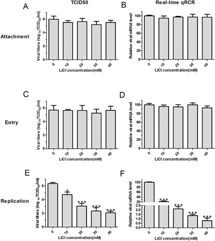
The assay of FMDV inhibition stage by TCID50 and Real‐time qPCR. Viral inhibition time course of LiCl against FMDV was determined by FMDV life cycle: attachment stage (A and B), entry stage (C and D), and replication stage (E and F). The viral yields of the cells were detected by TCID50 and RT‐qPCR in every stage
3.4. LiCl can affect FMDV replication stage
The viral replication assay was analyzed to determine whether LiCl affected the FMDV replication stage in BHK‐21 cells. The mean viral titers (TCID50) of mock‐treated and 10, 20, 30, and 40 mM drug‐treated cells were 6.33, 4.73, 3.06, 2.33, and 2.07, respectively (Fig. 2E); The mean relative mRNA yields of mock‐treated and 10, 20, 30, and 40 mM drug‐treated cells were 100.00, 21.33, 2.13, 1.37, and 0.8 (with mock‐treated cells set at 100), respectively (Fig. 2F). The results of viral yields have indicated that LiCl has antiviral effect on replication of FMDV.
3.5. LiCl affect FMDV replication at early stages
The replication time course assay was performed to determine which time course of FMDV replication was influenced by LiCl. The LiCl was added to the cells at a series of time interval (0‐2, 2‐4, 4‐6, 6‐8, 8‐10, 10‐12, 12‐14, 14‐16, 16‐18, and 18‐24 h). The mean viral titers (TCID50) of time interval 0‐2, 2‐4, 4‐6, 6‐8, 8‐10, 10‐12, 12‐14, 14‐16, 16‐18, and 18‐24 h with 40 mM drug‐treated cells were 5.88, 2.14, 2.12, 2.30, 2.46, 3.66, 4.78, 5.49, 5.55, 5.97, and 5.89, respectively (Fig. 3A); The mean relative mRNA yields of time interval 0‐2, 2‐4, 4‐6, 6‐8, 8‐10, 10‐12, 12‐14, 14‐16, 16‐18, and 18‐24 h with 40 mM drug‐treated cells were 100.00, 0.53, 0.70, 0.70, 0.80, 1.67, 11.67, 93.67, 99.33, 97.33, and 96.33 (with mock‐treated cells set at 100), respectively (Fig. 3B). The results of viral yields have indicated that LiCl has antiviral effect at early stages of FMDV replication.
Figure 3.

The assay of FMDV inhibition course time by TCID50 and Real‐time qPCR. The replication time course assay was performed to determine which time course of influenced of FMDV replication by LiCl. The LiCl was added to the cells at a series of time interval (0‐2, 2‐4, 4‐6, 6‐8, 8‐10, 10‐12, 12‐14, 14‐16, 16‐18, and 18‐24 h). The assay of FMDV inhibition course time by TCID50 (A) and Real‐time qPCR (B)
4. DISCUSSION
As previously reported, LiCl has antiviral effects agaist serval virus in vitro, such as IBV, an avian coronavirus, TGEV, herpes simplex virus and pseudorabies herpesvirus, EV‐A7, and PRRSV and so on,9, 10, 11, 12, 13, 14, 17, 18 which indicated that LiCl had potential to be an antiviral agent.
In this study, When the concentration of LiCl in the range of 40 mM, compared with mock‐treated cells, the cells showed no significant toxicity and no significant difference in cell morphology. Firstly, the LiCl can inhibit FMDV replication when FMDV infected BHK‐21 cells. Secondly, different viral cycle treat with LiCl to further investigate which viral cycle of FMDV infection was sensitive to LiCl. We have found that the LiCl had no effects on FMDV attachment and entry in cells, which indicated that the LiCl did not directly affect the connection between the virus and cell receptor or the passageway of viruses into cells. Nevertheless, LiCl had significantly inhibited FMDV replication stage in cells with dose‐dependent. Thirdly, to further investigate what time course of FMDV replication stage was affected by LiCl treatment, cells were exposed to LiCl at several points in the viral replication stage. We have found that LiCl reduced FMDV replication when treated within 12 hpi, suggesting that the early time of viral replication is the target for the antiviral effect of LiCl. The early time of viral replication stage is the time that viral RNA and viral protein were synthesized, which indicated that the LiCl has inhibited the viral RNA and viral protein synthesized.
LiCl is a often prescribed drug in the modern pharmacopoeia.19, 20, 21 It resulted in reductions in both viral protein synthesis and the yield of released viral progeny in EV‐A71‐infected cells with LiCl, but it only led to a decrease in progeny virus but had no effect on viral protein synthesis.22 As the treatment might be used for food animal, the food safety of using LiCl as a medicine is also important problem. In order to minimize the risk of LiCl toxicity, careful monitoring, and adjustment of LiCl dosage is especially important.
In conclusion, the FMDV replication stage was inhibited by LiCl with dose‐dependent. Besides, the LiCl target of the antiviral effect was the early phase of FMDV replication. FMDV causes an economically important and highly contagious disease of cloven‐hoofed animals. The use of FMDV vaccines to protect early infection is limited, and the FMDV mutates frequently to escape the immune system.3, 4, 23 The resulst reveals tha LiCl has potential as an effective anti‐FMDV drug. Therefore, LiCl may be an effective drug for the control of FMDV. Based on that, the mechanism of the antiviral effect of LiCl on FMDV infection is need to in‐depth research in vivo.
ACKNOWLEDGMENTS
This work was supported by grants from the Key Technologies R&D Program of Gansu Province of China (grant number 1604NKCA045‐1), and the National Pig Industrial System (CARS‐36‐06B), and the Open Fund of the Key Laboratory of Fujian Province livestock epidemic prevention and control and biological technology (2016KL03).
CONFLICTS OF INTEREST
None.
Zhao F‐R, Xie Y‐L, Liu Z‐Z, et al. Lithium chloride inhibits early stages of foot‐and‐mouth disease virus (FMDV) replication in vitro. J Med Virol. 2017;89: 2041–2046. 10.1002/jmv.24821
REFERENCES
- 1. Domingo E, Escarmís C, Baranowski E, et al. Evolution of foot‐and‐mouth disease virus. Virus Res. 2003; 91:47–63. [DOI] [PubMed] [Google Scholar]
- 2. Knowles NJ, Samuel AR. Molecular epidemiology of foot‐and‐mouth disease virus. Virus Res. 2003; 91:65–80. [DOI] [PubMed] [Google Scholar]
- 3. Pereira HG. 1981. Foot‐and‐Mouth Disease Animals. New York, NY: Academic Press, pp 333–363. [Google Scholar]
- 4. Golde WT, Pacheco JM, Duque H, et al. Vaccination against foot‐and‐mouth disease virus confers complete clinical protection in 7 days and partial protection in 4 days: use in emergency outbreak response. Vaccine. 2005; 23:5775–5782. [DOI] [PubMed] [Google Scholar]
- 5. Manji HK, Potter WZ, Lenox RH. Signal transduction pathways. Molecular targets for lithium's actions. Arch Gen Psychiatry. 1995; 52:531–543. [DOI] [PubMed] [Google Scholar]
- 6. Chen Y, Whetstone HC, Lin AC, et al. Beta‐catenin signaling plays a disparate role in different phases of fracture repair: implications for therapy to improve bone healing. PLoS Med. 2007; 31:e249. [DOI] [PMC free article] [PubMed] [Google Scholar]
- 7. Lucas KC, Hart DA, Becker RW. Porcine proximal tubular cells (LLC‐PK1) are able to tolerate high levels of lithium chloride in vitro: assessment of the influence of 1‐20 mM LiCl on cell death and alterations in cell biology and biochemistry. Cell Biol Int. 2010; 34:225–233. [DOI] [PubMed] [Google Scholar]
- 8. Muñoz‐Montaño JR, Moreno FJ, Avila J, Diaz‐Nido J. Lithium inhibits Alzheimer's disease‐like tau protein phosphorylation in neurons. FEBS Lett. 1997; 411:83–88. [DOI] [PubMed] [Google Scholar]
- 9. Harrison SM, Tarpey I, Rothwell L, Kaiser P, Hiscox JA. Lithium chloride inhibits the coronavirus infectious bronchitis virus in cell culture. Avian Pathol. 2007; 36:109–114. [DOI] [PMC free article] [PubMed] [Google Scholar]
- 10. Li J, Yin J, Sui X, Li G, Ren X. Comparative analysis of the effect of glycyrrhizin diammonium and lithium chloride on infectious bronchitis virus infection in vitro. Avian Pathol. 2009; 38:215–221. [DOI] [PubMed] [Google Scholar]
- 11. Ren X, Meng F, Yin J, et al. Action mechanisms of lithium chloride on cell infection by transmissible gastroenteritis coronavirus. PLoS ONE. 2011; 6:e18669. [DOI] [PMC free article] [PubMed] [Google Scholar]
- 12. Hung HC, Shih SR, Chang TY, Fang MY, Hsu JT. The combination effects of licl and the active leflunomide metabolite, A771726, on viral‐induced interleukin 6 production and EV‐A71 replication. PLoS ONE 2014; 9:e111331. [DOI] [PMC free article] [PubMed] [Google Scholar]
- 13. Cui J, Xie J, Gao M, et al. Inhibitory effects of lithium chloride on replication of type II porcine reproductive and respiratory syndrome virus in vitro. Antivir Ther. 2015; 20:565–572. [DOI] [PubMed] [Google Scholar]
- 14. Hao HP, Wen LB, Li JR, et al. LiCl inhibits PRRSV infection by enhancing Wnt/β‐catenin pathway and suppressing inflammatory responses. Antiviral Res. 2015; 117:99–109. [DOI] [PubMed] [Google Scholar]
- 15. Chen Y, Kong D, Cai G, et al. Novel antiviral effect of lithium chloride on mammalian orthoreoviruses in vitro. Microb Pathog. 2016; 93:152–257. [DOI] [PubMed] [Google Scholar]
- 16. Livak KJ, Schmittgen TD. Analysis of relative gene expression data using real‐time quantitative PCR and the 2(‐Delta Delta C(T)) Method. Methods. 2001; 25:402–408. [DOI] [PubMed] [Google Scholar]
- 17. Cernescu C, Popescu L, Constantinescu S, Cernescu S. Antiviral effect of lithium chloride. Virologie. 1988; 39:93–101. [PubMed] [Google Scholar]
- 18. Skinner GR, Hartley C, Buchan A, Harper L, Gallimore P. The effect of lithium chloride on the replication of herpes simplex virus. Med Microbiol Immunol. 1980; 168:139–148. [DOI] [PubMed] [Google Scholar]
- 19. D'Souza R, Rajji TK, Mulsant BH, Pollock BG. Use of lithium in the treatment of bipolar disorder in late‐life. Curr Psychiatry Rep. 2011; 13:488–492. [DOI] [PubMed] [Google Scholar]
- 20. Freland L, Beaulieu JM. Inhibition of GSK3 by lithium, from single molecules to signaling networks. Front Mol Neurosci. 2012; 5:14. [DOI] [PMC free article] [PubMed] [Google Scholar]
- 21. Hernandez F, Lucas JJ, Avila J. GSK3 and tau: two convergence points in Alzheimer's disease. J Alzheimers Dis. 2013; 33:S141–S144. [DOI] [PubMed] [Google Scholar]
- 22. Yuan J, Zhang J, Wong BW, et al. Inhibition of glycogen synthase kinase 3beta suppresses coxsackievirus‐induced cytopathic effect and apoptosis via stabilization of beta‐catenin. Cell Death Differ. 2005; 12:1097–1106. [DOI] [PubMed] [Google Scholar]
- 23. Mason PW, Grubman MJ, Baxt B. Molecular basis of pathogenesis of FMDV. Virus Res. 2003; 91:9–32. [DOI] [PubMed] [Google Scholar]