

Abstract
Sigmoid volvulus in children is a potentially disastrous situation, still remaining rare in terms of occurrence. We hereby present a case report of a 10-year-old male, having admitted in our department complaining about abdominal pain, who finally proved to suffer from sigmoid volvulus.
Key words: volvulus, torsion, sigmoid, children
Introduction
Torsion of the sigmoid colon in children still remains in low frequency levels, although with severe, life-threatening results. The occurrence is due to torsion around the mesenteric axis of an already dilated colon. It is mandatory mentioning that clinical signs and symptoms may be obscure and consecutive physical examination may prove difficult in terms of final diagnosis. Therefore, a high clinical suspicion gives high chances of uncomplicated outcome.
Case Report
History of present illness
A 10-year old male was referred to our department complaining about abdominal pain. The medical history starts about 12 hours ago, with an episode of vomiting just after his breakfast, following acute pain in the mesogastric area. The patient also noted some diarrheic stool of small amount after the vomiting.
Physical examination
On physical examination, the child appeared as a heavily sick patient, sweaty with weakness. His abdomen was flatulent in the two upper quadrants and totally flat and stiff in the two lower ones. Bowel sounds were absent. Digital rectal examination revealed an empty rectum. Laboratory exams in the emergency department revealed a leukocytosis with neutrophilic shift (WBC: 15400, Neut: 88%), while an erect abdominal X-Ray showed a hugely enlarged colic area, without free air in the peritoneal cavity (Figure 1). Emergency CT revealed a sigmoid torsion, with the largest diameter of the twisted sigmoid reaching 14.5 centimeters and the characteristic “whirlpool” sign (Figure 2).
The child was immediately introduced to the operating theater. An exploratory laparotomy was carried out, with a medial hypersubumbilical incision and access. The preoperative imaging of a sigmoid volvulus diagnosis was confirmed. The sigmoid colon was twice twisted in a clockwise direction, with no macroscopic signs of perforation (Figure 3). A counterclockwise detorsion was initially performed. Attempts of restoration of normal blood flow in the previously ischemic sigmoid colon with warm compresses had no signs of amelioration. A sigmoidectomy with a primary endto- end anastomosis of the descending colon with the upper rectum plus appendectomy were performed (Figure 4).
Having the operation ended, the child was transferred intubated to the pediatric intensive care unit. He had a normal postoperative course. He had a low inotropic support during the first postoperative day. He was extubated during the second postoperative day and total parenteral nutrition was also initiated. He had his first defecation during the second day postoperatively. He returned to our department during the third postoperative day under triple antibiotic chemoprophylaxis (cephalosporin, metronidazole, amikacin) with normal laboratory examination. He was discharged at 12th postoperative day. He also had an episode of acute abdominal pain about six months ago, which resolved spontaneously during his two-day hospitalization for observation.
Discussion
Sigmoid torsion in children’s group is a rather rare but potentially life-threatening condition. Therefore, high suspicion is needed in cases of acute abdominal pain with subsequent distension of the abdominal wall and episode of vomiting.1 The incidence of sigmoid volvulus is much common in South America, Africa and Asia, where there is high consumption of high-fiber diets.2 It is suggested that these kinds of diet contribute to sigmoid colon elongation, creating a predisposition for torsion.3 In elderly adults, it is referred as the third cause of colonic obstruction, after neoplasmatic disease and diverticulitis.4 In large bowel volvulus, highest frequency is noted in the sigmoid colon (60%-85%) and caecum as well as transverse colon follow.4 In pediatric age group, 7 years old is the median age of occurrence, ranging from 4 years to 18 years old. More frequent is the occurrence among boys, rather than girls (3.5/1).5
It is a common sense that, if left untreated, sigmoid volvulus probably leads to severe ischemic lesions, necrosis of the ischemic bowel, perforation and generalized peritonitis. First-line treatment remains the surgical one. The first step in terms of surgical treatment is the assessment of the twisted bowel. Especially when the sigmoid is volvulised, an attempt of endoscopic detorsion can be carried out. However, when the twisted bowel proves to have gangrenous lesions due to severe ischemia, endoscopic detorsion remains a contraindication, favoring the classic surgical exploration, with resection of the volvulised segment. 6 Regarding our own case, our male patient indeed appeared at the emergency department as heavily sick patient, indicating that the torsion episode had already been settled a couple of hours ago. Therefore, there was a high suspicion of an – at least – severely ischemic bowel (and perhaps perforated). This is the reason for overriding an endoscopic attempt for detorsion and only a rectal tube was placed. Moreover, the CT scan was selected as a primary diagnostic tool, because we had a cooperative (due to his age) patient and it is a cheap, non-interventional and quick examination, revealing properly the arterial and venous congestion of the detorsed bowel segment and a probable leading point for the volvulised segment, thus suggesting our operative plan.
Figure 1.
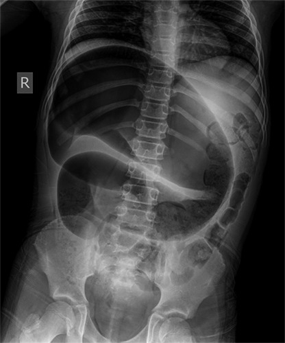
Erect abdominal X-Ray, showing a hugely enlarged colic area, without free air in the peritoneal cavity.
Figure 2.

Abdominal CT scan, revealing the characteristic “whirlpool” sign of the detorsed sigmoid colon.
Figure 3.

The exploratory laparotomy revealed a twice twisted sigmoid colon in a clockwise direction, with no macroscopic signs of perforation.
Figure 4.

A sigmoidectomy with a primary end-to-end anastomosis of the descending colon with the upper rectum plus appendectomy were performed.
Conclusions
Sigmoid volvulus in children remains a rare, but potentially disastrous situation, if left untreated. Therefore, a high index of suspicion must be maintained, in terms of observation, diagnosis and treatment of episodes of acute abdominal pain. An excellent prognosis is certain if a timely diagnosis is made.
Funding Statement
Funding: None.
References
- 1.Haider F, Al Asheeri N, Ayoub B, et al. Sigmoid volvulus in children: a case report. J Med Case Rep 2017;11:286. [DOI] [PMC free article] [PubMed] [Google Scholar]
- 2.Raveenthiran V, Madiba TE, Atamanalp SS, De U. Volvulus of the sigmoid colon. Colorectal Dis 2010;12:e1-17 [DOI] [PubMed] [Google Scholar]
- 3.Kapadia M. Volvulus of the small bowel and colon. Clin Colon Rectal Surg 2017;30:40-5 [DOI] [PMC free article] [PubMed] [Google Scholar]
- 4.Patel RV, Njere I, Campbell A, et al. Sigmoid volvulus in an adolescent girl: staged management with emergency colonoscopic reduction and decompression followed by elective sigmoid colectomy. BMJ Case Rep 2014. doi:10.1136/bcr-2014-206003 [DOI] [PMC free article] [PubMed] [Google Scholar]
- 5.Salas S, Angel CA, Salas N, et al. Sigmoid volvulus in children and adolescents. J Am Coll Surg 2000;190:722. [DOI] [PubMed] [Google Scholar]
- 6.Parolini F, Orizio P, Bulotta AL, et al. Endoscopic management of sigmoid volvulus in children. World J Gastrointest Endosc 2016;8:439-43. [DOI] [PMC free article] [PubMed] [Google Scholar]