Background:
Posterior elbow capsulotomy plus triceps lengthening facilitates passive elbow flexion in children with arthrogryposis multiplex congenita, allowing independent function for activities of daily living, such as feeding and self-care of the face and hair.
Description:
The posterior aspect of the distal end of the humerus and the olecranon are identified by palpation and exposed via a curvilinear incision over the posterior aspect of the elbow. Identifying the osseous landmarks can be challenging in some patients. The ulnar nerve is identified and protected. The triceps tendon is isolated, and z-lengthening is performed. Next, the posterior elbow capsule is incised proximal to the tip of the olecranon to expose the joint surface, and the arthrotomy is continued incrementally along the medial and lateral capsule until elbow flexion increases by ≥40°, or past 90° (maximum, 120°), with contact between the lengthened ends of the triceps tendon for repair. The triceps tendon is then repaired in the elongated position. After the wound is closed, the elbow is placed in flexion and immobilized in a cast.
Alternatives:
Alternative treatments include passive stretching exercises to increase elbow flexion.
Rationale:
Elbow extension contractures result in substantial limitations in the activities of daily living for children with arthrogryposis multiplex congenita. Those who fail to attain at least 90° of elbow flexion with passive stretching in the first year of life benefit from posterior elbow release and triceps lengthening. In addition, children with <30° of passive elbow flexion are at risk of developing valgus instability of the elbow from passive flexion exercises because the axis of rotation of the elbow is difficult to detect. Once passive elbow flexion is attained, such children may be candidates for tendon transfers allowing active elbow flexion.
Introductory Statement
Posterior elbow capsulotomy plus triceps lengthening allows passive elbow flexion in children with arthrogryposis multiplex congenita (AMC) and elbow extension contractures, facilitating independence and self-care, with a low rate of complications and contracture recurrence1.
Indications & Contraindications
Indications
Elbow extension contracture due to AMC with <90° of passive elbow flexion after the age of 1 year.
Inability to reach the hand to the mouth.
Preserved elbow joint space (lateral elbow. radiograph does not show radiohumeral synostosis).
An age of <3 years (relative indication).
Contraindications
More than 90° of passive elbow flexion.
Osseous deformity or heterotopic ossification limiting elbow flexion (on elbow radiographs).
Medical conditions or anesthetic risks precluding surgery.
Step-by-Step Description of Procedure (Video 1)
Step 1: Positioning and Marking
Position the patient and mark anatomic landmarks and the planned incision.
Place the patient on the operating table in the supine position for bilateral simultaneous posterior release or the lateral decubitus position for unilateral release.
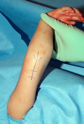
Palpate and mark the medial epicondyle, lateral epicondyle, and olecranon with a surgical marking pen; the medial epicondyle can be mistaken for the olecranon because of the internally rotated position of the shoulder, which places the ulnar nerve at risk (Fig. 1).
Prepare and drape the involved extremity circumferentially from the shoulder to the fingertips.
Place a well-padded sterile tourniquet as far proximal as possible on the involved arm.
Measure the elbow range of motion (Fig. 2-A).
Fig. 1.

The posterior longitudinal incision is marked over the humerus. The location of the olecranon is marked with a curved line.
Figs. 2-A and 2-B A patient with AMC who was managed with posterior elbow capsulotomy and triceps lengthening.
Fig. 2-A.

The preoperative maximum passive elbow flexion.
Fig. 2-B.

The postoperative maximum passive elbow flexion.
Video 1.
Critical steps of the posterior elbow capsulotomy and triceps lengthening, including preoperative range of motion, mobilizing and protecting the ulnar nerve with a vessel loop, marking and incising the step cut in the triceps tendon, making the posterior capsulotomy, and testing the range of motion after triceps lengthening and posterior capsulotomy and again after triceps tendon repair and skin closure.
Step 2: Incision
Use a posterior approach to the elbow.
Make a longitudinal incision along the posterior aspect of the humerus, curving radially around the olecranon.
Elevate full-thickness flaps of skin and subcutaneous tissue from the triceps fascia.
Identify, decompress, mobilize, and protect the ulnar nerve from the upper arm medial intermuscular septum to the deep fascia of the flexor carpi ulnaris in the proximal aspect of the forearm. Transpose the ulnar nerve anterior to the medial epicondyle if it tends to subluxate into the elbow joint during range of motion, after the posterior capsule is released.
The radial nerve does not routinely need to be identified and protected.
Step 3: Triceps Lengthening
Identify and mobilize the triceps tendon from the humerus and incise the triceps tendon near its insertion.
Identify and mobilize the triceps tendon from the humerus medially and laterally while preserving the insertion on the olecranon.
Incise the triceps tendon near its insertion in a step-cut or z-shaped fashion. Make the 2 resultant limbs as long as possible to allow for adequate lengthening. The actual length depends on the size of the child; usually the step cut is 2 to 4 cm long. Alternatively, a V-Y or W lengthening of the triceps tendon can be performed; however, in our experience, more length and a stronger repair, due to a larger contact area, can be obtained with a step-cut tenotomy.
Step 4: Posterior Elbow Capsulotomy
Identify the posterior elbow capsule deep to the triceps tendon and place an incision proximal to the tip of the olecranon to expose the joint surface.
Identify the posterior elbow capsule deep to the triceps tendon and incise it transversely just proximal to the tip of the olecranon to expose the joint surface.
Extend the arthrotomy medially and laterally as needed to achieve as much flexion as possible, preferably an increase of at least 40°, or past 90° (maximum, 120°), while maintaining contact between the limbs of the triceps tendon. Partial transection of the medial and lateral collateral ligaments may be necessary to achieve adequate flexion, but do not completely transect them.
Avoid forceful elbow flexion, which may result in a transphyseal fracture.
Avoid extreme elbow flexion as this may compromise vascular supply.
Reapproximate the limbs of the triceps tendon in the elongated position using multiple interrupted nonabsorbable sutures (e.g., 2-0 Ethibond; Ethicon). Determine the position in which the elbow is to be immobilized while visualizing the triceps tendon repair, measuring and recording in the operative report the maximum amount of flexion (<120°) that does not place tension on the triceps repair.
Step 5: Skin Closure
Close the skin with interrupted absorbable subcutaneous sutures and measure the maximum elbow flexion after closure.
Close the skin with interrupted absorbable subcutaneous sutures (e.g., 4-0 Vicryl; Ethicon) and running subcuticular absorbable suture (e.g., 4-0 Monocryl; Ethicon).
Measure the maximum elbow flexion after closure. (However, do not attempt to flex more than was measured at the end of Step 4, or beyond the point at which vascular supply is compromised to either the arm or the skin edges of the wound) (Fig. 2-B).
Step 6: Dressing
Apply a bulky dressing with the elbow in flexion and cover with a fiberglass cast.
Apply a bulky dressing with the elbow in the optimal position of flexion, as determined in Step 4, and cover with a fiberglass cast.
Step 7: Postoperative Therapy
Remove the long arm cast after 4 weeks and fabricate a static elbow splint, which is worn with the elbow in maximum flexion for an additional 4 weeks and is removed only for performing exercises and hygiene.
Leave the long arm cast in place for 4 weeks with the elbow in maximum flexion.
Remove the cast after 4 weeks and fabricate a static elbow splint in the position of maximum flexion. The child should wear the flexion splint full time for 4 more weeks except for range-of-motion exercises and hygiene. Start range-of-motion exercises including active and gentle passive elbow extension and flexion. Allow weight-bearing in the splint (e.g., using a platform walker, if indicated).
At 2 months postoperatively, discontinue daytime splinting. Fabricate a static elbow splint in the position of maximum extension. The child should wear flexion and extension elbow splints on alternate nights for 1 month. If the child develops an elbow flexion contracture, increase the frequency of wear of the extension splint. Continue the home exercise program (active and passive elbow flexion and extension), and add triceps strengthening if the child is old enough to cooperate with a strengthening program. Allow weight-bearing out of the splint.
At 3 months postoperatively, assess the elbow range of motion and need for a splint; continue the use of a nighttime flexion splint if the child does not yet have at least as much flexion as was attained intraoperatively, and/or an extension splint, if the child has developed a flexion contracture. It may be necessary to alternate flexion and extension splints to maintain the full arc of passive motion. Continue the home exercise program as noted above and add more vigorous elbow flexion stretching if indicated.
At 4, 6, and 12 months postoperatively and annually thereafter, reassess the range of motion and splinting needs and evaluate for changes in function, especially the ability of the child to reach the hand to the face. Continue the use of splinting and the home exercise program if indicated.
Results
Triceps lengthening for the treatment of elbow extension contracture in 29 elbows of 23 children with AMC successfully increased passive elbow flexion and the arc of elbow motion, allowing them to perform hand-to-mouth activities1.
Children with AMC frequently have reduced mobility because of lower-extremity involvement, and they may have limited spine motion. They should be evaluated preoperatively by a team that includes an experienced occupational therapist and a pediatric orthopaedist to make sure that the child is a good candidate for surgery, that the timing of surgery is coordinated to take the ambulatory status and the need for assistive devices into account, and that anesthetic exposure is reduced by performing upper and lower-extremity operations during the same anesthetic session, if indicated.
Posterior elbow capsulotomy and triceps lengthening can reliably improve passive elbow flexion to >90°. Passive flexion may enable independent self-feeding and self-care of the face and hair because the child will use the contralateral arm, a leg, or a tabletop to passively flex the elbow. This operation has become a well-accepted and standard technique to improve elbow flexibility, with improved range of motion lasting ≥5 years1. Elbow motion should be monitored throughout growth, as elbow flexion contracture may occur; this can usually be treated with nighttime splinting. Children with this condition are also frequently candidates for other operations to improve the position of the hand and upper extremity, including an index rotation flap2 to reduce a flexion-adduction contracture of the thumb, dorsal carpal wedge osteotomy3-5 to improve wrist position and potentially grasp, and humeral external rotation osteotomy6 for internal rotation contracture of the shoulder. Posterior capsulotomy and triceps lengthening can be performed during the same anesthetic session as a dorsal carpal wedge osteotomy and index dorsal rotation flaps because the rehabilitation for these 3 procedures is compatible; however, it should not be performed at the same time as external rotation humeral osteotomy as this combination of procedures results in worse outcomes6.
Once passive elbow flexion is achieved, muscle transfer to achieve active elbow flexion can be considered if expendable donor muscles are available (as discussed below). Improved passive flexion of the elbow alone often provides such substantial improvements in daily activities that an active elbow tendon transfer may not be necessary. Children with AMC often also have stiff, weak fingers, making 1-handed grasp difficult and reducing the benefit of active elbow flexion. They also frequently have hypoplasia or aplasia of potential donor muscles, precluding transfer. The triceps muscle is usually reasonably strong in children with AMC, and the long head of the triceps can be transferred to restore active elbow flexion7; however, triceps lengthening (while necessary to provide flexibility) may preclude subsequent long head of the triceps transfer because it weakens the muscle, rendering it a less effective donor. Other options for restoration of active flexion include the Steindler flexorplasty8, which requires a strong flexor-pronator mass, and latissimus dorsi transfer9. Pectoralis major transfer was previously used to gain active elbow flexion for children with AMC, but long-term results have been reported to be poor10.
Pitfalls & Challenges
It is easy to confuse the medial epicondyle for the olecranon because of the internally rotated position of the shoulder. Failure to differentiate the medial epicondyle and the olecranon prior to incision places the ulnar nerve at risk of iatrogenic injury. This is compounded by the fact that the nerve is typically hypoplastic. The best way to avoid this pitfall is to locate the olecranon by palpating the subcutaneous border of the ulna, which is contiguous with it.
Avoid forceful passive elbow flexion, which may result in a transphyseal fracture.
It is challenging to maintain elbow flexion gains while recovering triceps strength, and children may develop elbow flexion contractures after this procedure. These concerns are the rationales for the therapy program detailed in Step 7.
Published outcomes of this procedure can be found at: J Bone Joint Surg Am. 2008 Jul;90(7):1517-23.
Investigation performed at Shriners Hospital for Children-Northern California, Sacramento, California
Disclosure: The authors indicated that no external funding was received for any aspect of this work. On the Disclosure of Potential Conflicts of Interest forms, which are provided with the online version of the article, one or more of the authors checked “yes” to indicate that the author had a relevant financial relationship in the biomedical arena outside the submitted work (http://links.lww.com/JBJSEST/A287).
References
- 1.Van Heest A, James MA, Lewica A, Anderson KA. Posterior elbow capsulotomy with triceps lengthening for treatment of elbow extension contracture in children with arthrogryposis. J Bone Joint Surg Am. 2008. July;90(7):1517-23. [DOI] [PubMed] [Google Scholar]
- 2.Ezaki M, Oishi SN. Index rotation flap for palmar thumb release in arthrogryposis. Tech Hand Up Extrem Surg. 2010. March;14(1):38-40. [DOI] [PubMed] [Google Scholar]
- 3.Ezaki M, Carter PR. Carpal wedge osteotomy for the arthrogrypotic wrist. Tech Hand Up Extrem Surg. 2004. December;8(4):224-8. [DOI] [PubMed] [Google Scholar]
- 4.Van Heest AE, Rodriguez R. Dorsal carpal wedge osteotomy in the arthrogrypotic wrist. J Hand Surg Am. 2013. February;38(2):265-70. Epub 2012 Dec 23. [DOI] [PubMed] [Google Scholar]
- 5.Foy CA, Mills J, Wheeler L, Ezaki M, Oishi SN. Long-term outcome following carpal wedge osteotomy in the arthrogrypotic patient. J Bone Joint Surg Am. 2013. October 16;95(20):e150. [DOI] [PubMed] [Google Scholar]
- 6.Zlotolow DA, Kozin SH. Posterior elbow release and humeral osteotomy for patients with arthrogryposis. J Hand Surg Am. 2012. May;37(5):1078-82. Epub 2012 Apr 5. [DOI] [PubMed] [Google Scholar]
- 7.Gogola GR, Ezaki M, Oishi SN, Gharbaoui I, Bennett JB. Long head of the triceps muscle transfer for active elbow flexion in arthrogryposis. Tech Hand Up Extrem Surg. 2010. June;14(2):121-4. [DOI] [PubMed] [Google Scholar]
- 8.Goldfarb CA, Burke MS, Strecker WB, Manske PR. The Steindler flexorplasty for the arthrogrypotic elbow. J Hand Surg Am. 2004. May;29(3):462-9. [DOI] [PubMed] [Google Scholar]
- 9.Zargarbashi R, Nabian MH, Werthel JD, Valenti P. Is bipolar latissimus dorsi transfer a reliable option to restore elbow flexion in children with arthrogryposis? A review of 13 tendon transfers. J Shoulder Elbow Surg. 2017. November;26(11):2004-9. Epub 2017 Jul 6. [DOI] [PubMed] [Google Scholar]
- 10.Lahoti O, Bell MJ. Transfer of pectoralis major in arthrogryposis to restore elbow flexion: deteriorating results in the long term. J Bone Joint Surg Br. 2005. June;87(6):858-60. [DOI] [PubMed] [Google Scholar]


