Love at first site: The endoplasmic reticulum and Flavivirus replication
The endoplasmic reticulum (ER) is a continuous intracellular membrane system that composes the nuclear envelope and radiates out to the peripheries of the cell, transitioning from ribosome-studded, flat cisternal sheets to highly curved reticulated tubules. It has been implicated in a diverse array of cellular functions, including production, trafficking, and degradation of proteins; synthesis and distribution of lipids and steroids; calcium sequestration and release; cell signaling and innate immunity; carbohydrate metabolism; and detoxification of harmful substances [1]. Such a ubiquitous and functionally versatile organelle is parasitized by viruses in order to facilitate their life cycle. Rotavirus, vaccinia virus (VV), hepatitis C virus (HCV), as well as members of the Flavivirus genus—dengue virus (DENV), West Nile virus(WNV), yellow fever virus (YFV), and Zika virus (ZIKV)—are all intimately associated with the ER [2]. The flaviviruses replicate on the ER membranes and form immature viral particles that bud into the ER lumen. These particles are then trafficked through the ER–Golgi network and undergo a maturation process brought about by pH alteration and cleavage by the host-protease Furin. This viral takeover of the ER requires the co-opting of cellular proteins as well as the active remodeling of the ER membrane to create a cellular environment more conducive to replication [3]. Several ER proteins have been identified as host factors in flavivirus replication [4], but a class of resident proteins that shape and maintain the dynamic ER architecture are of particular interest. These “ER-shaping” proteins could potentially serve as host factors that support viral replication or restriction factors that protect the ER. Interactome and CRISPR screens have produced somewhat discordant results, but the less stringent screens suggest that ER-shaping proteins may be targets of viral proteins [5] [6] [7]. This Pearl review will explore the virus-induced ER-membrane rearrangement and the relationship between ER-shaping proteins and the flavivirus life cycle.
Virulent and manipulative passions: Virus-induced manipulation to the ER structure
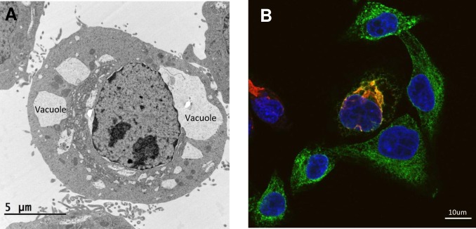
Virus-induced morphological changes to the ER membrane are brought about by active restructuring facilitated by interactions between viral proteins and host factors. The functional benefits of membrane alterations include the spatial compartmentalization of the viral-replication machinery—increasing the accessibility and concentration of necessary host factors—protection from the innate immune response, and the possibility of increased viral spread [3] [8]. The characteristics of the replication-supporting ER-membrane structures differ between viruses. HCV forms a “membranous web” and double-membrane vesicles, and VV manipulates the ER into wrapping around the cytosolic site of viral-DNA replication [9] [10]. Flaviviruses form invaginations in the ER referred to as vesicle packets (VPs), which are clusters of vesicle membranes housing the viral-replication machinery [3] [11] [12] [13] [14]. Immature flavivirus virions form paracrystalline arrays within the ER lumen [11]. In some cell types, flaviviruses also form convoluted membranes (CMs), which are potential sites for the translation and processing of viral proteins [3] [11] [12]. Interestingly, ZIKV infection in certain cell types produces large ER-derived vacuoles, which are followed by an implosive cell death (Fig 1A) [8]. Several Flavivirus proteins and host factors (i.e., reticulophagy factor FAM134B [15]) have been implicated in the ER-modification process. Of recent interest are ER-shaping proteins, that can be operationally subdivided into proteins that help form the structure and curvature of the ER and fusogens that maintain the reticulated ER network.
Fig 1. Microscopy images of ZIKV-infected HeLa Cells 24 hours after infection.
(A) Electron micrograph of ER-derived vacuoles in a ZIKV-infected cell. (B) Confocal microscopy image showing the colocalization (yellow/orange) of ER-shaping protein ATL3 (green) with the ZIKV protein NS3 (red). ATL3, Atlastin-3; ER, endoplasmic reticulum; NS3, nonstructural protein 3; ZIKV, Zika virus.
Curvy and sturdy: ER curvature-stabilizing proteins and virus interactions
The class of ER-shaping proteins labeled as curvature-stabilizing proteins are subdivided into two families, the receptor expression-enhancing protein (REEP)/DP1/yop1p and the reticulons [1]. The REEPs are a family of six proteins that interact with microtubules through an extended C-terminal cytoplasmic domain. By facilitating the association of the ER membrane to cytoskeletal dynamics, they provide the mechanics necessary to extend ER tubules, as well as to form the positive curvature of the ER membrane [16] [1]. Mutations in the REEP1 proteins are associated with hereditary spastic paraplegias (HSP), a family of inherited neurological disorders characterized by spastic weakness in the extremities, which is partly reminiscent of symptoms induced by neurotropic flaviviruses [1] [16]. The effect of the REEP family on flavivirus replication has not been thoroughly explored, with one investigation reporting that the depletion of REEP1 does not affect DENV replication [17].
The reticulon family consists of four (Reticulon 1 [RTN1] to RTN4) membrane-bound proteins that contain hydrophobic domains occupying the outer leaflet of the ER membrane. The resultant hydrophobic wedging, and possibly the scaffolding and protein–protein crowding formed by reticulon oligomers, contributes to the curvature of the ER membrane [1]. Several studies have identified reticulons as host factors in viral replication; Enterovirus 71 protein 2C and brome mosaic virus protein 1a both interact with RTNs to facilitate replication [18] [19]. Contrarily, RTN3 acts as a restriction factor in HCV replication by preventing nonstructural protein 4A (NS4A) self-interaction [20].
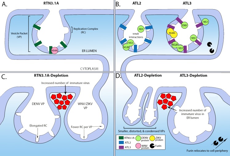
DENV, WNV, and ZIKV recruit RTN3.1A to the viral-replication site [21]. RTN3.1A depletion reduces viral titers and decreases viral RNA and protein levels [21]. The RTN3.1A colocalizes with the NS4A proteins of DENV, ZIKV, and WNV, but it only directly interacts with WNV NS4A via its N-terminal transmembrane domain (Fig 2A) [21]. The expression of WNV and DENV NS4A alone is sufficient to induce ER rearrangements [22] [23]. Depletion of RTN3.1A differentially alters the formation of the ER-derived replication organelles between flaviviruses. In ZIKV-infected and WNV-infected cells, there is a noticeable decrease in the amount of vesicle membranes present per VP (Fig 2C) [21]. While amount of vesicle membranes per VP was not altered in DENV-infected cells, their morphology became more elongated (Fig 2C), and there was an increase in immature viral particles upon RTN3.1A depletion (Fig 2C) [21]. Overall, while RTN3.1A may differently influence the life cycle of specific flaviviruses, it interacts with viral proteins and induces morphological changes to the ER in order to promote replication.
Fig 2. The relationship between ER-shaping proteins and flaviviruses during infection.
A simplified schematic showing the interaction between different flavivirus proteins and ER-shaping proteins: (A) RTN3.1A and (B) ATL2 (left) and ATL3 (right). (C) Depleting RTN3.1 results in elongated RCs in DENV-infected cells and fewer RCs per VP in WNV-infected and ZIKV-infected cells. (D) Depleting ATL2 (left) results in VPs that are smaller, distorted, and condensed but does not change the overall number of VPs, whereas ATL3 depletion results in the host-protease Furin relocating from the perinuclear region to the cell periphery and accumulation of immature virus particles in the ER lumen. Immature virus particles are represented smaller than they would be in relation to VPs. ATL2, Atlastin-2; ATL3, Atlastin-3; DENV, dengue virus; ENV, envelope; ER, endoplasmic reticulum; NS1, nonstructural protein 1; RC, replication complex; RTN3.1A, Reticulon 3.1A; VP, vesicle packet; WNV, West Nile virus; ZIKV, Zika virus.
Together at-last-in each other’s embrace: Virus interaction with the atlastin fusogens
The atlastins (ATLs) are a family of three membrane-bound, dynamin-related guanosine triphosphate (GTP)ases that mediate the construction of the reticulated ER network. The hydrolysis of GTP dissociates cis-membrane dimers and supports the tethering and dimerization with ATLs situated on a different ER membrane [24]. The homotypic fusion of ER membranes results in the formation of three-way junctions, shaping the smooth ER into an intricate and dynamic web of tubules. The depletion of ATL reduces the density of the ER network [25], and mutations in ATLs are also associated with HSP. In addition to their role as an ER-shaping protein, ATLs also influence protein targeting to the inner nuclear membrane and the biogenesis of nuclear pore complexes, regulate lipid droplet size, and facilitate selective autophagy [26] [27] [28]. Thus, ATLs could potentially be expedient host factors for several pathogens. A previous study implicated ATL3 in remodeling the ER to promote the formation of vacuoles that facilitate the intracellular replication of Legionella pneumophila [29].
Two recent investigations examined the relationship between the three ATLs and different flaviviruses and found varying effects based on the virus [17] [30]. Silencing ATL2 resulted in a significant reduction of DENV, WNV, and ZIKV titers and viral RNA [17]. ATL2 depletion affected formation of ER-derived DENV-replication organelles, distorting the size and shape of the vesicles and condensing them into a small perinuclear region (Fig 2D) [17]. The depletion of ATL3 reduced the titers of ZIKV and DENV (but not WNV) and had no effect on viral-RNA levels [17] [30]. ATL3 depletion did not change the morphology of the DENV-induced vesicles but resulted in an increased accumulation of intracellular viral particles in the ER lumen (Fig 2D) [17]. The GTPase function of ATLs is important for DENV and ZIKV replication [30] [17].
The different ATL proteins exhibit variation in their interaction with flavivirus proteins. ATL3 relocates to the ZIKV-replication site and directly interacts with nonstructural protein 3 (NS3; Fig 1B) and NS2B3, that belong to the viral-replication complex (Fig 2B) [30]. Both ATL2 and ATL3 interact with DENV NS3, NS5, and NS2B proteins, though interactions with ATL2 are relatively weak (Fig 2B) [17]. DENV NS1 protein and the envelope and capsid proteins also interact with ATL3 (Fig 2B) [17]. ATL3 is recruited to DENV-replication organelles and is enriched in the membrane-surrounding virions, but it is not necessarily associated with the site of viral-RNA replication [17]. In DENV-infected cells, ATL3 interacts with ADP-ribosylation factor 4 (ARF4), a protein that is associated with trafficking of proteins and vesicle processing [17]. Depletion of AFR4 and the related ARF5 protein impairs DENV assembly and release. The depletion of ATL3 results in the relocalization of Furin from the perinuclear region to the cell periphery (Fig 2D), suggesting that ATL3 may play a role in providing immature viral particles access to Furin (Fig 2D) in addition to a potential direct role on assembly and trafficking of viral particles [17]. Overall, these studies show that ATL3 is associated with the cytoplasmic transport of vesicles and is intimately involved with flavivirus assembly and maturation [17].
Happily, ever after: How the future could shape out
ER-shaping proteins impact the life cycle of flaviviruses by interacting with viral proteins, influencing viral assembly and maturation and promoting the formation of ER-derived factories. Future investigations could examine the role of other ER-shaping proteins including Spastin, Lunapark, and other REEP proteins that are involved in the regulation of ER shape through microtubule dynamics, stabilization of three-way ER tubular junctions, and formation and stabilization of the ER curvature, respectively. It will be worth investigating if ER-shaping proteins are involved in ZIKV-induced cytoskeleton modifications [12] and the formation of vacuoles to understand if these proteins are related to ER stress, innate sensing of infection, and viral replication [30] [8] [29]. The interplay between different ER-shaping proteins [31] within the context of viral infection is also of potential interest. Finally, since mutations in several of the ER-shaping proteins are associated with inherited neurological complications, it would be interesting to determine if they are implicated in the pathology induced by neurotropic flaviviruses in relevant cell and animal models [32].
Acknowledgments
The authors would like to thank the UtechS Photonic BioImaging (Imagopole), C2RT, Institut Pasteur (supported by the French National Research Agency; France BioImaging; ANR-10–INSB–04; Investments for the Future) for confocal imaging and Philippe Roingeard’s team (Université de Tours) for generating the electron microscope image. The authors also thank Beatrice de Cougny for her graphic design assistance and Ludivine Grzelak for her artistic insights.
Funding Statement
M.M.R is supported by the Pasteur-Paris University (PPU) International Doctoral Program. Work in the O.S. lab is funded by Institut Pasteur, ANRS, Sidaction, the Vaccine Research Institute (ANR-10-LABX-77), Labex IBEID (ANR-10-LABX-62-IBEID), TIMTAMDEN ANR-14-CE14-0029, CHIKV-Viro-Immuno ANR-14-CE14-0015-01, and the Gilead HIV cure program. B.M. is supported by the Pasteur-Roux/Pasteur-Cantarini fellowship from Institut Pasteur (Paris). The funders had no role in study design, data collection and analysis, decision to publish, or preparation of the manuscript.
References
- 1.Goyal U, Blackstone C. Untangling the web: mechanisms underlying ER network formation. Biochim Biophys Acta. 2013;1833(11):2492–8. Epub 2013/04/23. 10.1016/j.bbamcr.2013.04.009 [DOI] [PMC free article] [PubMed] [Google Scholar]
- 2.Inoue T, Tsai B. How viruses use the endoplasmic reticulum for entry, replication, and assembly. Cold Spring Harb Perspect Biol. 2013;5(1):a013250 Epub 2013/01/04. 10.1101/cshperspect.a013250 [DOI] [PMC free article] [PubMed] [Google Scholar]
- 3.Chatel-Chaix L, Bartenschlager R. Dengue virus- and hepatitis C virus-induced replication and assembly compartments: the enemy inside—caught in the web. J Virol. 2014;88(11):5907–11. Epub 2014/03/14. 10.1128/JVI.03404-13 [DOI] [PMC free article] [PubMed] [Google Scholar]
- 4.Rothan HA, Kumar M. Role of Endoplasmic Reticulum-Associated Proteins in Flavivirus Replication and Assembly Complexes. Pathogens. 2019;8(3). Epub 2019/09/25. 10.3390/pathogens8030148 [DOI] [PMC free article] [PubMed] [Google Scholar]
- 5.Zhang R, Miner JJ, Gorman MJ, Rausch K, Ramage H, White JP, et al. A CRISPR screen defines a signal peptide processing pathway required by flaviviruses. Nature. 2016;535(7610):164–8. Epub 2016/07/08. 10.1038/nature18625 [DOI] [PMC free article] [PubMed] [Google Scholar]
- 6.Marceau CD, Puschnik AS, Majzoub K, Ooi YS, Brewer SM, Fuchs G, et al. Genetic dissection of Flaviviridae host factors through genome-scale CRISPR screens. Nature. 2016;535(7610):159–63. Epub 2016/07/08. 10.1038/nature18631 [DOI] [PMC free article] [PubMed] [Google Scholar]
- 7.Coyaud E, Ranadheera C, Cheng D, Goncalves J, Dyakov BJA, Laurent EMN, et al. Global Interactomics Uncovers Extensive Organellar Targeting by Zika Virus. Mol Cell Proteomics. 2018;17(11):2242–55. Epub 2018/07/25. 10.1074/mcp.TIR118.000800 [DOI] [PMC free article] [PubMed] [Google Scholar]
- 8.Monel B, Compton AA, Bruel T, Amraoui S, Burlaud-Gaillard J, Roy N, et al. Zika virus induces massive cytoplasmic vacuolization and paraptosis-like death in infected cells. EMBO J. 2017;36(12):1653–68. 10.15252/embj.201695597 [DOI] [PMC free article] [PubMed] [Google Scholar]
- 9.Romero-Brey I, Merz A, Chiramel A, Lee JY, Chlanda P, Haselman U, et al. Three-dimensional architecture and biogenesis of membrane structures associated with hepatitis C virus replication. PLoS Pathog. 2012;8(12):e1003056 Epub 2012/12/14. 10.1371/journal.ppat.1003056 [DOI] [PMC free article] [PubMed] [Google Scholar]
- 10.Tolonen N, Doglio L, Schleich S, Krijnse Locker J. Vaccinia virus DNA replication occurs in endoplasmic reticulum-enclosed cytoplasmic mini-nuclei. Mol Biol Cell. 2001;12(7):2031–46. Epub 2001/07/14. 10.1091/mbc.12.7.2031 [DOI] [PMC free article] [PubMed] [Google Scholar]
- 11.Welsch S, Miller S, Romero-Brey I, Merz A, Bleck CK, Walther P, et al. Composition and three-dimensional architecture of the dengue virus replication and assembly sites. Cell Host Microbe. 2009;5(4):365–75. 10.1016/j.chom.2009.03.007 . [DOI] [PMC free article] [PubMed] [Google Scholar]
- 12.Cortese M, Goellner S, Acosta EG, Neufeldt CJ, Oleksiuk O, Lampe M, et al. Ultrastructural Characterization of Zika Virus Replication Factories. Cell Rep. 2017;18(9):2113–23. Epub 2017/03/02. 10.1016/j.celrep.2017.02.014 [DOI] [PMC free article] [PubMed] [Google Scholar]
- 13.Miorin L, Romero-Brey I, Maiuri P, Hoppe S, Krijnse-Locker J, Bartenschlager R, et al. Three-dimensional architecture of tick-borne encephalitis virus replication sites and trafficking of the replicated RNA. J Virol. 2013;87(11):6469–81. Epub 2013/04/05. 10.1128/JVI.03456-12 [DOI] [PMC free article] [PubMed] [Google Scholar]
- 14.Gillespie LK, Hoenen A, Morgan G, Mackenzie JM. The endoplasmic reticulum provides the membrane platform for biogenesis of the flavivirus replication complex. J Virol. 2010;84(20):10438–47. Epub 2010/08/06. 10.1128/JVI.00986-10 [DOI] [PMC free article] [PubMed] [Google Scholar]
- 15.Lennemann NJ, Coyne CB. Dengue and Zika viruses subvert reticulophagy by NS2B3-mediated cleavage of FAM134B. Autophagy. 2017;13(2):322–32. Epub 2017/01/20. 10.1080/15548627.2016.1265192 [DOI] [PMC free article] [PubMed] [Google Scholar]
- 16.Park SH, Zhu PP, Parker RL, Blackstone C. Hereditary spastic paraplegia proteins REEP1, spastin, and atlastin-1 coordinate microtubule interactions with the tubular ER network. J Clin Invest. 2010;120(4):1097–110. 10.1172/JCI40979 [DOI] [PMC free article] [PubMed] [Google Scholar]
- 17.Neufeldt CJ, Cortese M, Scaturro P, Cerikan B, Wideman JG, Tabata K, et al. ER-shaping atlastin proteins act as central hubs to promote flavivirus replication and virion assembly. Nat Microbiol. 2019. Epub 2019/10/23. 10.1038/s41564-019-0586-3 . [DOI] [PMC free article] [PubMed] [Google Scholar]
- 18.Tang WF, Yang SY, Wu BW, Jheng JR, Chen YL, Shih CH, et al. Reticulon 3 binds the 2C protein of enterovirus 71 and is required for viral replication. J Biol Chem. 2007;282(8):5888–98. Epub 2006/12/22. 10.1074/jbc.M611145200 . [DOI] [PubMed] [Google Scholar]
- 19.Diaz A, Wang X, Ahlquist P. Membrane-shaping host reticulon proteins play crucial roles in viral RNA replication compartment formation and function. Proc Natl Acad Sci U S A. 2010;107(37):16291–6. Epub 2010/09/02. 10.1073/pnas.1011105107 [DOI] [PMC free article] [PubMed] [Google Scholar]
- 20.Wu MJ, Ke PY, Hsu JT, Yeh CT, Horng JT. Reticulon 3 interacts with NS4B of the hepatitis C virus and negatively regulates viral replication by disrupting NS4B self-interaction. Cell Microbiol. 2014;16(11):1603–18. Epub 2014/06/06. 10.1111/cmi.12318 . [DOI] [PubMed] [Google Scholar]
- 21.Aktepe TE, Liebscher S, Prier JE, Simmons CP, Mackenzie JM. The Host Protein Reticulon 3.1A Is Utilized by Flaviviruses to Facilitate Membrane Remodelling. Cell Rep. 2017;21(6):1639–54. Epub 2017/11/09. 10.1016/j.celrep.2017.10.055 . [DOI] [PubMed] [Google Scholar]
- 22.Roosendaal J, Westaway EG, Khromykh A, Mackenzie JM. Regulated cleavages at the West Nile virus NS4A-2K-NS4B junctions play a major role in rearranging cytoplasmic membranes and Golgi trafficking of the NS4A protein. J Virol. 2006;80(9):4623–32. Epub 2006/04/14. 10.1128/JVI.80.9.4623-4632.2006 [DOI] [PMC free article] [PubMed] [Google Scholar]
- 23.Miller S, Sparacio S, Bartenschlager R. Subcellular localization and membrane topology of the Dengue virus type 2 Non-structural protein 4B. J Biol Chem. 2006;281(13):8854–63. Epub 2006/01/27. 10.1074/jbc.M512697200 . [DOI] [PubMed] [Google Scholar]
- 24.Liu TY, Bian X, Romano FB, Shemesh T, Rapoport TA, Hu J. Cis and trans interactions between atlastin molecules during membrane fusion. Proc Natl Acad Sci U S A. 2015;112(15):E1851–60. Epub 2015/04/01. 10.1073/pnas.1504368112 [DOI] [PMC free article] [PubMed] [Google Scholar]
- 25.Zhao G, Zhu PP, Renvoise B, Maldonado-Baez L, Park SH, Blackstone C. Mammalian knock out cells reveal prominent roles for atlastin GTPases in ER network morphology. Exp Cell Res. 2016;349(1):32–44. Epub 2016/09/28. 10.1016/j.yexcr.2016.09.015 . [DOI] [PubMed] [Google Scholar]
- 26.Liang JR, Lingeman E, Ahmed S, Corn JE. Atlastins remodel the endoplasmic reticulum for selective autophagy. J Cell Biol. 2018;217(10):3354–67. Epub 2018/08/26. 10.1083/jcb.201804185 [DOI] [PMC free article] [PubMed] [Google Scholar]
- 27.Klemm RW, Norton JP, Cole RA, Li CS, Park SH, Crane MM, et al. A conserved role for atlastin GTPases in regulating lipid droplet size. Cell Rep. 2013;3(5):1465–75. Epub 2013/05/21. 10.1016/j.celrep.2013.04.015 [DOI] [PMC free article] [PubMed] [Google Scholar]
- 28.Pawar S, Ungricht R, Tiefenboeck P, Leroux JC, Kutay U. Efficient protein targeting to the inner nuclear membrane requires Atlastin-dependent maintenance of ER topology. Elife. 2017;6 Epub 2017/08/23. 10.7554/eLife.28202 [DOI] [PMC free article] [PubMed] [Google Scholar]
- 29.Steiner B, Swart AL, Welin A, Weber S, Personnic N, Kaech A, et al. ER remodeling by the large GTPase atlastin promotes vacuolar growth of Legionella pneumophila. EMBO Rep. 2017;18(10):1817–36. 10.15252/embr.201743903 [DOI] [PMC free article] [PubMed] [Google Scholar]
- 30.Monel B, Rajah MM, Hafirassou ML, Sid-Ahmed S, Burlaud-Gaillard J, Zhu PP, et al. The Atlastin ER-shaping proteins facilitate Zika virus replication. J Virol. 2019. Epub 2019/09/20. 10.1128/JVI.01047-19 [DOI] [PMC free article] [PubMed] [Google Scholar]
- 31.Espadas J, Pendin D, Bocanegra R, Escalada A, Misticoni G, Trevisan T, et al. Dynamic constriction and fission of endoplasmic reticulum membranes by reticulon. Nat Commun. 2019;10(1):5327 Epub 2019/11/24. 10.1038/s41467-019-13327-7 [DOI] [PMC free article] [PubMed] [Google Scholar]
- 32.Zhao J, Hedera P. Hereditary spastic paraplegia-causing mutations in atlastin-1 interfere with BMPRII trafficking. Mol Cell Neurosci. 2013;52:87–96. 10.1016/j.mcn.2012.10.005 [DOI] [PMC free article] [PubMed] [Google Scholar]


