PLOS One logoLink to PLOS One
. 2020 Apr 16;15(4):e0231376. doi: 10.1371/journal.pone.0231376

Exploring differentially expressed genes between anagen and telogen secondary hair follicle stem cells from the Cashmere goat (Capra hircus) by RNA-Seq

Nimantana He 1,2, Rui Su 1,3,4,5, Zhiying Wang 1,5, Yanjun Zhang 1,5, Jinquan Li 1,3,4,5,*
Editor: Irina Polejaeva6
PMCID: PMC7162518  PMID: 32298297

Abstract

Hair follicle stem cells (HFSCs) have been shown to be essential in the development and regeneration of hair follicles (HFs). The Inner Mongolia Cashmere goat (Capra hircus) has two types of HFs, primary and secondary, with cashmere being produced from the secondary hair follicle. To identify the genes associated with cashmere growth, transcriptome profiling of anagen and telogen secondary HFSCs was performed by RNA-Seq. The RNA-Seq analysis generated over 58 million clean reads from each group, with 2717 differentially expressed genes (DEGs) detected between anagen and telogen, including 1500 upregulated and 1217 downregulated DEGs. A large number of DEGs were predominantly associated with cell part, cellular process, binding, biological regulation and organelle. In addition, the PI3K-Akt, MAPK, Ras and Rap1 signaling pathways may be involved in the growth of HFSCs cultured in vitro. The RNA-Seq results showed that the well-defined HFSC signature genes and cell cycle-associated genes showed no significant differences between anagen and telogen HFSCs, indicating a relatively quiescent cellular state of the HFSCs cultured in vitro. These results are useful for future studies of complex molecular mechanisms of hair follicle cycling in cashmere goats.

Introduction

Hair follicles, as skin appendages, play crucial role in skin homeostasis, and are also important in thermal regulation, social communication and sensory processes. In the 1980s, the location and characteristics of hair follicle stem cells were first identified using isotope labeling in mice [1], and subsequent studies have suggested that HFSCs play crucial roles in HF morphogenesis and cycling [2,3]. The results of intensive research in stem cell biology suggest that HFSCs are an excellent cell lineage to study stem cell plasticity [4], since most adult stem cells lose their self-renewal capacity and irreversibly differentiate, whereas hair follicles undergo continuous cycling throughout the lifetime. The morphogenesis and regeneration of hair follicle is a well-organized, spatiotemporal mesenchymal-epithelial interaction that requires complex signal transmission between the dermis and epidermis [5,6]. Hair follicle morphogenesis can be divided into three phases: induction, organogenesis and cytodifferentiation. During induction, Wnt mediated signal transduction first arises in mesenchymal cells directing the thickening of overlying epithelial cells to form a placode. The organogenesis stage consists of complex interplay of signals between the epidermis and the underlying dermis; epithelial placode cells direct the underlying dermal cells to proliferate and form a dermal condensate, which in turn signals the epithelial cells to proliferate and grow downwards into the dermis. In cytodifferentiation, the dermal condensate is enveloped with follicular epithelial cells thus forming distinct dermal papilla, which instruct the ectoderm to shape the entire HF through the action of morphogens and growth factors [7,8]. Based on the morphological characteristics, the hair cycle is divided into three major stages, including anagen (growth phase), catagen (regression phase) and telogen (resting phase) [9,10]. During anagen, bulge stem cell progeny populates the lower outer root sheath (ORS) towards the bulb matrix, constituting a specialized population of highly proliferative transit-amplifying (TA) cells and produce the hair shaft and the inner root sheath (IRS). Hair growth stops when matrix cells exhaust their proliferative capacity and transited into catagen phase. The apoptosis of matrix cells leads to the destruction of the lower two-thirds of the follicle, including the IRS, ORS, and hair bulb, whereas the bulge region remains intact. Catagen is followed by telogen phase. HFs remain quiescent, with the dermal papilla remaining in close proximity to the bulge throughout the telogen phase. When hair growth activating signals are generated, HFSCs are activated and initiate a new hair cycle [1113].

In case of skin injury, bulge HFSCs become active and rapidly adopt differentiation programs that differ from normal homeostasis and involve the upward migration of HFSCs to repair damaged epidermis in vivo. These cells have been speculated to be tightly regulated, as they cease activity once the wound is healed [14, 15].

The Inner Mongolia Cashmere goat has two types of HFs that have a similar structure but differ in fineness and produce two distinct fibers, guard hairs produced by primary HFs and soft down produced by secondary HFs (Fig 1). Cashmere is produced from secondary HFs. The secondary hair follicles which exhibit a notable photoperiod-based cycle that changes throughout the year, are suitable for hair cycle research. Histological studies on secondary hair follicles from Inner Mongolia Cashmere goats have shown that anagen lasts from May to December, catagen from December to January, and telogen until the end of April [16]. To further investigate the molecular mechanism of hair follicle cycling, it is important to perform gene expression profiling of secondary hair follicle stem cells.

Fig 1. Culture of Inner Mongolia Cashmere goat secondary HFSCs.

Fig 1

(a): Inner Mongolia cashmere goat primary (PHF) and secondary (SHF) hair follicles. (b, d): telogen phase SHF. (c): SHF from anagen phase. (e): in vitro cultured secondary HFSCs of anagen phase. (f): in vitro cultured secondary HFSCs of telogen phase. Scale bars 100 μm.

Transcriptome research is the basis of gene function and structure research. Through the high-throughput sequencing, almost all transcriptome information of a specific tissue or organ in a certain state can be obtained quickly and comprehensively. RNA-Seq is a commonly used high-throughput sequencing method used to detect differences in gene expression between two groups, which has been widely used in the fields of basic research and clinical diagnosis.

In our previous work, we isolated and identified primary HFSCs from Inner Mongolia Cashmere goat [17, 18]. In the present study, we used RNA-Seq to perform a genome-wide expression analysis of Inner Mongolia Cashmere goat HFSCs in the anagen and telogen phase to identify differentially expressed genes (DEGs). The results of this study will be useful for molecular and cellular analyses of hair follicle cycling.

Materials and methods

Ethics statement

In this study, adult Arbas Cashmere goat skin samples used for cell isolation were collected in accordance with the International Guiding Principles for Biomedical Research Involving Animals and was approved by the Animal Ethics Committee of the Inner Mongolia Academy of Agriculture and Animal Husbandry Sciences (approval number IMAAAHS#1215000046002373XP) that is responsible for Animal Care and Use in the Inner Mongolia Autonomous Region of China. In our study, no specific permissions were required for these activities and the animals did not involve endangered or protected species. The catagen phase of the Inner Mongolia Cashmere goat hair follicle is much shorter than anagen and telogen phase, and it is hard to isolate the cells. We focused on the differentially expressed genes in growth and quiescence state, so we collect anagen and telogen phase HFs for cell culture.

Cell isolation

Anagen- and telogen-phase HFSCs (ana-SHFSCs and tel-SHFSCs) were isolated as previously described [17]. Briefly, HFs were separated from skin samples after digested by dispase and adhered to collagen IV-treated dishes for culturing. After tissue adherence, the medium (DMEM/F12 supplemented with 5% FBS, 1% penicillin–streptomycin mixture, 10 ng/mL EGF, 10 ng/mL insulin and 0.4 μg/mL hydrocortisone) was changed every 3 days, and cell growth was observed microscopically. Telogen HFs were treated as the same with the anagen HFs. Immunocytochemistry staining of widely used HFSC markers like Krt15, Krt19 and Sox9 was performed to identify the cells [19,20]. In brief, cells were fixed in 4% paraformaldehyde for 10 min, and permeabilized with 0.1% Triton X-100 in PBS for 10 min. Blocked with 10% goat serum for 1 h at room temperature. Primary antibodies against Krt15, Krt19 and Sox9(1:300) were added separately to the cells, and incubated at 37 for 1 h. Control was incubated with 10% goat serum instead of primary antibodies. After washing, cells were incubated with secondary antibody (1:500) in the dark for 45 min. The nuclei were counterstained with DAPI (1:1000) for 5 min in the dark. The cells were visualized using a confocal microscope (A1, Nikon, Tokyo, Japan). Antibodies were purchased from Abcam (Cambridge, MA, USA).

RNA extraction and quality analysis

Total RNA was extracted from approximately 1.0×10⁷ ana-SHFSCs and 1.0×10⁷ tel-SHFSCs from the second passage using TRIzol reagent (Invitrogen, USA) following the manufacturer’s instructions. The RNA concentration, purity and integrity were determined using a microspectrophotometer NanoDrop 2000 (IMPLEN, CA, USA), a Qubit® 3.0 Flurometer (Life Technologies, CA, USA), and an Agilent RNA 6000 Nano Kit (Agilent Technologies, CA, USA), respectively.

Library preparation and sequencing

After quality determination, mRNA was enriched with oligo (dT) magnetic beads and then broken into 350-bp fragments in fragmentation buffer. Using the mRNA fragments as templates, first strand cDNA was synthesized using random hexamer primers. Subsequently, dNTPs, RNase H, DNA polymerase I and buffer were added to synthesize second cDNA. Double-stranded cDNA was purified using a QiaQuick PCR kit, and eluted with EB buffer for end repair and single nucleotide adenine (A) addition. Finally, adaptors were ligated to the fragments. The target fragments were purified and PCR amplified to construct the library, which was sequenced on an Illumina platform.

Mapping to the reference genome

High quality clean reads were obtained from the raw reads by removing low quality and adapter-contaminated reads. The clean reads were used for subsequent analysis and were aligned to the NCBI goat reference genome for Capra hircus ARS1. Bowtie2 v2.2.3 was used for building the genome index, and Clean Data was then aligned to the reference genome using HISAT2 v2.1.0. HISAT2 is the successor to TopHat2, which uses a modified BWT algorithm to convert reference to index for faster speed and fewer resources. The distribution in the gene region refers to the number and proportion of unique sequences aligned to the three functional elements of the genes (exons, introns and intergenic regions) according to the annotation file.

Expression annotation

For gene expression analysis, the number of uniquely matched reads was calculated and normalized to FPKM (fragments per kilobase per million mapped fragments). The level of expression for each gene in the two groups was compared to identify differences in expression using DEGseq as described by Wang et al., 2010 [21]. DEGseq is proposed based on MA-plot and widely used for differential gene expression analysis. The P-value could be assigned to each gene and adjusted by the Benjamini and Hochberg`s approach for controlling the false discovery rate. Genes with q≤0.05 and |log2_ratio|≥1 are identified as differentially expressed genes (DEGs). Cluster analysis of gene expression patterns was performed by Hierarchical Cluster, and showed as heat map. In this study, we compared anagen to telogen, that is anagen data as sample, telogen data as control.

GO (Gene Ontology) enrichment analysis could exhibits the biological functions of the DEGs. The GO (http://geneontology.org/) enrichment of DEGs was implemented by the hypergeometric test, in which p-value is calculated and adjusted as q-value, and data background is genes in the whole genome. GO terms with q<0.05 were considered to be significantly enriched. GO functional enrichment analysis was performed by Blast2GO. Pathway enrichment analysis was carried out using KEGG (Kyoto Encyclopedia of Genes and Genomes) database. KEGG (http://www.kegg.jp/), a database resource containing a collection of manually drawn pathway maps representing our knowledge on the molecular interaction and reaction networks. The KEGG enrichment of DEGs was implemented by the hypergeometric test, in which p-value was adjusted by multiple comparisons as q-value. KEGG terms with q<0.05 were considered to be significantly enriched.

Validation of RNA-Seq data

Total RNA was extracted from ana-SHFSs and tel-SHFSCs using a TRIzol Plus RNA Purification Kit (Invitrogen) following the manufacturer’s protocols. The concentration of RNA was measured using a UV spectrophotometer (NanoDrop 2000, Thermo Scientific, Hudson, NH, USA), and reverse transcription to cDNA. Six differentially expressed genes were selected randomly for validation of RNA-Seq data. qRT-PCR was performed using an ABI 7300 real-time PCR system (Applied Biosystems, Foster City, CA, USA) with a SYBR Premix Ex Taq II kit (Takara, Dalian, China). The primers used for qRT-PCR are listed in Table 1, and β-actin was used as a reference gene. The qRT-PCR thermocycling parameters were as follows: 95°C for 30 s followed by 40 cycles of 95°C for 30 s and 60°C for 30 s. The specificity of the SYBR green PCR signal was confirmed by melting curve analysis. The relative level of mRNA expression for each gene from triplicate experiments was calculated using the 2-ΔΔCt method.

Table 1. Primer sequences for qRT-PCR.

gene NCBI accession sequence
ACTB NM_001314342.1 forward TCCTGCGTCTGGACCTGG
Reverse CCTTGATGTCACGGACGATTC
LHX2 XM_005881527.2 forward GATGCGGAGCACCTGGAC
reverse TCGGGGTTGTGGTTAATGG
SOX9 XM_012109896.2 forward ACAAGTTCCCCGTCTGCATC
reverse GTGCGGCTTGTTCTTGCTC
LGR5 XM_005679712.3 forward GTAGCTGGCTGATCCGAATTG
reverse CCCATGAGCATGTTGACCG
RUNX2 XM_012664647.1 forward TGGACGAGGCAAGAGTTTCAC
reverse CTTCTGGGTTCCCGAGGTC
BMP4 NM_007554.3 forward TCCCAAGCATCACCCACAG
reverse GCCACAATCCAATCATTCCAG
KLF4 XM_006079408.2 forward GTCCCACCGCTCCATTACC
reverse ACCGCCTTCCCCTCTTTG
SLC6A6 XM_012114225.2 forward CAAGGGGTGGACATTGCTG
reverse CACTTCCACAAACTGGCTATCC
KRTAP3-1 NM_001285774.1 forward CCTGCCACCACCATCTGC
reverse CACAGAAAGTGGGCTGGAGG

Results

Cell culture

Cell morphology of the anagen and telogen secondary HFSCs were alike. The cells showed typical morphology of hair follicle stem cells, including a cobblestone and nest appearance, high refractive index, good adhesion ability, small cell size, centralized, round and large nuclei with more than two nucleoli (Fig 1).

Immunocytochemistry staining showed that Krt15, Krt19 and Sox9 were positively expressed in the ana-SHFSCs and tel-SHFSCs (Fig 2).

Fig 2. Identification of the in vitro cultured HFSCs.

Fig 2

(a): Immunocytochemistry staining of Krt15, Krt19 and Sox9 in the anagen secondary HFSCs. (b): Immunocytochemistry staining of Krt15, Krt19 and Sox9 in the telogen phase secondary HFSCs. Scale bars 100 μm. Control was HFSCs incubated with 10% goat serum instead of primary antibody; red, Cy3–conjugated goat anti-rabbit IgG; green, Fluorescein (FITC)–conjugated goat anti-rabbit IgG; blue, DAPI staining.

Alignment to the reference genome

To elucidate the gene expression patterns of ana-SHFSCs and tel-SHFSCs, we constructed cDNA libraries for the two independently cultured cell lines and performed deep sequencing using an Illumina HiSeq 2000 platform, resulting in 64,858,310 and 60,223,076 raw reads, respectively. After filtering out the adaptors and low-quality sequences, 63,393,498 and 58,734,944 clean reads were generated from the ana-SHFSCs and tel-SHFSCs representing 97.74 and 97.53% of the raw reads, respectively. Of these reads, 61,558,263 and 56,797,731 could be mapped to the goat reference genome (NCBI, Capra_hircus_ARS1, ftp://ftp.ncbi.nlm.nih.gov/genomes/all/GCF/001/704/415/GCF_001704415.1_ARS1/GCF_001704415.1_ARS1_genomic.fna.gz, and annotation file, ftp://ftp.ncbi.nlm.nih.gov/genomes/all/GCF/001/704/415/GCF_001704415.1_ARS1/GCF_001704415.1_ARS1_genomic.gff.gz), with mapping rates of 97.11 and 96.7%, respectively (Table 2).

Table 2. Summary of mapped read and mapping rates based on the RNA-Seq data.

ana-SHFSCs tel-SHFSCs
Raw reads 64858310 60,223,076
Clean reads 63,393,498 58,734,944
Clean reads rate (%) 97.74% 97.53%.
Mapped Reads 61,558,263 56,797,731
Mapping Rate (%) 97.11% 96.7%
UnMapped Reads 1,835,235 1,937,213
MultiMap Reads 2,775,221 3,284,301
MultiMap Rate (%) 4.38% 5.59%

Gene expression profiles between anagen and telogen secondary HFSCs

The expression levels of individual genes from two groups were compared to assess differences in expression using DEGSeq. In this study, we compared anagen RNAseq data to telogen RNAseq data. It is like anagen data as sample, telogen data as reference/control.

From both the ana-SHFSCs and tel-SHFSCs, 16,849 transcripts were identified as being expressed genes from the FPKM values, of which 2717 genes were significantly differentially expressed between the two groups, including 1500 upregulated and 1217 downregulated genes (Fig 3, S1 Table), respectively.

Fig 3. Hierarchical cluster analysis of gene expression based on log ratio FPKM data.

Fig 3

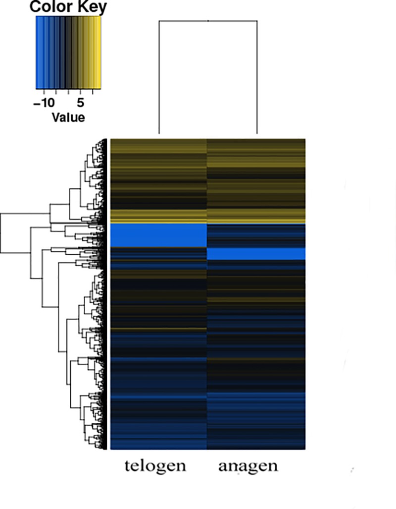
Yellow indicates the genes with greater expression, and blue indicates the genes with lower expression. There were clusters with relatively minor differences between anagen and telogen secondary HFSCs.

Interestingly, among the well-known HFSC signature genes reported to be important for hair follicle stem cell biology, including SOX9, LHX2, NFATC1, FGF18, RUNX1 and VDR [22], LHX2 and NFATC1 were observed to be differentially expressed in the RNA-Seq data (S2 Table). Most of the cell cycle genes, such as CCNA2, CCNB2, CCNB1, CDKN1A and MCM5 were all expressed but did not exhibit differences in expression (S2 Table).

Previous studies have shown that KRT and KRTAP are major structural proteins of the hair fiber and sheath, and their content is important for fleece quality [23]. We identified a set of keratins (KRT) and keratin-associated protein (KRTAP) genes that were also annotated in cashmere goat (Capra hircus) hair follicles [24] (Table 3). Most of the differentially expressed KRT and KRTAP were down regulated in the telogen phase HFSCs.

Table 3. KRT and KRTAP, which were annotated in Cashmere goat (Capra hircus) hair follicles.

Gene Fold Change(ana-tel) Description
KRT4 63.777962 PREDICTED: keratin, type II cytoskeletal 4 [Capra hircus]
KRT79 12.391147 PREDICTED: keratin, type II cytoskeletal 79 isoform X2 [Ovis aries musimon]
KRT7 6.381597 PREDICTED: keratin, type II cytoskeletal 7 isoform X3 [Ovis aries musimon]
LOC102180595 4.1000119 PREDICTED: keratin, type I cytoskeletal 18 isoform X2 [Ovis aries musimon]
LOC102179515 3.1967979 PREDICTED: keratin, type I cytoskeletal 15 [Ovis aries musimon]
KRT8 2.1871825 PREDICTED: keratin, type II cytoskeletal 8 [Capra hircus]
LOC102180424 0.3971521 PREDICTED: keratin, type I cytoskeletal 16 [Capra hircus]
KRT1 0.3814496 PREDICTED: keratin, type II cytoskeletal 1 isoform X2 [Ovis aries musimon]
KRT78 0.3057293 PREDICTED: keratin, type II cytoskeletal 78 isoform X1 [Capra hircus]
KRTAP11-1 0.2014041 keratin-associated protein 11–1 [Capra hircus]
KRT23 0.1741835 PREDICTED: keratin, type I cytoskeletal 23 [Capra hircus]
KRT82 0.1518523 PREDICTED: keratin, type II cuticular Hb2 isoform X1 [Capra hircus]
KPRP 0.1447063 PREDICTED: keratinocyte proline-rich protein-like [Capra hircus]
KRT10 0.1368225 PREDICTED: keratin, type I cytoskeletal 10 [Pantholops hodgsonii]
KRTAP3-1 0.0759261 keratin-associated protein 3–1 [Capra hircus]
LOC102182669 0.0364445 PREDICTED: keratin, type II cytoskeletal 5-like [Ovis aries]
LOC102176685 0.0206982 PREDICTED: keratin, type II cytoskeletal 75 isoform X2 [Ovis aries musimon]
LOC108635997 0.0200734 PREDICTED: keratin, type II cytoskeletal 6A [Capra hircus]

Functional classification

To better understand the HF cycle, DEGs were categorized into three gene ontology categories: cellular components, biological processes, and molecular functions. DEGs between the ana-SHFSCs and tel-SHFSCs were categorized into 55 functional groups based on sequence homology. In the three main GO classification categories, 26, 16, and 13 functional groups were identified, respectively (Fig 4). The top five functional categories for the upregulated and downregulated DEGs included cell part, cellular process, binding, biological regulation and organelle. Among these groups, the terms cell part, cellular process and binding were dominant in each of the three categories, respectively. We also noticed a high percentage of genes in membrane part, metabolic process, organelle part, catalytic activity, and developmental process. The GO analysis results showed that the functions of the identified DEGs were involved in various biological processes, such as cellular process, biological regulation, metabolic process, developmental process and response to stimulus, and were associated with HFs and skin development. For example, the DEGs LHX2, LGR5 [25] and FOXC1 [26], which were identified as being important in hair follicle development, were enriched in transcriptional activator, hair morphogenesis and hair cycle process categories, respectively.

Fig 4. GO classification of DEGs.

Fig 4

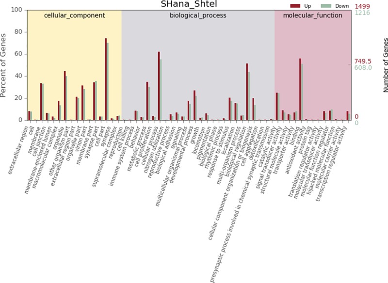
The results are summarized in three main categories: biological process, cellular component and molecular function. The X-axis indicates the second level term of gene ontology; The Y-axis shows the percentage of genes. red, up regulated genes; green, down regulated genes.

The KEGG analysis results predicted that the DEGs were significantly enriched in pathways such as PI3K-Akt, MAPK, Ras and Rap1 (Table 4).

Table 4. KEGG pathway analysis of DEGs.

Pathway term FDR Up Count Down Count
MAPK signaling pathway 1.806E-05 38 17
Protein digestion and absorption 1.806E-05 20 7
PI3K-Akt signaling pathway 2.128E-05 47 21
Ras signaling pathway 0.0001668 31 16
Rap1 signaling pathway 0.0002316 30 10
Serotonergic synapse 0.0008405 16 13
Drug metabolism—cytochrome P450 0.0013631 8 13
Thyroid hormone synthesis 0.0015981 11 8
ECM-receptor interaction 0.0020869 15 4
Relaxin signaling pathway 0.0023643 20 7
Focal adhesion 0.0042909 25 9
Aldosterone synthesis and secretion 0.0048675 16 2
Arachidonic acid metabolism 0.0048675 9 10
Melanogenesis 0.0048675 15 6
Regulation of lipolysis in adipocytes 0.0048675 8 6
Signaling pathways regulating pluripotency of stem cells 0.0048675 16 10
TNF signaling pathway 0.0048675 16 6
Glutathione metabolism 0.0071094 4 11
Calcium signaling pathway 0.0080587 22 10
Basal cell carcinoma 0.0085833 11 4

qRT-PCR validation of the RNA-Seq data

To validate the results from the transcriptomic analysis, eight differentially expressed genes were randomly selected and assayed by qRT-PCR. When anagen compared to telogen (anagen as sample, telogen as control), the qRT-PCR results verified that these genes were differentially expressed in HFSCs at the anagen and telogen phases, consistent with the RNA-Seq results (Fig 5). Thus, the RNA-Seq results provided reliable data for the mRNA differential expression analysis.

Fig 5. qRT-PCR validation of the RNA-Seq data.

Fig 5

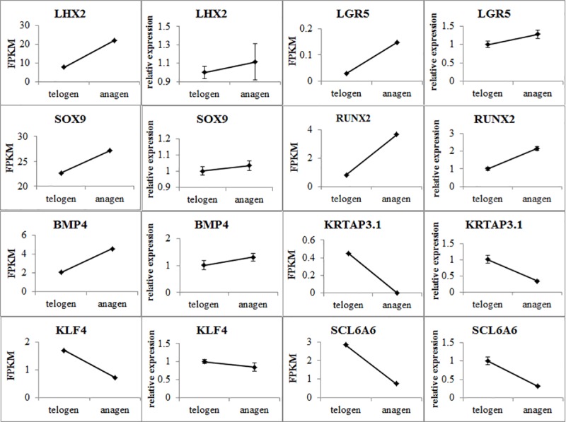
The qRT-PCR data are shown as the means ± standard error (SE) of three replicates. FPKM values from the RNA-Seq analysis are shown as the means and SE of three replicates. The left side shows the qRT-PCR data, while the right side shows the FPKM values from the RNA-Seq results.

Discussion

Because of differences between the breed, heredity and living environment of domestic animals, our molecular and morphological understanding of hair follicle biology relies a great deal on mouse hair follicle research. In recent years, studies on Cashmere goat hair follicles has increased [2729], providing valuable information for further research on the molecular mechanisms associated with these cells.

In this study, we performed gene expression profiling between anagen and telogen hair follicle stem cells of the Inner Mongolia Cashmere goat. With respect to the HFSC signature genes, we observed that LHX2, a master regulator of HFSC quiescence, was differentially expressed. LHX2 has been shown to govern both cellular quiescence and differentiation [30]. We observed higher LHX2 expression in anagen HFSCs in our data, indicating that LHX2 is also associated with the activity of HFSCs, consistent with the results of a previous study [3133]. SOX9, a master regulator of HFSCs, orchestrates the dynamics of cell fate decision, stem cell plasticity and the active-quiescent transition of HFSCs [3436]. In our previous study, we showed that SOX9 is crucial in HFSC feature maintenance [37]. In this study, no significant difference in SOX9 expression was observed between anagen and telogen HFSCs. Combining with mouse and human HFSC research [38,39], we hypothesize that during the distinct anagen and telogen phases of the Inner Mongolia Cashmere goat hair follicle, the HFSCs were under a relatively cellular quiescent state, but when activated at the initiation of the hair cycle or in response to injury, HFSCs begin to differentiate, indicating that SOX9 plays a crucial role in HFSC maintenance. In agreement with this result, the cell cycle-related genes in our data showed no significant difference, indicating that the cells were in a relatively quiescent cellular state.

Hair follicle development and homeostasis are maintained by various signaling pathways. In our data, DEGs were significantly enriched in pathways that included the MAPK, PI3K-Akt, Ras, and Rap1 signaling pathways, which regulate the pluripotency of stem cells and cytokine-cytokine receptor interaction as well as cell adhesion molecules (CAMs). The PI3K pathway has been suggested to potentially regulate the transition of HFSCs from proliferation to quiescence [40]. Signaling pathways such as Wnt, TGF-β, Hedgehog and Notch, which are important in hair follicle development [41], were not significantly enriched in this study, possibly because the HFSCs were cultured in vitro, and the significantly enriched genes and pathways primarily contribute to the cellular process, maintaining HFSC features rather than developmental processes. Hair growth is a highly complex biological process that is affected by the environment, heredity, nutrition, cytokines and hormones. HFSCs reside in a distinct microenvironment, balancing stem cell maintenance with neighboring cells, and because extracellular matrix and signals are derived from these compartments, it is hard to mimic the in vivo microenvironment under in vitro culture conditions. Therefore, an in vitro culture system that can simulate the in vivo microenvironment is needed. In addition, further investigation into the molecular mechanism of the hair cycle is also needed.

From this study, we showed that the Inner Mongolia Cashmere goat HFSCs maintained a relatively cellular quiescent in vitro, in agreement with the quiescent nature of HFSCs [42, 43]. Maintaining cellular quiescence may allow cells to retain stemness or a high enough cell population for self-renewal and regeneration throughout the lifetime. Key challenges in the future are to identify the molecular mechanisms that control the hair follicle cycle, the dynamics between quiescence and stemness, and HFSC homeostasis. The results obtained using anagen and telogen phase secondary HFSCs in our study will provide new data related to gene expression profiles in hair follicle cycling in cashmere goats.

Supporting information

S1 Table. DEGs in ana-SHFSCs and tel-SHFSCs.

(XLSX)

S2 Table. List of reported HFSC signature genes and reported cell cycle genes identified from the RNA-Seq data.

(XLSX)

Acknowledgments

We are grateful to the Inner Mongolia white Cashmere goat farm for the kind help and cooperation during the animal experiments.

Data Availability

The sequencing reads of each sequencing libraries have been deposited under NCBI with BioProject ID PRJNA588350 (BioSample: SAMN13241935, RNA-seq of cashmere goat anagen HFSC, SRA: SRR10417580).

Funding Statement

Nimantana He, 2017M613263XB, project funded by China Postdoctoral Science Foundation; Nimantana He, 2019BS03012, project funded by Natural Science Foundation of Inner Mongolia; Jinquan Li, DC1900004124, project funded by Inner Mongolia Agricultural University. The funders had no role in study design, data collection and analysis, decision to publish or preparation of the manuscript.

References

  • 1.Cotsarelis G, Sun TT, Lavker RM. Label-retaining cells reside in the bulge area of pilosebaceous unit: implications for follicular stem cells, hair cycle, and skin carcinogenesis. Cell. 1990; 61:1329–1337. 10.1016/0092-8674(90)90696-c [DOI] [PubMed] [Google Scholar]
  • 2.Greco Valentina, Chen Ting, Rendl Michael, Schober Markus, Pasolli H. Amalia, Stokes Nicole, et al. A Two-Step Mechanism for Stem Cell Activation during Hair Regeneration. Cell Stem Cell. 2009; 4:155–169. 10.1016/j.stem.2008.12.009 [DOI] [PMC free article] [PubMed] [Google Scholar]
  • 3.Toyoshima Koh-ei, Asakawa Kyosuke, Ishibashi naoko, Hiroshi Toki, ogawa miho, Hasegawa Tomoko, et al. Fully functional hair follicle regeneration through the rearrangement of stem cells and their niches. Nature Communications. 2012; 3:784 10.1038/ncomms1784 [DOI] [PMC free article] [PubMed] [Google Scholar]
  • 4.Blanpain C & Fuchs E. Stem cell plasticity, Plasticity of epithelial stem cells in tissue regeneration. Science. 2014; 344:1242281 10.1126/science.1242281 [DOI] [PMC free article] [PubMed] [Google Scholar]
  • 5.Liu Shuang, Zhang Huishan, and Duan Enkui. Epidermal Development in Mammals: Key Regulators, Signals from Beneath, and Stem Cells. Int J Mol Sci. 2013; 14, 10869–10895. 10.3390/ijms140610869 [DOI] [PMC free article] [PubMed] [Google Scholar]
  • 6.Sennett R, Rendl M. Mesenchymal-epithelial interactions during hair follicle morphogenesis and cycling. Semin Cell Dev Biol. 2012; 23(8):917–927. 10.1016/j.semcdb.2012.08.011 [DOI] [PMC free article] [PubMed] [Google Scholar]
  • 7.Schmidt-Ullrich R, Paus R. Molecular principles of hair follicle induction and morphogenesis. Bioessays. 2005; 27(3):247–61. 10.1002/bies.20184 [DOI] [PubMed] [Google Scholar]
  • 8.Pisal Rishikaysh., Kapil Dev., Daniel Diaz., Mohiuddin Shaikh Qureshi Wasay., Stanislav Filip., Jaroslav Mokry. Signaling involved in hair follicle morphogenesis and development. Int J Mol Sci. 2014; 15(1), 1647–70. 10.3390/ijms15011647 [DOI] [PMC free article] [PubMed] [Google Scholar]
  • 9.Cq Li, Yin J, Zhang Y-j, Guo Z, Zhang W, Gao A, et al. Comparative study on skin and hair follicles cycling between Inner Mongolia and Liaoning cashmere goats. ACTA VETERINARIA ET ZOOTECHNICA SINICA. 2005; 36(7):674. [Google Scholar]
  • 10.Wu JH, Zhang YJ, Zhang JX, Chang ZL, Li JQ, Yan ZW, et al. Hoxc13/β-catenin correlation with hair follicle activity in Cashmere goat. J Integrat Agric. 2012; 11(7):1159–66. 10.1016/s2095-3119(12)60110-5 [DOI] [Google Scholar]
  • 11.Blanpain Cédric and Fuchs Elaine. Epidermal homeostasis: a balancing act of stem cells in the skin. Nature reviews. 2009; 10:207–217. 10.1038/nrm2636 [DOI] [PMC free article] [PubMed] [Google Scholar]
  • 12.Fuchs E. Scratching the surface of skin development. Nature. 2007; 445(7130):834–842. 10.1038/nature05659 [DOI] [PMC free article] [PubMed] [Google Scholar]
  • 13.Nowak JA, Polak L, Pasolli HA & Fuchs E. Hair follicle stem cells are specified and function in early skin morphogenesis. Cell Stem Cell. 2008; 3:33–43. 10.1016/j.stem.2008.05.009 [DOI] [PMC free article] [PubMed] [Google Scholar]
  • 14.Ito M, Liu Y, Yang Z, Nguyen J, Liang F, Morris RJ, et al. Stem cells in the hair follicle bulge contribute to wound repair but not to homeostasis of the epidermis. Nature Med. 2005; 11:1351–1354. 10.1038/nm1328 [DOI] [PubMed] [Google Scholar]
  • 15.Levy V, Lindon C, Harfe BD & Morgan BA. Distinct stem cell populations regenerate the follicle and interfollicular epidermis. Dev Cell. 2005; 9:855–861. 10.1016/j.devcel.2005.11.003 [DOI] [PubMed] [Google Scholar]
  • 16.Li YR, Fan WB, Li CQ, Yin J, Zhang YJ, Li JQ. Histomorphology research of the secondary follicle cycling of Inner Mongolia Cashmere goat. Scientia Agricultura Sinica. 2008; 41(11):3920–6. [Google Scholar]
  • 17.He Nimantana, Dong Zhenguo, Tao Li, Zhao Sirguleng, Bou Shorgan, Liu Dongjun. Isolation and characterization of hair follicle stem cells from Arbas Cashmere goat. Cytotechnology. 2016; 68:2579–2588. 10.1007/s10616-016-9981-2 [DOI] [PMC free article] [PubMed] [Google Scholar]
  • 18.He Nimantana, Dong Zhenguo, Zhu Bing, Nuo Mingtu, Bou Shorgan, Liu Dongjun. Expression of Pluripotency Markers in Arbas Cashmere Goat Hair Follicle Stem Cells. In Vitro Cell.Dev.Biol.—Animal. 2016; 52:782–788. 10.1007/s11626-016-0023-3 [DOI] [PubMed] [Google Scholar]
  • 19.Purba Talveen S., Haslam Iain S., Enrique Poblet., Francisco Jiménez., Alberto Gandarillas., Ander Izeta., et al. Human epithelial hair follicle stem cells and their progeny: current state of knowledge, the widening gap in translational research and future challenges. Bioessays, 2014; 36(5):513–25. 10.1002/bies.201300166 [DOI] [PubMed] [Google Scholar]
  • 20.Nowak Jonathan A., Lisa Polak., Amalia Pasolli H., Elaine Fuchs. Hair follicle stem cells are specified and function in early skin morphogenesis. Cell Stem Cell, 2008; 3(1):33–43. 10.1016/j.stem.2008.05.009 [DOI] [PMC free article] [PubMed] [Google Scholar]
  • 21.Wang L, Feng Z, Wang X, Wang X & Zhang X. DEGseq: an r package for identifying differentially expressed genes from RNA-seq data. Bioinformatics. 2010; 26:136–138. 10.1093/bioinformatics/btp612 [DOI] [PubMed] [Google Scholar]
  • 22.Fuchs Elaine. Scratching the surface of skin development. Nature. 2007; 445(7130): 834–842. 10.1038/nature05659 [DOI] [PMC free article] [PubMed] [Google Scholar]
  • 23.Langbein Lutz and Schweizer Jürgen. Keratins of the human hair follicle. Int Rev Cytol. 2005; 243:1–78. 10.1016/S0074-7696(05)43001-6 [DOI] [PubMed] [Google Scholar]
  • 24.Dong Yang, Xie Min, Jiang Yu, Xiao Nianqing, Du Xiaoyong, Zhang Wenguang, et al. Sequencing and automated whole-genome optical mapping of the genome of a domestic goat (Capra hircus). Nature biotechnology. 2013; 13:135–142. 10.1038/nbt.2478 [DOI] [PubMed] [Google Scholar]
  • 25.Barker N, van Es JH, Jaks V, Kasper M, Snippert H, Toftgård R, et al. Very long-term self-renewal of small intestine, colon, and hair follicles from cycling Lgr5+ve stem cells. Cold Spring Harb. Symp Quant Biol. 2008; 73:351–356. 10.1101/sqb.2008.72.003 [DOI] [PubMed] [Google Scholar]
  • 26.Lay Kenneth, Kume Tsutomu, and Fuchs Elaine. FOXC1 maintains the hair follicle stem cell niche and governs stem cell quiescence to preserve long-term tissue-regenerating potential. 2016; 113(11):E1506–15. 10.1073/pnas.1601569113 [DOI] [PMC free article] [PubMed] [Google Scholar]
  • 27.Liu B, Gao F, Guo J, Wu D, Hao B, Li Y, et al. A Microarray-Based Analysis Reveals that a Short Photoperiod Promotes Hair Growth in the Arbas Cashmere Goat. PLoS ONE. 2016; 11(1):e0147124 10.1371/journal.pone.0147124 [DOI] [PMC free article] [PubMed] [Google Scholar]
  • 28.Yang Min, Song Shen, Dong Kunzhe, Chen XiaoFei, Liu Xuexue, Rouzi Marhaba, et al. Skin transcriptome reveals the intrinsic molecular mechanisms underlying hair follicle cycling in Cashmere goats under natural and shortened photoperiod conditions. Scientific reports. 2017; 7:13502 10.1038/s41598-017-13986-w [DOI] [PMC free article] [PubMed] [Google Scholar]
  • 29.Geng R, Yuan C, Chen Y. Exploring Differentially Expressed Genes by RNA-Seq in Cashmere Goat (Capra hircus) Skin during Hair Follicle Development and Cycling. PLoS ONE. 2013; 8(4): e62704 10.1371/journal.pone.0062704 [DOI] [PMC free article] [PubMed] [Google Scholar]
  • 30.Mardaryev AN, Meier N, Poterlowicz K, Sharov AA, Sharova TY, Ahmed MI, et al. Lhx2 differentially regulates Sox9, Tcf4 and Lgr5 in hair follicle stem cells to promote epidermal regeneration after injury. Development. 2011; 138:4843–4852. 10.1242/dev.070284 [DOI] [PMC free article] [PubMed] [Google Scholar]
  • 31.Rhee H, Polak L, Fuchs E. Lhx2 maintains stem cell character in hair follicles. Science. 2006; 312(5782):1946–1949. 10.1126/science.1128004 [DOI] [PMC free article] [PubMed] [Google Scholar]
  • 32.Tiede S, Paus R. Lhx2—decisive role in epithelial stem cell maintenance, or just the "tip of the iceberg"? Bioessays. 2006; 28(12):1157–1160. 10.1002/bies.20506 [DOI] [PubMed] [Google Scholar]
  • 33.Yi Rui. Mechanisms of Quiescent Hair Follicle Stem Cell Regulation. Stem Cells. 2017; 35(12): 2323–2330. 10.1002/stem.2696 [DOI] [PMC free article] [PubMed] [Google Scholar]
  • 34.Vidal VP, Chaboissier MC, Lützkendorf S, Cotsarelis G, Mill P, Hui CC, et al. Sox9 Is Essential for Outer Root Sheath Differentiation and the Formation of the Hair Stem Cell Compartment. Current Biology. 2005; 15(15):1340–51. 10.1016/j.cub.2005.06.064 [DOI] [PubMed] [Google Scholar]
  • 35.Kadaja Meelis, Keyes Brice E., Lin Mingyan, Pasolli H. Amalia, Genander Maria, Polak Lisa, et al. SOX9: a stem cell transcriptional regulator of secreted niche signaling factors. Genes Dev. 2014; 28(4): 328–341. 10.1101/gad.233247.113 [DOI] [PMC free article] [PubMed] [Google Scholar]
  • 36.Nikolova, Oristian Daniel S., Polak Lisa, Kadaja Meelis, Asare Amma, Zheng Deyou, et al. Pioneer factors govern super-enhancer dynamics in stem cell plasticity and lineage choice Nature. 2015; 521(7552):366–370. 10.1038/nature14289 [DOI] [PMC free article] [PubMed] [Google Scholar]
  • 37.He Nimantana, Dong Zhenguo, Tai Dapeng, Guo Xudong, Cang Ming, Liang Hao, et al. The role of Sox9 in maintaining the characteristics and pluripotency of Arbas Cashmere goat hair follicle stem cells. Cytotechnology. 2018; 70:1155–1165. 10.1007/s10616-018-0206-8 [DOI] [PMC free article] [PubMed] [Google Scholar]
  • 38.Elaine Fuchs. Skin Stem Cells in Silence, Action, and Cancer. Stem Cell Reports. 2018; 10(5):1432–1438. 10.1016/j.stemcr.2018.04.008 [DOI] [PMC free article] [PubMed] [Google Scholar]
  • 39.Yejing Ge, Gomez Nicholas C., Adam Rene C., Maria Nikolova., Hanseul Yang., Akanksha Verma., et al. Stem Cell Lineage Infidelity Drives Wound Repair and Cancer. Cell. 2017; 169(4):636–650.e14. 10.1016/j.cell.2017.03.042 [DOI] [PMC free article] [PubMed] [Google Scholar]
  • 40.Wang Dongmei, Zhang Zhaojie, O’Loughlin Evan, Wang Li, Fan Xiying, Eric C., et al. MicroRNA-205 controls neonatal expansion of skin stem cells by modulating the PI(3)K pathway. Nat Cell Biol. 2013; 15:1153–1163. 10.1038/ncb2827 [DOI] [PMC free article] [PubMed] [Google Scholar]
  • 41.Carlos Andrés Chacón-Martínez Markus Klose, Niemann Catherin, Glauche Ingmar &Wickström Sara A. Hair follicle stem cell cultures reveal self-organizing plasticity of stem cells and their progeny. The EMBO Journal. 2017; 36:151–164. 10.15252/embj.201694902 [DOI] [PMC free article] [PubMed] [Google Scholar]
  • 42.Morris RJ, Liu Y, Marles L, Yang Z, Trempus C, Li S, et al. Capturing and profiling adult hair follicle stem cells. Nature Biotech, 2004, 22,:411–417. 10.1038/nbt950 [DOI] [PubMed] [Google Scholar]
  • 43.Tumbar Tudorita, Guasch Geraldine, Greco Valentina, Blanpain Cedric, Lowry William E, Rend Michael, et al. Defining the epithelial stem cell niche in skin. Science. 2004; 303(5656):359–363. 10.1126/science.1092436 [DOI] [PMC free article] [PubMed] [Google Scholar]

Decision Letter 0

Irina Polejaeva

9 Oct 2019

PONE-D-19-19805

Exploring differentially expressed genes between anagen and telogen secondary hair follicle stem cells of Cashmere goat (capra hircus) by RNA-seq

PLOS ONE

Dear Dr. He,

Thank you for submitting your manuscript to PLOS ONE. After careful consideration, we feel that it has merit but does not fully meet PLOS ONE’s publication criteria as it currently stands. Therefore, we invite you to submit a revised version of the manuscript that addresses the points raised during the review process.

You only randomly selected 6 genes and used the qPCR for RNA seq data validation. You should use qPCR to validate all important genes that you reference in your manuscript discussion section. Additinally,  instead of listing all the differentially expressed genes between the anagen HFSC and telogen HFSC (Table 2-4), present this  differentially expressed gene info as a heat map graph with genes in the same category clustered together. Please, also address the reviewers comments included below.

We would appreciate receiving your revised manuscript by November 15, 2019. When you are ready to submit your revision, log on to https://www.editorialmanager.com/pone/ and select the 'Submissions Needing Revision' folder to locate your manuscript file.

If you would like to make changes to your financial disclosure, please include your updated statement in your cover letter.

To enhance the reproducibility of your results, we recommend that if applicable you deposit your laboratory protocols in protocols.io, where a protocol can be assigned its own identifier (DOI) such that it can be cited independently in the future. For instructions see: http://journals.plos.org/plosone/s/submission-guidelines#loc-laboratory-protocols

Please include the following items when submitting your revised manuscript:

  • A rebuttal letter that responds to each point raised by the academic editor and reviewer(s). This letter should be uploaded as separate file and labeled 'Response to Reviewers'.

  • A marked-up copy of your manuscript that highlights changes made to the original version. This file should be uploaded as separate file and labeled 'Revised Manuscript with Track Changes'.

  • An unmarked version of your revised paper without tracked changes. This file should be uploaded as separate file and labeled 'Manuscript'.

Please note while forming your response, if your article is accepted, you may have the opportunity to make the peer review history publicly available. The record will include editor decision letters (with reviews) and your responses to reviewer comments. If eligible, we will contact you to opt in or out.

We look forward to receiving your revised manuscript.

Kind regards,

Irina Polejaeva, PhD

Academic Editor

PLOS ONE

Journal Requirements:

When submitting your revision, we need you to address these additional requirements.

Please ensure that your manuscript meets PLOS ONE's style requirements, including those for file naming. The PLOS ONE style templates can be found at

http://www.journals.plos.org/plosone/s/file?id=wjVg/PLOSOne_formatting_sample_main_body.pdf and http://www.journals.plos.org/plosone/s/file?id=ba62/PLOSOne_formatting_sample_title_authors_affiliations.pdf

1. Thank you for including your ethics statement: Adult Arbas Cashmere goat was obtained from the Inner Mongolia YIWEI white Cashmere Goat Farm. Skin samples using in cell isolation were collected in accordance with the Animal Ethics Committee of the Inner Mongolia Academy of Agriculture and Animal Husbandry Sciences that is responsible for Animal Care and Use in the Inner Mongolia Autonomous Region of China.

Please amend your current ethics statement to confirm that your named ethics committee specifically approved this study.

For additional information about PLOS ONE submissions requirements for animal ethics, please refer to http://journals.plos.org/plosone/s/submission-guidelines#loc-animal-research  

Once you have amended this/these statement(s) in the Methods section of the manuscript, please add the same text to the “Ethics Statement” field of the submission form (via “Edit Submission”).

2. We note that you are reporting an analysis of a microarray, next-generation sequencing, or deep sequencing data set. PLOS requires that authors comply with field-specific standards for preparation, recording, and deposition of data in repositories appropriate to their field. Please upload these data to a stable, public repository (such as ArrayExpress, Gene Expression Omnibus (GEO), DNA Data Bank of Japan (DDBJ), NCBI GenBank, NCBI Sequence Read Archive, or EMBL Nucleotide Sequence Database (ENA)). In your revised cover letter, please provide the relevant accession numbers that may be used to access these data. For a full list of recommended repositories, see http://journals.plos.org/plosone/s/data-availability#loc-omics or http://journals.plos.org/plosone/s/data-availability#loc-sequencing.

[Note: HTML markup is below. Please do not edit.]

Reviewers' comments:

Reviewer's Responses to Questions

Comments to the Author

1. Is the manuscript technically sound, and do the data support the conclusions?

The manuscript must describe a technically sound piece of scientific research with data that supports the conclusions. Experiments must have been conducted rigorously, with appropriate controls, replication, and sample sizes. The conclusions must be drawn appropriately based on the data presented.

Reviewer #1: Partly

Reviewer #2: Partly

**********

2. Has the statistical analysis been performed appropriately and rigorously?

Reviewer #1: N/A

Reviewer #2: N/A

**********

3. Have the authors made all data underlying the findings in their manuscript fully available?

The PLOS Data policy requires authors to make all data underlying the findings described in their manuscript fully available without restriction, with rare exception (please refer to the Data Availability Statement in the manuscript PDF file). The data should be provided as part of the manuscript or its supporting information, or deposited to a public repository. For example, in addition to summary statistics, the data points behind means, medians and variance measures should be available. If there are restrictions on publicly sharing data—e.g. participant privacy or use of data from a third party—those must be specified.

Reviewer #1: Yes

Reviewer #2: Yes

**********

4. Is the manuscript presented in an intelligible fashion and written in standard English?

PLOS ONE does not copyedit accepted manuscripts, so the language in submitted articles must be clear, correct, and unambiguous. Any typographical or grammatical errors should be corrected at revision, so please note any specific errors here.

Reviewer #1: Yes

Reviewer #2: Yes

**********

5. Review Comments to the Author

Please use the space provided to explain your answers to the questions above. You may also include additional comments for the author, including concerns about dual publication, research ethics, or publication ethics. (Please upload your review as an attachment if it exceeds 20,000 characters)

Reviewer #1: In this article, the authors compared transcriptome profiles between anagen and telogen secondary HFSCs using RNA-Seq technique, and found 2717 differentially expressed genes (DEGs). The RNA-Seq results were verified by RT-PCR, in which 6 DEGs selected randomly were amplified and analyzed. The data in this article are obviously useful for researchers working in this field. The article is organized and written in standard English. The data have not been published elsewhere.

Abstract

1. Line 4-6: Cashmere only grows in the second hair follicles (HFs). Both anagen and telogen HFSCs were isolated from the second HFs (the authors did not state how to isolate HFSCs at different stages); therefore, the results in this paper can not disclose the differentially expressed genes between primary and secondary HFs.

2. Next page, Line 3: ‘in agreement with the quiescent nature of HFSCs’ is confusing.

Introduction

1. There are 3 important stages in hair cycle, growth (anagen), regression (catagen), and relative quiescence (telogen). The authors analyzed gene expressing profiles in both anagen and telogen stages. Why not include data in the catagen stage?

2. Page2, Last sentence: ‘Cashmere is the primary commercial product of the Inner Mongolia Cashmere goat, the secondary hair follicles from which exhibit a notable photoperiod-based cycle that changes throughout the year, making them suitable for hair cycle research.’ I suggest this sentence should be improved.

3. Page3: ‘RNA-Seq is a commonly used high-throughput sequencing method used to detect differences in gene expression between two groups.’ There is only one sentence in this paragraph. I suggest the authors either combine this sentence with last paragraph or include more information concerning RNA-Seq technique.

Materials and Methods

1. 1st paragraph: The isolation and identification of ana-SHFSCs and tel-SHFSCs should be written in a single paragraph with detailed descriptions. Are there any markers that can be used to identify the ana-SHFSCs and tel-SHFSCs?

2. Check the sentence: ‘The clean reads were used in for subsequent analysis and were aligned to the NCBI goat reference genome …’. The ‘in’ should be deleted.

Results

1. Please provide the isolation and identification results in the main text, not in the Supplement. How did the authors differentiate ana-SHFSCs and tel-SHFSCs in their exp.., with immunohistochemistry staining?

2. The figure S1: Some pictures in S1 Fig. look like those published in Reference 16, such as, morphology of HFSCs, Krt15 and Krt19 staining. Also there are no pictures showing different stages of HFSCs.

3. Check the sentence: ‘resulting in 64,858,310 and 60,223,076 raw reads being obtained, respectively (Table 1).’. The ‘being obtained’ should be deleted.

4. The figure S2: A typo in ‘Percentage’.

Discussion

1. Please provide the reference for this sentence, ‘indicating that LHX2 is also associated with the activity of HFSCs, consistent with the results of a previous study’. Also briefly introduce the results in the paper published.

2. No data support the conclusion in this sentence, ‘but when activated at the initiation of the hair cycle or in response to injury, HFSCs begin to differentiate, indicating that SOX9 plays a crucial role in HFSC maintenance.’. Please provide the reference and briefly introduce the results in the paper published.

3. It the ‘PI3K-Akt’ the same as the ‘PI(3)K’?

Reviewer #2: In this manuscripts, the authors aimed to investigate the transcriptome differences anagen and telogen secondary hair follicle stem cells in goats. They found 2717 genes were differentially expressed in these two populations of hair follicle stem cells. I found this type of study intriguing since it could provide a lot of information that not only could contribute to the cashmere production but also to our knowledge of the regenerative characteristics of stem cell. However, this experiment only has the RNA sequencing data. It will be more convincing and useful if the authors could do some follow-up experiment related to the differentially expressed genes found using RNA seq.

1. The only data this manuscript have is the RNA seq data. To validate the RNA seq, the authors randomly picked 6 genes and did the qPCR. I suggest the authors use qPCR to validate all these important genes that you made your discussion on.

2. Table 2-4 listed all the differentially expressed genes between the anagen HFSC and telogen HFSC. I found this is a very inefficient and confusing way to present RNA seq data. I highly suggest the authors put this differentially expressed gene info into a heat map graph with genes in the same category clustered together. The results part included too much technique information that should be put into the methods section. For example, the summary of mapped read and mapping rates based on the RNA-Seq, seems really irrelevant to the study aim. In results, the authors to list the experiment outcome that contribute to test the hypothesis of the study.

3. Tabe3,4. I don’t think it’s necessary to mention they genes has different reads but has no significant difference.

**********

6. PLOS authors have the option to publish the peer review history of their article (what does this mean?). If published, this will include your full peer review and any attached files.

If you choose “no”, your identity will remain anonymous but your review may still be made public.

Do you want your identity to be public for this peer review? For information about this choice, including consent withdrawal, please see our Privacy Policy.

Reviewer #1: No

Reviewer #2: No

[NOTE: If reviewer comments were submitted as an attachment file, they will be attached to this email and accessible via the submission site. Please log into your account, locate the manuscript record, and check for the action link "View Attachments". If this link does not appear, there are no attachment files to be viewed.]

While revising your submission, please upload your figure files to the Preflight Analysis and Conversion Engine (PACE) digital diagnostic tool, https://pacev2.apexcovantage.com/. PACE helps ensure that figures meet PLOS requirements. To use PACE, you must first register as a user. Registration is free. Then, login and navigate to the UPLOAD tab, where you will find detailed instructions on how to use the tool. If you encounter any issues or have any questions when using PACE, please email us at figures@plos.org. Please note that Supporting Information files do not need this step.

PLoS One. 2020 Apr 16;15(4):e0231376. doi: 10.1371/journal.pone.0231376.r002

Author response to Decision Letter 0


3 Dec 2019

Reviewer #1

Abstract

1. Line 4-6: Cashmere only grows in the second hair follicles (HFs). Both anagen and telogen HFSCs were isolated from the second HFs (the authors did not state how to isolate HFSCs at different stages); therefore, the results in this paper cannot disclose the differentially expressed genes between primary and secondary HFs.

In this paper, we were aimed to explore the differentially expressed genes between anagen and telogen phase of the secondary hair follicle, so there is no data or result about differentially expressed genes between primary and secondary HFs. We revised the statement.

Isolation of the cells from anagen and telogen phase hair follicle was detailed in the Materials and Methods section.

2. Next page, Line 3: ‘in agreement with the quiescent nature of HFSCs’ is confusing.

This statement was deleted.

Introduction

1. There are 3 important stages in hair cycle, growth (anagen), regression (catagen), and relative quiescence (telogen). The authors analyzed gene expressing profiles in both anagen and telogen stages. Why not include data in the catagen stage?

The hair cycle of Inner Mongolia Cashmere goat can divided into anagen, catagen, and telogen three stages. We focused on the differentially expressed genes in growth and quiescence state. In addition, the catagen phase of the Inner Mongolia Cashmere goat hair follicle is much shorter than anagen and telogen phase, and it is hard to isolate the cells. So we chose anagen and telogen.

2. Page2, Last sentence: ‘Cashmere is the primary commercial product of the Inner Mongolia Cashmere goat, the secondary hair follicles from which exhibit a notable photoperiod-based cycle that changes throughout the year, making them suitable for hair cycle research.’ I suggest this sentence should be improved.

We improved the sentence as ‘The secondary hair follicles which exhibit a notable photoperiod-based cycle that changes throughout the year, are suitable for hair cycle research.’

3. Page3: ‘RNA-Seq is a commonly used high-throughput sequencing method used to detect differences in gene expression between two groups.’ There is only one sentence in this paragraph. I suggest the authors either combine this sentence with last paragraph or include more information concerning RNA-Seq technique.

We combined this sentence with last paragraph.

Materials and Methods

1. 1st paragraph: The isolation and identification of ana-SHFSCs and tel-SHFSCs should be written in a single paragraph with detailed descriptions. Are there any markers that can be used to identify the ana-SHFSCs and tel-SHFSCs?

The isolation of the ana-SHFSCs and tel-SHFSCs were detailed in the cell isolation. We are not sure if there are any markers that can be identify the ana-SHFSCs and tel-SHFSCs nowadays. In this paper, we use well-known HFSC markers to identify the cells.

2. Check the sentence: ‘The clean reads were used in for subsequent analysis and were aligned to the NCBI goat reference genome …’. The ‘in’ should be deleted.

Yes, the ‘in’ has been deleted.

Results

1. Please provide the isolation and identification results in the main text, not in the Supplement. How did the authors differentiate ana-SHFSCs and tel-SHFSCs in their exp.., with immunohistochemistry staining?

Thank you. Isolation and identification results were put in the main text. Cell morphology of ana-SHFSCs and tel-SHFSCs were much alike under microscope observation. Isolation, culture and RNA extraction of ana-SHFSCs and tel-SHFSCs were carried out separately, avoiding from mixed throughout the experiment.

2. The figure S1: Some pictures in S1 Fig. look like those published in Reference 16, such as, morphology of HFSCs, Krt15 and Krt19 staining. Also there are no pictures showing different stages of HFSCs.

I am the first author of the reference 16. We isolated HFSCs from primary and secondary (anagen and telogen) HFs, and carried out a series of experiments. The morphology of HFSCs from the primary and secondary (anagen and telogen) were with high similarity, and markers used to identify the HFSCs were the same, so we chose different secondary antibody (FITC and Cy3) in reference 16 and this paper.

3. Check the sentence: ‘resulting in 64,858,310 and 60,223,076 raw reads being obtained, respectively (Table 1)’. The ‘being obtained’ should be deleted.

It has been deleted.

4. The figure S2: A typo in ‘Percentage’.

Yes. Figure S2 is showed as percentage.

Discussion

1. Please provide the reference for this sentence, ‘indicating that LHX2 is also associated with the activity of HFSCs, consistent with the results of a previous study’. Also briefly introduce the results in the paper published.

Mardaryev AN, Meier N, Poterlowicz K, Sharov AA, Sharova TY, Ahmed MI, et al. Lhx2 differentially regulates Sox9, Tcf4 and Lgr5 in hair follicle stem cells to promote epidermal regeneration after injury. Development. 2011; 138:4843–4852. doi: 10.1242/dev.070284. PMID: 22028024

In this paper, they found that Lhx2+ cells reside in the stem cell-enriched epithelial compartments (bulge, secondary hair germ) and co-express selected stem cell markers (Sox9, Tcf4 and Lgr5). Cell proliferation in the bulge and the number of Sox9+ and Tcf4+ cells in the HFs closely adjacent to the wound in Lhx2+/– mice are decreased in comparison with wild-type controls. Chip-on-chip/ChIP-qPCR and reporter assay analyses identified Sox9, Tcf4 and Lgr5 as direct Lhx2 targets. These data strongly suggest that Lhx2 positively regulates Sox9 and Tcf4 in the bulge cells, and promotes wound re-epithelization. Thus, Lhx2 operates as an important regulator of epithelial stem cell activity in the skin response to injury.

Törnqvist G, Sandberg A, Hägglund AC, Carlsson L. Cyclic expression of lhx2 regulates hair formation. PLoS Genet. 2010:8;6(4):e1000904. doi: 10.1371/journal.pgen.1000904. PMID: 20386748

In this paper, they found Lhx2 is primarily expressed by precursor cells outside of the bulge region where the HF stem cells are located. They hypothesis this developmental, stage- and cell-specific expression of Lhx2 regulates the generation and regeneration of hair. Moreover, transgenic expression of Lhx2 in postnatal HFs is sufficient to induce anagen. Thus, Lhx2 is an essential positive regulator of hair formation.

Combining the results in the papers mentioned above and our data, it suggests that Lhx2 is associated with the activity of the HFSCs. HFSC is a major cell population that participated in the morphogenesis and regeneration of the HFs.

2. No data support the conclusion in this sentence, ‘but when activated at the initiation of the hair cycle or in response to injury, HFSCs begin to differentiate, indicating that SOX9 plays a crucial role in HFSC maintenance’. Please provide the reference and briefly introduce the results in the paper published.

In our manuscript, the entire sentence is ‘We hypothesize that during the distinct anagen and telogen phases, the HFSCs were under a relatively cellular quiescent state, but when activated at the initiation of the hair cycle or in response to injury, HFSCs begin to differentiate, indicating that Sox9 plays a crucial role in HFSC maintenance.’

The following references may explain our conjectures, and we add these papers in the references.

Sox9 Is Essential for Outer Root Sheath Differentiation and the Formation of the Hair Stem Cell Compartment. Current Biology. 2005;15:1340–1351. DOI 10.1016/j.cub.2005.06.064)

Using tissue-specific inactivation of Sox9, the authors demonstrate that this gene serves a crucial role in hair differentiation and that skin deleted for Sox9 lacks external hair. Sox9 knock hair show severe proliferative defects and the stem cell niche never forms. They summarized that Sox9 directs differentiation of the ORS and is required for the formation of the hair stem cell compartment.

Jonathan A. Nowak, Lisa Polak, H. Amalia Pasolli, and Elaine Fuchs. Hair Follicle Stem Cells Are Specified and Function in Early Skin Morphogenesis. Cell Stem Cell.2008; 3, 33–43. DOI 10.1016/j.stem.2008.05.009.

From this paper they found that the progeny of Sox9-expressing cells contribute to all skin epithelial lineages and Sox9 is required for SC specification.

Meelis Kadaja, Brice E. Keyes, Mingyan Lin, H. Amalia Pasolli, Maria Genander,Lisa Polak, Nicole Stokes, Deyou Zheng, and Elaine Fuchs. SOX9: a stem cell transcriptional regulator of secreted niche signaling factors. Genes Dev. 2014; 28: 328-341. doi:10.1101/gad.233247.113

By conditionally targeting Sox9 in adult HFSCs, they show that SOX9 is essential for maintaining them. The findings reveal roles for SOX9 in regulating adult HFSC maintenance and suppressing epidermal differentiation in the niche.

Rene C. Adam, Hanseul Yang, Shira Rockowitz, Samantha B. Larsen, Maria Nikolova,Daniel S. Oristian, Lisa Polak, Meelis Kadaja, Amma Asare, Deyou Zheng, and Elaine Fuchs. Pioneer factors govern super-enhancer dynamics in stem cell plasticity and lineage choice Nature. 2015 May 21; 521(7552): 366–370. doi:10.1038/nature14289.

In this paper, it has been identified SOX9 as a crucial chromatin rheostat of HFSC super-enhancers, and provide functional evidence that super-enhancers are dynamic, dense TF-binding platforms which are acutely sensitive to pioneer master regulators whose levels define not only spatial and temporal features of lineage-status, but also stemness, plasticity in transitional states and differentiation.

Hair follicles (HFs) undergo cyclical periods of growth, which are fueled by stem cells (SCs) at the base of the resting follicle. HFSC formation occurs during HF development and Sox9 is essential for the HFSCs specification, maintenance and hair differentiation.

3. It the ‘PI3K-Akt’ the same as the ‘PI(3)K’?

Yes, they are the same, and we unify the writing as PI3K.

Reviewer #2

1. The only data this manuscript have is the RNA seq data. To validate the RNA seq, the authors randomly picked 6 genes and did the qPCR. I suggest the authors use qPCR to validate all these important genes that you made your discussion on.

Thank you very much. We appreciate your suggestion. When we carried out the qPCR, we just randomly chose, and just focused on the signature genes, so picked LHX2, LGR5, SOX9 and RUNX2. qPCR validation of SOX9 and RUNX2 was now added to the manuscript.

We carried out qPCR to validate the RNA-seq data, and the qPCR result of those genes was in consistent with the RNA-seq data, and it provided reliable data for the mRNA differential expression analysis. What`s more, lacking of nucleotide sequences of capra hircus genes in the NCBI, so it is hard to validate all those genes we mentioned in the discussion.

2. Table 2-4 listed all the differentially expressed genes between the anagen HFSC and telogen HFSC. I found this is a very inefficient and confusing way to present RNA seq data. I highly suggest the authors put this differentially expressed gene info into a heat map graph with genes in the same category clustered together. The results part included too much technique information that should be put into the methods section. For example, the summary of mapped read and mapping rates based on the RNA-Seq, seems really irrelevant to the study aim. In results, the authors to list the experiment outcome that contribute to test the hypothesis of the study.

We reformat the tables and maps as your suggestion.

3. Tabe3,4. I don’t think it’s necessary to mention they genes has different reads but has no significant difference.

We put table3,4 as supplemental material.

Attachment

Submitted filename: Reply to the Reviewers.docx

Decision Letter 1

Irina Polejaeva

23 Jan 2020

PONE-D-19-19805R1

Exploring differentially expressed genes between anagen and telogen secondary hair follicle stem cells of Cashmere goat (capra hircus) by RNA-seq

PLOS ONE

Dear Dr. He,

Thank you for submitting your manuscript to PLOS ONE. After careful consideration, we feel that it has merit but does not fully meet PLOS ONE’s publication criteria as it currently stands. Therefore, we invite you to submit a revised version of the manuscript that addresses the points raised during the review process.

You addressed most of the comments from the previous reviewers. However, there are still recommendations that were not addressed. Please carefully read review 3 comments and address them by either making changes in the manuscript or providing answers in a response to the reviewers document.

We would appreciate receiving your revised manuscript by March 1, 2020. When you are ready to submit your revision, log on to https://www.editorialmanager.com/pone/ and select the 'Submissions Needing Revision' folder to locate your manuscript file.

If you would like to make changes to your financial disclosure, please include your updated statement in your cover letter.

To enhance the reproducibility of your results, we recommend that if applicable you deposit your laboratory protocols in protocols.io, where a protocol can be assigned its own identifier (DOI) such that it can be cited independently in the future. For instructions see: http://journals.plos.org/plosone/s/submission-guidelines#loc-laboratory-protocols

Please include the following items when submitting your revised manuscript:

  • A rebuttal letter that responds to each point raised by the academic editor and reviewer(s). This letter should be uploaded as separate file and labeled 'Response to Reviewers'.

  • A marked-up copy of your manuscript that highlights changes made to the original version. This file should be uploaded as separate file and labeled 'Revised Manuscript with Track Changes'.

  • An unmarked version of your revised paper without tracked changes. This file should be uploaded as separate file and labeled 'Manuscript'.

Please note while forming your response, if your article is accepted, you may have the opportunity to make the peer review history publicly available. The record will include editor decision letters (with reviews) and your responses to reviewer comments. If eligible, we will contact you to opt in or out.

We look forward to receiving your revised manuscript.

Kind regards,

Irina Polejaeva, PhD

Academic Editor

PLOS ONE

[Note: HTML markup is below. Please do not edit.]

Reviewers' comments:

Reviewer's Responses to Questions

Comments to the Author

1. If the authors have adequately addressed your comments raised in a previous round of review and you feel that this manuscript is now acceptable for publication, you may indicate that here to bypass the “Comments to the Author” section, enter your conflict of interest statement in the “Confidential to Editor” section, and submit your "Accept" recommendation.

Reviewer #1: All comments have been addressed

Reviewer #3: (No Response)

**********

2. Is the manuscript technically sound, and do the data support the conclusions?

The manuscript must describe a technically sound piece of scientific research with data that supports the conclusions. Experiments must have been conducted rigorously, with appropriate controls, replication, and sample sizes. The conclusions must be drawn appropriately based on the data presented.

Reviewer #1: Yes

Reviewer #3: Partly

**********

3. Has the statistical analysis been performed appropriately and rigorously?

Reviewer #1: N/A

Reviewer #3: No

**********

4. Have the authors made all data underlying the findings in their manuscript fully available?

The PLOS Data policy requires authors to make all data underlying the findings described in their manuscript fully available without restriction, with rare exception (please refer to the Data Availability Statement in the manuscript PDF file). The data should be provided as part of the manuscript or its supporting information, or deposited to a public repository. For example, in addition to summary statistics, the data points behind means, medians and variance measures should be available. If there are restrictions on publicly sharing data—e.g. participant privacy or use of data from a third party—those must be specified.

Reviewer #1: Yes

Reviewer #3: Yes

**********

5. Is the manuscript presented in an intelligible fashion and written in standard English?

PLOS ONE does not copyedit accepted manuscripts, so the language in submitted articles must be clear, correct, and unambiguous. Any typographical or grammatical errors should be corrected at revision, so please note any specific errors here.

Reviewer #1: Yes

Reviewer #3: Yes

**********

6. Review Comments to the Author

Please use the space provided to explain your answers to the questions above. You may also include additional comments for the author, including concerns about dual publication, research ethics, or publication ethics. (Please upload your review as an attachment if it exceeds 20,000 characters)

Reviewer #1: All my comments have been addressed.

The authors did a great job. The results in this study are important to elucidate the molecular mechanisms of hair follicle cycling in goats.

Reviewer #3: In the revision of manuscript “Exploring differentially expressed genes between anagen and telogen secondary hair follicle stem cells of Cashmere goat (capra hircus) by RNA-seq”, the authors answered previous reviewers’ questions, but there are still several important issues need to be clearly addressed:

1. What’s the differences between hair follicle stem cells at anagen and telogen stages? Even though authors included the immunostaining of two different stages, but still no differences had been found/addressed.

2. In the other publication, “Exploring Differentially Expressed Genes by RNA-Seq in Cashmere Goat (Capra hircus) Skin during Hair Follicle Development and Cycling (Geng et al. Plos one 2013, reference 25)”, same methods/strategies (RNA-Seq) were used as current study to investigate differentiation during three stages of hair follicle development. Have authors compared the similar/differences of gene expression at same stage between current and previous study? Also, as mentioned in the manuscript “Cashmere is the primary commercial product of the Inner Mongolia Cashmere goat” ,how would current study potentially help cashmere production in the future, prolong the anagen stage?

3. Methods and software used for statistical analysis in this study is not clear.

4. In Results, most of description is confused whether authors compared differentiated expression between two stages of HFSCs, or compared two stages HFSCs to other stem cells.

Specific comments:

1. Please add page number and line number in the manuscript, which is easier for reviewer to refer.

2. Please list the full name of all abbreviations, when first mentioned, e.g. GO, KEGG. Please check the whole manuscript.

3. Introduction, paragraph 2, “During anagen… the telogen stage”, could be shorten. Since this study is focused on HFSCs, so a little more information about how they have been activated and initiate hair follicle cycling should be included.

4. “Cashmere is the primary commercial product of the Inner Mongolia Cashmere goat”, it’s better refer Cashmere is produced from secondary HFs, for readers who don’t know.

5. I think authors should add one or two more sentences about RNA-seq technique.

6. M&M, Cell isolation, add reference after 1st sentence. Add references for HFSC markers- Krt15, Krt19 and Sox9, staining procedure, company of antibodies (or add references). Moreover, authors explained the reason of excluding catagen phase in this study, and I think this information should be included in M&M.

7. Statistical analysis should be separated part in M&M.

8. Results, please move all the legends to the end of text. Fig 2, what cells were used for control staining? add scale bar for Fig. 2.

9. Gene expression profiles in Results, “of which 2717 genes were significantly differentially expressed between the two groups”, I wonder all these 2717 genes were differentially expressed in which group of cells.

10. “Interestingly, among the well-known HFSC signature genes reported to be important for hair follicle stem cell biology, including SOX9, LHX2, NFATC1, FGF18, RUNX1 and VDR [19], LHX2 and NFATC1 were observed to be differentially expressed in the RNA-Seq data (Table S2). Most of the cell cycle genes, such as CCNA2, CCNB2, CCNB1, CDKN1A and MCM5 were all expressed but did not exhibit differences in expression (Table S2)”, again, were all these genes differentially expressed in anagen or telogen phase?

11. “The qRT-PCR results verified that these genes were differentially expressed in HFSCs at the anagen and telogen phases, consistent with the RNA-Seq results”, so these genes were differentially expressed in HFSCs at both stages? Then what was the reference sample?

12. Fig.4, different colors of column represent what?

13. “We hypothesize that during the distinct anagen and telogen phases of the Inner Mongolia Cashmere goat hair follicle, the HFSCs were under a relatively cellular quiescent state, but when activated at the initiation of the hair cycle or in response to injury, HFSCs begin to differentiate,”, any references to support? Or similar finding in other species?

**********

7. PLOS authors have the option to publish the peer review history of their article (what does this mean?). If published, this will include your full peer review and any attached files.

If you choose “no”, your identity will remain anonymous but your review may still be made public.

Do you want your identity to be public for this peer review? For information about this choice, including consent withdrawal, please see our Privacy Policy.

Reviewer #1: No

Reviewer #3: No

[NOTE: If reviewer comments were submitted as an attachment file, they will be attached to this email and accessible via the submission site. Please log into your account, locate the manuscript record, and check for the action link "View Attachments". If this link does not appear, there are no attachment files to be viewed.]

While revising your submission, please upload your figure files to the Preflight Analysis and Conversion Engine (PACE) digital diagnostic tool, https://pacev2.apexcovantage.com/. PACE helps ensure that figures meet PLOS requirements. To use PACE, you must first register as a user. Registration is free. Then, login and navigate to the UPLOAD tab, where you will find detailed instructions on how to use the tool. If you encounter any issues or have any questions when using PACE, please email us at figures@plos.org. Please note that Supporting Information files do not need this step.

PLoS One. 2020 Apr 16;15(4):e0231376. doi: 10.1371/journal.pone.0231376.r004

Author response to Decision Letter 1


27 Feb 2020

Reply to the Reviewers

Thank you for your kindly and academic suggestions and advises. We made reply to reviewer#3 and specific comments below, and we revised the manuscript according to comments.

Reviewer #3:

In the revision of manuscript “Exploring differentially expressed genes between anagen and telogen secondary hair follicle stem cells of Cashmere goat (capra hircus) by RNA-seq”, the authors answered previous reviewers’ questions, but there are still several important issues need to be clearly addressed:

1. What’s the differences between hair follicle stem cells at anagen and telogen stages? Even though authors included the immunostaining of two different stages, but still no differences had been found/addressed.

We first obtained HFs at anagen and telogen phase, and cultured cells. In vitro cultured HFSCs from anagen and telogen positively expressed well defined HFSC markers, and with similar morphology. Then we carried out RNAseq. From the RNAseq data, we found that most of the genes associate with cell cycle and HFSC significant genes were with no differences. Functional categories for the DEGs (upregulated and downregulated) included cell part, cellular process, binding, biological regulation and organelle. From KEGG analysis, it is found that DEGs were enriched in PI3K-Akt, MAPK, Ras and Rap1 signaling pathways, and these pathways were mainly participated in the cellular process and biological regulation. Pathways like Wnt/β-catenin, TGF, and SHH, which are play roles in the development of hair follicle, were not significantly enriched. In our research, we explored DEGs in HFSC in its different status. We noticed a high percentage of genes in terms of membrane part, metabolic process, organelle part, catalytic activity, and developmental process.

2. In the other publication, “Exploring Differentially Expressed Genes by RNA-Seq in Cashmere Goat (Capra hircus) Skin during Hair Follicle Development and Cycling (Geng et al. Plos one 2013, reference 25)”, same methods/strategies (RNA-Seq) were used as current study to investigate differentiation during three stages of hair follicle development. Have authors compared the similar/differences of gene expression at same stage between current and previous study? Also, as mentioned in the manuscript “Cashmere is the primary commercial product of the Inner Mongolia Cashmere goat”, how would current study potentially help cashmere production in the future, prolong the anagen stage?

We have read reference 25(Geng et al. Plos one 2013). In our present paper, we did not compare the gene expression.

Cashmere production is affected by many factors, such as breed, nutrition, and environment. We aimed to find differentially expressed genes in in vitro cultured bulge stem cells. If functional genes can be found out, we could try genetic approach to prolong the anagen phase.

In reference 25 the authors explored differentially expressed genes in skin samples according to the hair cycle, not in hair follicles.

I found that many published articles on exploring genes which play important roles in cashmere goat hair follicle development or hair cycle all using skin sample. Skin is composed of epidermal, dermal and subcutaneous tissue. As an appendage of skin, hair follicle is defined as a mini organ with its stem cell niche in the bulge, and bulge stem cells are critical for the hair morphogenesis and hair cycle. So, I think using adult skin sample to explore differentially expressed genes in hair development is not specific. We know it is very hard to isolate hair follicle cells from domestic animal like cashmere goat and find wood sheep, and it is a limitation of in vitro study.

3. Methods and software used for statistical analysis in this study is not clear.

We added methods and software used for statistical analysis in the text.

4. In Results, most of description is confused whether authors compared differentiated expression between two stages of HFSCs, or compared two stages HFSCs to other stem cells.

In this article, we compared differentiated expression between two stages of HFSCs.

Specific comments:

1. Please add page number and line number in the manuscript, which is easier for reviewer to refer.

Page number and line number were added in the manuscript.

2. Please list the full name of all abbreviations, when first mentioned, e.g. GO, KEGG. Please check the whole manuscript.

Full name of abbreviations was added when first mentioned.

3. Introduction, paragraph 2, “During anagen… the telogen stage”, could be shorten. Since this study is focused on HFSCs, so a little more information about how they have been activated and initiate hair follicle cycling should be included.

Information about hair follicle morphogenesis and hair cycle were added.

4. “Cashmere is the primary commercial product of the Inner Mongolia Cashmere goat”, it’s better refer Cashmere is produced from secondary HFs, for readers who don’t know.

It has been revised.

5. I think authors should add one or two more sentences about RNA-seq technique.

It has been added.

6. M&M, Cell isolation, add reference after 1st sentence. Add references for HFSC markers- Krt15, Krt19 and Sox9, staining procedure, company of antibodies (or add references). Moreover, authors explained the reason of excluding catagen phase in this study, and I think this information should be included in M&M.

References for markers and ICC procedure, and explanation of excluding catagen phase were added in the M&M.

7. Statistical analysis should be separated part in M&M.

Statistical analysis was included in each step of the M&M, and it is easy to understand how we analysis the data step by step. So, we tend to include statistical analysis in each step if it is not necessary.

8. Results, please move all the legends to the end of text. Fig 2, what cells were used for control staining? add scale bar for Fig. 2.

All legends were moved to the end of text. Fig.2 was revised.

9. Gene expression profiles in Results, “of which 2717 genes were significantly differentially expressed between the two groups”, I wonder all these 2717 genes were differentially expressed in which group of cells.

10. “Interestingly, among the well-known HFSC signature genes reported to be important for hair follicle stem cell biology, including SOX9, LHX2, NFATC1, FGF18, RUNX1 and VDR [19], LHX2 and NFATC1 were observed to be differentially expressed in the RNA-Seq data (Table S2). Most of the cell cycle genes, such as CCNA2, CCNB2, CCNB1, CDKN1A and MCM5 were all expressed but did not exhibit differences in expression (Table S2)”, again, were all these genes differentially expressed in anagen or telogen phase?

11. “The qRT-PCR results verified that these genes were differentially expressed in HFSCs at the anagen and telogen phases, consistent with the RNA-Seq results”, so these genes were differentially expressed in HFSCs at both stages? Then what was the reference sample?

Explain 9-11

In this paper, DEGs were shown as anagen compared to telogen. It is like anagen data as sample, telogen data as reference/control, and we revised in the manuscript.

12. Fig.4, different colors of column represent what?

In fig.4 up regulated genes in red, and down regulated genes in green. We added it in the fig.4 legend.

13. “We hypothesize that during the distinct anagen and telogen phases of the Inner Mongolia Cashmere goat hair follicle, the HFSCs were under a relatively cellular quiescent state, but when activated at the initiation of the hair cycle or in response to injury, HFSCs begin to differentiate,”, any references to support? Or similar finding in other species?

I read the article “Fuchs Elaine. Skin Stem Cells in Silence, Action, and Cancer. Stem Cell Reports. 2018; 10(5), 1432-1438”, and combined with our result get to the hypothesize that during the distinct anagen and telogen phases of the Inner Mongolia Cashmere goat hair follicle, the HFSCs were under a relatively cellular quiescent state. Some articles mention about the quiescent of HFSCs in the mouse and human are list below, and we add two of these to the Reference in the manuscript.

Fuchs Elaine. Skin Stem Cells in Silence, Action, and Cancer. Stem Cell Reports. 2018; 10(5), 1432-1438. doi:10.1016/j.stemcr.2018.04.008. PMID:29742389

Lay, K., Kume, T., and Fuchs, E. FOXC1 maintains the hair follicle stem cell niche and governs stem cell quiescence to preserve long-term tissue-regenerating potential. Proc. Natl. Acad. Sci. USA. 2016; 113(11), E1506-15. doi:10.1073/pnas.1601569113. PMID:26912458

Horsley Valerie, Aliprantis Antonios O., Polak Lisa, Glimcher Laurie H., Fuchs Elaine. NFATc1 balances quiescence and proliferation of skin stem cells. Cell. 2008; 132(2), 299-310. doi:10.1016/j.cell.2007.11.047. PMID:18243104

Lien Wen-Hui., Fuchs Elaine. Wnt some lose some: transcriptional governance of stem cells by Wnt/β-catenin signaling. Genes Dev. 2014; 28(14), 1517-1532. doi:10.1101/gad.244772.114. PMID:25030692

Adam Rene C., Yang Hanseul., Rockowitz Shira., Larsen Samantha B., Nikolova Maria., Oristian Daniel S., Polak Lisa., Kadaja Meelis., Asare Amma., Zheng Deyou., Fuchs Elaine. Pioneer factors govern super-enhancer dynamics in stem cell plasticity and lineage choice. Nature. 2015; 521(7552), 366-370. doi:10.1038/nature14289. PMID:25799994

Ge Yejing, Gomez Nicholas C., Adam Rene C., Nikolova Maria., Yang Hanseul., Verma Akanksha., Lu Catherine Pei-Ju., Polak Lisa., Yuan Shaopeng., Elemento Olivier., Fuchs Elaine. Stem Cell Lineage Infidelity Drives Wound Repair and Cancer. Cell. 2017; 169(4), 636-650.e14. doi:10.1016/j.cell.2017.03.042. PMID:28434617

Attachment

Submitted filename: Reply to the Reviewers20200227.docx

Decision Letter 2

Irina Polejaeva

20 Mar 2020

PONE-D-19-19805R2

Exploring differentially expressed genes between anagen and telogen secondary hair follicle stem cells of Cashmere goat (capra hircus) by RNA-seq

PLOS ONE

Dear Dr. He,

Thank you for submitting your manuscript to PLOS ONE. After careful consideration, we feel that it has merit but does not fully meet PLOS ONE’s publication criteria as it currently stands. Therefore, we invite you to submit a revised version of the manuscript that addresses the points raised during the review process.

Please incorporate minor suggestions from the reviewer included below.

We would appreciate receiving your revised manuscript by March 24. When you are ready to submit your revision, log on to https://www.editorialmanager.com/pone/ and select the 'Submissions Needing Revision' folder to locate your manuscript file.

If you would like to make changes to your financial disclosure, please include your updated statement in your cover letter.

To enhance the reproducibility of your results, we recommend that if applicable you deposit your laboratory protocols in protocols.io, where a protocol can be assigned its own identifier (DOI) such that it can be cited independently in the future. For instructions see: http://journals.plos.org/plosone/s/submission-guidelines#loc-laboratory-protocols

Please include the following items when submitting your revised manuscript:

  • A rebuttal letter that responds to each point raised by the academic editor and reviewer(s). This letter should be uploaded as separate file and labeled 'Response to Reviewers'.

  • A marked-up copy of your manuscript that highlights changes made to the original version. This file should be uploaded as separate file and labeled 'Revised Manuscript with Track Changes'.

  • An unmarked version of your revised paper without tracked changes. This file should be uploaded as separate file and labeled 'Manuscript'.

Please note while forming your response, if your article is accepted, you may have the opportunity to make the peer review history publicly available. The record will include editor decision letters (with reviews) and your responses to reviewer comments. If eligible, we will contact you to opt in or out.

We look forward to receiving your revised manuscript.

Kind regards,

Irina Polejaeva, PhD

Academic Editor

PLOS ONE

[Note: HTML markup is below. Please do not edit.]

Reviewers' comments:

Reviewer's Responses to Questions

Comments to the Author

1. If the authors have adequately addressed your comments raised in a previous round of review and you feel that this manuscript is now acceptable for publication, you may indicate that here to bypass the “Comments to the Author” section, enter your conflict of interest statement in the “Confidential to Editor” section, and submit your "Accept" recommendation.

Reviewer #3: All comments have been addressed

**********

2. Is the manuscript technically sound, and do the data support the conclusions?

The manuscript must describe a technically sound piece of scientific research with data that supports the conclusions. Experiments must have been conducted rigorously, with appropriate controls, replication, and sample sizes. The conclusions must be drawn appropriately based on the data presented.

Reviewer #3: Yes

**********

3. Has the statistical analysis been performed appropriately and rigorously?

Reviewer #3: Yes

**********

4. Have the authors made all data underlying the findings in their manuscript fully available?

The PLOS Data policy requires authors to make all data underlying the findings described in their manuscript fully available without restriction, with rare exception (please refer to the Data Availability Statement in the manuscript PDF file). The data should be provided as part of the manuscript or its supporting information, or deposited to a public repository. For example, in addition to summary statistics, the data points behind means, medians and variance measures should be available. If there are restrictions on publicly sharing data—e.g. participant privacy or use of data from a third party—those must be specified.

Reviewer #3: Yes

**********

5. Is the manuscript presented in an intelligible fashion and written in standard English?

PLOS ONE does not copyedit accepted manuscripts, so the language in submitted articles must be clear, correct, and unambiguous. Any typographical or grammatical errors should be corrected at revision, so please note any specific errors here.

Reviewer #3: Yes

**********

6. Review Comments to the Author

Please use the space provided to explain your answers to the questions above. You may also include additional comments for the author, including concerns about dual publication, research ethics, or publication ethics. (Please upload your review as an attachment if it exceeds 20,000 characters)

Reviewer #3: The manuscript has been improved, and most of points have been addressed clearly. But I was a bit confused the two versions of manuscript in this submission. Some revision can’t be found in the first one, but only shown in the one with track on: for example, “telogen data as reference/control” could not been found in the clean version (this information should be included in the manuscript); Legend of figure were missing in the first one, too. Please check which is the final version for revision.

Besides that, I only have a few minor comments:

1. L123, provide country of Abcam products produced.

2. L167, move “Gene Ontology, http://geneontology.org/” to L166, after first GO mentioned in this paragraph.

3. L234-235, add reference.

4. All the names of genes should be italic. Please check the discussion part.

5. L292-293, any references to support SOX9 expression was correlated to differentiation of HFSCs, which caused by initiation of hair cycle or injury?

**********

7. PLOS authors have the option to publish the peer review history of their article (what does this mean?). If published, this will include your full peer review and any attached files.

If you choose “no”, your identity will remain anonymous but your review may still be made public.

Do you want your identity to be public for this peer review? For information about this choice, including consent withdrawal, please see our Privacy Policy.

Reviewer #3: No

[NOTE: If reviewer comments were submitted as an attachment file, they will be attached to this email and accessible via the submission site. Please log into your account, locate the manuscript record, and check for the action link "View Attachments". If this link does not appear, there are no attachment files to be viewed.]

While revising your submission, please upload your figure files to the Preflight Analysis and Conversion Engine (PACE) digital diagnostic tool, https://pacev2.apexcovantage.com/. PACE helps ensure that figures meet PLOS requirements. To use PACE, you must first register as a user. Registration is free. Then, login and navigate to the UPLOAD tab, where you will find detailed instructions on how to use the tool. If you encounter any issues or have any questions when using PACE, please email us at figures@plos.org. Please note that Supporting Information files do not need this step.

PLoS One. 2020 Apr 16;15(4):e0231376. doi: 10.1371/journal.pone.0231376.r006

Author response to Decision Letter 2


21 Mar 2020

1.Some revision can’t be found in the first one, but only shown in the one with track on: for example, “telogen data as reference/control” could not been found in the clean version (this information should be included in the manuscript); Legend of figure were missing in the first one, too. Please check which is the final version for revision.

Sorry for this. I checked two version, and found that I was mistaken the track version as clean version. This time I got them right, and marked the revised section in the manuscript with track.

2.L123, provide country of Abcam products produced.

We added the provide country of antibodies from Abcam(Cambridge, MA,USA).

3.L167, move “Gene Ontology, http://geneontology.org/” to L166, after first GO mentioned in this paragraph.

“Gene Ontology, http://geneontology.org/” in the line 167 was moved to the beginning of the paragraph after first GO mentioned.

4.L234-235, add reference.

There are many articles from 1990s on keratins and skin or hair follicle, and we just added one review in the reference list as “23. Lutz Langbein and Jürgen Schweizer. Keratins of the human hair follicle. Int Rev Cytol. 2005; 243:1-78. doi:10.1016/S0074-7696(05)43001-6. PMID: 15797458”.

5.All the names of genes should be italic. Please check the discussion part.

Yes, we check the entire manuscript, and revised all the gene names in italic.

6.L292-293, any references to support SOX9 expression was correlated to differentiation of HFSCs, which caused by initiation of hair cycle or injury?

References from 33-39 can support the hypothesis that expression of Sox9 is associated with the differentiation and activation of HFSCs during the hair follicle initiation stage or injury.

Attachment

Submitted filename: Reply to the Reviewers20200321.docx

Decision Letter 3

Irina Polejaeva

24 Mar 2020

Exploring differentially expressed genes between anagen and telogen secondary hair follicle stem cells of Cashmere goat (capra hircus) by RNA-seq

PONE-D-19-19805R3

Dear Dr. He,

We are pleased to inform you that your manuscript has been judged scientifically suitable for publication and will be formally accepted for publication once it complies with all outstanding technical requirements.

Within one week, you will receive an e-mail containing information on the amendments required prior to publication. When all required modifications have been addressed, you will receive a formal acceptance letter and your manuscript will proceed to our production department and be scheduled for publication.

Shortly after the formal acceptance letter is sent, an invoice for payment will follow. To ensure an efficient production and billing process, please log into Editorial Manager at https://www.editorialmanager.com/pone/, click the "Update My Information" link at the top of the page, and update your user information. If you have any billing related questions, please contact our Author Billing department directly at authorbilling@plos.org.

If your institution or institutions have a press office, please notify them about your upcoming paper to enable them to help maximize its impact. If they will be preparing press materials for this manuscript, you must inform our press team as soon as possible and no later than 48 hours after receiving the formal acceptance. Your manuscript will remain under strict press embargo until 2 pm Eastern Time on the date of publication. For more information, please contact onepress@plos.org.

With kind regards,

Irina Polejaeva, PhD

Academic Editor

PLOS ONE

Additional Editor Comments (optional):

Reviewers' comments:

Acceptance letter

Irina Polejaeva

6 Apr 2020

PONE-D-19-19805R3

Exploring differentially expressed genes between anagen and telogen secondary hair follicle stem cells from the Cashmere goat (Capra hircus) by RNA-Seq

Dear Dr. He:

I am pleased to inform you that your manuscript has been deemed suitable for publication in PLOS ONE. Congratulations! Your manuscript is now with our production department.

If your institution or institutions have a press office, please notify them about your upcoming paper at this point, to enable them to help maximize its impact. If they will be preparing press materials for this manuscript, please inform our press team within the next 48 hours. Your manuscript will remain under strict press embargo until 2 pm Eastern Time on the date of publication. For more information please contact onepress@plos.org.

For any other questions or concerns, please email plosone@plos.org.

Thank you for submitting your work to PLOS ONE.

With kind regards,

PLOS ONE Editorial Office Staff

on behalf of

Dr Irina Polejaeva

Academic Editor

PLOS ONE

Associated Data

    This section collects any data citations, data availability statements, or supplementary materials included in this article.

    Supplementary Materials

    S1 Table. DEGs in ana-SHFSCs and tel-SHFSCs.

    (XLSX)

    S2 Table. List of reported HFSC signature genes and reported cell cycle genes identified from the RNA-Seq data.

    (XLSX)

    Attachment

    Submitted filename: Reply to the Reviewers.docx

    Attachment

    Submitted filename: Reply to the Reviewers20200227.docx

    Attachment

    Submitted filename: Reply to the Reviewers20200321.docx

    Data Availability Statement

    The sequencing reads of each sequencing libraries have been deposited under NCBI with BioProject ID PRJNA588350 (BioSample: SAMN13241935, RNA-seq of cashmere goat anagen HFSC, SRA: SRR10417580).


    Articles from PLoS ONE are provided here courtesy of PLOS

    RESOURCES