

Abstract
The human interosseous membrane (IOM) is a fundamental stabilizer during forearm rotation. To investigate the dynamic aspects of forearm stability, we analyzed sensory nerve endings in the IOM. The distal oblique bundle (DOB), the distal accessory band (DAB), the central band (CB), the proximal accessory band (PAB), the dorsal oblique accessory cord (DOAC) and the proximal oblique cord (POC) were dissected from 11 human cadaver forearms. Sensory nerve endings were analyzed at two levels per specimen as total cell amount/mm2 after immunofluorescence staining with low‐affinity neurotrophin receptor p75, protein gene product 9.5, S‐100 protein and 4′,6‐diamidino‐2‐phenylindole on an Apotome microscope, according to Freeman and Wyke’s classification. Sensory nerve endings were significantly more commonly found to be equally distributed throughout the structures, rather than being epifascicular, interstitial, or close to the insertion into bone (P ≤ 0.001, respectively). Free nerve endings were the predominant mechanoreceptor in all six structures, with highest density in the DOB, followed by the POC (P ≤ 0.0001, respectively). The DOB had the highest density of Pacini corpuscles. The DOAC and CB had the lowest amounts of sensory innervation. The high density of sensory corpuscles in the DOB, PAB and POC indicate that proprioceptive control of the compressive and directional muscular forces acting on the distal and proximal radioulnar joints is monitored by the DOB, PAB and POC, respectively, due to their closed proximity to both joints, whereas the central parts of the IOM act as structures of passive restraint.
Keywords: histology, immunofluorescence, interosseous membrane, forearm, proprioception
The human interosseous membrane (IOM) is a fundamental stabilizer during forearm rotation. Sensory nerve endings were analyzed in the IOM in order to investigate the dynamic aspects of forearm stability.
Introduction
The interosseous membrane (IOM) of the forearm has always been considered to be a fundamental static stabilizer during rotation and axial loading of the forearm, as well as during manual weight lifting (Hotchkiss et al. 1989; Gabl et al. 1998; Kitamura et al. 2011; Werner et al. 2011; Anderson et al. 2015; Gutowski et al. 2017; Hackl et al. 2017). The possible sensory role of the IOM in the dynamic muscular stability of the forearm has not yet been investigated. The stability of the distal (DRUJ) and proximal radioulnar joints (PRUJ) is mediated not only statically by ligaments, but also dynamically by muscles (Hagert, 1992; Hagert & Hagert, 2010). The basis of radioulnar joint stability is best described by the concept of 'tensegrity', which means stability through a synergy of tension and compression forces (Buckminster, 1961; Hagert & Hagert, 2010). This enables harmonic rotation of the forearm (Hagert, 1992; Hagert & Hagert, 2010). The IOM plays an important role in this synergy. The role of proprioception in dynamic neuromuscular control of the forearm stability is important, therefore, for the treatment of forearm injuries involving bone, ligaments and joint capsules (Essex‐Lopresti, 1951; Sabo & Watts, 2012; Grassmann et al. 2014; Miller et al., 2016; Gaspar et al. 2018).
The innervation of periarticular structures is characterized by specific sensory nerve endings, which have been studied extensively in the wrist (Hagert et al. 2005; Hagert et al. 2012; Rein et al. 2015), as well as in the finger joints (Chikenji et al. 2011), elbow (Petrie et al. 1998; Kholinne et al. 2018; Kholinne et al. 2018), and shoulder joints (Morisawa, 1998) of the upper extremity. Recent studies have shown that the combination of the immunohistochemical marker of S100 protein (S100), low‐affinity neurotrophin receptor p75 (p75) and protein gene product 9.5 (PGP 9.5) allows a precise differentiation of specific neural and perineural structures in sensory nerve endings (Hagert et al. 2004; Rein et al. 2012; Rein et al. 2015). Immunofluorescence techniques allow simultaneous presentations of different entities (Lee et al. 2012). The aim of the present study, therefore, was to perform multifluorescence imaging of S100, p75, PGP 9.5 and 4′,6‐diamidino‐2‐phenylindole (DAPI) in order to answer the following questions: 'Are there sensory nerve endings in the different portions of the IOM?'; 'In which part of the structure are sensory nerve endings located?'; 'Which pattern and type of mechanoreceptors can be found?'; and 'Do correlations exist between the different types in density of sensory nerve endings?'. The answers to these questions will provide novel insights into the proprioceptive functions of the IOM and its dynamic forearm stabilization role.
Materials and methods
Cadaver specimens
All protocols in the present study were approved by the local ethics committee review board (Medical Association of Saxony‐Anhalt, approval number: 33/17). Eleven cadaveric forearms from six women and one man, with a median (minimum–maximum) age of 72 (67–75) years were included in this study. Five left and six right cadaveric forearms, frozen at −20 °C pending dissection, were analyzed. Specimens were sourced from the Department of Anatomy of the University of Barcelona. Causes of death included pulmonary embolism, myocardial infarct, multi‐organ failure after abdominal surgery and cardiac failure.
All forearms were assessed macroscopically and radiographically to exclude ligament injury, structural abnormality, post‐traumatic changes, wrist or elbow arthritis, and/or bony lesions. The IOM was harvested with its six parts: the distal oblique bundle (DOB); distal accessory band (DAB); central band (CB); proximal accessory band (PAB); proximal oblique cord (POC); and dorsal oblique accessory cord (DOAC; Noda et al. 2009), whereas the DOB and DOAC were absent in one IOM and the POC in two IOMs, respectively.
Immunofluorescence
Specimens were immediately fixed in 4% buffered paraformaldehyde solution (pH = 7.4), rinsed in phosphate buffer saline (pH = 7.4), and embedded in paraffin with the aid of a tissue processor device (Leica ASP200S; Leica Biosystems, Nussloch, Germany). Sections of 4 μm were cut and mounted onto silane‐coated slides for conventional staining and immunofluorescence. All structures were cut at two levels, with a 50‐μm cutting interval between both levels.
The mounted sections were dehydrated, beginning with xylol in decreasing concentrations. Sections were then rehydrated with distilled water. Slides were incubated in 1% trypsin for antigen retrieval over 10 min at 37 °C and were rinsed for 3 × 4 min in distilled water. Subsequently, samples were incubated with Image‐iT™ FX Signal Enhancer (code: 136933; Thermo Fisher Scientific, Waltham, MA, USA) for 30 min in a humid chamber at room temperature. After removal of the Image‐iT FX, blocking reagents, consisting of tris‐buffered saline (TBS, pH = 7.4), 1% bovine serum albumin, 10% normal donkey serum (code: ab7475; Abcam, Cambridge, UK), and 0.1% triton X 100, were applied for 60 min in a humid chamber box at 37 °C, followed by incubation with primary antibodies overnight at 4 °C (Table 1). After rinsing with TBS for 3 × 5 min, the secondary antibodies and DAPI were applied for 30 min in a humid chamber at room temperature (Table 1). The sections were washed in TBS 3 × 5 min then 5 min with distilled water.
Table 1.
Details of the antibodies used in the study.
| Antibody | Source | Dilution | Characteristics | |
|---|---|---|---|---|
| Primary | S100B | Code: Z 0311, nbp2‐53188; Novus Biologicals Europe, Abingdon, United Kingdom | 1:250 | Monoclonal rabbit antisera against S100B |
| p75 | Code: am1842a; Abgent, San Diego, CA, USA | 1:250 | Monoclonal mouse anti‐nerve growth factor receptor p75 | |
| PGP 9.5 | Code: nb100‐1640; Novus Biologicals Europe, Abingdon, United Kingdom | 1:1000 | Polyclonal guinea pig antisera against PGP 9.5 | |
| DAPI | Code: 6335.1, Carl Roth, Karlsruhe, Germany | 1:1000 | Labeling deoxyribonucleic acid (DNA) | |
| Secondary | Cy5 | Code: 706‐175‐148, Dianova GmbH, Hamburg, Germany | 1:800 | Cy5‐AffiniPure Donkey anti‐guinea pig IgG |
| Cy3 | Code: 711‐165‐152, Dianova GmbH, Hamburg, Germany | 1:800 | Cy3‐AffiniPure donkey anti‐rabbit IgG | |
| Alexa Fluor® 488 | Code: 715‐545‐150, Dianova GmbH, Hamburg, Germany | 1:800 | Alexa Fluor® 488‐AffiniPure donkey anti‐mouse IgG | |
Samples were air‐dried over 15 min before being covered and mounted with ProLong Gold Anti‐Fade Reagent. Samples were stored in a dark slide box at 4 °C for 1–3 days prior to imaging under the microscope.
As a negative control, identical staining was performed, deleting the primary antibodies and replacing them with TBS. A further negative control was used, with identical staining, deleting secondary antibodies and replacing them with TBS.
A positive control was formed using spinal cord samples from C57/B6 mice, which were stained in parallel, in series, together with all three primary antibodies and their corresponding secondary antibodies as well as with each primary antibody and its corresponding secondary antibody alone.
Morphological analysis and cell counting
Specimens were blinded for histological analysis. Hematoxylin and eosin (H&E)‐stained slices were used for determination of tissue morphology, and all structures were evaluated to exclude signs of collagen lesions before starting the mechanoreceptor analysis because ligamentous lesions lead to a gradually decreased number of sensory nerve endings over time (Denti et al. 1994; Rein et al. 2012). Stained slides were stored in dark slide boxes at 4 °C until image analysis using fluorescence microscopy (Apotom.2, Carl Zeiss Microscopy, Jena, Germany) and Zen 2 pro software (Carl Zeiss Microscopy). An oil immersion lens 40×, aperture 1.3, was used for documentation purposes.
Spinal cord samples from mice were imaged first for evaluation of the positive and negative controls. After confirmation of positive staining success, structures of the IOM of the forearm were evaluated.
The area of the analyzed tissue was measured using Zen software (version 2.0, Carl Zeiss) and reported in mm2. Specimens were analyzed at 40× at a wavelength of 532 nm for cell counting. Positive immunoreactive structures were captured and studied for each staining separately, as well as in a simultaneous presentation of all four stainings.
Sensory nerve endings were classified according to Freeman and Wyke (Freeman & Wyke, 1967), modified by Hagert (Hagert, 2008). Ruffini, Pacini, Golgi‐like, free nerve endings, and unclassifiable corpuscles were counted at both levels with respect to total cell count per section. Sensory corpuscles that could not be clearly defined as Ruffini, Pacini, Golgi‐like or free nerve endings were counted as unclassifiable corpuscles according to Hagert (Hagert, 2008). The localization of the sensory corpuscles in the structures was differentiated as follows: epifascicular, interstitial and equal distribution, as well as insertion into bone, central and equal distribution (Rein et al. 2012). The number of sensory corpuscles was adjusted to the size of the analyzed tissue, determining the density of sensory nerve endings/mm2.
Statistical analysis
Medians (minimum–maximum) and 95% confidence interval (CI) were used for descriptive statistics. Raw data of all sensory nerve ending counts in every region across the samples, as well as the size of the analyzed tissue observed in each sample, were tested for normal distribution using the Shapiro–Wilk normality test. Although the sizes of the analyzed tissue were normally distributed across the samples, the analysis showed a non‐normal distribution of all sensory nerve ending counts; therefore, we performed our calculations using tests that do not assume a Gaussian distribution in all further analyses.
The primary aim of the study was to examine the localization of sensory nerve endings in the structures of the IOM, classifying this as equal, epifascicular, interstitial or close to insertion into bone localization. A chi‐squared test was used, with a final level of significance set at P ≤ 0.05. A secondary aim was to analyze the general distribution of sensory nerve endings in all structures (n = 62). A Kruskal–Wallis test, followed by Dunn’s multiple comparisons, were used. A post‐hoc test for multiple comparisons was used to test for statistical significance between the five different types of sensory nerve endings, with a final significance set at P < 0.005. We also compared the quantity of sensory nerve endings found in the six different portions of the IOM. Two‐way anova with Tukey’s multiple comparison test was used to assess statistical significance.
In addition, we tested correlations with regard to the quantity of nerve endings. Correlation analysis was performed with Spearman’s rho coefficient with a two‐sided significance level of P ≤ 0.05. All 62 portions of the IOM were examined together for the correlation analysis.
Results
Acute and chronic lesions were excluded in all IOM structures when analyzing the following histological features: autolysis, necrosis, bleeding, fibrin exudation, granulation tissue, bleeding residuals, focal intrafascicular vascularization inside connective collagen tissue, fibroblast proliferation, hyalinization of collagen fibers, scar tissue, chondroid metaplasia outside the insertion area, calcification, and ossification, using H&E. In terms of histological signs of old lesions, only a few focal, not > 1 high‐resolution power field, reparative or scar tissue changes, which would be interpreted as an expression of degeneration or state after microtrauma, were observed. The overall collagen structure was intact. No morphological correlate for an old complete or partial rupture was found. Granulation tissue and focal intrafascicular vascularization was observed in one POC. Two CBs, one PAB, and one DOB showed hyalinization of collagen fibers. Focal intrafascicular vascularization inside the connective collagen tissue was observed in one DOAC.
Immunofluorescent quadruple staining with S100, p75, PGP 9.5 and DAPI allowed clear differentiation between Ruffini, Pacini, Golgi‐like corpuscles, and free nerve endings. Ruffini endings were distinguished by their dendritic nerve terminals within the corpuscle, which was immunoreactive for PGP 9.5 and S100 (Fig. 1). Pacini corpuscles had a characteristic lamellar and thick capsule, which displayed immunoreactivity for p75. The central axon of Pacini corpuscles was demarcated by PGP 9.5 immunoreactivity. Since S100 is located in Schwann‐like cells, the central axon displayed S100 positive immunoreactivity (Fig. 2). Golgi‐like endings were large and fusiform, containing smaller grouped corpuscles within the body. These smaller grouped corpuscles were immunoreactive to S100 and p75 (Fig. 3). Axons with Schwann‐like cells of free nerve endings showed a marked immunoreactivity to S100, whereas nerve sheaths had p75 immunoreactivity (Fig. 4). Simultaneous presentation of the sensory corpuscles further facilitated the morphological analysis, as shown in Fig. 5.
Figure 1.

Ruffini ending. A Ruffini ending in the proximal oblique cord as stained with (A) protein gene product 9.5 (PGP 9.5), (B) S100 protein (S100), (C) low‐affinity neurotrophin receptor p75 (p75) and (D) 4′,6‐diamidino‐2‐phenylindole (DAPI). Specific immunoreactivity of terminal nerve endings is seen for PGP 9.5 and S100 (arrows in A and B). The capsule has immunoreactivity for p75 (arrowhead in C). Original magnification 400×; scale bar size: 20 μm.
Figure 2.

Pacini corpuscle. Immunofluorescence stainings of a Pacini corpuscle in a proximal accessory band using (A) protein gene product 9.5 (PGP 9.5), (B) S100 protein (S100), (C) low‐affinity neurotrophin receptor p75 (p75) and (D) 4′,6‐diamidino‐2‐phenylindole (DAPI) are seen. The onion‐layered capsule is immunoreactive to p75 (arrowhead in C). The central axon is immunoreactive for S100 and PGP 9.5, whereas S100 is located in Schwann‐like cells (arrows in A and B). Original magnification 400×; scale bar size: 100 μm
Figure 3.

Golgi‐like ending. A Golgi‐like ending in a distal oblique bundle as stained with (A) protein gene product 9.5 (PGP 9.5), (B) S100 protein (S100), (C) low‐affinity neurotrophin receptor p75 (p75) and (D) 4′,6‐diamidino‐2‐phenylindole (DAPI). Smaller corpuscles within the Golgi‐like ending are seen, each of them containing terminal nerve endings (arrows in B and C). Original magnification 400×; scale bar size: 100 μm
Figure 4.

Free nerve ending. Section of a free nerve ending from a central band, cut longitudinally, is stained with (A) protein gene product 9.5 (PGP 9.5), (B) S100 protein (S100), (C) low‐affinity neurotrophin receptor p75 (p75) and (D) 4′,6‐diamidino‐2‐phenylindole (DAPI). Axons are immunoreactive for PGP 9.5. S100 shows positive immunoreactivity for Schwann‐like cells (arrow in A and B). Nerve sheaths are immunoreactive to p75 (arrow in C). Original magnification 400×; scale bar size: 100 μm
Figure 5.

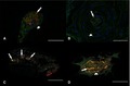
Simultaneous immunofluorescence presentation of sensory nerve endings. Simultaneous presentations of immunofluorescence staining of sensory nerve endings using S100 protein (S100), low‐affinity neurotrophin receptor p75 (p75), protein gene product 9.5 (PGP 9.5) and 4′,6‐diamidino‐2‐phenylindole (DAPI) are shown. (A) The Ruffini ending is characterized by PGP 9.5 and S100‐immunoreactive dendritic nerve endings (arrow), and a thin, at times partial encapsulation of the corpuscle, which is immunoreactive to p75 (arrowhead). (B) In contrast, the Pacini corpuscle has an onion‐layered p75‐immunoreactive capsule (arrowhead) and S100‐immunoreactive Schwann or Schwann‐related cells of the central axon (arrow). (C) The Golgi‐like ending is larger with typically smaller groups of terminal nerve endings within the corpuscle (arrows). (D) Schwann or Schwann‐like cells of axons of free nerve endings are S100 immunoreactive (arrow), whereas the membrane of Schwann cells and the perineurium of axons are p75‐immunoreactive (arrowhead). Original magnification 400×; (A) scale bar size: 20 μm; (B, C, D) scale bar size: 100 μm.
General distribution
No polarization was observed between epifascicular and interstitial distribution or between insertion into bone and central localization. Statistically, sensory nerve endings were distributed equally throughout the structures (P ≤ 0.001, respectively).
Free nerve endings [0.02 (0–0.3) per mm2 (95% CI 0.03–0.07)] were the predominant mechanoreceptor, followed by unclassifiable corpuscles [0 (0–0.04) per mm2 (95% CI 0.002–0.007)], Pacini corpuscles [0 (0–0.1) per mm2 (95% CI 0.0002–0.008)], Ruffini [0b (0–0.028) per mm2 (95% CI 0.0003–0.003)] and Golgi‐like endings [0 (0–0.27) per mm2 (95% CI −0.0001–0.002); P ≤ 0.0001, respectively (Fig. 6)]. Furthermore, the occurrence of unclassifiable corpuscles [0 (0–0.04) per mm2 (95% CI 0.002–0.007)] was significantly higher than that of Golgi‐like endings [0 (0–0.27) per mm2 (95% CI −0.0001–0.002); P ≤ 0.05 (Fig. 6)].
Figure 6.

General distribution of sensory nerve endings (n = 62). Free nerve endings (FNE) had a significantly higher density than all other corpuscles (****P < 0.0001). In addition, unclassifiable corpuscles (UC) had a significant higher density than Golgi‐like endings (*P < 0.05).
Only nine IOM structures contained Ruffini endings, ten IOM portions Pacini corpuscles, and four IOM structures Golgi‐like endings. Free nerve endings were observed in 48 out of 62 IOM structures. Twenty‐four specimens (21%) contained unclassifiable corpuscles. No sensory corpuscles were found in 13 structures, namely, one DAB, two CBs, two PABs, six DOACs and two POCs.
Interstructural distribution
Free nerve endings were significantly more abundantly distributed in the DOB [0.1 (0–0.29) per mm2 (95% CI 0.08–0.12)] than in the DAB [0.02 (0–0.2) per mm2 (95% CI 0.04–0.07); P = 0.0025], the CB [0.01 (0–0.06) per mm2 (95% CI 0.008–0.01); P < 0.00001], the PAB [0.02 (0–0.24) per mm2 (95% CI 0.02–0.06); P = 0.0001] and the DOAC [0 (0–0.07) per mm2 (95% CI −0.0001–0.01); P < 0.00001], respectively. Furthermore, the DOAC [0 (0–0.07) per mm2 (95% CI −0.0001–0.01)] contained fewer free nerve endings than the DAB [0.02 (0–0.2) per mm2 (95% CI 0.04–0.07); P = 0.00001], the PAB [0.02 (0–0.24) per mm2 (95% CI 0.02–0.06); P = 0.0003] and the POC [0.03 (0–0.4) per mm2 (95% CI 0.06–0.14); P < 0.00001, respectively (Fig. 7)].
Figure 7.

Interstructural distribution of free nerve endings. The density of free nerve endings was significant higher in the distal oblique bundle (DOB), as compared to all other structures except for the proximal oblique cord (POC; ***P < 0.0001), and significant lower in the dorsal oblique accessory cord (DOAC) as compared to all other structures except for the central band (CB; ****P < 0.001). DAB, distal accessory band; PAB, proximal accessory band.
No significant differences among all six portions of the IOM were observed with regard to Ruffini endings, Golgi‐like endings or unclassifiable corpuscles (Fig. 8). The DOB had the highest number of Pacini corpuscles. Ruffini endings were found in the DOB, DAB, PAB and POC (Fig. 8).
Figure 8.

Interstructural distribution of non‐free nerve ending receptors. No significant differences between Ruffini endings, Golgi‐like endings, and unclassifiable corpuscles were found within each structure; however, the corpuscles differed significantly in their distribution between the interosseous membrane (IOM) structures and the corpuscle types (P < 0.0001). The density of the Pacini corpuscles was significantly higher in the distal oblique bundle (DOB) than in any other corpuscle in the DOB (***P < 0.001) and higher than in any other structure of the IOM (****P < 0.0001). CB, central band; DAB, distal accessory band; DOAC, dorsal oblique accessory cord; PAB, proximal accessory band POC, proximal oblique cord; UC, unclassifiable corpuscles.
Apart from free nerve endings, Pacini corpuscles were encountered significantly more often in the DOB [0 (0–0.03) per mm2 (95% CI 0.0006–0.006)] than in any other structure of the IOM [DAB: 0 (0–0.03) per mm2 (95% CI 0.0003–0.004); CB: 0 (0–0.007) per mm2 (95% CI −0.0002–0.0005); PAB: 0 (0–0.02) per mm2 (95% CI 0.00004–0.003); DOAC: 0 (0–0) per mm2 (95% CI 0–0) per mm2; POC: 0 (0–0.04) per mm2 (95% CI −0.0002–0.006); P < 0.0001, respectively]. In addition, Pacini corpuscles [0 (0–0.03) per mm2 (95% CI 0.0006–0.006)] were the most frequent type of other sensory endings within the DOB [Ruffini: 0 (0–0.03) per mm2 (95% CI 0.0006–0.006) Golgi‐like endings: 0 (0–0.03) per mm2 (95% CI 0.00008–0.005); unclassifiable corpuscles: 0 (0–0.07) per mm2 (95% CI 0.002–0.01); P < 0.001, respectively; Fig. 8]. None of the other corpuscles showed a significant difference in distribution within one structure or between different structures of the IOM (Fig. 8).
Correlation analysis
A higher density of free nerve endings correlated significantly with a higher number of Ruffini endings (P = 0.0004; r = 0.22), Pacini corpuscles (P < 0.00001, r = 0.37), and unclassifiable corpuscles (P = 0.00008; r = 0.25). Furthermore, a higher density of unclassifiable corpuscles correlated significantly with a higher number of Golgi‐like endings [P = 0.001; r = 0.2].
Discussion
With regard to general distribution of sensory nerve endings, free nerve endings were the predominant receptor type, which is in accordance with the literature. Studies investigating ankle ligaments (Rein et al. 2012) and the triangular fibrocartilage complex (Rein et al. 2015) also report free nerve endings as the predominant receptor. This indicates that nociception has great importance in forearm rotation, axial forearm loading and weight lifting, reacting to harmful inflammatory, mechanical or chemical stimuli. A polarization of the nerve ending distribution was expected based on previous works (Morisawa, 1998; Del Valle et al. 1998; Tomita et al. 2007; Rein et al. 2012). Surprisingly, that is not what we found. Statistically, sensory nerve endings were distributed equally throughout the structures. One explanation for this is that the IOM not only provides longitudinal but also transversal stability of the forearm (Essex‐Lopresti, 1951; Gutowski et al. 2017). Furthermore, the strain distribution within the IOM depends on the forearm position: the highest overall strain exists in the neutral position, whereas the strain focus shifts proximally with pronation and distally with supination (Manson et al. 2000). An equal distribution of sensory nerve endings in the IOM, therefore, allows them to act sensitively as monitors of applied tension in this dynamic strain distribution condition.
The DOB and POC had a significant higher density of sensory nerve endings than any other structure of the IOM. The CB and DOAC had the lowest innervation. A higher density of mechanoreceptors in the DOB and POC close to the DRUJ and PRUJ, respectively, allows them to act more sensitively as monitors of tension applied to forearm rotation, as compared to the CB situated in the center of forearm (Werner et al. 2011). No significant differences were found among any of the investigated structures with regard to Ruffini, Pacini and Golgi‐like corpuscles. The DOB had the highest number of Pacini corpuscles, which react to joint acceleration and deceleration (Skoglund, 1956), providing neuromuscular control during forearm pronosupination.
Positive correlations between the number of free nerve endings and Ruffini, Pacini, Golgi corpuscles and unclassifiable corpuscles, as well as between Golgi‐like endings and unclassifiable corpuscles, have been observed. The occurrence of different receptor types ensures sensorimotor monitoring of all joint activities. Furthermore, the different mechanoreceptor types complement each other due to their defined physiological properties, resulting in sensorimotor control of all complex joint movements.
Limitations of the present study include the fact that only a small number of specimens were used in the cadaver study. Furthermore, unclassifiable corpuscles were the second most frequently observed receptor in the study, which is a limitation of the existing 'gold standard' classification of Freeman and Wyke (Freeman & Wyke, 1967). In addition, two‐dimensional analysis does not allow complete representation of whole sensory corpuscles; however, even three‐dimensional imaging of sensory nerve endings has shown a high morphological variability of sensory corpuscles, resulting in a large number of unclassifiable corpuscles in the dorsal radiocarpal ligaments (Tomita et al. 2007). Donor age, and post mortem conditions and interval were further limitations of the study.
Many techniques have been described for the reconstruction of the CB in chronic Essex‐Lopresti injuries, including: bone‐patellar tendon‐bone graft (Gaspar et al. 2018); synthetic grafts (Sabo & Watts, 2012); tightrope tenodesis (Brin et al. 2014); tendons grafts, such as the anterior tibial (Miller et al. 2016), palmaris longus (Tejwani et al. 2005), flexor carpi radialis (Skahen et al. 1997), semitendinosus (Soubeyrand et al. 2006), Achilles tendon (Stabile et al. 2005), fascia lata (Bigazzi et al. 2017), and pronator teres rerouting (Chloros et al. 2008). Based on the present study, the CB is sparsely innervated, which accords with its static stability functions (Werner et al. 2011; Anderson et al. 2015), and reconstruction could be performed with non‐sensory tissue (Skahen et al. 1997; Tejwani et al. 2005; Soubeyrand et al. 2006; Miller et al. 2016) or material (Sabo & Watts, 2012; Brin et al. 2014) from the proprioceptive point of view.
In contrast, reconstruction of the richly innervated DOB with tendon grafts (Riggenbach et al. 2015) may not address the sensory function of that IOM bundle (Kim et al. 2012), and consequently fails to account for the dynamic aspects of DRUJ stability. Alternatively, the DOB could be reconstructed with brachioradialis distal tendon rerouting (Aita et al. 2018) or bone‐patella tendon‐bone interposition. A histological study has shown that the patellar tendon contains sensory nerve endings (Cabuk & Kusku‐Cabuk, 2016); however, the bone‐patellar tendon‐bone graft has been only performed for reconstruction of the CB (Marcotte & Osterman, 2007; Gaspar et al. 2018). The DOB is an important stabilizer of the DRUJ (Kitamura et al. 2011; Arimitsu et al. 2011); therefore, an injury to the richly innervated DOB may lead to sensorimotor control impairment due to disruption of the ligament‐muscular pathways that mediate joint neuromuscular control (Hagert et al. 2009). The pronator quadratus and the extensor carpi ulnaris muscles as well as the triangular fibrocartilage complex offer dynamic proprioceptive support at the DRUJ during forearm rotation, which is why both muscles play a key role in dynamic stabilization exercises to address DRUJ instability (Hagert & Hagert, 2010; Esplugas et al. 2016).
In conclusion, the IOM contains all five types of sensory nerve endings, which are precisely delineated with the immunofluorescence combination of S100, p75, PGP9.5 and DAPI. Consequently, the IOM plays a crucial role in dynamic forearm stabilization, in which nociception is key. The DOB and POC have pronounced proprioceptive functions; therefore, dynamic stabilization of the forearm mainly takes place in the vicinity of the DRUJ and PRUJ, whereas the CB and DOAC have primary static functions.
Conflict of interest
The authors declare that they have no competing interests. This study was financially supported by Deutsche Gesetzliche Unfallversicherung, Sankt Augustin, Germany (grant number: FR‐0272) and Bauerfeind AG, Zeulenroda‐Triebes, Germany (grant number: 8344). The authors disclose any financial conflicts of interest that may influence interpretation of this study and/ or results.
Supporting information
Fig S1 Unclassifiable corpuscle
Acknowledgements
We thank Arthur Eisenkrein for photographical work, Manuel Llusa, MD, PhD (Department of Anatomy, University of Barcelona, Barcelona, Spain) for generous assistance in the laboratory work, and Christian Retschke (Leipzig, Germany) for logistic support. We thank the BioImaging Core Facility BCF (http://www.maginlab.eu/home-bcf.html).
Data availability statement
We confirm the absence of shared data.
References
- Aita MA, Mallozi RC, Ozaki W, et al. (2018) Ligamentous reconstruction of the interosseous membrane of the forearm in the treatment of instability of the distal radioulnar joint. Rev Bras Ortop 53(2), 184–191. [DOI] [PMC free article] [PubMed] [Google Scholar]
- Anderson A, Werner FW, Tucci ER, et al. (2015) Role of the interosseous membrane and annular ligament in stabilizing the proximal radial head. J Shoulder Elb Surg 24(12), 1926–1933. [DOI] [PubMed] [Google Scholar]
- Arimitsu S, Moritomo H, Kitamura T, et al. (2011) The Stabilizing effect of the distal interosseous. J Bone Jt Surg Am 93(21), 2022–2030. [DOI] [PubMed] [Google Scholar]
- Bigazzi P, Marenghi L, Biondi M, et al. (2017) Surgical treatment of chronic essex‐lopresti lesion. Tech Hand Up Extrem Surg 21(1), 2–7. [DOI] [PubMed] [Google Scholar]
- Brin YS, Palmanovich E, Bivas A, et al. (2014) Treating acute Essex‐lopresti injury with the TightRope device: a case study. Tech Hand Up Extrem Surg 18(1), 51–55. [DOI] [PubMed] [Google Scholar]
- Buckminster Fuller R (1961) Tensegrity. Portf Arts News Annu 4, 112–127. [Google Scholar]
- Cabuk H, Kusku‐Cabuk F (2016) Mechanoreceptors of the ligaments and tendons around the knee. Clin Anat 29(6), 789–795. [DOI] [PubMed] [Google Scholar]
- Chikenji T, Berger RA, Fujimiya M, et al. (2011) Distribution of nerve endings in human distal interphalangeal joint and surrounding structures. J Hand Surg Am 36(3), 406–412. [DOI] [PubMed] [Google Scholar]
- Chloros GD, Wiesler ER, Stabile KJ, et al. (2008) Reconstruction of essex‐lopresti injury of the forearm: technical note. J Hand Surg Am 33(1), 124–130. [DOI] [PubMed] [Google Scholar]
- Del Valle ME, Harwin SF, Maestro A, et al. (1998) Immunohistochemical analysis of mechanoreceptors in the human posterior cruciate ligament. J Arthroplasty 13(8), 916–922. [DOI] [PubMed] [Google Scholar]
- Denti M, Monteleone M, Berardi A, et al. (1994) Anterior cruciate ligament mechanoreceptors. Histologic studies on lesions and reconstruction. Clin Orthop Relat Res 308, 29–32. [PubMed] [Google Scholar]
- Esplugas M, Garcia‐Elias M, Lluch A, et al. (2016) Role of muscles in the stabilization of ligament‐deficient wrists. J Hand Ther 29(2), 166–174. [DOI] [PubMed] [Google Scholar]
- Essex‐Lopresti P (1951) Fractures of the radial head with distal radio‐ulnar dislocation. Report of two cases. J Bone Joint Surg Br 33(2), 244–247. [PubMed] [Google Scholar]
- Freeman MA, Wyke B (1967) The innervation of the ankle joint. An anatomical and histological study in the cat. Acta Anat (Basel) 68(3), 321–333. [DOI] [PubMed] [Google Scholar]
- Gabl M, Zimmermann R, Angermann P, et al. (1998) The interosseous membrane and its influence on the distal radioulnar joint: an anatomical investigation of the distal tract. J Hand Surg Eur 23(2), 179–182. [DOI] [PubMed] [Google Scholar]
- Gaspar MP, Adams JE, Zohn RC, et al. (2018) Late reconstruction of the interosseous membrane with bone‐patellar tendon‐bone graft for chronic essex‐lopresti injuries. J Bone Joint Surg Am 100(5), 416–427. [DOI] [PubMed] [Google Scholar]
- Grassmann JP, Hakimi M, Gehrmann SV, et al. (2014) The treatment of the acute essex‐lopresti injury. Bone Joint J 96B(10), 1385–1391. [DOI] [PubMed] [Google Scholar]
- Gutowski CJ, Darvish K, Ilyas AM, et al. (2017) Interosseous ligament and transverse forearm stability: a biomechanical cadaver study. J Hand Surg Am 42(2), 87–95. [DOI] [PubMed] [Google Scholar]
- Hackl M, Andermahr J, Staat M, et al. (2017) Suture button reconstruction of the central band of the interosseous membrane in Essex‐Lopresti lesions: a comparative biomechanical investigation. J Hand Surg Eur 42(4), 370–376. [DOI] [PubMed] [Google Scholar]
- Hagert CG (1992) The distal radioulnar joint in relation to the whole forearm. Clin Orthop Relat Res 275, 56–64. [PubMed] [Google Scholar]
- Hagert E (2008) Wrist ligaments‐ innervation patterns and ligamento‐muscular reflexes. PhD thesis 2008; Stockholm, Sweden: Karolinska Institutet, p. 1–51. [Google Scholar]
- Hagert E, Hagert CG (2010) Understanding stability of the distal radioulnar joint through an understanding of its anatomy. Hand Clin 26(4), 459–466. [DOI] [PubMed] [Google Scholar]
- Hagert E, Ljung BO, Forsgren S (2004) General innervation pattern and sensory corpuscles in the scapholunate interosseous ligament. Cells Tissues Organs 177(1), 47–54. [DOI] [PubMed] [Google Scholar]
- Hagert E, Forsgren S, Ljung BO (2005) Differences in the presence of mechanoreceptors and nerve structures between wrist ligaments may imply differential roles in wrist stabilization. J Orthop Res 23(4), 757–763. [DOI] [PubMed] [Google Scholar]
- Hagert E, Persson JKE, Werner M, et al. (2009) Evidence of wrist proprioceptive reflexes elicited after stimulation of the scapholunate interosseous ligament. J Hand Surg Am 34(4), 642–651. [DOI] [PubMed] [Google Scholar]
- Hagert E, Lee J, Ladd AL (2012) Innervation patterns of thumb trapeziometacarpal joint ligaments. J Hand Surg Am 37(4), 706–714. [DOI] [PubMed] [Google Scholar]
- Hotchkiss RN, An K‐N, Sowa DT, et al. (1989) An anatomic and mechanical study of the interosseous membrane of the forearm: pathomechanics of proximal migration of the radius. J Hand Surg Am 14(2), 256–261. [DOI] [PubMed] [Google Scholar]
- Kholinne E, Lee HJ, Kim GY, et al. (2018) Mechanoreceptors distribution in the human medial collateral ligament of the elbow. Orthop Traumatol Surg Res 104(2), 251–255. [DOI] [PubMed] [Google Scholar]
- Kholinne E, Lee HJ, Lee YM, et al. (2018) Mechanoreceptor profile of the lateral collateral ligament complex in the human elbow. Asia‐Pacific J Sport Med Arthrosc Rehabil Technol 14, 17–21. [DOI] [PMC free article] [PubMed] [Google Scholar]
- Kim SH, Chun CH, Chun KC, et al. (2012) Histological assessment of mechanoreceptors in achilles allografts after anterior cruciate ligament reconstruction. Am J Sports Med 40(9), 2061–2065. [DOI] [PubMed] [Google Scholar]
- Kitamura T, Moritomo H, Arimitsu S, et al. (2011) The biomechanical effect of the distal interosseous membrane on distal radioulnar joint stability: a preliminary anatomic study. J Hand Surg Am 36(10), 1626–1630. [DOI] [PubMed] [Google Scholar]
- Lee J, Ladd A, Hagert E (2012) Immunofluorescent triple‐staining technique to identify sensory nerve endings in human thumb ligaments. Cells Tissues Organs 195(5), 456–464. [DOI] [PubMed] [Google Scholar]
- Manson TT, Pfaeffle HJ, Herndon JH, et al. (2000) Forearm rotation alters interosseous ligament strain distribution. J Hand Surg Am 25(6), 1058–1063. [DOI] [PubMed] [Google Scholar]
- Marcotte AL, Osterman AL (2007) Longitudinal radioulnar dissociation: identification and treatment of acute and chronic injuries. Hand Clin 23(2), 195–208. [DOI] [PubMed] [Google Scholar]
- Miller AJ, Naik TU, Seigerman DA, et al. (2016) Anatomic interosseus membrane reconstruction utilizing the biceps button and screw tenodesis for essex‐lopresti injuries. Tech Hand Up Extrem Surg 20(1), 6–13. [DOI] [PubMed] [Google Scholar]
- Morisawa Y (1998) Morphological study of mechanoreceptors on the coracoacromial ligament. J Orthop Sci 3(2), 102–110. [DOI] [PubMed] [Google Scholar]
- Noda K, Goto A, Murase T, et al. (2009) Interosseous membrane of the forearm: an anatomical study of ligament attachment locations. J Hand Surg Am 34(3), 415–422. [DOI] [PubMed] [Google Scholar]
- Petrie S, Collins JG, Solomonow M, et al. (1998) Mechanoreceptors in the human elbow ligaments. J Hand Surg Am 23(3), 512–518. [DOI] [PubMed] [Google Scholar]
- Rein S, Hagert E, Hanisch U, et al. (2012) Immunohistochemical analysis of sensory nerve endings in ankle ligaments: a cadaver study. Cells Tissues Organs 197(1), 64–76. [DOI] [PubMed] [Google Scholar]
- Rein S, Semisch M, Garcia‐Elias M, et al. (2015) Immunohistochemical mapping of sensory nerve endings in the human triangular fibrocartilage complex. Clin Orthop Relat Res 473(10), 3245–3253. [DOI] [PMC free article] [PubMed] [Google Scholar]
- Riggenbach MD, Wright TW, Dell PC (2015) Reconstruction of the distal oblique bundle of the interosseous membrane: a technique to restore distal radioulnar joint stability. J Hand Surg Am 40(11), 2279–2282. [DOI] [PubMed] [Google Scholar]
- Sabo MT, Watts AC (2012) Reconstructing the interosseous membrane. Tech Hand Up Extrem Surg 16(4), 187–193. [DOI] [PubMed] [Google Scholar]
- Skahen JR, Palmer AK, Werner FW, et al. (1997) Reconstruction of the interosseous membrane of the forearm in cadavers. J Hand Surg Am 22(6), 986–994. [DOI] [PubMed] [Google Scholar]
- Skoglund S (1956) Anatomical and physiological studies of knee joint innervation in the cat. Acta Physiol Scand 36(Suppl 124), 1–100. [PubMed] [Google Scholar]
- Soubeyrand M, Oberlin C, Dumontier C, et al. (2006) Ligamentoplasty of the forearm interosseous membrane using the semitendinosus tendon: anatomical study and surgical procedure. Surg Radiol Anat 28(3), 300–307. [DOI] [PubMed] [Google Scholar]
- Stabile KJ, Pfaeffle J, Saris I, et al. (2005) Structural properties of reconstruction constructs for the interosseous ligament of the forearm. J Hand Surg Am 30(2), 312–318. [DOI] [PubMed] [Google Scholar]
- Tejwani SG, Markolf KL, Benhaim P (2005) Graft reconstruction of the interosseous membrane in conjunction with metallic radial head replacement: a cadaveric study. J Hand Surg Am 30(2), 335–342. [DOI] [PubMed] [Google Scholar]
- Tomita K, Berger EJ, Berger RA, et al. (2007) Distribution of nerve endings in the human dorsal radiocarpal ligament. J Hand Surg Am 32(4), 466–473. [DOI] [PubMed] [Google Scholar]
- Werner FW, Taormina JL, Sutton LG, et al. (2011) Structural properties of 6 forearm ligaments. J Hand Surg Am 36(12), 1981–1987. [DOI] [PubMed] [Google Scholar]
Associated Data
This section collects any data citations, data availability statements, or supplementary materials included in this article.
Supplementary Materials
Fig S1 Unclassifiable corpuscle
Data Availability Statement
We confirm the absence of shared data.