

Abstract
Introduction:
Anogenital area is a small compartment in the human body. Recognition of various dermatological conditions affecting this area in children is essential for any physician involved in pediatric examination and evaluation.
Aim:
To assess the nature, diagnoses, and gender differences of Anogenital lesions in pediatric patients presented to Royal Medical Services (RMS) general dermatology clinics, who were five year old and younger.
Methods:
The authors reviewed patients’ medical records in the period between September 2015 and September 2018. The inclusion criteria were children of both genders who were five year old or younger and presented with papular lesions in the Anogenital area. Those patients visited the general dermatology clinics of The Royal Medical Services Hospitals from the Eastern and Southern regions of Jordan.
Results:
Over a period of 3 years, a total of 514 patients were five year old or younger presented with various Anogenital papular lesions were evaluated and treated in general dermatology clinics. 35% of the patients presented with Perianal Psuedoverrucous Papules and nodules, 21% presented with Anogenital warts, 16% presented with Molluscum Contageosum. Moreover, 10% were presented with Epidermal nevi, 6% presented with Pearly Penile Papules, 7% presented with Infantile Perineal Pyramidal Protrusion, 4% presented with Vulvar Vestibular Papillomatosis, 0.2% presented with Juvenile Xanthogranuloma, 0.2% presented with lymphangioma Circumscriptum, and 0.2% presented with median raphe cyst. In addition, gender differences were noticed among Genital Warts, Mollascum Contageosum, Pearly Penile Papules, and Vulvar Vestibular Papillomatosis.
Conclusion:
Anogenital papules in children have variable clinical presentations and can be caused by multiple number of infectious and non-infectious factors. The presence of such lesions can be a source of a major concern for parents, and might be mistakenly assumed as a result of sexual assaults. Proper recognition of these papules is of paramount importance for all physicians involved in children examination, to appropriately reassure parents and avoid unnecessary investigations and psychological distress.
Keywords: The Royal Medical Services (RMS), Anogenital, Perianal Pseudoverrucous Papules and Nodules (PPPN)
1. INTRODUCTION
Many infectious and non-infectious factors can cause Anogenital papules in children. Proper assessment is crucial for the accurate diagnosis and management. The Perianal Pseudoverrucous Papules and Nodules (PPPN) are smooth verrucous papules and nodules in the perianal, genital, suprapubic area, and the buttocks (1-3). They develop as a result of chronic irritation from prolonged exposure to stool and/or urine. PPPN may occur in patients with incontinence, persistent diarrhea and congenital megacolon (1-4). The diagnosis is based on the history and the clinical picture of the patient (5). These lesions usually persist until complete cessation of diarrhea or treatment of the underlying problem (2, 3, 6).
Anogenital warts (Condylomata Acuminata), is an infection caused by human papilloma virus (HPV) in the Anogenital area (7). HPV is a double stranded DNA virus of more than 100 types; certain types are responsible about infection at specific site (8, 9). This virus has long incubation period ranging from 3 weeks to 8 months (7, 10). Clinically, the infection is manifested as flesh colored, brown or pink verrucous papules or plaques (the cauliflower appearance) (7, 11). The possible ways for having HPV infection in children include the following: hetero-inoculation or autoinoculation (which mean that the virus can be transmitted form the hands of caregiver or from the hands of a child him/herself if they touch the genital area during hygiene or changing diapers). Perinatal and prenatal transmission and sexual abuse are other ways of HPV transmission among children.
The incidence of sexual abuse in children with HPV varies widely across countries from <10% up to 90% (7, 10, 12), and it increases with increase in child’s age (9, 12, 13). Molluscum Contagiosum (MC) is an infection caused by molluscum contagiosum virus that is a member of Poxviridae family, this virus has long incubation period that may last up to 6 months. Clinically the MC lesions appear as small shiny papules with central umbilication (14, 15). MC virus is easily transmitted between children by non-sexual skin contact as in bathing or swimming. In children, MC is frequently seen on the face, extremities, and trunk. However, it is rarely occurs in the Anogenital area (15, 16). These lesions may resolve spontaneously, although treatment is better to prevent further spread of the virus (15). Epidermal nevi are benign cutaneous hamartoma characterized by hyper pigmented verrucous papules and plaques that follow linear arrangement along Blaschko’s line over face, trunk, extremities, and rarely over genitalia (17-19). Epidermal nevi usually appear in the first year of life as flat lesions that get more verrucous with age (20-22). The diagnosis is made by clinical picture. The skin biopsy, if needed, will show epidermal hyperplasia, hyperkeratosis, acanthosis, papillomatosis, and variable parakeratosis (21). Medical management is extremely variable, but usually does not result in complete resolution (19, 20).
Pearly Penile Papules (PPP) are a small dome shaped, skin colored papules distributed all the way around the sulcus or corona of the glans penis, which is considered as a normal anatomic variant (23). PPP are asymptomatic lesions and can be diagnosed clinically without further lab work (24). No treatment is required (25, 26). Infantile Perineal Pyramidal Protrusion (IPPP) is a skin colored soft tissue swelling located in the perineal median raphae anterior to the anus. It may be presented at birth or appear soon after (27, 28).
Three subtypes of IPPP are encountered (29-31). The first one is the constitutional or congenital form that appears early in life with unknown cause. The acquired type is the one associated with persistent constipation. The third subtype is the lichen sclerosis et atrophicus associated type. The diagnosis of IPPP is made clinically. It usually resolves spontaneously and no treatment is required (29, 30).
Vulvar Vestibular papillomatosis (VVP) is a normal anatomic variant in the vulva that is the female counterpart of the pearly penile papules (33, 34). VVP presents as diffuse symmetrical micropapillary projections seen on the medial aspect of the labia minora, introitus, and lower vagina; it does not cluster as in warts (33-35). The typical clinical picture makes the diagnosis of this lesion. However, these papules may regress spontaneously if left untreated (33-35). Juvenile xanthogranuloma (JXG) is a benign tumor of non-langerhan’s cell histiocytosis (36). JXG presented as a yellowish-waxy colored papulonodular solitary or multiple lesions. JXG usually seen over trunk, extremities, head and neck, and rarely reported over genital area (36-38). The diagnosis is made by the typical clinical picture. Lymphangioma circumscriptum is a congenital malformation of lymphatic vessels, presented since early childhood as psuedovesicular, nodular or warty lesions. Lesions are rarely seen in the Anogenital area (39, 40). Median raphe cyst is a rare congenital anomaly, clinically presented as cyst or linear lesion over the ventral surface of penis and scrotum but may occur anywhere from the urethral meatus to the anus (41, 42). Diagnosis is based on typical clinical picture and location of the lesion. No treatment is required (42).
2. AIM
The study aims to assess the nature, diagnoses, and gender differences of Anogenital lesions in pediatric patients presented to RMS general dermatology clinics, who were five year old and younger.
3. MATERIAL AND METHODS
The authors collected and reviewed patients’ medical records of pediatric patients who were five year old or younger and presented with papular lesions in the anogenital area. Those patients visited The Royal Medical Services’ general dermatology clinics in the period between September 2015 and September 2018. RMS general dermatology clinics serve people across Jordan. The current study covered records of the Eastern and Southern clinics of Jordan. Detailed personal and family history were taken and complete physical examination was performed for each patient. All lesions were diagnosed upon clinical diagnoses by expert clinical dermatologists. No biopsies seemed necessary at the time of examination. Photo of the lesions were taken with consent from parents. No conflict of interest to declare and ethical approval from the Ethical Committee at the Royal Medical Services was obtained prior to the data collection phase. The patients’ permission was taken from their parents and all information obtained was kept confidential.
4. RESULTS
Over the period of 3 years, between September 2015 to and September 2018, the total number of patients evaluated in RMS clinics was about 44,640 patients, 4355 patients (10% of the total number of patients seen in our clinics) were kids below the age of 5 years. 514 patients (about 12% of pediatric patients and 1% of the total number of patients seen in our clinics) were five year old or younger and presented with Anogenital papular lesions. Among this group; 182 patients (35%) presented with Perianal Pseudoverrucous Papules and Nodules (PPPN), 110 patients (21%) presented with genital warts. Moreover, 81 patients (16%) presented with Molluscum Contageosum, 54 patients (10%) presented with epidermal nevi, 30 patients (6%) presented with Pearly Penile Papules (PPP), 34 patients (7%) presented with Infantile Perineal Pyramidal Protrusion (IPPP), 21 patients (4%) presented with Vulvar Vestibular Papillomatosis (VVP). One patient (0.2%) presented with Juvenile Xanthogranuloma, one patient (0.2%) presented with lymphangioma Circumscriptum, and one patient (0.2%) presented with median raphe cyst (Table 1).
Table 1. The Anogenital Lesions Encountered in our clinics.
| Diagnosis | Number of Patients | Percentage |
|---|---|---|
| Perianal Psuedoverrucous Papules and Nodules | 182 | 35% |
| Genital Warts | 110 | 21% |
| Mollascum Contageosum | 81 | 16% |
| Epidermal Nevi | 54 | 10% |
| Pearly Penile Papules | 30 | 6% |
| Infantile Perineal Pyramidal Protrusion | 34 | 7% |
| Vulvar Vestibular PapilloMatosis | 21 | 4% |
| Juvenile Xanthogranuolma | 1 | 0.2% |
| Lymphangioma Circumscriptum | 1 | 0.2% |
| Median Raphe Cyst | 1 | 0.2% |
Gender differences was tested with frequency of Anogenital papular lesions and the results showed that there are significant differences between males and females in the following types; Mollascum Contageosum (X2 = 14.75, P = 0.000), Pearly Penile Papules (X2 = 15.69, P = 0.000), Vulvar Vestibular Papillomatosis (X2 = 15.69, P = 0.000) and Genital Warts (X2 = 5.14, P = 0.02) (Table 2). Table 3 presents the response to Anogenital Papular Lesions treatment. The results showed that different dosages of treatment were effective and significant at p value ≤ .01 for Perianal Psuedoverrucous Papules and Nodules, Genital Warts, Mollascum Contageosum, and Juvenile Xanthogranuolma. However, treatment were effective and significant at p value ≤ .05 for Epidermal Nevi.
Table 2. Gender Differences in Study Variables.
| Diagnosis | # of Females | # of Males | X2 | P value |
|---|---|---|---|---|
| Perianal Psuedoverrucous Papules and Nodules | 95 | 87 | 0.35 | .55 |
| Genital Warts | 65 | 44 | 5.14 | .02* |
| Mollascum Contageosum | 57 | 24 | 14.75 | .000** |
| Epidermal Nevi | 33 | 21 | 3.07 | .80 |
| Pearly Penile Papules | 2 | 28 | 15.69 | .000** |
| Infantile Perineal Pyramidal Protrusion | 20 | 14 | 1 | .31 |
| Vulvar Vestibular Papillomatosis | 19 | 2 | 15.69 | .000** |
| Juvenile Xanthogranuolma | 0 | 1 | NA | NA |
| Lymphangioma Circumscriptum | 1 | 0 | NA | NA |
Table 3. Effectiveness of Treatment for Study Variables.
| Diagnosis | Treatment Effectiveness | P value |
|---|---|---|
| Perianal Psuedoverrucous Papules and Nodules | 90% | ≤.01 |
| Genital Warts | 87% | ≤.01 |
| Mollascum Contageosum | 92% | ≤.01 |
| Epidermal Nevi | 72% | ≤.05 |
| Pearly Penile Papules | No need for treatment | ≥ .05 |
| Infantile Perineal Pyramidal Protrusion | No need for treatment | ≥ .05 |
| Vulvar Vestibular Papillomatosis | No need for treatment | ≥ .05 |
| Juvenile Xanthogranuolma | Surgical excision | ≤.01 |
| Lymphangioma Circumscriptum | CO2 laser ( not effective) | ≥ .05 |
| Median Raphe cyst | No need for treatment | ≥ .05 |
5. DISCUSSION
Anogenital lesions in children represent a major concern to parents. One percent of patients attended our general dermatology clinics were children 5-year-old or younger and presented with Anogenital papular lesions. To the best of our knowledge, we did not find this kind of analysis and statistics in the literature that collectively studied this population with these possible diagnoses. In addition, the literature is lacking statistical data to compare with our results.
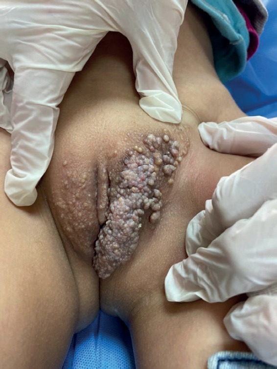
The Perianal Pseudoverrucous Papules and Nodules (PPPN) were the most commonly encountered in our clinics (35% of cases). PPPN were mentioned in literature as case reports without mentioning the actual incidence (1-6). In the study group, PPPN was found to be relatively common. This may be related to the low socioeconomic status and hygienic level of the patients in those underserved and poor area of Jordan. The diagnosis is based on the history and the clinical picture. All of these patients had history of recent chronic diarrhea, and clinically presented with verrucous papules or nodules over erythematous macerated base in the Anogenital area. These lesions did not improve unless complete cessation of diarrhea was achieved (Figure 1a, 1b, 1c). Anogenital warts were the second most common cause of genital lesions (21% of cases) encountered in study study group. The incidence of HPV infections in children varies in literature due to different definition of HPV (the presence of HPV DNA or the presence of clinical lesions (8-10). Anogenital warts may be acquired through sexual or non- sexual contact (7, 10). Although the concern of child abuse is a major concern in kids, most kids under the age of four may acquire HPV infection via non-sexual contact (7, 10, 12). In literature, the incidence of genital warts increases with increase in patient’s age (12). The diagnosis of genital warts in kids is based mainly on the clinical picture. Histopathology as well as viral serology are infrequently required for diagnosis (7, 9, 10). It is important to note that no test is available to tell exactly what is the mode of viral transmission and the source of infection (7, 10-12). The study cases were diagnosed based on the typical clinical picture of a flesh colored, brown or pink verrucous papules or plaques in the Anogenital area (Figure 2a, 2b). Molluscum Contageosum was encountered in 16% of the study patients. Molluscum contageosum is commonly acquired by children from infected children via non-sexual skin contact as in bathing or swimming. In children, MC is frequently seen on the face, extremities and trunk, but rarely occurs in genital area (14-16). If present in genitalia, MC appears as small papules with central umbilication. These lesions may resolve spontaneously but treatment is better to prevent further spread of the virus (14, 15) (Figure 3). Epidermal nevi were encountered in 10% of the study patients. In literature, there are only few reports of epidermal nevi occurring over Anogenital area without documented exact prevalence (17-19). Epidermal nevi present as linear hyper pigmented verrucous grouped papules and plaques (22). The diagnosis was made by the typical history and clinical picture (Figure 4a, 4b). Pearly Penile Papules (PPP) were seen in 6% of our patients. The reported prevalence of PPP ranges from 14%-48% of males, and it is rarely seen in children (23, 24). The incidence in current study group is less than what has been reported in literature. This may be related to the younger age group reviewed in the study. PPP are asymptomatic lesions and are diagnosed by the typical clinical appearance of dome shaped, small, skin colored papules distributed around the sulcus or corona of the glans penis, it is considered as a normal anatomic variant (23-26) (Figure 5).
Figure 1a. Perianal Pseudoverrucous papules and nodules over erythematous macerated base in the Anogenital area.

Figure 1b. Perianal Psuedoverrucous papules and nodules in a female patient.

Figure 1c. Perianal Pseudoverrucous papules and nodules.

Figure 2a. Anogenital warts.

Figure 2b. Perianal warts, typical clinical picture of a flesh colored, brown or pink verrucous papules or plaques in the Anogenital area.

Figure 3. Anogenital Molluscum Contageosum. These lesions may resolve spontaneously but treatment is better to prevent further spread of the virus.

Figure 4 a. Linear Epidermal Nevus over vulva in a female patient with CHILD syndrom.

Figure 4,b. Linear epidermal nevus over buttocks, the diagnosis was made by the typical history and clinical picture.
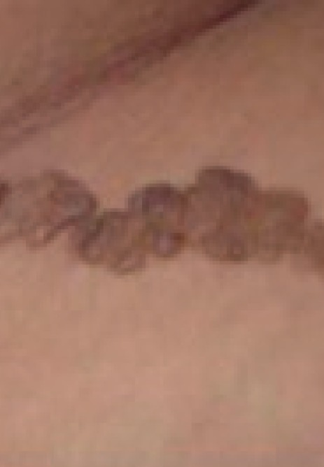
Figure 5. Pearly penile papules, small, skin colored papules distributed around the sulcus or corona of the glans penis.

Infantile Perineal Pyramidal Protrusion (IPPP) was seen in 7% of our patients. It is rare and reported in literature as case reports with unknown exact prevalence (28-30). IPPP is a skin colored soft tissue swelling located in the perineal median raphae anterior to the anus, which may present at birth or soon after (28, 29, 31). The diagnosis of IPPP is made by clinical recognition (Figure 6).
Figure 6. Infantile perineal pyramidal protrusion, the diagnosis of IPPP is made by clinical recognition.

Vulvar Vestibular papillomatosis (VVP) was seen in 4% of the study patients. VVP prevalence range from 1%-33% in literature (34, 35). The number of cases in the current study group matched the reported prevalence. VVP present as diffuse symmetrical micropapillary projections seen on the medial aspect of labia minora, introitus, and lower vagina, not clustered as seen in warts (33-35). The typical clinical picture makes the diagnosis of this lesion. One patient (0.1%) presented with Juvenile xanthogranuloma (JXG) over suprapubic area. JXG is a benign tumor of non-langerhan’s cell histiocytosis (36). JXG usually seen over trunk, extremities, head and neck, and rarely reported over anogenital area (36-38). The typical clinical picture of a yellowish-waxy colored solitary nodule diagnosed our case over pubic area. After 4 months, the parents asked for excision of this lesion, the histopathology was consistent with our clinical diagnosis (Figure 7). One patient (0.1%) presented with lymphangioma circuscriptum over vulva. Lymphangioma circumscriptum is a congenital malformation of lymphatic vessels, presented since early childhood as psuedovesicular, warty lesions over vulva. Lesions not commonly affecting the vulva (39, 40) (Figure 8).
Figure 7. Juvenile Xanthogranuloma, typical clinical picture of a yellowish-waxy coloured solitary nodule over pubic area.

Figure 8. Lymphangioma Circumscriptum presented since early childhood as Psuedovesicular warty lesions over vulva.
Finally, one patient (0.1%) presented with median raphe cyst (MRC). MRC is a rare congenital anomaly that present as a cyst or linear lesion over the ventral surface of penis and scrotum, but may occur anywhere from the urethral meatus to the anus (41, 42). Our case was diagnosed based on the typical clinical picture of a linear protrusion over the ventral surface of perineum extending from scrotum to anus (Figure 9).
Figure 9. Median Raphae cyst Linear protrusion over the ventral surface of perineum extending from scrotum to anus.

6. CONCLUSIONS
Anogenital papules in children can be caused by multiple number of infectious and non-infectious factors, with variable clinical presentations. The presence of such lesions can be a source of major concern for parents and may lead to be mistakenly assumed as sexual assaults. Proper recognition of the nature of these papules is of paramount importance for all physicians involved in children’s examination in order to properly reassure the parents, and to avoid unnecessary investigations and psychological distress. The authors believed that the current study is unique, comprehensive, and can be considered a good addition to the body of the literature.
Acknowledgments
Authors are thankful to Dr. Mahmoud Al-Hussami for his valuable contributions to the study.
Declaration of patient consent
The authors certify that they obtained all appropriate patient consent forms.
Author’s contribution
All authors were included in all steps of preparation this article. The first author made the final proof reading.
Conflict of Interest
The authors have no conflicts of interest to disclose
Financial support and sponsorship
No funding was obtained for this study.
REFERENCES
- 1.Dandale A, Dhurat R, Ghate S. Perianal pseudoverrucous papules and nodule. Indian J Sex Transm Dis AIDS. 2013 Jan-Jun;34(1):44–46. doi: 10.4103/2589-0557.112939. [DOI] [PMC free article] [PubMed] [Google Scholar]
- 2.Goldberg NS, Esterly NB, Rothman KF, Fallon JD, Cropley TG, Szaniawski W, et al. Perianal pseudoverrucous papules and nodules in children. Arch Dermatol. 1992;128:240–242. [PubMed] [Google Scholar]
- 3.Rodriguez CI, Garcia-patos BV, Pedragosa JR, Castells RA. Perianal pseudoverrucous papules and nodules after surgery for Hirschsprung disease. J Pediatr. 1994;125:914–916. doi: 10.1016/s0022-3476(05)82008-6. [DOI] [PubMed] [Google Scholar]
- 4.Coppo P, Salomone R. Pseudoverrucous papules: an aspect of incontinence in children. Journal of the European Academy of Dermatology and Venereology. 2002;16(4) doi: 10.1046/j.1468-3083.2002.00418.x. [DOI] [PubMed] [Google Scholar]
- 5.Frasier LD. The anogenital exam: Diagnostic dilemmas, Mimics of abuse. In: Finkel MA, editor. Medical evaluation of child sexual abuse: A practical guide. 2nd. Vol. 168. California: Sage publication; 2002. [Google Scholar]
- 6.Amiry S, Pride H, Tyler W. Perianal psuedoverrucose papules and nodules mimicking Condylomata acuminate and sexual abuse. CUTIS. 2001 Apr;67:335–338. [PubMed] [Google Scholar]
- 7.Gearhart P. Human papillomavirus. 2014. Aug 4, http://emedicine.medscape.com/article/219110-overview.
- 8.Myhre AK, Dalen A, Berntzen K, Bratlid D. Anogenital human papillomavirus in non-abused preschool children. Acta Paediatr. 2003 Dec;92(12):1445–1452. [PubMed] [Google Scholar]
- 9.Honor G. Anogenital warts in children: sexual abuse or not? Journal of Pediatric Health Care. 2014;18(4):165–170. doi: 10.1016/j.pedhc.2003.01.001. [DOI] [PubMed] [Google Scholar]
- 10.Jayasinghe Y, Garland SM. Genital warts in children: what do they mean? Arch Dis Child. 2006 Aug;91(8):696–700. doi: 10.1136/adc.2005.092080. [DOI] [PMC free article] [PubMed] [Google Scholar]
- 11.Willey D, Douglas J, Beutner K, Cox T, Fife K, Fukumoto L. External Genital warts: Diagnosis, Treatment and Prevention. Clinical infectious Diseases. 2002;35(2):210–224. doi: 10.1086/342109. [DOI] [PubMed] [Google Scholar]
- 12.Benjamin L, Levy M, Ofori A. Condylomata acuminate (Anogenital warts) in children. [2014]. Available on: www.uptodate.com.
- 13.Syrjanen S. HPV infections in children. Papillomavirus Report. :20031493–110. [Google Scholar]
- 14.Rane V. Penile appearance lumps and bumps. The Royal Australian College of General Practitiones. 2013;42(5):270–274. Available on: http://www.racgp.org.au/afp/2013/may/ [PubMed] [Google Scholar]
- 15.Zhuang KW, Ran Y, Xu F, Lama J. Atypical infantile genital Molluscum contagiosum. An Bras Dermatol. 2015;90(3):403–405. doi: 10.1590/abd1806-4841.20153298. [DOI] [PMC free article] [PubMed] [Google Scholar]
- 16.Fischer G, Rogers M. Vulvar disease in children: a clinical audit of 130 cases. Pediatr Dermatol. 2000;17:1–6. doi: 10.1046/j.1525-1470.2000.01701.x. [DOI] [PubMed] [Google Scholar]
- 17.Bandyopadhyay D, Saha A. Geintal/perigenital inflammatory linear verrucouc epidermal nevus: A case series. Indian J Dermatol. 2015 Nov-Dec;60(6):592–595. doi: 10.4103/0019-5154.169132. [DOI] [PMC free article] [PubMed] [Google Scholar]
- 18.Kaur T, Kataria AS, Sethi A. Verrucous epidermal nevus on female genitalia: A rare presentation. Indian J Paediatr Dermatol. 2015;16:110–1. [cited 2019 Mar 16] [Google Scholar]
- 19.Avcioglu S, Altinkaya S, Kucuk M, Yaksel H, Demircan-Sezer S, Ucar G. Vulvar and perianal Condyloma Superimposed Inflammatory Linear Verrucous Epidermal Nevus: A Case Report and Review of the literature. Case reports in Dermatological Medicine. 2013;13:3. doi: 10.1155/2013/261574. Article ID 261574. [DOI] [PMC free article] [PubMed] [Google Scholar]
- 20.Le K, Wong L, Fischer G. Vulvar and perianal inflammatory linear verrucous epidermal naevus. Australian Journal of Dermatology. 2009;50(2):115–117. doi: 10.1111/j.1440-0960.2009.00518.x. [DOI] [PubMed] [Google Scholar]
- 21.Rogers M, McCrossin I, Commens C. Epidermal nevi and epidermal nevus syndrome: a review of 131 cases. J. Am. Acad. Dermatol. 1989;20:476–88. doi: 10.1016/s0190-9622(89)70061-x. [DOI] [PubMed] [Google Scholar]
- 22.Abu al-haija H, Al-Zoubi A, Assaf J, Smadi R, Abu al-haija B. CHILD Syndrome in a mother and her daughter: Acase Report. JRMS. 2015 Jun;22(2):51–54. doi: 10.12816/0011369. [DOI] [Google Scholar]
- 23.Aldahan AS, Brah TK, Nouri K. Diagnosis and Management of Pearly Penile Papules. Am J Mens Health. 2018 May;12(3):624–627. doi: 10.1177/1557988316654138. [DOI] [PMC free article] [PubMed] [Google Scholar]
- 24.Yildiz H, Demirer Z, Ozmen I. The Prevalence of Penile Pearly Papules among Young Men. Acta Dermatovenerol Croat. 2017 Apr;25(1):46–49. [PubMed] [Google Scholar]
- 25.Leung A, Barankin B. Pearly penile papules. The Journal of Pediatrics. 2014 Augest;165(2):409. doi: 10.1016/j.jpeds.2014.03.019. [DOI] [PubMed] [Google Scholar]
- 26.Duffill M. Pearly Penile Papules. DermNet NZ; 2013. http://www.deretnz.org/site-age-specific/penile-papules.html. [Google Scholar]
- 27.Lamberti A, Filippou G, Adinolfi A, Fimiani M, Rubegni P. Infantile perianal pyramidal protrusion: a case report with dermoscopy and ultrasound findings. Dermatol Pract Concept. 2015 Apr;5(2):125–128. doi: 10.5826/dpc.0502a25. [DOI] [PMC free article] [PubMed] [Google Scholar]
- 28.Zavras N, Christianakis E, Tsamoudaki S, Velaoras K. Infantile Perianal Pyramidal Protrusion: A Report of 8 New Cases and a Review of the Literature. Case Rep Dermatol. 2012 Sep-Dec;4(3):202–206. doi: 10.1159/000342954. [DOI] [PMC free article] [PubMed] [Google Scholar]
- 29.Khachemoune A, Guldbakke KK, Ehrsam E. Infantile perineal protrusion. J Am Acad Dermatol. 2006;54:1046–1049. doi: 10.1016/j.jaad.2006.02.029. [DOI] [PubMed] [Google Scholar]
- 30.Kayashima K, Kitoh M, Ono T. Infantile perianal pyramidal protrusion. Arch Dermatol. 1996;132:1481–1484. [PubMed] [Google Scholar]
- 31.Verma S, Wollina U. Infantile perianal pyramidal protrusion with coexisting perineal and perianal hemangioma: A fortuitous association or incomplete PELVIS syndrome? Indian Journal of dermatology. 2014;59(1):71–74. doi: 10.4103/0019-5154.123503. [DOI] [PMC free article] [PubMed] [Google Scholar]
- 32.Cruces M, Torre C, Losada A, Ocampo C, Garcia-Doval I. Infantile Pyramidal Protrusion as a manifestation of Lichen Sclerosus et Atrophicus. Arch Dermatol. 1998;134(9):1118–1120. doi: 10.1001/archderm.134.9.1118. [DOI] [PubMed] [Google Scholar]
- 33.Gonzales J, Luna E, Romero A, Hernandez A, Cherit J. Vestibular papillomatosis as a normal vulvar anatomical condition. Dermatology online journal. 2013 Oct;19(10) [PubMed] [Google Scholar]
- 34.Wollina U, Verma S. Vulvar vestibular papillomatosis. Indian J Dermatol Venereol Leprol. 2010;76:270–272. doi: 10.4103/0378-6323.62971. [DOI] [PubMed] [Google Scholar]
- 35.Kakkar S, Sharma P. Benign vulvar vestibular papillomatosis: An underreported condition in Indian dermatological literature. Indian Dermatol Online J. 2017 Jan-Feb;8(1):63–65. doi: 10.4103/2229-5178.198777. [DOI] [PMC free article] [PubMed] [Google Scholar]
- 36.Gupta B, Yaadav S, Khurana N, Sharma M. Juvenile Xanthogranuloma in Vulva of a 10-Year-Old Child. J Clin Diagn Res. 2016 Nov;10(11):ED21–ED22. doi: 10.7860/JCDR/2016/22266.8930. [DOI] [PMC free article] [PubMed] [Google Scholar]
- 37.DeLuca IJ, Grossman ME. Vulvar necrobiotic xanthogranuloma. Journal of the American Academy of Dermatology. 2014;71(6):e247–48. doi: 10.1016/j.jaad.2014.04.039. [DOI] [PubMed] [Google Scholar]
- 38.Tran DT, Wolgamot GM, Olerud J, Hurst S, Argenyi Z. An ‘eruptive’ variant of juvenile xanthogranuloma associated with langerhans cell histiocytosis. Journal of Cutaneous Pathology. 2008;35:50–55. doi: 10.1111/j.1600-0560.2007.00959.x. [DOI] [PubMed] [Google Scholar]
- 39.Gonul M, Cakmak SK, Soylu S, Kilic A, Gunduz H, Gul U, et al. Lymphangioma circumscriptum of the vulva: A case report. Acta Derm Venereol. 2009;89:180–181. doi: 10.2340/00015555-0574. [DOI] [PubMed] [Google Scholar]
- 40.Sinha A, Phukan J, Jalan S, Pal S. Lymphangioma circumscriptum of the vulva: Report of a rare case. J Mid-Life Health. 2015 Apr-Jun;6(2):91–93. doi: 10.4103/0976-7800.158968. [DOI] [PMC free article] [PubMed] [Google Scholar]
- 41.Park C, Chun E, Lee J. Median raphe cyst on the scrotum and perineum. JAAD. 2006 Nov;55(5):S114–S115. doi: 10.1016/j.jaad.2005.07.008. Supplement. [DOI] [PubMed] [Google Scholar]
- 42.Syed M, Amatya B, Sitaula S. Median raphe cyst of the penis: a case report and review of the literature. J Med Case Rep. 2019;13:214. doi: 10.1186/s13256-019-2133-5. [DOI] [PMC free article] [PubMed] [Google Scholar]