Abstract
Aim
To develop an effective oral vaccine against the very virulent infectious bursal disease virus (vvIBDV), we generated two recombinant Lactobacillus plantarum strains (pPG612‐VP2/LP and pPG612‐T7g10‐VP2/LP, which carried the T7g10 translational enhancer) that displayed the VP2 protein on the surface, and compared the humoral and cellular immune responses against vvIBDV in chickens.
Methods and Results
We genetically engineered the L. plantarum strains pPG612‐VP2/LP and pPG612‐T7g10‐VP2/LP constitutively expressing the VP2 protein of vvIBDV. We found that the T7g10 enhancer efficiently upregulates VP2 expression in pPG612‐T7g10‐VP2/LP. Orally administered, pPG612‐T7g10‐VP2/LP exhibited significant levels of protection (87·5%) against vvIBDV in chickens, indicating improved immunogenicity. Chickens in the pPG612‐T7g10‐VP2/LP group produced higher levels of interferons (IFN‐γ) and interleukins (IL‐2 and IL‐4) than those in the pPG612‐VP2/LP group. CD8+ and CD4+ lymphocyte counts indicated greater stimulation in the pPG612‐T7g10‐VP2/LP group (13·3 and 21·0% respectively) than in the pPG612‐VP2/LP group (10·4 and 14·0% respectively). Thus, pPG612‐T7g10‐VP2/LP could induce strong humoral and cellular immune responses against vvIBDV.
Conclusions
The recombinant L. plantarum that expresses pPG612‐T7g10‐VP2 is a promising candidate for oral vaccine development against vvIBDV.
Significance and Impact of the Study
The recombinant Lactobacillus delivery system provides a promising strategy for vaccine development against vvIBDV in chickens.
Keywords: immune response, immunogenicity, infectious bursal disease virus, Lactobacillus plantarum, recombinant oral vaccine, translational enhancer, VP2 protein
Introduction
In the poultry industry, infectious bursal disease virus (IBDV) is a key source of economic losses and may be responsible for almost 30–60% of mortality in young chickens (Berg 2000). IBDV mainly suppresses immunity by the severe destruction of B lymphocytes in the bursa of Fabricius (BF), thereby reducing the effect of vaccines. IBDV has been classified into two serotypes, serotype I found in chickens, and serotype II found in turkeys. IBDV belongs to the genus Avibirnavirus of the family Birnaviridae that are nonenveloped, icosahedral double‐stranded RNA viruses enclosing two segments, A and B (Azad et al. 1985). The short segment B (2·8 kb) comprises viral protein 1 (VP1, ‘an RNA‐dependent RNA polymerase’; 97 kDa; Macreadie and Azad 1993), while segment A (3·17 kb) encodes the major components of the virus and contains two partially overlapping open reading frames (ORFs; Spies et al. 1989; Kibenge et al. 1991). The first ORF encodes the nonstructural viral protein VP5 (17 kDa) and the second ORF encodes a polyprotein precursor (pVP2–VP4–VP3, 110 kDa) that can be trans‐cleaved by VP4 (28 kDa) to release pVP2 (512 residues, 54·4 kDa) and VP3 (32 kDa) (Lejal et al. 2000). The puromycin‐sensitive aminopeptidase (PurSA) and VP4 cleave pVP2 at its C‐terminus to produce the transitional pVP2 (452 residues) (Irigoyen et al. 2012). pVP2 is further cleaved to produce the mature VP2 (441 residues) (Saugar et al. 2005; Irigoyen et al. 2009). VP2 (206 to 350 aa residues) is encoded by the hypervariable region (HVR) of IBDV and is responsible for antigenic variation (Vakharia et al. 1994). In addition to homologous recombination in segments, mutations in the HVR of VP2 and genetic reassortment events are also responsible for the variation in IBDV (Islam et al. 2001; Wei et al. 2006; He et al. 2009).
Replication of IBDV is promoted by chondroitin sulphate N‐acetylgalactosaminyltransferase‐2 (CSGalNAcT2), a type II transmembrane protein, which is located in the Golgi apparatus and aids viral glycosylation (Zhang et al. 2015). The structural VP2 protein in IBDV is considered a significant host‐protective antigen. Indeed, vaccination with VP2 was able to induce protection against IBDV (Fahey et al. 1989). VP2 consists of at least two neutralizing epitopes and assists in the activation of virus‐neutralizing antibodies in chickens to protect them from vvIBDV. In addition, antigenic variation, tissue‐culture adaptation, and viral virulence are all associated with VP2 (Brandt et al. 2001).
The bacterial surface display of foreign proteins has gained attention in several fields of science, particularly in the development of vaccines (Samuelson et al. 2002; Lee et al. 2005; Rutherford and Mourez 2006). Narita et al. (2006) documented increased expression of the active forms of proteins on the surface of Escherichia coli by using the surface display expression system (HCE‐PgsA) and the Geobacillus toebi‐derived HCE promoter that drives highly constitutive expression (Poo et al. 2002). Due to the evolution of new strain variants, the traditional practice of using inactivated or attenuated vaccines has become less effective in controlling IBDV. However, compared to traditional vaccines, the recombinant vaccines adopted against IBDV have successfully minimized the risk of induction of bursal pathology or the reversal to virulence. In addition, recombinant vaccines are also helpful in providing protection against multiple infectious agents, reducing stress in vaccinated birds, and decreasing labour and vaccination costs (Mahgoub 2012). Thus, recombinant vaccines offer several advantages to the poultry industry, where vaccination is practiced extensively. IBDV recombinant vaccines with VP2 proteins have been developed using different methods and expression systems. However, whole bacterial cell vaccines are considered to have better immunogenicity than that of intracellular secreted antigens (Stover et al. 1993; Grode et al. 2002; Lee et al. 2005).
Lactobacilli are attractive vaccine candidates because they have been successfully used as hosts for bacterial and viral antigen expression and can induce immune responses by oral administration (Maassen et al. 1999; Grangette et al. 2001; Reveneau et al. 2002; Scheppler et al. 2002; Ho et al. 2005; Ma et al. 2018). In addition, lactobacilli have demonstrated intrinsic adjuvant activity (Perdigón et al. 2001; Ogawa et al. 2005; Yu et al. 2017). Lactobacillus strains serve as live bacterial vehicles and can colonize the intestinal tract (Medina and Guzmán 2001). Oral vaccination has been proven to be an effective way to achieve better colonization of bacteria and successful diffusion within the systemic circulation (Mestecky 1987; Mcghee et al. 1992). Additionally, oral immunization induced both humoral and cellular immune responses (Li et al. 2010). In the present study, we investigated a novel recombinant Lactobacillus delivery system against vvIBDV. The pPG612 vector, containing a surface display expression system (HCE‐PgsA), was used to construct recombinant vectors expressing VP2 protein on the surface of the bacteria with or without the translational enhancer T7g10 (Olins and Rangwala 1989); these vectors were named pPG612‐T7g10‐VP2 and pPG612‐VP2 respectively. We compared the humoral and cellular immune responses induced by pPG612‐VP2 and pPG612‐T7g10‐VP2 vectors in chickens. To date, the HCE‐PgsA system and T7g10 enhancer‐based vaccine strategy has not been reported for vvIBDV. To our knowledge, this is the first study investigating the humoral and cellular immune responses of chicken immunized with pPG612‐VP2/LP and pPG612‐T7g10‐VP2/LP as well as the immunogenicity of a Lactobacillus system expressing the vvIBDV‐VP2 protein.
Materials and methods
Virus and experimental animals
The VP2 gene was amplified from a very virulent IBDV DB11 strain isolated by our laboratory in 2013. DB11 was propagated in 9‐day‐old‐specific, pathogen‐free (SPF) embryonated chicken eggs, and the virus was harvested 72 h postinfection. The Reed and Muench method was adopted to measure the 50% embryo infectious dose (EID50) of virus suspension (Muench 1938). A total of 40 SPF chickens were taken from the Harbin Veterinary Research Institute in China and kept in germicidal/antiseptic experimental cages with free access to feed and water.
Bacterial species and plasmids
Lactobacillus plantarum was used as an expression system and was successfully electrotransformed with genetically engineered surface expression vectors (i) pPG612‐HCE‐PgsA‐VP2‐rrnBT1T2 (pPG612‐VP2) and (ii) pPG612‐HCE‐T7g10‐PgsA‐VP2‐rrnBT1T2 (pPG612‐T7g10‐VP2), consisting of the VP2 gene, a HCE promoter, a T7g10 enhancer, a PgsA anchor, and a chloramphenicol (Cm) antibiotic resistance gene (Fig. S1 and Table S1). The two engineered plasmids were constructed from a pPG612 shuttle expression vector comprising a HCE promoter, a PgsA anchor and a chloramphenicol antibiotic resistance gene. Vector construction and electroporation were carried out as described previously by Iram et al. (2016).
Evaluation of VP2 protein expression by immunofluorescence and Western blot
VP2 protein surface expression in pPG612/LP (negative control), pPG612‐VP2/LP and pPG612‐T7g10‐VP2/LP vectors were evaluated using immunofluorescence as described previously (Min et al. 2012). Briefly, transformants were grown in DeMan‐Rogosa‐Sharpe (MRS) medium (Sigma, St Louis, MO, USA) containing 5 μg ml−1 Chloramphenicol (Cm) at 37°C for 20 h. The cells were harvested by centrifugation at 5000 g for 5 min at 4°C, and the cell pellets were washed three times with phosphate‐buffered saline (1× PBS). The pellets were resuspended with 5 μg ml−1 of the chicken anti‐VP2 monoclonal primary antibody (prepared in our laboratory) and incubated for 1 h at 37°C. Next, cells were harvested and washed three times with sterile PBS plus 0·05% Tween 20 (PBST). Samples were then incubated with 2 μg ml−1 of FITC‐conjugated goat anti‐mouse secondary antibody (Invitrogen, Life Technologies, Carlsbad, CA, USA) for 1 h at 37°C in the absence of light, and after another round of washing, stained with 4′,6′‐diamidino‐2‐phenylindole (DAPI) (Invitrogen) for 20 min at 4°C. Samples were rewashed three times, resuspended in 500 μl PBS and analysed under a laser confocal microscope (Leica, Wetzlar, Germany).
SDS‐PAGE and Western blot was used to further confirm the VP2 protein expression of pPG612‐VP2/LP and pPG612‐T7g10‐VP2/LP. The recombinants were grown in MRS medium containing Cm overnight at 37°C, then cells were harvested following centrifugation at 12 000 g for 2 min. Sterile PBS was used to wash the cells twice, which were then incubated at 37°C with 10 mg ml−1 lysozyme for 60 min. Lysed cells were washed two more times, and pellets were incubated for 10 min with 2% SDS loading buffer in a boiling water bath. The proteins were separated on 15% SDS‐PAGE to examine protein expression, and then confirmed by Western blot, following transfer to a polyvinylidene fluoride (PVDF) membrane for 70 min at 50 A. The blocking of the membrane was performed overnight at 4°C with 5% skimmed milk and then washed three times with sterile PBS for 10 min each. Next, the immunoblot membrane was probed for 2 h at 37°C with the chicken anti‐VP2 monoclonal primary antibody (5 μg ml−1). The PVDF membrane was then washed three times and incubated with 2 μg ml−1 HRP‐conjugated goat anti‐mouse IgG (Takara, Tokyo, Japan) as a secondary antibody for 2 h at 37°C. Anti‐GAPDH antibody (Sigma) was used as a loading control. The target protein was visualized using a chemiluminescent substrate reagent (Pierce, Rockford, IL) according to the manufacturer's instructions.
Immunization
In this study, 40 chickens were used for the immunization trial. SPF chickens were randomly arranged into groups A (pPG612‐VP2/LP), B (pPG612‐T7g10‐VP2/LP), C (pPG612/LP), D (PBS) and E (unchallenged control) comprising eight chickens per group. All animals were isolated from feed and water for 4 h before vaccination. Bacterial cells containing pPG612‐VP2/LP, pPG612‐T7g10‐VP2/LP and pPG612/LP were harvested by centrifugation. The cell pellets were washed once using sterile PBS and resuspended in sterile PBS at a concentration of 109 colony‐forming units (CFU) per ml. A total of three vaccinations were administered to each animal. The first oral vaccination at a dosage of 1 ml was achieved by oral gavage to 8‐day‐old chickens for three consecutive days. The second booster vaccination was administered when the chickens were 20, 21 and 22 days old. The third booster vaccination was administered to 33‐, 34‐ and 35‐day‐old chickens. Blood was collected from wing veins at 0 10 22 35 and 44 days after the first oral vaccination. Then, chickens were orally challenged with 500 μl vvIBDV strain DB11 at the 50% embryo infectious dose (106 EID50) at 10 days after the last immunization. Daily observation of clinical signs and mortality were recorded for 7 days postchallenge, and the protection rate of the vaccine was evaluated. The surviving chickens were euthanized, and the vaccine protection rate in both surviving and dead chickens was calculated by gross examination of the BF. Blood, spleen and bursa samples were collected from the surviving chickens for testing the humoral and cellular immune responses.
Determination of anti‐IBDV‐specific IgG antibodies by ELISA
Serum was separated from collected blood samples and preserved at −20°C for evaluation of IgG antibody titres. Antigen–antibody complexes were observed using a commercially prepared antigen‐coated ELISA Kit (IDEXX, Laboratories, Westbrook, ME). The serum anti‐IBDV antibody titre of each sample was evaluated twice. A positive (diluted chicken anti‐IBDV serum) and negative (diluted chicken serum nonreactive to IBDV) control was provided with the kit, and the ELISA procedure was followed according to manufacturer's instructions. Serum anti‐IBDV samples were diluted at 1 : 500 with the sample diluent provided with the kit. Diluted samples (100 μl) were dispensed into appropriate ELISA plate wells (viral antigen coated) and incubated for 30 min (±2 min) at 22°C. The solution was then removed, and each well was washed five times with approximately 350 μl of distilled water. Plates were prevented from drying between plate washings and before the addition of the next reagent. After the final wash, plates were tapped onto absorbent material to get rid of any residual fluid. Next, 100 μl of the conjugate (goat anti‐chicken: HRPO conjugate) was dispensed into each well and incubated for 30 min (±2 min) at 22°C. After another round of washing, 100 μl of tetramethylbenzidine substrate solution was added into each well and incubated for 15 min (±1 min) at 18–26°C. Stop solution (100 μl) was dispensed into each well to stop the reaction. The presence of vvIBDV antibodies was determined by comparing the absorbance A (650 nm) of the unknown sample to the positive control mean. The positive control was standardized and represented a significant IBDV antibody level in chicken serum. The relative level of antibodies in the sample was determined by calculating the sample‐to‐positive (S/P) ratio according to kit instructions, and results with S/P >0·20 were considered positive. The log10 IBDV antibody titre was calculated to illustrate the antibody response. The following equation related the S/P at a 1 : 500 dilution to an endpoint titre: log10 titre = 1·09 (log10 S/P) + 3·36.
CD4+, CD8+ and cytokine detection
The cellular immune response was evaluated after challenging the chickens with IBDV. Our objective was to compare the effects of different recombinant Lactobacillus strains (pPG612‐VP2/LP and pPG612‐T7g10‐VP2/LP) on T‐cell responses upon viral attack. Direct flow cytometry was used to identify lymphocyte responses. Splenic cells stimulated with IBDV were purified as previously described (Kim et al. 2000). The cell suspension was adjusted to a concentration of 2 × 10⁶ cells and then incubated with 5 μg ml−1 mouse anti‐chicken CD4+ (ab25420), and CD8+ (ab24899) monoclonal antibodies (Abcam Inc., Cambridge, MA) at 4°C for 30 min. Cells were washed three times by centrifugation at 400 g for 5 min. Cells were resuspended in 500 μl of cold PBS containing 10% FCS and 1% sodium azide, and the cells were analysed using a flow cytometer. In addition, levels of cytokines, including chicken interleukin 2 (IL‐2), interleukin 4 (IL‐4) and interferon (IFN‐γ), in the serum were detected using a TBD ELISA Kit (Tianjin Haoyang Biological Manufacture Co., Ltd, Tianjin, China). The concentrations of IL‐2, IL‐4 and IFN‐γ were then determined by comparing the optical density (OD) of the samples to a standard curve. The OD of the samples was measured at 450 nm with an automatic ELISA reader (EIX 800 Bio‐Tek Instruments, Highland Park, IL, USA).
Statistical analysis
Data were statistically analysed by one‐way anova (Mahmood et al. 2007), using graphpad prism ver. 5.0 software. A P‐value of less than 0·05 was considered statistically significant.
Results
Expression of the PgsA‐VP2 protein
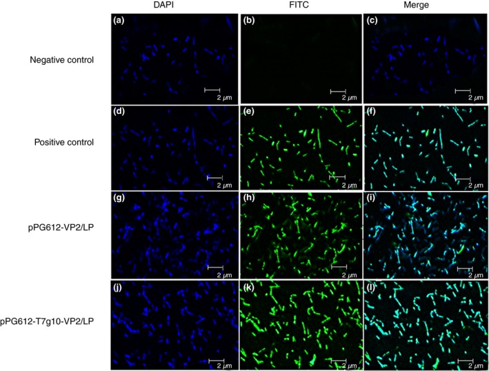
Laser confocal microscopy was used to confirm the surface expression of VP2 on recombinant Lactobacillus pPG612‐VP2/LP (pPG612‐HCE‐PgsA‐VP2‐rrnBT1T2/LP) and pPG612‐T7g10‐VP2/LP (pPG612‐HCE‐T7g10‐PgsA‐VP2‐rrnBT1T2/LP). pPG612‐VP2/LP and pPG612‐T7g10‐VP2/LP treated with FITC‐conjugated goat anti‐mouse antibody with DAPI were evident through green fluorescence emission on the cell surface. There was no green fluorescence detected on the surface of the pPG612/LP (Fig. 1). The SDS‐PAGE and western blot assays were used to identify recombinant Lactobacillus expressing the surface‐displayed VP2 protein using a highly active constitutive promoter. Protein expression was determined from lysed cell pellets of pPG612‐VP2/LP and pPG612‐T7g10‐VP2/LP cultured for 24 h. A PgsA anchor‐VP2 fusion protein of approximately 90 kDa size (45 kDa of the PgsA anchor and 44 kDa of VP2 protein) was detected in both recombinant Lactobacillus strains and compared to the positive control (pPG612‐VP2/Lactobacillus casei 393). The highest level of VP2 protein was expressed in pPG612‐T7g10‐VP2/LP followed by the pPG612‐VP2/LP strain, and no expression was observed in the pP612/LP (Fig. 2). These results confirmed the successful expression of the VP2 protein on the bacterial surface and proved that T7g10 was able to enhance VP2 protein expression.
Figure 1.

Confocal laser scanning microscopic detection of surface expression of VP2 protein. Our results illustrate that there is a green fluorescent signal on the cell surface of strains pPG612‐VP2/LP, pPG612‐T7g10‐VP2/LP and positive control but no signals on negative control (pPG612/LP), indicating that the protein of interest is expressed and displayed successfully on the surface of recombinant Lactobacillus. a, b, c: pPG612/LP (negative control); d, e, f: pPG612‐VP2/Lactobacillus casei 393 (positive control); g, h, i: pPG612‐VP2/LP; j, k, l: pPG612‐T7g10‐VP2/LP. [Colour figure can be viewed at http://wileyonlinelibrary.com]
Figure 2.

Western blot analysis of the expression of recombinant PgsA anchor‐VP2 protein (90 kDa) in pPG612‐VP2/LP and pPG612‐T7g10‐VP2/LP groups. Negative control: pPG612/LP; positive control: pPG612‐VP2/Lactobacillus casei 393. The expression levels were normalized to that of GAPDH as an internal control.
Stimulation of humoral immune response
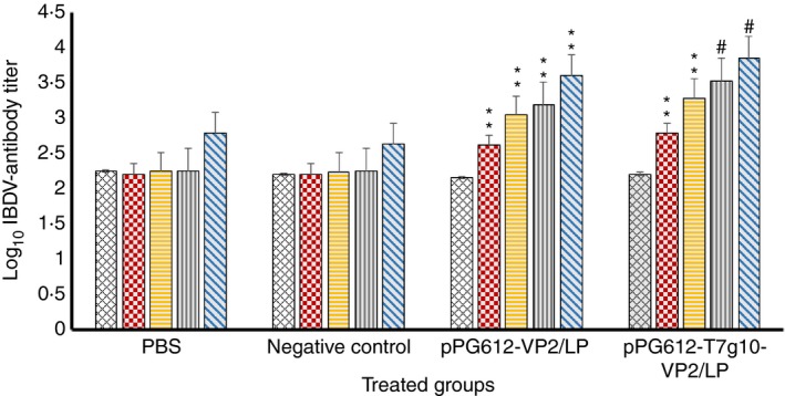
vvIBDV‐VP2 antibodies were measured to evaluate the humoral immune response and level of protection provided. Serum samples were collected on days 0, 10, 22, 35 and 44 following the first oral vaccination and analysed through ELISA (Fig. 3). The chickens that received pPG612‐VP2/LP and pPG612‐T7g10‐VP2/LP showed detectable antibody levels from day 10 (P < 0·01) that considerably increased with time to day 44. At 35 and 44 days after the first oral vaccination, significantly higher levels of specific IBDV‐VP2 antibodies were induced in chickens orally immunized with pPG612‐T7g10‐VP2/LP in comparison with the pPG612‐VP2/LP group (P < 0·05).
Figure 3.

Log10
IBDV antibody titre of different groups immunized with various vaccines. Eight‐day‐old chickens vaccinated orally with 1 ml of 109
CFU per ml PBS, Negative control (pPG612/LP), pPG612‐VP2/LP or pPG612‐T7g10‐VP2/LP. Antibodies were detected in serum collected at 0, 10, 22, 35 and 44 days following the first oral vaccination. Titres were calculated by comparing the absorbance (A
650) values of the unknown sample to that of the positive control sample. **P < 0·01 vs
pPG612/LP, #
P < 0·05 vs
pPG612‐VP2/LP group. ( ) 0 DPI; (
 ) 10 DPI; (
 ) 22 DPI; (
 ) 35 DPI; (
 ) 44 DPI. [Colour figure can be viewed at http://wileyonlinelibrary.com]
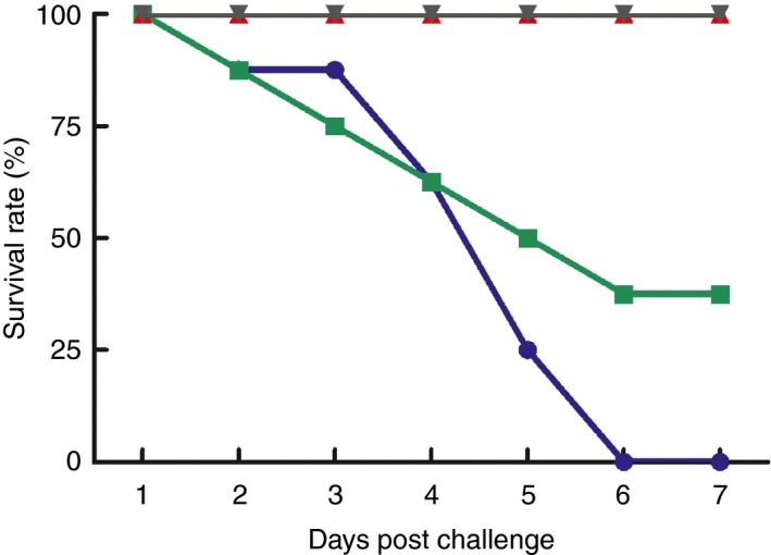
The chickens were challenged with 500 μl (106 EID50) vvIBDV strain DB11 to evaluate the protection rate of the vaccine. The protection rate in chickens of group B (pPG612‐T7g10‐VP2/LP) was 87·5% while that in group A (pPG612‐VP2/LP) was 75% (Table 1). As expected, no significant IBDV antibodies were detected in the pPG612/LP and PBS group. Furthermore, the protection rates in these groups were 37·5 and 0%, respectively, and these chickens showed clinical signs like swollen BF, watery diarrhoea, depression, dehydration and bursal atrophy. In addition, moderate bursal atrophy was observed in the vaccinated groups when compared to the pPG612/LP group. The titre of IBDV antibodies after the challenge was significantly different (P < 0·01) in vaccinated groups immunized with pPG612‐T7g10‐VP2/LP and pPG612‐VP2/LP when compared to the pPG612/LP group (Fig. 3). These outcomes confirmed the improved immunogenicity of pPG612‐T7g10‐VP2/LP against vvIBDV in chickens and showed 100% survival rate after vvIBDV challenge (Fig. 4).
Table 1.
Protection rate against IBDV challenge in each group of chickens
| Groups | Vaccines (given at 8 days old) | Dose | B/B ratio (mean ± SD)a | Survival rateb (%) | Protection ratec (%) |
|---|---|---|---|---|---|
| A | pPG612‐VP2/LP | 109 CFU | 3·15 ± 0·43d | 8/8 (100) | 6/8 (75) |
| B | pPG612‐T7g10‐VP2/LP | 109 CFU | 4·06 ± 0·41e | 8/8 (100) | 7/8 (87·5) |
| C | pPG612/LP | 109 CFU | 1·68 ± 0·11 | 3/8 (37·5) | 3/8 (37·5) |
| D | PBS | 1 ml | 1·53 ± 0·08 | 0/8 (0) | 0/8 (0) |
| E | Unchallenged control | N/A | 3·88 ± 0·54d | 8/8 (100) | N/A |
N/A, group for B/B ratio comparison and not considered for protection.
IBDV, infectious bursal disease virus; PBS, phosphate‐buffered saline; SD, standard deviation.
Bursal/body weight ratio was calculated as (bursa weight)/(body weight) × 1000.
Survival rate was calculated as the number of chickens that survived viral challenge/total number of chickens per group.
Protection rate was calculated as the number of protected chicken/total number of chicken in each group.
P < 0·01 vs pPG612/LP group.
P < 0·05 vs pPG612‐VP2/LP group.
This article is being made freely available through PubMed Central as part of the COVID-19 public health emergency response. It can be used for unrestricted research re-use and analysis in any form or by any means with acknowledgement of the original source, for the duration of the public health emergency.
Figure 4.

Survival rates of chickens were observed from day (D) 1 to 7 days postchallenge with vvIBDV. Survival rates were calculated by dividing the number of chickens that survived by the total number of chickens per group (n = 8). ( ) PBS; (
 ) Negative control; (
 ) pPG612‐VP2/LP; (
 ) pPG612‐T7g10‐VP2/LP. [Colour figure can be viewed at http://wileyonlinelibrary.com]
Lymphocyte proliferation
Spleen samples were taken after vvIBDV infection from immunized and unimmunized animals to assess the cell‐mediated immune response of the chickens. The lymphocyte response of splenic cells stained with anti‐CD4+ and CD8+ was analysed (Fig. 5a). The cellular immune response in the entire vaccinated groups was significantly higher (P < 0·01) compared to that in the negative control group. pPG612‐T7g10‐VP2/LP showed a higher production of CD8+ and CD4+ cells (13·3 and 21·0% respectively) compared to pPG612‐VP2/LP (10·4 and 14·0% respectively). Among these, the production of CD4+ cells was significantly higher (P < 0·01) in pPG612‐T7g10‐VP2/LP compared to pPG612‐VP2/LP. In both groups, CD4+ cells were stimulated to a greater extent than CD8+ cells (Fig. 5b).
Figure 5.

CD4+ and CD8+ T cells identified by flow cytometry. Lymphocyte proliferation was identified from splenic cells stained with anti‐CD4+ and CD8+ antibodies. CD4+ and CD8+ T cells were sorted based on expression and activation with anti‐CD4+ and CD8+ antibodies. Q1: CD8+; Q4: CD4+. Negative control = pPG612/LP (a). **P < 0·01 vs
pPG612/LP, ##
P < 0·01 vs
pPG612‐VP2/LP group (b). ( ) CD8; (
 ) CD4. [Colour figure can be viewed at http://wileyonlinelibrary.com]
Cytokine levels
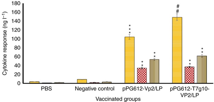
IL‐2, IL‐4 and IFN‐γ cytokine levels in the experimental chickens were evaluated from serum samples using the TBD ELISA kit. Higher levels of IFN‐γ, IL‐2 and IL‐4 were detected in groups A and B than in groups C and D (Fig. 6). Groups A and B exhibited the same trend in cytokine production: IFN‐γ > IL‐2 ˃ IL‐4, although group A cytokine production of IFN‐γ, IL‐2 and IL‐4 was lower than that in group B; among the three cytokines, the expression of IFN‐γ was significant higher (P < 0·01) in group B (pPG612‐T7g10‐VP2/LP) than in group A (pPG612‐VP2/LP). Since we observed a low concentration of Th2 cytokines (IL‐4), expression analysis of Th1/Th2 cytokines supports that vvIBDV induced an immune response via the Th1 pathway.
Figure 6.

Concentrations of IFN‐γ, IL‐2 and IL‐4 in serum samples of chickens vaccinated with pPG612‐VP2/LP, pPG612‐T7g10‐VP2/LP were detected by ELISA. Negative control: pPG612/LP; phosphate‐buffered saline: PBS. Values were calculated by comparing the OD of samples to the standard curve. **P < 0·01 and ***P < 0·001 vs
pPG612/LP, ##
P < 0·01 vs
pPG612‐VP2/LP group. ( ) IFN‐γ; (
 ) IL‐4; (
 ) IL‐2. [Colour figure can be viewed at http://wileyonlinelibrary.com]
Discussion
Broiler production on a commercial level is still facing serious problems due to IBDV. Vaccines available in the market show partial defence against virulent and highly virulent IBDV strains. Previously, several studies have reported that DNA‐based vaccines successfully induced cellular and humoral immune responses against pathogens (Kodihalli et al. 1999; Fan et al. 2002; Serezani et al. 2002). Deoxyribonucleic acid vaccines against IBDV that employed the VP2 protein have been shown to induce antibody production, but the expression and protection conferred have been found to vary (Becht et al. 1988). In the current study, we evaluated the immune response to VP2 protein expressed on the surface of bacteria using the HCE‐PgsA system, since higher immunological responses have been reported from surface‐displayed antigens compared with intracellularly secreted antigens (Stover et al. 1993; Jong‐Soo et al. 2000; Grode et al. 2002). Lee et al. (2006) reported both systemic and mucosal immune responses against acute respiratory syndrome‐associated coronavirus with the S antigen (SARS‐CoV S) using the PgsA system in the L. casei expression system. OprF (a dominant outer membrane protein of Pseudomonas aeruginosa), FadL (an outer membrane protein involved in the transport of long‐chain fatty acids in E. coli) and PgsA have all been used as anchor proteins (Poo et al. 2002; Lee et al. 2004, 2005), but PgsA has been shown to be a better anchor for lactic acid bacteria. However, to date, no candidate vaccine against vvIBDV has been developed using the HCE‐PgsA system. Therefore, we tested the PgsA gene product as an anchor for the surface display of antigens. The HCE‐PgsA system has been used in vaccine development against tumours and influenza viruses, showing positive results. However, it has been reported that the HCE‐PgsA system expresses low levels of recombinant protein (Poo et al. 2006). Therefore, we introduced an extra‐ribosomal binding site (RBS) T7g10 (enhancer) in the HCE‐PgsA system and compared VP2 protein expression and protection against vvIBDV in the presence and absence of this enhancer. This synthetic sequence was derived from gene 10 of bacteriophage T7 called ‘Epsilon’ (enhancer of protein synthesis initiation) (Olins and Rangwala 1989). It has been reported that this sequence can improve the expression of poorly expressed heterologous genes. Studies have proven that the efficacy of gene 10 is due to the presence of a nine‐base sequence upstream of the Shine–Dalgarno region. It has been documented that T7g10‐derived RBS displays 40‐fold greater expression than the typical RBS. Mammalian, plant and bacterial proteins have been expressed at high levels by the construction of a plasmid vector with a T7g10 leader sequence (Olins et al. 1988).
To express the antigenic protein on the surface of bacteria, antigenic proteins should be fused with an anchor protein that is naturally expressed on the surface of the bacteria. Therefore, we constructed two recombinant surface‐displayed expression vectors, pPG612‐VP2 and pPG612‐T7g10‐VP2, expressed in L. plantarum. In this study, indirect immunofluorescence confirmed VP2 protein expression on the surface of the recombinant bacteria. We used the translational enhancer T7g10 to enhance the constitutive protein expression of the vector. Expression analysis by Western blot indicated that pPG612‐T7g10‐VP2/LP expressed higher levels of VP2 protein when compared to pPG612‐VP2/LP and pPG612/LP.
Immunogenicity studies were performed after the expression of VP2 protein on the surface of L. plantarum had been confirmed. Antibody levels in sera and production of cytokines were measured to evaluate the immune responses of the chickens.
It has been reported that antibody levels determine protection against vvIBDV in chickens (Berg 2000). In this study, we determined that the recombinant Lactobacillus delivery system induces a strong antibody response in both the pPG612‐VP2/LP‐ and pPG612‐T7g10‐VP2/LP‐treated groups. Moreover, the immune response to pPG612‐T7g10‐VP2/LP was significantly higher (P < 0·05) than to pPG612‐VP2/LP. We also determined the bursal/body weight ratio (B/B ratio), survival and protective rates against IBDV challenge. The B/B ratios of the pPG612‐VP2/LP and pPG612‐T7g10‐VP2/LP groups were both significantly higher than that of the pPG612/LP group (P < 0·01), and the value of the pPG612‐T7g10‐VP2/L group was significantly higher than that of the pPG612‐VP2/L group (P < 0·05). Although the protection rate of the pPG612‐T7g10‐VP2/LP group was higher than that of the pPG612‐VP2/LP group (87·5 vs 75%), no significant difference was found between the two groups, which may be caused by the small sample size in this study. It has been proven that not only the humoral immune response but also the cellular immune response mediated by T cells plays a significant role in the control of vvIBDV infection (Rautenschlein et al. 2002). During the IBDV replication phase, CD4+ and CD8+ T cells show an influx into the BF (the site of virus replication) and other adjacent lymphoid organs such as the caecal tonsils and the spleen (Sharma et al. 1989; Kim et al. 1999, 2000). We examined the effect of vaccination on T‐cell responses upon viral attack, showing that the number of T cells significantly increased after virus infection. Cyclosporin A treatment was responsible for reducing circulating T cells and was involved in T‐cell mitogenesis, which in turn was responsible for viral burden in the bursae of IBDV‐infected chickens (Kim et al. 2000). T cell‐mediated responses have a significant role in viral clearance and are involved in the recovery from viral infection (Kim et al. 2000). The cell‐mediated immune system activates macrophages and natural killer cells in response to an antigen (Lukacs and Kurlander 1989; Harty and Bevan 1992). In IBDV‐infected chickens, there was an increase in the number of intrabursal T cells, while the bursae of uninfected chickens had very few resident T cells (Khan and Hashimoto 1996; Lütticken 1997; Kim et al. 1999, 2000). Our results indicate that T7g10 may be responsible for the increased expression of the VP2 protein, thereby increasing antibody production that in turn produces a strong humoral immune response and subsequently maximum protection from viral challenge. Furthermore, a greater amount of VP2 stimulates the cell‐mediated immune response, which shows a better CD4+ and CD8+ T‐cell response. CD8+ T cells are cytotoxic T cells which are involved in the lysis of virally infected cells, tumour cells and allografts, and play a crucial role in the immune response (Sharma et al. 1989). Upon activation, helper CD4+ T cells function as “middlemen” and trigger secretion and proliferation of various cytokines.
Cytokine production regulates host responses. Th1 cells are responsible for the secretion of IFN‐γ and IL‐2 cytokines and are involved in effective cell‐mediated immunity leading to the successful elimination of intracellular pathogens (Kunzendorf et al. 1998). Th2 cells are involved in the control of certain parasitic infections through the production of cytokines such as IL‐4, IL‐5 and IL‐13 (Avery et al. 2004). The bursa of virally infected chickens is involved in the activation of T cells and upregulates the expression of other cytokine genes such as IL‐1b, IL‐6 and IFN‐γ (Eldaghayes et al. 2006). Cellular immune responses mediated by cytokines against vvIBDV in chickens are poorly illustrated and have not yet been broadly studied. In our study, higher concentrations of IFN‐γ, IL‐2 and IL‐4 cytokines were detected in pPG612‐T7g10‐VP2/LP compared to pPG612‐VP2/LP; among these, concentrations of IFN‐γ in pPG612‐T7g10‐VP2/LP were significantly higher (P < 0·01) compared to pPG612‐VP2/LP. IBDV‐infected chickens had upregulated gamma interferon gene expression in spleen and bursal cells compared to virus‐free chickens (Khan and Hashimoto 1996; Lütticken 1997). It has been documented that IL‐2 has a significant role in regulating the function and development of T cells (Malek 2003). In mice, targeted deletion in the IL‐2 gene has been shown to affect multiple target organs thus supporting that IL‐2 plays a role in immunodeficiency and fatal autoimmune inflammatory disease (Schorle and Hunig 1991; Kündig et al. 1993; Sadlack et al. 1993).
Th1 or Th2 cytokine production in vaccinated chickens is the outcome of an adaptive immune response. Th2 cytokines direct B cells to produce the anti‐allergen IgE, and they also inhibit Th1 cell function and prevent the production of IL‐2 and IFN‐γ that are crucial for the development of cytotoxic T cells (Berg 2000). IL‐4 is a multifunctional pleiotropic cytokine involved in apoptosis and gene expression in various cells including lymphocytes, macrophages and fibroblasts, as well as epithelial and endothelial cells. Investigation of IL‐4 is required for the understanding of the Th2 phenotype of lymphocytes and for regulating cell proliferation (Luzina et al. 2012). Among the three cytokines studied, we observed low production of the Th2 cytokine IL‐2, and the highest expression of the Th1 cytokine IFN‐g. The expression analysis of Th1 and Th2 cytokines indicated that this recombinant Lactobacillus delivery system induces an immune response via the Th1 pathway.
To conclude, our results demonstrated that VP2 protein was expressed on the surface of bacteria using the HCE‐PgsA system and proved that the T7g10 enhancer improved the level of VP2 expression and induced a greater immune response against vvIBDV. Therefore, bacteria are better recognized by the immune system when antigens are expressed on its surface rather than intracellularly (Lee et al. 2006). To the best of our knowledge, this HCE‐PgsA and T7g10 enhancer‐based vaccine strategy has not been previously reported for vvIBDV. Our results indicate that this recombinant Lactobacillus delivery system was more successful than has been previously achieved, demonstrating a 100% survival rate and being suitable for administration (Wu et al. 2007; Park et al. 2009; Arnold et al. 2012; He et al. 2018). Considerably less bursal atrophy was observed compared to other DNA vaccines (Kapczynski et al. 2003; Haygreen et al. 2006; Hsieh et al. 2007), which indicates that this strategy provides more protection from the virus. These results taken together demonstrate that this recombinant Lactobacillus delivery system is immunogenic and can confer protection against vvIBDV in chickens.
Conflict of Interest
The authors declare that there is no conflict of interest.
Supporting information
Figure S1 Map of pPG612‐HCE‐T7g10‐PgsA‐VP2‐rrnBT1T2.
Table S1 pPG612‐HCE‐T7g10‐PgsA‐VP2‐rrnBT1T2 information.
Acknowledgements
First and foremost, we are indebted to all of our coworkers who assisted in achieving this fact‐finding. The National Technology and Research Project of China supported this work (grant no. 2015BAD12B01‐4).
Iram Maqsood and Wen Shi contributed equally to this work.
[Correction added on 15 October 2018, after first online publication: details of the corresponding author has been changed in this version.]
Contributor Information
L. Tang, Email: tanglijie@163.com.
Y. Li, Email: yijingli@163.com.
References
- Arnold, M. , Durairaj, V. , Mundt, E. , Schulze, K. , Breunig, K.D. and Behrens, S.E. (2012) Protective vaccination against infectious bursal disease virus with whole recombinant Kluyveromyces lactis yeast expressing the viral VP2 subunit. PLoS ONE 7, e42870. [DOI] [PMC free article] [PubMed] [Google Scholar]
- Avery, S. , Rothwell, L. , Degen, W.D. , Schijns, V.E. , Young, J. , Kaufman, J. and Kaiser, P. (2004) Characterization of the first nonmammalian T2 cytokine gene cluster: the cluster contains functional single‐copy genes for IL‐3, IL‐4, IL‐13, and GM‐CSF, a gene for IL‐5 that appears to be a pseudogene, and a gene encoding another cytokinelike transcript, KK34. J Interferon Cytokine Res 24, 600–610. [DOI] [PubMed] [Google Scholar]
- Azad, A.A. , Barrett, S. and Fahey, K. (1985) The characterization and molecular cloning of the double‐stranded RNA genome of an Australian strain of infectious bursal disease virus. Virology 143, 35–44. [DOI] [PubMed] [Google Scholar]
- Becht, H. , Müller, H. and Müller, H.K. (1988) Comparative studies on structural and antigenic properties of two serotypes of infectious bursal disease virus. J Gen Virol 69, 631–640. [DOI] [PubMed] [Google Scholar]
- Berg, T.P.V.D. (2000) Acute infectious bursal disease in poultry: a review. Avian Pathol 29, 175–194. [DOI] [PubMed] [Google Scholar]
- Brandt, M. , Yao, K. , Liu, M. , Heckert, R.A. and Vakharia, V.N. (2001) Molecular determinants of virulence, cell tropism, and pathogenic phenotype of infectious bursal disease virus. J Virol 75, 11974–11982. [DOI] [PMC free article] [PubMed] [Google Scholar]
- Eldaghayes, I. , Rothwell, L. , Williams, A. , Withers, D. , Balu, S. , Davison, F. and Kaiser, P. (2006) Infectious bursal disease virus: strains that differ in virulence differentially modulate the innate immune response to infection in the chicken bursa. Viral Immunol 19, 83–91. [DOI] [PubMed] [Google Scholar]
- Fahey, K.J. , Erny, K. and Crooks, J. (1989) A conformational immunogen on VP‐2 of infectious bursal disease virus that induces virus‐neutralizing antibodies that passively protect chickens. J Gen Virol 70, 1473–1481. [DOI] [PubMed] [Google Scholar]
- Fan, M. , Bian, Z. , Peng, Z. , Guo, J. , Jia, R. and Chen, Z. (2002) DNA vaccine encoding Streptococcus mutans surface protein protected gnotobiotic rats from caries. Chin J Stomatol 37, 4–7. [PubMed] [Google Scholar]
- Grangette, C. , Müller‐Alouf, H. , Goudercourt, D. , Geoffroy, M.C. , Turneer, M. and Mercenier, A. (2001) Mucosal immune responses and protection against tetanus toxin after intranasal immunization with recombinant Lactobacillus plantarum . Infect Immun 69, 1547–1553. [DOI] [PMC free article] [PubMed] [Google Scholar]
- Grode, L. , Kursar, M. , Fensterle, J. , Kaufmann, S.H. and Hess, J. (2002) Cell‐mediated immunity induced by recombinant Mycobacterium bovis Bacille Calmette‐Guerin strains against an intracellular bacterial pathogen: importance of antigen secretion or membrane‐targeted antigen display as lipoprotein for vaccine efficacy. J Immunol 168, 1869–1876. [DOI] [PubMed] [Google Scholar]
- Harty, J.T. and Bevan, M.J. (1992) CD8+ T cells specific for a single nonamer epitope of Listeria monocytogenes are protective in vivo. J Exp Med 175, 1531–1538. [DOI] [PMC free article] [PubMed] [Google Scholar]
- Haygreen, E. , Kaiser, P. , Burgess, S. and Davison, T. (2006) In ovo DNA immunisation followed by a recombinant fowlpox boost is fully protective to challenge with virulent IBDV. Vaccine 24, 4951–4961. [DOI] [PubMed] [Google Scholar]
- He, C.Q. , Ma, L.Y. , Wang, D. , Li, G.R. and Ding, N.Z. (2009) Homologous recombination is apparent in infectious bursal disease virus. Virology 384, 51–58. [DOI] [PubMed] [Google Scholar]
- He, W.S. , Wang, H.H. , Jing, Z.M. , Cui, D.D. , Zhu, J.Q. , Li, Z.J. and Ma, H.L. (2018) Highly efficient synthesis of hydrophilic phytosterol derivatives catalyzed by ionic liquid. J Am Oil Chem Soc 95, 89–100. [Google Scholar]
- Ho, P. , Kwang, J. and Lee, Y. (2005) Intragastric administration of Lactobacillus casei expressing transmissible gastroenteritis coronavirus spike glycoprotein induced specific antibody production. Vaccine 23, 1335–1342. [DOI] [PMC free article] [PubMed] [Google Scholar]
- Hsieh, M.K. , Wu, C.C. and Lin, T.L. (2007) Priming with DNA vaccine and boosting with killed vaccine conferring protection of chickens against infectious bursal disease. Vaccine 25, 5417–5427. [DOI] [PubMed] [Google Scholar]
- Iram, M. , Wang, X. , Zhao, H. , Mohsin, B.S. , Nadeem, A. , Saima, K. , Tang, L.J. and Jing, L.Y. (2016) Ameliorate maneuver for transformation of lactobacillus strains by electroporation with ibdv‐vp2 chemically engineered expression vector. J Anim Plant Sci 26, 814–822. [Google Scholar]
- Irigoyen, N. , Garriga, D. , Navarro, A. , Verdaguer, N. , Rodríguez, J.F. and Castón, J.R. (2009) Autoproteolytic activity derived from the infectious bursal disease virus capsid protein. J Biol Chem 284, 8064–8072. [DOI] [PMC free article] [PubMed] [Google Scholar]
- Irigoyen, N. , Castón, J.R. and Rodríguez, J.F. (2012) Host proteolytic activity is necessary for infectious bursal disease virus capsid protein assembly. J Biol Chem 287, 24473–24482. [DOI] [PMC free article] [PubMed] [Google Scholar]
- Islam, M. , Zierenberg, K. and Müller, H. (2001) The genome segment B encoding the RNA‐dependent RNA polymerase protein VP1 of very virulent infectious bursal disease virus (IBDV) is phylogenetically distinct from that of all other IBDV strains. Arch Virol 146, 2481–2492. [DOI] [PMC free article] [PubMed] [Google Scholar]
- Jong‐Soo, L. , Shin, K.S. , Jae‐Gu, P. and Kim, C.J. (2000) Surface‐displayed viral antigens on Salmonella carrier vaccine. Nat Biotechnol 18, 645. [DOI] [PubMed] [Google Scholar]
- Kapczynski, D.R. , Hilt, D.A. , Shapiro, D. , Sellers, H.S. and Jackwood, M.W. (2003) Protection of chickens from infectious bronchitis by in ovo and intramuscular vaccination with a DNA vaccine expressing the S1 glycoprotein. Avian Dis 47, 272–285. [DOI] [PubMed] [Google Scholar]
- Khan, M.Z. and Hashimoto, Y. (1996) An immunohistochemical analysis of T‐cell subsets in the chicken bursa of Fabricius during postnatal stages of development. J Vet Med Sci 58, 1231–1234. [DOI] [PubMed] [Google Scholar]
- Kibenge, F.S. , Mckenna, P.K. and Dybing, J.K. (1991) Genome cloning and analysis of the large RNA segment (segment A) of a naturally avirulent serotype 2 infectious bursal disease virus. Virology 184, 437–440. [DOI] [PubMed] [Google Scholar]
- Kim, I.J. , Gagic, M. and Sharma, J.M . (1999) Recovery of antibody‐producing ability and lymphocyte repopulation of bursal follicles in chickens exposed to infectious bursal disease virus. Avian Dis, 43, 401–413. [PubMed] [Google Scholar]
- Kim, I.J. , You, S.K. , Kim, H. , Yeh, H.Y. and Sharma, J.M. (2000) Characteristics of bursal T lymphocytes induced by infectious bursal disease virus. J Virol 74, 8884–8892. [DOI] [PMC free article] [PubMed] [Google Scholar]
- Kodihalli, S. , Goto, H. , Kobasa, D.L. , Krauss, S. , Kawaoka, Y. and Webster, R.G. (1999) DNA vaccine encoding hemagglutinin provides protective immunity against H5N1 influenza virus infection in mice. J Virol 73, 2094–2098. [DOI] [PMC free article] [PubMed] [Google Scholar]
- Kündig, T.M. , Schorle, H. , Bachmann, M.F. , Hengartner, H. , Zinkernagel, R.M. and Horak, I. (1993) Immune responses in interleukin‐2‐deficient mice. Science‐New York Then Washington 262, 1059–1061. [DOI] [PubMed] [Google Scholar]
- Kunzendorf, U. , Tran, T.H. and Bulfone‐Paus, S. (1998) The Th1‐Th2 paradigm in 1998: law of nature or rule with exceptions. Nephrol Dial Transplant 13, 2445–2448. [DOI] [PubMed] [Google Scholar]
- Lee, S.H. , Choi, J.I. , Park, S.J. , Lee, S.Y. and Park, B.C. (2004) Display of bacterial lipase on the Escherichia coli cell surface by using FadL as an anchoring motif and use of the enzyme in enantioselective biocatalysis. Appl Environ Microbiol 70, 5074–5080. [DOI] [PMC free article] [PubMed] [Google Scholar]
- Lee, S.H. , Choi, J.I. , Han, M.J. , Choi, J.H. and Lee, S.Y. (2005) Display of lipase on the cell surface of Escherichia coli using OprF as an anchor and its application to enantioselective resolution in organic solvent. Biotechnol Bioeng 90, 223–230. [DOI] [PubMed] [Google Scholar]
- Lee, J.S. , Poo, H. , Han, D.P. , Hong, S.P. , Kim, K. , Cho, M.W. , Kim, E. , Sung, M.H. et al (2006) Mucosal immunization with surface‐displayed severe acute respiratory syndrome coronavirus spike protein on Lactobacillus casei induces neutralizing antibodies in mice. J Virol 80, 4079–4087. [DOI] [PMC free article] [PubMed] [Google Scholar]
- Lejal, N. , Da Costa, B. , Huet, J.C. and Delmas, B. (2000) Role of Ser‐652 and Lys‐692 in the protease activity of infectious bursal disease virus VP4 and identification of its substrate cleavage sites. J Gen Virol 81, 983–992. [DOI] [PubMed] [Google Scholar]
- Li, Y.J. , Ma, G.P. , Li, G.W. , Qiao, X.Y. , Ge, J.W. , Tang, L.J. , Liu, M. and Liu, L.W . (2010) Oral vaccination with the porcine rotavirus VP4 outer capsid protein expressed by Lactococcus lactis induces specific antibody production. Biomed Res Int 2010, 708460. [DOI] [PMC free article] [PubMed] [Google Scholar]
- Lukacs, K. and Kurlander, R. (1989) Lyt‐2+ T cell‐mediated protection against listeriosis. Protection correlates with phagocyte depletion but not with IFN‐gamma production. J Immunol 142, 2879–2886. [PubMed] [Google Scholar]
- Lütticken, D. (1997) Viral diseases of the immune system and strategies to control infectious bursal disease by vaccination. Acta Vet Hung 45, 239–249. [PubMed] [Google Scholar]
- Luzina, I.G. , Keegan, A.D. , Heller, N.M. , Rook, G.A. , Shea‐Donohue, T. and Atamas, S.P. (2012) Regulation of inflammation by interleukin‐4: a review of “alternatives”. J Leukoc Biol 92, 753–764. [DOI] [PMC free article] [PubMed] [Google Scholar]
- Ma, S. , Wang, L. , Huang, X. , Wang, X. , Chen, S. , Shi, W. , Qiao, X. , Jiang, Y. et al (2018) Oral recombinant Lactobacillus vaccine targeting the intestinal microfold cells and dendritic cells for delivering the core neutralizing epitope of porcine epidemic diarrhea virus. Microb Cell Fact 17, 20–32. [DOI] [PMC free article] [PubMed] [Google Scholar]
- Maassen, C. , Laman, J. , Den Bak‐Glashouwer, M.H. , Tielen, F. , van Holten‐Neelen, J. , Hoogteijling, L. , Antonissen, C. , Leer, R. et al (1999) Instruments for oral disease‐intervention strategies: recombinant Lactobacillus casei expressing tetanus toxin fragment C for vaccination or myelin proteins for oral tolerance induction in multiple sclerosis. Vaccine 17, 2117–2128. [DOI] [PubMed] [Google Scholar]
- Macreadie, I.G. and Azad, A.A. (1993) Expression and RNA dependent RNA polymerase activity of birnavirus VP1 protein in bacteria and yeast. Biochem Mol Biol Int 30, 1169–1178. [PubMed] [Google Scholar]
- Mahgoub, H.A. (2012) An overview of infectious bursal disease. Arch Virol 157, 2047–2057. [DOI] [PubMed] [Google Scholar]
- Mahmood, M. , Hussain, I. , Siddique, M. , Akhtar, M. and Ali, S. (2007) DNA vaccination with VP2 gene of very virulent infectious bursal disease virus (vvIBDV) delivered by transgenic E. coli DH5α given orally confers protective immune responses in chickens. Vaccine 25, 7629–7635. [DOI] [PubMed] [Google Scholar]
- Malek, T.R. (2003) The main function of IL‐2 is to promote the development of T regulatory cells. J Leukoc Biol 74, 961–965. [DOI] [PubMed] [Google Scholar]
- Mcghee, J.R. , Mestecky, J. , Dertzbaugh, M.T. , Eldridge, J.H. , Hirasawa, M. and Kiyono, H. (1992) The mucosal immune system: from fundamental concepts to vaccine development. Vaccine 10, 75–88. [DOI] [PubMed] [Google Scholar]
- Medina, E. and Guzmán, C.A. (2001) Use of live bacterial vaccine vectors for antigen delivery: potential and limitations. Vaccine 19, 1573–1580. [DOI] [PubMed] [Google Scholar]
- Mestecky, J. (1987) The common mucosal immune system and current strategies for induction of immune responses in external secretions. J Clin Immunol 7, 265–276. [DOI] [PubMed] [Google Scholar]
- Min, L. , Li, Z.L. , Wei, G.J. , Yuan, Q.X. , Jing, L.Y. and Qiu, L.D. (2012) Immunogenicity of Lactobacillus‐expressing VP2 and VP3 of the infectious pancreatic necrosis virus (IPNV) in rainbow trout. Fish Shellfish Immunol 32, 196–203. [DOI] [PubMed] [Google Scholar]
- Muench, H.R. (1938) A simple method of estimating 50 per cent end points. Am J Hyg 27, 493–497. [Google Scholar]
- Narita, J. , Okano, K. , Tateno, T. , Tanino, T. , Sewaki, T. , Sung, M.H. , Fukuda, H. and Kondo, A. (2006) Display of active enzymes on the cell surface of Escherichia coli using PgsA anchor protein and their application to bioconversion. Appl Microbiol Biotechnol 70, 564–572. [DOI] [PubMed] [Google Scholar]
- Ogawa, T. , Asai, Y. , Yasuda, K. and Sakamoto, H. (2005) Oral immunoadjuvant activity of a new synbiotic Lactobacillus casei subsp casei in conjunction with dextran in BALB/c mice. Nutr Res 25, 295–304. [Google Scholar]
- Olins, P.O. and Rangwala, S. (1989) A novel sequence element derived from bacteriophage T7 mRNA acts as an enhancer of translation of the lacZ gene in Escherichia coli . J Biol Chem 264, 16973–16976. [PubMed] [Google Scholar]
- Olins, P.O. , Devine, C.S. , Rangwala, S.H. and Kavka, K.S. (1988) The T7 phage gene 10 leader RNA, a ribosome‐binding site that dramatically enhances the expression of foreign genes in Escherichia coli . Gene 73, 227–235. [DOI] [PubMed] [Google Scholar]
- Park, J.H. , Sung, H.W. , Yoon, B.I. and Kwon, H.M. (2009) Protection of chicken against very virulent IBDV provided by in ovo priming with DNA vaccine and boosting with killed vaccine and the adjuvant effects of plasmid‐encoded chicken interleukin‐2 and interferon‐γ. J Vet Sci 10, 131–139. [DOI] [PMC free article] [PubMed] [Google Scholar]
- Perdigón, G. , Fuller, R. and Raya, R. (2001) Lactic acid bacteria and their effect on the immune system. Curr Issues Intest Microbiol 2, 27–42. [PubMed] [Google Scholar]
- Poo, H. , Song, J.J. , Hong, S.P. , Choi, Y.H. , Yun, S.W. , Kim, J.H. , Lee, S.C. , Lee, S.G. et al (2002) Novel high‐level constitutive expression system, pHCE vector, for a convenient and cost‐effective soluble production of human tumor necrosis factor‐α. Biotechnol Lett 24, 1185–1189. [Google Scholar]
- Poo, H. , Pyo, H.M. , Lee, T.Y. , Yoon, S.W. , Lee, J.S. , Kim, C.J. , Sung, M.H. and Lee, S.H. (2006) Oral administration of human papillomavirus type 16 E7 displayed on Lactobacillus casei induces E7 specific antitumor effects in C57/BL6 mice. Int J Cancer 119, 1702–1709. [DOI] [PubMed] [Google Scholar]
- Rautenschlein, S. , Yeh, H.Y. and Sharma, J. (2002) The role of T cells in protection by an inactivated infectious bursal disease virus vaccine. Vet Immunol Immunopathol 89, 159–167. [DOI] [PubMed] [Google Scholar]
- Reveneau, N. , Geoffroy, M.C. , Locht, C. , Chagnaud, P. and Mercenier, A. (2002) Comparison of the immune responses induced by local immunizations with recombinant Lactobacillus plantarum producing tetanus toxin fragment C in different cellular locations. Vaccine 20, 1769–1777. [DOI] [PubMed] [Google Scholar]
- Rutherford, N. and Mourez, M. (2006) Surface display of proteins by Gram‐negative bacterial autotransporters. Microb Cell Fact 5, 22. [DOI] [PMC free article] [PubMed] [Google Scholar]
- Sadlack, B. , Merz, H. , Schorle, H. , Schimpl, A. , Feller, A.C. and Horak, I. (1993) Ulcerative colitis‐like disease in mice with a disrupted interleukin‐2 gene. Cell 75, 253–261. [DOI] [PubMed] [Google Scholar]
- Samuelson, P. , Gunneriusson, E. , Nygren, P.A. and Stahl, S. (2002) Display of proteins on bacteria. J Biotechnol 96, 129–154. [DOI] [PubMed] [Google Scholar]
- Saugar, I. , Luque, D. , Oña, A. , Rodríguez, J.F. , Carrascosa, J.L. , Trus, B.L. and Castón, J.R. (2005) Structural polymorphism of the major capsid protein of a double‐stranded RNA virus: an amphipathic α helix as a molecular switch. Structure 13, 1007–1017. [DOI] [PubMed] [Google Scholar]
- Scheppler, L. , Vogel, M. , Zuercher, A.W. , Zuercher, M. , Germond, J.E. , Miescher, S.M. and Stadler, B.M. (2002) Recombinant Lactobacillus johnsonii as a mucosal vaccine delivery vehicle. Vaccine 20, 2913–2920. [DOI] [PubMed] [Google Scholar]
- Schorle, H. and Hunig, T. (1991) Development and function of T cells in mice rendered interleukin‐2 deficient by gene targeting. Nature 352, 621. [DOI] [PubMed] [Google Scholar]
- Serezani, C.H.C. , Franco, A.R. , Wajc, M. , Yokoyama‐Yasunaka, J.K.U. , Wunderlich, G. , Borges, M.M. and Uliana, S.R.B. (2002) Evaluation of the murine immune response to Leishmania meta 1 antigen delivered as recombinant protein or DNA vaccine. Vaccine 20, 3755–3763. [DOI] [PubMed] [Google Scholar]
- Sharma, J. , Dohms, J. and Metz, A . (1989) Comparative pathogenesis of serotype 1 and variant serotype 1 isolates of infectious bursal disease virus and their effect on humoral and cellular immune competence of specific‐pathogen‐free chickens. Avian Dis, 33, 112–124. [PubMed] [Google Scholar]
- Spies, U. , Müller, H. and Becht, H. (1989) Nucleotide sequence of infectious bursal disease virus genome segment A delineates two major open reading frames. Nucleic Acids Res 17, 7982. [DOI] [PMC free article] [PubMed] [Google Scholar]
- Stover, C.K. , Bansal, G.P. , Hanson, M.S. , Burlein, J. , Palaszynski, S. , Young, J. , Koenig, S. , Young, D. et al (1993) Protective immunity elicited by recombinant bacille Calmette‐Guerin (BCG) expressing outer surface protein A (OspA) lipoprotein: a candidate Lyme disease vaccine. J Exp Med 178, 197–209. [DOI] [PMC free article] [PubMed] [Google Scholar]
- Vakharia, V.N. , He, J. , Ahamed, B. and Snyder, D.B. (1994) Molecular basis of antigenic variation in infectious bursal disease virus. Virus Res 31, 265–273. [DOI] [PubMed] [Google Scholar]
- Wei, Y. , Li, J. , Zheng, J. , Xu, H. , Li, L. and Yu, L. (2006) Genetic reassortment of infectious bursal disease virus in nature. Biochem Biophys Res Commun 350, 277–287. [DOI] [PubMed] [Google Scholar]
- Wu, J. , Yu, L. , Li, L. , Hu, J. , Zhou, J. and Zhou, X. (2007) Oral immunization with transgenic rice seeds expressing VP2 protein of infectious bursal disease virus induces protective immune responses in chickens. Plant Biotechnol J 5, 570–578. [DOI] [PubMed] [Google Scholar]
- Yu, M. , Qi, R. , Chen, C. , Yin, J. , Ma, S. , Shi, W. , Wu, Y. , Ge, J. et al (2017) Immunogenicity of recombinant Lactobacillus casei expressing F4 (K88) fimbrial adhesin FaeG in conjunction with a heat‐labile enterotoxin A (LTAK63) and heat labile enterotoxin B (LTB) of enterotoxigenic Escherichia coli as an oral adjuvant in mice. J Appl Microbiol 122, 506–515. [DOI] [PubMed] [Google Scholar]
- Zhang, L. , Ren, X. , Chen, Y. , Gao, Y. , Wang, N. , Lu, Z. , Gao, L. , Qin, L. et al (2015) Chondroitin sulfate N‐acetylgalactosaminyltransferase‐2 contributes to the replication of infectious bursal disease virus via interaction with the capsid protein VP2. Viruses 7, 1474–1491. [DOI] [PMC free article] [PubMed] [Google Scholar]
Associated Data
This section collects any data citations, data availability statements, or supplementary materials included in this article.
Supplementary Materials
Figure S1 Map of pPG612‐HCE‐T7g10‐PgsA‐VP2‐rrnBT1T2.
Table S1 pPG612‐HCE‐T7g10‐PgsA‐VP2‐rrnBT1T2 information.


