Abstract
We report a rare case of adenoviral pneumonia in a previously healthy pregnant woman at 26+4 weeks' gestation. She presented with persistent high fever, cough for 5 days, and developed progressive dyspnea with hypoxemic respiratory failure and bilateral pulmonary infiltrates with pleural effusions. Aggressive supportive care and timely obstetrical management saved the mother and prevented preterm delivery and fetal anomaly.
Keywords: adenovirus, immunocompetent, pneumonia, pregnancy
Introduction
Pregnant women are prone to respiratory infection, due to alterations in the immune system and mechanical changes involving the chest and abdominal cavities.1 Pneumonia also increases the risk of adverse pregnancy outcomes, such as low birthweight and preterm birth, compared to unaffected women.2 However, adenoviral pneumonia in pregnant patients has not been reported before. Here, we report a case of severe community‐acquired adenoviral pneumonia in a previously healthy pregnant woman who was managed successfully.
Case Report
A 28‐year‐old pregnant woman was admitted because of fever and cough for 5 days at 26+4 weeks' gestation. The patient had been well until 5 days earlier, when fever developed with temperatures of up to 38 °C (100.4 °F). Four days prior to admission, her temperature had risen to 39 °C (102.2 °F), accompanied by rigors, malaise and cough with purulent sputum. She was seen in the obstetrics clinic and was given oral azithromycin 0.5 g once daily for 1 day and intravenous (i.v.) cefotiam 1 g twice daily for another 2 days. One day before admission, i.v. ceftriaxone 2.0 g daily was administered.
The patient had previously been healthy. During the first trimester of pregnancy, she had reported transient threatened abortion and was properly managed. Two weeks prior, her parents had had fever, one after the other, which had resolved spontaneously within 3 days and 1 day, respectively. The patient's mother is a clinical laboratory technician in the fever clinic in our hospital. The patient did not smoke, drink alcohol, or use illicit drugs and denied recent travel and exposure to live poultry.
On examination at admission (day 1), her temperature was 40.4 °C (104.72 °F), blood pressure was 133/69 mmHg, respiratory rate was 24 breaths/min, and pulse was 133 beats/min. Oxygen saturation was 93–97% while she was breathing 5 L/min of oxygen by nasal cannula. On auscultation of the chest, reduced air entry was noted in the left lower lung. The remainder of the examination was unremarkable. The fetal heart rate was 180 beats/min. Uterine contractions were 5 times/min.
A rapid detection for influenza A and B antigens was negative from a throat swab. Serological tests were negative for IgM to Legionella pneumophila serotype 1 (LP1), mycoplasma, chlamydia, Q fever rickettsia, adenovirus, respiratory syncytial virus, parainfluenza virus and influenza A and B. Cultures of blood, urine and sputum were negative. Initial blood tests showed normal white blood cells (WBC) of 7 × 10⁹/L with decreased lymphocyte of 0.406 × 10⁹/L (reference 1.1–3.2 × 10⁹/L), procalcitonin was less than 0.25 ng/mL and C‐reactive protein was 182 mg/L (reference < 8 mg/L). Initial liver and renal function tests, glucose, electrolytes and urinalysis were all normal.
Community‐acquired pneumonia (CAP) and threatened abortion were diagnosed. From day 1 to day 3, i.v. ceftriaxone and azithromycin continued, and rectal indomethacin 100 mg followed by oral 25 mg Q4h were prescribed to relieve frequent uterine contractions. On day 2, the fetal fibronectin testing was negative and oral indomethacin was decreased to 25 mg Q6h after gradually diminished uterine contraction. However, the patient's condition deteriorated. Liver enzymes became deranged during the first 48 h and peaked on day 2 with alanine aminotransferase of 74 U/L (reference <40 U/L) and aspartate aminotransferase of 87 U/L (reference <30 U/L); albumin decreased to 21.7 g/L (reference 40–55 g/L), hemoglobin decreased to 98 g/L, lymphocyte decreased to 0.28 × 10⁹/L and platelet count decreased to 119 × 109/L. Meropenem 1.0 g every 8 h replaced ceftriaxone while azithromycin continued on day 3.
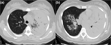
From day 4 to day 5, respiration was increasingly distressed. The fetal heart rate was between 160 and 180 beats/min. HIV and autoimmune antibodies were negative. Atypical pneumonia was suspected. Oral oseltamivir 150 mg twice daily, i.v. vancomycin 1.0 g every 12 h and i.v. immunoglobulin were administered empirically. Indomethacin was given intermittently to relieve high‐grade fever. Chest auscultation revealed bronchovesicular sounds in the left lower field. Chest ultrasound showed left‐sided moderate free‐flowing pleural effusion. Thoracentesis yielded 100 mL straw‐colored pleural effusion with 90% lymphocytes out of total nucleated cells of 350/mm3, total protein was 21.5 g/L (61.1 g/L in serum), albumin was 13.1 g/L, glucose was 10.2 mmol/L, lactate dehydrogenase (LDH) was 226 IU/L (266 IU/L in serum) and adenosine deaminase (ADA) was 9.8 U/L. Pleural fluid culture and acid fast stain were negative. High fever and increasing respiratory distress prompted a low‐dose chest computed tomography with consent from the family and abdomen protection, which revealed massive left lung consolidation and patchy infiltrates in the right middle and lower lobes (Fig. 1). Severe pneumonia was diagnosed. On day 5, the patient's respiratory rate was about 35 breaths/min with a trough SpO2 of 70% while breathing 12 L/min oxygen through a non‐rebreathing facemask. Non‐invasive ventilation was instituted and she was transferred to the respiratory intensive care unit.
Figure 1.

Low‐dose computed tomography of lung on day 4. Lung window (a) and (b) showing left lung consolidation and patchy infiltrates in right upper, middle and lower lobes.
From day 5 to day 9, non‐invasive ventilation and broad‐spectrum antibiotics (meropenem, vancomycin and azithromycin) were continued. Subcutaneous injection of thymosin Alpha‐1 1.6 mg every day for 3 days and 1.6 mg every 2 days for 3 days were given. The patient's temperature gradually decreased to normal, copious purulent sputum was expectorated, dyspnea relieved and non‐invasive ventilation was gradually switched to oxygen mask. The fetal heart rate was between 140 and 160 beats/min and there was no uterine contraction within 15 min. Gram stains and culture of sputum on 4 consecutive days from day 5 to 10 showed methicillin‐resistant Staphylococcus epidermidis (MRSE). Meropenem, oseltamivir and azithromycin were withdrawn on day 9. The patient was transferred to the general ward on day 10 and improved further. Vancomycin was continued until day 18. She fully recovered and was discharged on day 18. The lymphocytopenia resolved and liver enzymes returned to normal.
Throat swab and blood samples were sent to the Laboratory of Virology at the Capital Institute of Pediatrics on day 4. Polymerase chain reaction (PCR), nested PCR and gene sequencing of nasopharyngeal aspirates were positive for adenovirus type 7, which was confirmed by more than fourfold elevation of IgG titer of adenovirus type 7 (retrieved from the patient's blood sample stored in the clinical laboratory from hospital day 1 to day 7).
On follow‐up, the patient had a natural delivery at 39+2 weeks of gestation. The baby weighed 3.2 kg at birth, and is so far healthy. No abnormality in neurology, hearing or growth rate has been found at regular follow‐ups by 20 months.
Discussion
To the best of our knowledge, this is the first report of adenovirus 7 pneumonia occurring in a previously healthy pregnant woman. Human adenoviruses (HAdV) belong to the Adenoviridae family of DNA viruses with seven subgroups and 52 known serotypes. HAdV are a common cause of lower respiratory tract infection in children, immunocompromised adults3 and adults in closed settings (particularly those in the military).4 For immunocompetent adults, prospective studies in the published work (pregnant women were excluded) showed that the adenovirus, especially serotype 7, is an important cause of CAP, accounting for 3.7% (18/487), 4% (11/304), and 5% (48/969) of CAP.5, 6, 7 The seeming rarity of these cases might be due to the physicians' unawareness of viral infection and the unavailability of methods to detect this virus in most hospitals.
Our patient had prolonged fever, cough and dyspnea, normal WBC with lymphopenia, bilateral lung infiltrates, and pleural exudates dominated by mononuclear cells and unresponsive to initial 3‐day antibiotic treatment. Although non‐specific, it was in accordance with the scenario of adenoviral pneumonia as previously reported.5, 6, 7, 8 High‐grade fever (about 90%), cough (>90%), sputum and dyspnea were the most common symptoms and less than 30% of patients experienced diarrhea and/or neurologic symptoms. Crackles on chest auscultation were the most common abnormalities, followed by wheeze and rhonchi. The most common abnormal laboratory findings included lymphopenia, thrombocytopenia, and elevated AST, LDH and creatine kinase (CK) levels. About half of the patients had bilateral involvement on chest radiography, in which reticular nodular changes, consolidation, patchy infiltrate and ground‐glass opacities were the most common findings.
There have been no effective antiviral treatments for adenoviral pneumonia; although cidofovir has been most commonly used, no controlled trials have demonstrated its benefits. For our patient, adequate supportive care, such as oxygen therapy, non‐invasive mechanical ventilation, defervesce and obstetric treatment, led to ultimate success. The value of gamma globulin and thymopeptides needs further studies.
Fortunately, there were neither adverse pregnant outcomes for our patient nor teratogenesis of her baby, except transient threatened abortion, even though our patient had prolonged high fever, transient hypoxemia, and received antimicrobials, including vancomycin, meropenem and oseltamivir. The reported rate of acute respiratory distress syndrome of adenovirus‐associated CAP in the general population is 4.2%.5 Previous studies have shown that pregnant women with viral pneumonia, such as influenza A virus subtype H1N1 and severe acute respiratory syndrome (SARS) coronavirus, have a higher mortality rate and higher rates of both intubation and intensive care unit admission than the general population.9, 10 The increased severity of viral infections in pregnancy is believed to be related to physiologic changes in pregnancy and immunologic alterations result in a shift away from cell‐mediated immunity.1 In addition, increased adverse pregnancy outcomes, such as preterm delivery, low‐birthweight infants, spontaneous abortion and fetal death, are higher in pregnant patients with H1N1 pneumonia than those without.11, 12 The same cases were also reported in pregnant women with SARS.13 The effects of adenovirus on pregnant women and their fetuses need further large‐scale clinical studies.
The limitation of this case report is the lack of radiographic information at disease onset. In addition, although continuous sputum cultures of our patient showed MRSE and response to vancomycin and supportive treatment, secondary S. epidermidis pneumonia could not be confirmed due to a lack of more reliable evidence, such as culture from blood or bronchoalveolar lavage.
In conclusion, this case of adenoviral pneumonia emphasizes the importance of etiologic diagnosis. Aggressive therapies, appropriate supportive care and obstetrical management are key to clinical success.
Disclosure
The authors have no potential conflicts of interest to disclose.
Acknowledgments
The authors are grateful to Dr We Xiaoyu and Dr Shan Xuemin (Department of Gynecology and Obstetrics, Peking University First Hospital) for obstetrical management. We are thankful to Dr Yan Yan (Microbiology Laboratory, Peking University First Hospital) for his immediate smear of the sputum and consecutive cultures afterwards, and Qinwei Song (Laboratory of Virology of Capital Institute of Pediatrics, Beijing) for his contribution to the sample processing and viral identification.
Liao, J.‐p , Wang, G.‐f , Jin, Z. , Qian, Y. , Deng, J. , and Que, C.‐l (2016) Severe pneumonia caused by adenovirus 7 in pregnant woman: Case report and review of the literature. J. Obstet. Gynaecol. Res., 42: 1194–1197. doi: 10.1111/jog.13036.
References
- 1. Kourtis AP, Read JS, Jamieson DJ. Pregnancy and infection. N Engl J Med 2014; 370: 2211–2218. [DOI] [PMC free article] [PubMed] [Google Scholar]
- 2. Chen YH, Keller J, Wang IT, Lin CC, Lin HC. Pneumonia and pregnancy outcomes: A nationwide population‐based study. Am J Obstet Gynecol 2012; 207: 288 e1–288 e7. [DOI] [PMC free article] [PubMed] [Google Scholar]
- 3. Carrigan DR. Adenovirus infections in immunocompromised patients. Am J Med 1997; 102: 71–74. [DOI] [PubMed] [Google Scholar]
- 4. Vento TJ, Prakash V, Murray CK et al. Pneumonia in military trainees: A comparison study based on adenovirus serotype 14 infection. J Infect Dis 2011; 203: 1388–1395. [DOI] [PMC free article] [PubMed] [Google Scholar]
- 5. Sun B, He H, Wang Z et al. Emergent severe acute respiratory distress syndrome caused by adenovirus type 55 in immunocompetent adults in 2013: A prospective observational study. Crit Care 2014; 18: 456. [DOI] [PMC free article] [PubMed] [Google Scholar]
- 6. Gu L, Liu Z, Li X et al. Severe community‐acquired pneumonia caused by adenovirus type 11 in immunocompetent adults in Beijing. J Clin Virol 2012; 54: 295–301. [DOI] [PMC free article] [PubMed] [Google Scholar]
- 7. Jennings LC, Anderson TP, Beynon KA et al. Incidence and characteristics of viral community‐acquired pneumonia in adults. Thorax 2008; 63: 42–48. [DOI] [PubMed] [Google Scholar]
- 8. Clark TW, Fleet DH, Wiselka MJ. Severe community‐acquired adenovirus pneumonia in an immunocompetent 44‐year‐old woman: A case report and review of the literature. J Med Case Reports 2011; 5: 259. [DOI] [PMC free article] [PubMed] [Google Scholar]
- 9. Mosby LG, Rasmussen SA, Jamieson DJ. 2009 pandemic influenza A (H1N1) in pregnancy: A systematic review of the literature. Am J Obstet Gynecol 2011; 205: 10–18. [DOI] [PubMed] [Google Scholar]
- 10. Lam CM, Wong SF, Leung TN et al. A case‐controlled study comparing clinical course and outcomes of pregnant and non‐pregnant women with severe acute respiratory syndrome. BJOG 2004; 111: 771–774. [DOI] [PMC free article] [PubMed] [Google Scholar]
- 11. Centers for Disease Control and Prevention . Maternal and infant outcomes among severely ill pregnant and postpartum women with 2009 pandemic influenza A (H1N1)–United States, April 2009–August 2010. MMWR Morb Mortal Wkly Rep 2011; 60: 1193–1196. [PubMed] [Google Scholar]
- 12. McNeil SA, Dodds LA, Fell DB et al. Effect of respiratory hospitalization during pregnancy on infant outcomes. Am J Obstet Gynecol 2011; 204: S54–S57. [DOI] [PubMed] [Google Scholar]
- 13. Wong SF, Chow KM, Leung TN et al. Pregnancy and perinatal outcomes of women with severe acute respiratory syndrome. Am J Obstet Gynecol 2004; 191: 292–297. [DOI] [PMC free article] [PubMed] [Google Scholar]


