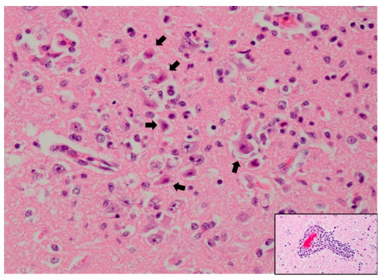
Figure 2.
Hippocampus of a WNV-infected sheep with neuronal necrosis and necrotic debris (arrows) and abundant mixed inflammation admixed with remaining neurons. Hematoxylin and eosin staining at 400× magnification. Inset: Perivascular cuffing with lymphocytes and plasma cells. Hematoxylin and eosin staining at 200× magnification. Slide courtesy of Dr. Chad B. Frank, Colorado State University.

