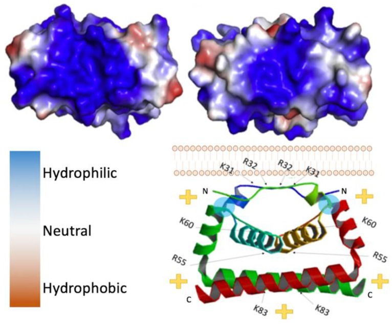
Figure 2.
Flavivirus capsid structure. (Top left) Zika virus (ZIKV) C bottom view, 5YGH.pdb. (Top right) West Nile virus (WNV) C bottom view. Color key provided bottom left, 1SFK.pdb. (Bottom right) side view of ZIKV C dimer with its orientation to the lipid bilayer, indicating the polarity of the complex. Blue circles indicate location of residues necessary for interaction with the lipid membrane (L50 and L54).

