

Abstract
Background
There are certain reported cases of unusual displacements of teeth involved in a maxillofacial trauma to the maxillary sinus, nasal cavity, gastrointestinal tract or the airway, if worst. All these usually lead to complication the worst being death. So enquiring about them is a very essential part of surveying a maxillofacial trauma patient involving dentition.
Case
This patient was referred to our department for a dental consultation for his ill-defined firm, mildly tender, non-suppurative, submental swelling unresponsive to medication. He had undergone a polytrauma involving his face two months back. An orthopantomogram was ordered which showed a lower central incisor from the fracture site had slipped through the fracture gap into the submental space possibly missed by the CT scan. There was also a lower border splaying at the fracture site. A layer-wise dissection was done extra-orally to retrieve the tooth.
Conclusion
This case envisages the importance of an OPG as an adjuvant to the basic radiographic study, in the emergency room, for a patient with oral and maxillofacial trauma. It also establishes the importance of a meticulous secondary survey, including counting the number of teeth and establishing a correct occlusion to avoid a complication and re-operation. Besides, a medical negligence lawsuit can also arise as some teeth may slip to lungs even.
Keywords: Orthopantomogram, Maxillofacial trauma, Tooth displacement, Secondary survey
Introduction
Most of the avulsed teeth are lost at the site of injury during a trauma involving the maxillofacial region. But, in some of the cases the tooth or part of it find way into the soft tissues mostly the upper and lower lip, reported in literature too often. [1] However in certain rare cases of maxillofacial trauma the teeth get unusually displaced into maxillary sinus [2] or the nasal cavity. [3] While as a few percentage of teeth find way into body cavities like airway or gastrointestinal tract. [4]
All such displacements are not without complications. Teeth or their fragments that get embedded in the soft tissues may behave as foreign bodies resulting in a discharging sinus tract, dehiscence of the wound, or a disfiguring fibrosis. [5] But sometimes the complications can be even worse. Their aspiration into lung can lead to laryngeal oedema or pneumothorax as immediate complication. While as a missed or delayed diagnosis can lead to lung abscess, pneumonia, and in worst cases, death. [6]
In most of the cases the management is straight forward but in cases of unusual displacements both the diagnosis and management thereof are unpredictable. This holds true especially in poly-trauma cases where most of the time of examination is given to primary surveys and a meticulous secondary survey is either delayed or ignored leading to late morbidity. The aim of this paper is to report a “unique” clinical case of a lower central incisor “unusually” displaced into the submental space as a result of ignored secondary survey.
Case
This patient was referred from the department of plastic surgery to the department of oral and maxillofacial surgery for a submental swelling unresponsive to antibiotics and analgesics.
Past history was remarkable with history of road traffic accident two months back with polytrauma involving lower limb and maxillofacial region. Accordingly a team of surgeons had operated on the patient with open reduction and internal fixation (ORIF) of the pan-facial trauma with miniplates.
On examination there was a swelling at the right submental region ill-defined on inspection, round, firm and localized on palpation. A submental space infection associated with infection of the fracture site was suspected keeping in view the history of parasymphysis fracture. The lesion was aspirated that revealed a blood tinged, thin, odourless exudate (Fig. 1). An orthopantomograph (OPG) was ordered to evaluate fracture site and status of the miniplates. But to an utter surprise a displaced central incisor was found lying at the base of parasymphysis near the fracture site. Other findings of the OPG were splaying of the fracture gap near the lower border (Fig. 2). But there was absolutely no mobility of the fractured fragments. Also there was a missing edentulous space corresponding to the displaced tooth but no expected complaints regarding his temporomandibular joint (TMJ) and occlusion.
Fig. 1.

Syringe containing exudate after aspiration of the swelling.
Fig. 2.

OPG showing the displaced tooth lying at base of parasymphysis fracture (blue arrow). Fracture gap at parasymphysis base (black arrow). It also shows miniplates used in ORIF of the Pan-facial fractures. (For interpretation of the references to color in this figure legend, the reader is referred to the web version of this article.)
It was decided to surgically explore the region with removal of the unusually displaced tooth. An incision was given extra-orally in the chin where the tooth was felt. Layer-wise dissection was done. The tooth was found to walled-off by a thick fibrous connective tissue capsule that came out as whole along with the tooth inside (Fig. 3). The wound was closed in layers (Fig. 4). The patient was asked for a follow up after 7 days for suture removal and to decide subsequent plan of management for the fracture gap. But the patient never returned.
Fig. 3.
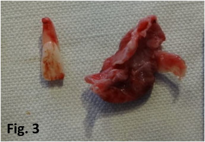
The displaced tooth along with the fibrous capsule removed.
Fig. 4.

Skin closure after layerwise suturing.
Discussion
For all the cases of maxillofacial injury, a meticulous clinical and radiographic assessment including counting the number of teeth, and most importantly establishing a correct occlusion is mandatory. It is because maxillofacial trauma can lead to tooth displacements that are benign [1], serious [2,3] or fatal at times [6].
This case of tooth displacement is unusual and probably unique as far as its location is concerned. The displacement of tooth through the fracture line is always a possibility and should be explored in event the tooth is not accounted for, in cases involving dental & maxillofacial trauma. Computerized tomography (CT) scan, as done in this case also, is usually a basic radiographic investigation in all cases of maxillofacial trauma. However, thicker slices can miss smaller things [7]. The missed/displaced tooth was later seen clearly on an OPG – a very cheap alternate radiographic investigation. As such OPG or cone beam CT (CBCT) can prove very beneficial as tools of investigation in every emergency room (ER). [8]
Another speculation for missing the tooth could be that attending surgeons at the primary surgery were not well aware about dentition or negligence of a meticulous secondary survey. Also the post-op gap at parasymphysis and the missing edentulous space corresponding to the displaced tooth, can be speculated that the surgeons might had made the fractured ends meet for plating in turn causing a splaying at the invisible lower border at the time of primary treatment. This should ideally be causing malocclusion but since the patient had no posterior teeth that complication was avoided. Why there were no TMJ symptoms can be explained by the fact that the splaying was very small.
Here comes the role of oral and maxillofacial surgeon as part of multidisciplinary team involved in management of dental & maxillofacial trauma. Literature has ample examples of patients needing secondary procedures, done by maxillofacial surgeons to correct malocclusion, who had previously been operated by otolaryngologists, neurosurgeons and plastic surgeons. [9]
All teeth displaced, reported in the literature, need and have been retrieved because they are usually laden with oral bacteria. However, certain authors propose that delaying the removal of such teeth for few weeks may allow the fibrosis and stabilize the tooth in position [10]. However mostly the ingested teeth don't need retrieval [4]. Also negligence and malpractice suits that may arise are also a matter of concern, if a tooth displacement is missed and slips into some body space causing complications.
Learning points
-
•
This case report illustrates the importance of a meticulous secondary survey without delay once the patient is stable.
-
•
An OPG should be part of every ER dealing with dental and maxillofacial trauma supplementary to the basic CT scan.
-
•
In case of any injury involving the maxillofacial regions an oral & maxillofacial surgeon should be part of the multidisciplinary team.
Declaration of competing interest
The authors report no conflicts of interest. The authors alone are responsible for the content and writing of the paper.
Acknowledgements
This research received no specific grant from any funding agency in the public, commercial, or not-for-profit sectors.
References
- 1.Michael J., Donald M.B., Swinburn Elizabeth. Tooth in the perioral tissues: a complication of craniofacial trauma. Am. J. Emerg. Med. January 2005;23(1):87–88. doi: 10.1016/j.ajem.2004.09.031. [DOI] [PubMed] [Google Scholar]
- 2.Wang H., Yang C.Y., Li Z. Traumatic displacement of teeth into maxillary sinus and the retrieval assisted by computer-assisted navigation: a case report. Medicine (Baltimore) December 2018;97(51) doi: 10.1097/MD.0000000000013865. [DOI] [PMC free article] [PubMed] [Google Scholar]
- 3.Chrcanovic B., Bueno S., Silveira D., Luis N., Custódio A. Traumatic displacement of maxillary permanent incisor into the nasal cavity. Oral Maxillofac. Surg. September 2010;14(3):175–182. doi: 10.1007/s10006-009-0191-3. [DOI] [PubMed] [Google Scholar]
- 4.Henderson C.T., Engel J., Schlesinger P. Foreign body ingestion: review and suggested guidelines for management. Endoscopy. March 1987;19(2):68–71. doi: 10.1055/s-2007-1018238. [DOI] [PubMed] [Google Scholar]
- 5.Cubukcu C.E., Aydin U., Ozbek S. Delayed removal of a primary incisor embedded in the upper lip after dental trauma: a case report about the importance of soft tissue examination. Dent. Traumatol. 2011;27(4):314–317. doi: 10.1111/j.1600-9657.2011.01000.x. [DOI] [PubMed] [Google Scholar]
- 6.Obi-Egbedi-Ejakpovi E.B., Ogbeide E. Delayed diagnosis of an aspirated tooth in an adolescent. Sahel Med. J. 2015;18(4):207–209. [Google Scholar]
- 7.Holmgren E.P., Dierks E.J., Homer L.D., Potter B.E. Facial computed tomography use in trauma patients who require a head computed tomogram. J. Oral Maxillofac. Surg. 2004 Aug;62(8):913–918. doi: 10.1016/j.joms.2003.12.026. [DOI] [PubMed] [Google Scholar]
- 8.Li G. Patient radiation dose and protection from cone-beam computed tomography. Imaging Sci. Dent. 2013;43(2):63–69. doi: 10.5624/isd.2013.43.2.63. [DOI] [PMC free article] [PubMed] [Google Scholar]
- 9.Lee S.S., Kim S.G., Moon S.Y., Oh J.S., You J.S. The treatment of malocclusion after open reduction of maxillofacial fracture: a report of three cases. J. Korean Assoc. Oral Maxillofac. Surg. 2014;40(2):91–95. doi: 10.5125/jkaoms.2014.40.2.91. [DOI] [PMC free article] [PubMed] [Google Scholar]
- 10.Medeiros N., Gaffrée G. Accidental displacement of inferior third molar into the lateral pharyngeal space: case report. J. Oral Maxillofac. Surg. March 2008;66(3):578–580. doi: 10.1016/j.joms.2005.10.042. [DOI] [PubMed] [Google Scholar]