Abstract
Objective To present the clinical and magnetic resonance imaging (MRI) characteristics of sphenoid bone osteomyelitis.
Procedures Two dogs (English Springer Spaniel – ESS, Golden Retriever – GR) and one cat (Domestic Long Haired) were presented with a 2–14‐day history of visual deficits and reduced pupillary light reflexes. Investigations included physical, ophthalmologic and neurological examination as well as hematology, serum biochemistry, MRI of the head and cerebrospinal fluid (CSF) analysis.
Results MRI changes included thickening of the sphenoid bone and a loss of normal bone marrow signal on T1W MRI. Enhancement of the sphenoid bones, ventral meninges and ventral surface of the brain was present using paramagnetic contrast medium. CSF analysis was abnormal in the two dogs with increased cellularity, neutrophilic pleocytosis, intracellular bacteria and increased total protein in one, and with lymphocytic pleocytosis in another. CSF analysis was normal in the cat.
An underlying cause for the osteomyelitis could not be identified. The use of broad‐spectrum antibiotics for 3–6 weeks combined with anti‐inflammatory medications proved effective. Full clinical recovery occurred with no relapse during the follow up time of 7 (ESS) and 4 (Domestic Long Haired) years. The GR relapsed 10 months after treatment and recovered following a second 3‐week course of broad‐spectrum antibiotics with no relapse during the following 3 years.
Conclusion Visual pathway deficits in dogs and cats may be due to sphenoid bone osteomyelitis. MRI and CSF analysis can assist diagnosing this potentially treatable condition. To the authors’ knowledge this is the first report of sphenoid bone osteomyelitis in these species.
Keywords: blindness, cat, dog, optic chiasm, sphenoid bone osteomyelitis
INTRODUCTION
Osteomyelitis mostly affects the appendicular skeleton of dogs. 1 , 2 Osteomyelitis of the skull is very rare. Mandibles, maxillary bones and tympanic bullae have been reported to be affected as a result of peridontal disease, bite wounds or otitis externa and media, respectively. 1 , 3 , 4 , 5 Stead (1984) reported an osteomyelitis affecting the skull without further describing localization of the disease. 4 One case report described a frontal bone osteomyelitis of unknown origin in a Great Dane. 6
The sphenoid bone forms the rostral base of the cranial cavity. It is composed of the rostral presphenoid bone that, in the cat but not in the dog, contains the sphenoid sinus, and the caudal basisphenoid bone. The chiasmatic fossa is located on the superior surface of these bones and is the anatomical site of the optic chiasm. The sphenoid bones also form the orbital fissures, which allow passage of the cranial nerves (CN) III, IV, VI and the ophthalmic branch of the CN V into the orbit. 7 , 8
In the present article we report sphenoid bone osteomyelitis affecting two dogs and one cat, which presented with visual pathway deficits.
CASE HISTORIES
Case 1
A 3.5‐year‐old, male English Springer spaniel (ESS) was referred with a 2‐week history of exophthalmos, chemosis, reduced pupillary light reflexes (PLR) and anisocoria with mydriasis affecting his right eye. Therapy by the referring veterinary surgeon with ampicillin (Amfipen tablets, Intervet, Milton Keynes, UK) 14 mg/kg BID PO was associated with mild improvement. The ophthalmic examination at the time of referral confirmed exophthalmos and mydriasis of the right eye. The direct PLR on the right was reduced, but the consensual PLR from right to left was present. Menace responses were present bilaterally. At this stage no visual deficits were noticed. The remainder of the ophthalmic examination and a neurological examination were unremarkable. The dog was bright alert and responsive. An ultrasound (Vingmed System 5, General Electric, Fairfield, CT) examination of the right eye was normal. Based on these findings damage to the right sided peripheral oculomotor nerve was suspected. Differential diagnoses included inflammatory/infectious, vascular and neoplastic diseases.
Routine hematology and serum biochemistry were unremarkable. A magnetic resonance imaging (MRI, 0.5 Tesla scanner, SMIS Ltd, Guildford, UK) of the head was performed with the dog under general anesthesia. Noncontiguous transverse images with a 6‐mm slice thickness and an interslice gap of 5 mm were generated with T1‐weighted (T1W) and T2‐weighted (T2W) spin echo pulse sequences in all three planes (transverse, dorsal, sagital) before and after administration of the paramagnetic contrast medium, gadobenate dimeglumin, (MultiHance®, Bracco S.p.A., Milano, Italy) 0.05 mmol/kg IV as a bolus. A right‐sided exophthalmos was documented which was caused by diffuse swelling of orbital tissues, affecting mainly the extra‐ocular muscles, with obliteration of the orbital fat. The pre‐ and basisphenoid bones were thickened and showed loss of normal bone marrow intensity on T1W images (Fig. 1). There was an abnormal, diffuse contrast enhancement of the extra ocular muscles. Less dramatic changes were also seen affecting the left orbit. Marked enhancement of the sphenoid bones, ventral meninges and also of the ventral surface of the brain was detected, surrounding the optic chiasm (Fig. 2). A cerebellomedullary cerebrospinal fluid (CSF) sample revealed a marked increased nucleated cell count (2610 cells/µL; reference range (ref.) 0–5 cells/µL), and a moderate to marked increase in protein (0.87 g/L; ref. 0.0–0.35 g/L), with no red blood cells present. A marked neutrophilic inflammation was documented in the cytospun CSF sample. In addition, intracellular structures consistent with bacterial rods were identified. CSF aerobic culture was negative. Polymerase chain reaction or serological evaluation for infectious diseases was not performed.
Figure 1.
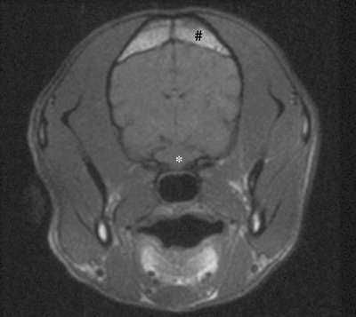
Transverse T1W MRI through the skull of the English Springer Spaniel at the level of the optic chiasm showing diffuse sphenoidal hypointensity (*) relative to normal fatty marrow as seen in the frontal bone (#).
Figure 2.
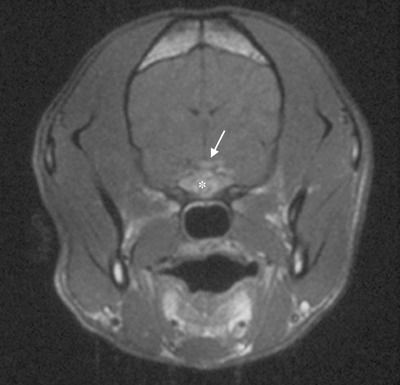
Transverse T1W MR image, post‐contrast through the skull of the English Springer Spaniel at the level of the optic chiasm showing diffuse contrast enhancement of the sphenoid bone (*), the ventral meninges and the ventral surface of the brain (arrow).
The dog was started on empiric treatment with amoxicillin/clavulanic acid (Synulox®, Pfizer, Sandwich, UK) 12.5 mg/kg BID PO and carprofen (Zenacarp®, Pfizer) 2 mg/kg BID PO. At re‐examination 4 days later the exophthalmos was less severe, but the dog showed bilateral visual impairment when performing an obstacle course. Menace responses remained normal bilaterally. Another re‐examination 3 days later still revealed reduced vision but with some improvement when accomplishing another obstacle course. At this point, the treatment was changed to cephalexin (Ceporex®, Schering‐Plough Animal Health, Harefield, UK) 20 mg/kg BID PO, lincomycin (Lincocin®, Pfizer) 11 mg/kg BID PO and carprofen (Zenacarp®, Pfizer) 2 mg/kg BID PO and continued for 3 weeks in total. On re‐examination 1 week later the dog's vision was still impaired, but had further improved, which was evident when performing an obstacle course. The PLR was present in both eyes, but a moderate mydriasis was still evident in the right eye. A rotary nystagmus was noted in the left eye suggestive of vestibular dysfunction, but no further diagnostic investigation was performed at this time. Six months later there was evidence of optic nerve head degeneration apparent on fundic examination, which remained unchanged over the 7‐year follow up period during which the dog did not show any signs of visual impairment.
Case 2
A 5‐year‐old, male neutered Golden Retriever was presented with a 3‐day history of sudden onset blindness and bilateral mydriasis. Ophthalmic examination revealed the absence of menace responses and reduction of direct and consensual PLRs bilaterally. Both optic nerve heads appeared swollen. In addition a neurological examination revealed a depressed mental status, disorientation and ataxia. The remainder of the ophthalmic and neurological examination was unremarkable. Electroretinography was unremarkable in both eyes. Therefore bilateral optic nerve or optic chiasm disease was considered with the differential diagnosis including inflammatory/infectious and neoplastic causes. Routine hematology and serum biochemistry were within normal limits. Polymerase chain reaction for canine distemper virus, Toxoplasma gondii and Neospora caninum were negative in blood and CSF. A MRI (1.5 T scanner, GE Signa, GE Medical Systems, Milwaukee, WI) of the head was performed. Imaging parameters included transverse T1W images and T1W images after administration of gadobenate dimeglumin, MultiHance®) in bolus of 0.05 mmol/kg IV in all three planes. Transverse T2W and gradient echo images were also obtained. Pathological changes identified included a uniform low‐signal intensity of the entire presphenoid bone (Fig. 3). There was a subtle decreased signal intensity of the adjacent optic chiasm. The ventral surface of the brain adjacent to the presphenoid bone was displaced dorsally with a thin hyperintense band of tissue between the brain and the bone. Post‐contrast images showed diffuse enhancement of the presphenoid bone surrounding the optic chiasm and the optic nerves and abnormal meningeal enhancement within the rostral aspect of the brain (Fig. 4). Cerebellomedullary CSF analysis revealed an elevated nucleated cell count (19 cells/µL; ref. 0–5 cells/µL) with a predominantly lymphocytic cell population. CSF culture was not performed. Differential diagnoses included sphenoid bone osteomyelitis or a neoplastic lesion.
Figure 3.
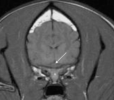
Transverse T1W MR image through the skull of the Golden Retriever at the level of the optic chiasm (arrow) showing diffuse hypointensity of the sphenoid bone (*) relative to normal fatty bone marrow.
Figure 4.
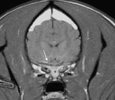
Transverse, T1W MR image post‐contrast through the skull of the Golden Retriever (case 2) at the level of the optic chiasm showing diffuse contrast enhancement of the sphenoid bone (*), the ventral meninges and the ventral surface of the brain (arrow).
The dog was treated empirically with trimethoprim/sulfadiazine (Tribrissen® 480, Schering‐Plough Animal Health) 15 mg/kg BID PO for 3 weeks and metronidazole (Metronidazole®, Metwest Pharmaceuticals, London, UK) 20 mg/kg BID PO for 3 weeks concurrently. After 10 months of complete remission the dog showed visual deficits again. Ophthalmic and neurological examination at this stage did not reveal any additional abnormalities. A repeat MRI study was performed including transverse T1W images, pre‐ and post‐paramagnetic contrast medium (gadobenate dimeglumin, MultiHance®) administration, T2W images in all three planes and transverse T2W FLAIR images. Despite mild clinical deterioration, marked improvement of the MRI appearance of the optic chiasm, presphenoid bone and meninges could be appreciated when compared to the previous study. The presphenoid bone exhibited a normal bone marrow signal, although swelling of the left optic nerve was visible (Fig. 6) when compared to previous images (Fig. 5). Post‐contrast images did not demonstrate sphenoid or meningeal enhancement but perineural enhancement of the left optic nerve suggestive of optic neuritis was detected. A cerebellomedullary CSF sample at this time was within normal limits. Clinical signs improved after another course of antibiotics as outlined above for 4 weeks. No further episodes of visual impairment occurred during the follow up period of 3 years.
Figure 6.
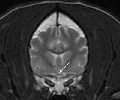
Transverse T2W MR image through the skull of the Golden Retriever at the level of the optic chiasm (arrow) 10 month after first presentation showing the sphenoid bone (*) returned to normal shape and marrow signal.
Figure 5.
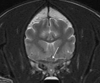
Transverse T2W MR image through the skull of the Golden Retriever at the level of the optic chiasm (arrow) showing diffuse hypointensity of the sphenoid bone relative to normal fatty marrow (*) at first presentation.
Case 3
A 9‐year‐old, female spayed Domestic Long Haired cat was presented with a 2‐day history of sudden onset blindness and depression. The ophthalmological examination revealed vision loss and slow, incomplete PLRs in both eyes. Multifocal bullous retinal detachments and retinal edema were seen in both eyes. Additionally bilateral horizontal nystagmus with the fast phase to the right was identified. The remainder of the ophthalmic and neurological examinations was normal. Routine hematology and serum biochemistry were unremarkable. Serological antibody titers for feline coronavirus were low (1 : 10) (Feline Virus Unit, Glasgow). A titer < 1 : 20 is considered highly indicative that the cat will not develop feline infectious peritonitis. 9 The Toxoplasma IgG antibody titer using an indirect immunofluorescence antibody test was 1 : 54 000 (School of Tropical Medicine, University of Liverpool). No further test results were available.
An MRI scan (1.5 T scanner, GE Signa, GE Medical Systems) of the head was performed obtaining transverse and sagittal T1W images before and after application of the paramagnetic contrast medium, gadodiamide (Omniscan®, GE Health Care, Oslo, Norway) 0.1 mmol/kg as a bolus, as well as transverse and sagittal T2W images. Pathological changes included enlarged sphenoid bones with loss of normal bone marrow signal on T1W images (Fig. 7). Abnormal contrast enhancement was noticed in the tissues surrounding the optic chiasm and optic nerves as well as widespread meningeal enhancement extending to the brainstem (8, 9). A spindle shaped structure dorsally elevated and compressed the brainstem. It peripherally enhanced and was hyperintense compared with normal brain tissue on T2W images, suggestive of a fluid ‘pocket’ (e.g. abscess, cyst, hemorrhage) (Fig. 10). There was bilateral orbital inflammation and enhancement of muscles ventral to the skull base. Cerebellomedullary CSF analysis and culture were unremarkable and negative, respectively. Based on these findings, meningitis, cellulitis and sphenoid osteomyelitis were suspected. The cat was treated with clindamycin (Antirobe®, Pfizer) 12.5 mg/kg BID PO for 6 weeks and prednisolone (Prednidale®, Arnolds Veterinary Products, Shrewsbury, UK) 1 mg/kg BID PO for 2 weeks, reducing the dose over the following 1 week. Six days after treatment initiation the cat was much brighter and marked vision improvement was noted. The PLRs were normal bilaterally and the nystagmus had resolved. The retinal lesions also showed signs of resolution. The antibiotic treatment was continued for 6 weeks. Prognosis was considered guarded due to the possibility of toxoplasmosis. The cat was euthanized 4 years after presentation, due to hepatic failure diagnosed by the referring veterinary surgeon. No relapse of visual deficits was noted until that event. The cat was not available for postmortem.
Figure 7.
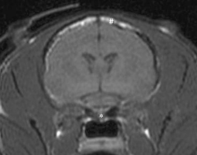
Transverse T1W MR image through the skull of the Domestic Long Haired cat at the level of the optic chiasm showing enlarged sphenoid bones (*), which are hypointense compared with normal fatty bone marrow as seen in the frontal bone (#).
Figure 8.
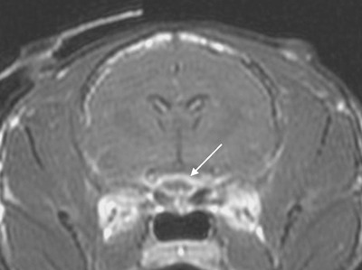
Transverse T1W post‐contrast MR image through the skull of the Domestic Long Haired cat, abnormal contrast enhancement is present within the enlarged sphenoid bones and the adjacent meninges (arrow).
Figure 9.
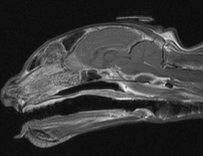
Sagittal T1W post‐contrast MR image through the skull of the Domestic Long Haired cat, showing abnormal contrast enhancement of the sphenoid bone (*) adjacent to the optic chiasm and optic nerves as well as widespread meningeal enhancement extending to the brainstem.
Figure 10.
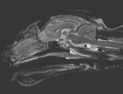
Sagittal T2W MR image through the skull of the Domestic Long Haired cat, showing enlarged sphenoidal bones with loss of normal marrow signal (*). Ventral to the brainstem is a spindle shaped structure, which is elevating and compressing the brainstem (#).
DISCUSSION
To the authors’ knowledge, this is the first report of sphenoid bone osteomyelitis in dogs or cats.
In humans, sphenoid bone or so‐called skull base osteomyelitis is an uncommon condition. 10 , 11 Clinical signs include persistent headache and eventual development of cranial nerve palsies. 12 Depending on the extension of the lesion, one or more of the cranial nerves IX–XII are affected; the cranial nerves III–V are less commonly involved. 12 , 13 Blindness and internal ophthalmoplegia are rarely reported. 14 Patients affected by this condition often have a predisposition to infection due to an underlying condition such as diabetes mellitus, human immunodeficiency virus infection or chronic corticosteroid use. 11 , 12 Skull base osteomyelitis in humans can be divided into two broad categories: typical and atypical. 15 Typical skull base osteomyelitis occurs due to otitis externa that spreads into the marrow of the temporal bone and also involves adjacent tissue. 16 The most common organism isolated in typical skull base osteomyelitis is Pseudomonas aeruginosa; however, Mycobacterium, Aspergillus and Candida spp. have all been documented. 10 Atypical skull base osteomyelitis occurs much less frequently and is not related to otitis externa. Classical signs of infection (fever, elevated WBC count, positive blood cultures) are notably absent. 12 In atypical skull base osteomyelitis, Staphylococcus aureus, coagulase‐negative staphylococcus, Salmonella spp., P. aeruginosa, Eikenella corrodens, Mycobacterium tuberculosis, and fungal agents such as Aspergillus spp., Mucor spp. and Cryptococcus neoformans have all been cultured. 12 , 13 , 17 , 18 , 19 MR imaging demonstrates the characteristic loss of T1 signal hyperintensity of skull base marrow fat. 20 To identify the causative organism a biopsy and culture of the bony lesion is necessary. This can be achieved by using a CT‐guided fine needle aspirate, endoscopic sphenoidotomy or open craniotomy. 12 , 13 Wide surgical debridement and intravenous antibiotics or antifungal treatment as well as hyperbaric oxygen therapy are effective in eradicating the infection. 10 , 14 , 16 Even though treatment is commonly successful, sphenoid bone osteomyelitis is still considered to have a high morbidity. 14
The dogs and cat in the current report showed visual and PLR deficits although the ESS first was presented with a unilateral CN III deficit and exophthalmos. Vision deficits and PLR loss in both eyes reflect a neurological lesion affecting the retinas, optic nerves, the optic chiasm and/or the rostral part of the optic tracts. In lesions of the optic nerves and chiasm the PLR will be preserved to some extent despite the loss of functional vision as fewer functioning optic nerve fibers are required. 21 An unilateral PLR deficit with a normal menace response was found in the ESS, reflecting a lesion of the ipsilateral CN III. The extension of the inflammatory process resulted subsequently in impaired function of visual pathway resulting in blindness. As in humans, classical signs of infections such as fever and an elevated WBC count were absent. A presumptive diagnosis was made predominantly based on MRI findings and CSF changes, if present. MRI abnormalities noted in our three cases include thickening and distortion of the sphenoid bone and diffuse sphenoid hypointensity relative to normal fatty bone marrow on T1W images. Varying surrounding tissue involvement was present such as meningitis and myositis of the extraocular muscles. The latter causing the exophthalmos seen in the ESS. Using paramagnetic contrast medium, a marked enhancement of the sphenoid bones, the ventral meninges and the ventral surface of the brain was present in all cases. This pathology was surrounding and involving the caudal part of the optic nerves and the optic chiasm. Cerebellomedullary CSF findings are not documented for skull base osteomyelitis diagnosis in humans, presumably as direct access to the lesion is possible for tissue sampling. In our cases, CSF results varied. Markedly increased cellularity, a moderate to marked increase of protein, marked neutrophilic inflammation and some intracellular structures highly suggestive of small bacterial rods where found in the ESS. In the GR, an elevated nucleated cell count and lymphocytic inflammation were present. These changes typically are associated with inflammatory central nervous system diseases although normal CSF results do not rule out an inflammatory process. 22 Elevated CSF protein may occur due to alteration of the blood–brain barrier, local necrosis and/or an interruption of normal CSF flow and absorption. 22 CSF analysis and culture in the cat were normal and negative, respectively.
Based on the MRI findings, differential diagnoses included sphenoid bone osteomyelitis or neoplastic lesions with adjacent meningitis and myositis of the extraocular muscles. A neoplastic lesion could not be ruled out at time of diagnostic imaging without biopsy. Although biopsies are taken routinely in humans, we considered this to be a high‐risk procedure for our patients. Therefore, the diagnosis of sphenoid bone osteomyelitis has to remain presumptive.
In the ESS, CN III was involved in the inflammatory process, which can be explained by it traversing the orbital fissure, which is formed by the sphenoid bones.
Retrospectively, it is unclear why case 1 showed a rotary pattern of nystagmus affecting the left eye later in the disease process. The cat showed bilateral horizontal nystagmus in the beginning of the disease. Inner ear disease, CN VIII deficits and lesions affecting the central vestibular nuclei and pathways within brainstem and cerebellum can cause nystagmus. Based on the results of our diagnostic procedures we can not explain the origin of the nystagmus.
An underlying cause for the suspected sphenoid bone osteomyelitis could not be identified in any of our cases. In general, most bone infections in dogs and cats are of bacterial origin, which can be acquired secondary to exposed traumatic wounds, extension from adjacent soft‐tissue infections or hematogenous spread. 1 , 23 Furthermore, multifactorial developmental forms of osteomyelitis (e.g. panosteitis) have been reported in young dogs. 24 , 25 A T. gondii titer of 54 000 in the cat suggested exposure but actual infection could not be proven. Further diagnostic tests (paired serum samples, convalescent samples, IgM titers) that could provide additional diagnostic information were not performed. Toxoplasma gondii has not been reported to cause osteomyelitis and is unlikely to be the causative agent. However, the present retinal changes have been reported to occur in ocular toxoplasmosis, which improved on specific treatment. 26 , 27
Treatment of osteomyelitis in dogs in general includes debridement, open wound drainage and the use of appropriate antimicrobial drugs for 4–6 weeks. The most common infectious agent isolated in cases of osteomyelitis, Staphylococcus spp., are most consistently sensitive to cloxacillin, cefazolin, clindamycin or amoxicillin‐clavulanate. 28 Most anaerobes are sensitive to metronidazole and clindamycin. 28 , 29 The apparent efficacy of antibiotic treatment would be consistent with an underlying bacterial etiology. The use of anti‐inflammatory medication may also have contributed to the clinical remission observed in these cases. The ESS was treated with carprofen, a non‐steroidal anti‐inflammatory drug. The GR received metronidazole, which is also thought to have immunomodulatory properties. 30 The cat responded well to a combination of clindamycin and prednisolone, which potentially could have a detrimental effect in cases of bacterial infection but was used for its anti‐inflammatory properties. Surgical management was not considered possible due to the anatomical location of the sphenoid bone.
To establish a definitive diagnosis more invasive surgical biopsies would be required. Whether this would be justified given the potentially morbidity associated with surgically exploring this region is debatable but this procedure could be substantiated in cases refractive to initial empiric treatment. All three patients underwent a full clinical recovery and appeared to have regained functional vision. However, vision assessment in animals is relatively insensitive and it is unclear how much of the clinical improvement is due to adaptation. Some visual deficits were expected in the ESS that developed a degree of optic nerve degeneration however, this was not clinically apparent.
CONCLUSION
Visual deficits in dogs and cats may be caused by sphenoid bone osteomyelitis. MRI investigation and CSF analysis should be considered to help diagnose this potentially treatable condition.
REFERENCES
- 1. Caywood DD, Wallace LJ, Braden TD. Osteomyelitis in the dog: a review of 67 cases. Journal of the American Veterinary Medical Association 1978; 172: 943–946. [PubMed] [Google Scholar]
- 2. Harari J. Osteomyelitis. Journal of the American Veterinary Medical Association 1984; 184: 101–102. [PubMed] [Google Scholar]
- 3. Johnson KA, Lomas GR, WOOD AKW. Osteomyelitis in dogs and cats caused by anaerobic bacteria. Australian Veterinary Journal 1984; 61: 57–61. [DOI] [PubMed] [Google Scholar]
- 4. Stead AC. Osteomyelitis in the dog and cat. Journal of Small Animal Practice 1984; 25: 1–13. [Google Scholar]
- 5. Seiler G, Rossi F, Vignoli M et al Computed tomographic features of skull osteomyelitis in four young dogs. Veterinary Radiology and Ultrasound 2007; 48: 544–549. [DOI] [PubMed] [Google Scholar]
- 6. Grahn BH, Szentimrey D, Battison A et al Exophthalmos associated with frontal sinus osteomyelitis in a puppy. Journal of the American Animal Hospital Association 1995; 31: 397–401. [DOI] [PubMed] [Google Scholar]
- 7. Seiferle E. Lehrbuch der Anatomie der Haustiere. Verlag Paul Parey, Berlin and Hamburg, 1992. [Google Scholar]
- 8. Evans HE. Miller's Anatomy of the Dog. W.B. Saunders Company, Philadelphia, 1993. [Google Scholar]
- 9. Addie DD, Jarrett O. Feline coronavirus infections. In: Infectious Diseases of the Dog and Cat, 3rd edn. (ed. Greene CE) Elsevier, St Louis, 2006. [Google Scholar]
- 10. Chandler JR, Grobman L, Quencer R, et al Osteomyelitis of the base of the skull. Laryngoscope 1986; 96: 245–251. [DOI] [PubMed] [Google Scholar]
- 11. Ducic Y. Management of osteomyelitis of the anterior skull base and craniovertebral junction. Otolaryngology – Head and Neck Surgery 2003; 128: 39–42. [DOI] [PubMed] [Google Scholar]
- 12. Chang PC, Fischbein NJ, Holliday RA. Central skull base osteomyelitis in patients without otitis externa: imaging findings. American Journal of Neuroradiology 2003; 24: 1310–1131. [PMC free article] [PubMed] [Google Scholar]
- 13. Malone D, O’Boynick PL, Ziegler DK et al Osteomyelitis of the skull base. Neurosurgery 1992; 30: 426–431. [DOI] [PubMed] [Google Scholar]
- 14. Singh A, Al Khabodi M, Hyder MJ. Skull base osteomyelitis: diagnostic and therapeutic challenges in atypical presentation. Otolaryngology – Head and Neck Surgery 2005; 133: 121–125. [DOI] [PubMed] [Google Scholar]
- 15. Grobman LR, Ganz W, Casiano R et al Atypical osteomyelitis of the skull base. Laryngoscope 1989; 99: 671–676. [DOI] [PubMed] [Google Scholar]
- 16. Djalilian HR, Shamloo B, Thakkar KH et al Treatment of culture‐negative skull base osteomyelitis. Otology and Neurotology 2006; 27: 250–255. [DOI] [PubMed] [Google Scholar]
- 17. Senegor M, Lewis HP. Salmonella osteomyelitis of the skull base. Surgical Neurology 1991; 36: 37–39. [DOI] [PubMed] [Google Scholar]
- 18. Chan LL, Singh S, Jones D et al Imaging of mucormycosis skull base osteomyelitis. American Journal of Neuroradiology 2000; 21: 828–831. [PMC free article] [PubMed] [Google Scholar]
- 19. Das JC, Singh K, Sharma P et al Tuberculous osteomyelitis and optic neuritis. Ophthalmic Surgery, Lasers and Imaging 2003; 34: 409–412. [PubMed] [Google Scholar]
- 20. Erdman WA, Tamburro F, Jayson HT et al Osteomyelitis: characteristics and pitfalls of diagnostic with MR imaging. Radiology 1991; 180: 533–539. [DOI] [PubMed] [Google Scholar]
- 21. Ferriera FM, Peterson‐Jones S. Neuro‐ophthalmology In: BSAVA Manual of Small Animal Ophthalmology, 2nd edn. (eds Peterson‐Jones SM, Crispin S) British Small Animal Veterinary Association, Gloucester, 2002; 257–275. [Google Scholar]
- 22. Chrisman CL. Cerebrospinal fluid analysis. The Veterinary Clinics of North America. Small Animal Practice 1992; 22: 781–810. [DOI] [PubMed] [Google Scholar]
- 23. Muir P, Johnson KA. Anaerobic bacteria isolated from osteomyelitis in dogs and cats. Veterinary Surgery 1992; 21: 463–466. [DOI] [PubMed] [Google Scholar]
- 24. La Fond E, Breur GJ, Austin CC. Breed susceptibility for developmental orthopedic diseases in dogs. Journal of the American Animal Hospital Association 2002; 38: 467–477. [DOI] [PubMed] [Google Scholar]
- 25. Demko J, McLaughlin R. Developmental orthopaedic disease. The Veterinary Clinics of North America. Small Animal Practice 2005; 35: 1111–1135. [DOI] [PubMed] [Google Scholar]
- 26. Davidson MG, English RV. Feline ocular toxoplasmosis. Veterinary Ophthalmology 1998; 1: 71–80. [DOI] [PubMed] [Google Scholar]
- 27. Davidson MG. Toxoplasmosis. The Veterinary Clinics of North America. Small Animal Practice 2000; 30: 1051–1062. [DOI] [PubMed] [Google Scholar]
- 28. Johnson KA. Osteomyelitis in dogs and cats. Journal of the American Veterinary Medical Association 1994; 205: 1882–1887. [PubMed] [Google Scholar]
- 29. Hirsh DC, Indiveri MC, Jang SS et al Changes in prevalence and susceptibility of obligate anaerobes in clinical veterinary practice. Journal of the American Veterinary Medical Association 1985; 186: 1086–1089. [PubMed] [Google Scholar]
- 30. Farajeh M, Mohammad MK, Bustanji Y et al Evaluation of immunosuppression induced by metronidazone in Balb/c mice and human peripheral blood lympnocytes. International Immunopharmacology 2008; 8: 341–350. [DOI] [PubMed] [Google Scholar]


