PLOS ONE logoLink to PLOS ONE
. 2020 Apr 20;15(4):e0231718. doi: 10.1371/journal.pone.0231718

Comparing the efficiency of open and enclosed filtration systems in environmental DNA quantification for fish and jellyfish

Sayaka Takahashi 1,2,*, Masayuki K Sakata 3, Toshifumi Minamoto 3, Reiji Masuda 2
Editor: Ruslan Kalendar4
PMCID: PMC7170242  PMID: 32310994

Abstract

Water sampling and filtration of environmental DNA (eDNA) analysis have been performed by several different methods, and each method may yield a different species composition or eDNA concentration. Here, we investigated the eDNA of seawater samples directly collected by SCUBA to compare two widely used filtration methods: open filtration with a glass filter (GF/F) and enclosed filtration (Sterivex). We referred to biomass based on visual observation data collected simultaneously to clarify the difference between organism groups. Water samples were collected at two points in the Sea of Japan in May, September and December 2018. The respective samples were filtered through GF/F and Sterivex for eDNA extraction. We quantified the eDNA concentration of five fish and two cnidarian species by quantitative polymerase chain reaction (qPCR) using species-specific primers/probe sets. A strong correlation of eDNA concentration was obtained between GF/F and Sterivex; the intercepts and slopes of the linear regression lines were slightly different in fish and jellyfish. The amount of eDNA detected using the GF/F filtration method was higher than that detected using Sterivex when the eDNA concentration was high; the opposite trend was observed when the eDNA concentration was relatively low. The concentration of eDNA correlated with visually estimated biomass; eDNA concentration per biomass in jellyfish was approximately 700 times greater than that in fish. We conclude that GF/F provides an advantage in collecting a large amount of eDNA, whereas Sterivex offers superior eDNA sensitivity. Both filtration methods are effective in estimating the spatiotemporal biomass size of target marine species.

Introduction

Environmental DNA (eDNA) analysis is attracting a great deal of attention as a more efficient and sensitive tool than conventional monitoring methods [1, 2]. After Ficetola [3] applied a newly developed eDNA method to detect bullfrogs in ponds, researchers have attempted to use it for detecting animals and plants, and for quantifying their abundance in the environment. Ocean field studies comparing the species-specific eDNA sequence method with conventional survey methods [4], or the eDNA metabarcoding analysis with the underwater visual census [5], bottom trawling [6] and net and trap [7] methods, suggest that the eDNA analysis is a promising tool for revealing the species composition of fish communities. By using the eDNA metabarcoding analysis, researchers managed to detect 93.3% of the fish species present in seawater samples from aquarium tanks [8].

For quantification using eDNA analysis, there was a positive correlation between eDNA concentration and fish biomass in ponds [9, 10] and tanks [11, 12]. The concentration of eDNA was also positively correlated with the size of fish [13], density [1416] and wet mass [17]. Previous studies comparing spatiotemporal change in abundance or biomass in aquatic species, based on traditional methods and eDNA concentration, found a significantly positive correlation between biomass and the amount of eDNA in visual observations via land or vessel based surveys [18], using commercial fish landing data [19], via captures by bottom trawl [6], and by monitoring using echo sounder technology [20] in marine environments. A similar correlation was detected in a snorkeling survey [21], net capture surveys [9, 10], and during mark-recapture experiments [14] in freshwater environments; however, some researchers reported that a quantitative relationship between biomass and eDNA abundance was not found [2224].

Water sampling, filtration, preservation and DNA extraction methods vary depending on the sample type and the research team conducting the eDNA analysis of species composition and quantification of aquatic organisms [25, 26]. Differences in filtration and sampling protocol affect the amount of eDNA detected [2730] and the detection rate [3134] of aquatic species, which is problematic. These studies elucidate the necessity to choose suitable filters, which vary depending on the target species, taking into account environmental factors and water sample types to establish an optimized and versatile protocol.

Recently, a simple, on-site eDNA analysis system was developed [11, 35]. For on-site filtration, an enclosed Sterivex filter is handier and more effective than an open filter; the latter requires a comparatively larger-scale filtration system. The transition from open filter (requiring handling, a filter funnel and a vacuum pump) to enclosed filter (enclosed in a capsule during filtration and DNA extraction) has advanced. Sterivex filtration is used as a method to examine the presence/absence of target species [36, 37]. Filtration time is shortened by combining the filter with a syringe, in situations where on-site filtration is required [38]. It has also been reported that eDNA is better conserved and a greater amount of it can be extracted when using an enclosed filter, as opposed to an open filter, to detect fish species in ponds [39]. This may be particularly true when using the eDNA metabarcoding method that utilizes MiFish PCR primers, whereby the number of species detected by using Sterivex filters, was significantly higher than the corresponding number obtained by using glass fiber filters (GF/F) [31]. On the other hand, the amount of eDNA obtained by open filtration was larger than that obtained by the precipitation method [27, 29]. When comparing open filtration systems, a greater amount of eDNA was extracted by using a cellulose nitrate filter [30] or by using a GF/F [27]; the most generally used pore sizes were 0.45 μm and 0.7 μm [26], the latter being used in this study. In the water, eDNA is considered to exist in various states and particle sizes [2], most abundantly in the 1–10 μm size class [40]. It is therefore efficient to use a 0.8 μm-pore size filter for filtration [38] and a 0.7 μm-pore size GF/F has been recommended for time and cost effectiveness [28], as shown in previous studies [1821, 35, 4143].

Some controversy exists as to whether there are any differences between the amount of eDNA detected by the two different filtration methods (GF/F and Sterivex) when performing quantitative analysis of eDNA. A direct comparison of eDNA concentrations obtained by the two filtration methods will clarify the differences between them. Based on such knowledge, adaptive usage of each filtration method will be possible, and this should facilitate the ability to estimate the biomass of target species.

The present research aimed to test whether there is a correlation between the eDNA concentrations obtained by GF/F and Sterivex, and between the eDNA concentration and biomass. To check for a potential bias between the upper and lower layers of sampling bags, the eDNA concentrations of these two layers were also compared. Comparing the estimated biomass using different eDNA methodologies and applications can be valuable for future eDNA studies, particularly for optimizing survey strategies [25].

Materials and methods

Ethics statement

The underwater visual survey was conducted in accordance with local and governmental laws and regulations. Underwater surveys in Nagahama were approved by the harbormaster of Maizuru Bay (No. 300 issued on July 6 and No. 405 issued on September 28, 2018). No approval was required for the surveys in Otomi where leisure diving is common. No fish or other animals were harmed for the purpose of this study, except for tissue sampling for genetic analyses (see 'PCR analysis' below). The research (observation, fish collection, tissue sampling and euthanasia) was performed according to the guidelines of Regulation on Animal Experimentation at Kyoto University (https://www.kyoto-u.ac.jp/en/research/research-compliance-ethics/animal-experiments.html, last accessed on November 14, 2019) and the Kyoto Prefecture Fishery Management Rules (https://www.pref.kyoto.jp/reiki/reiki_honbun/aa30006341.html, last accessed on November 14, 2019). No ethical approval was required for this procedure due to the common consumption of these fishes. Our field studies did not involve endangered or protected species.

Water sampling and filtration in Otomi

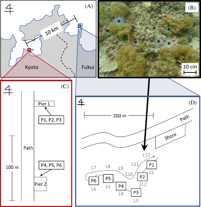
Seawater samples (3 L each) were collected using a water sampling bag, Lamizip (Standup Nylon Bag with Zipper, LZ-14, Seisannipponsha, Ltd., Tokyo, Japan) at approximately ten min intervals at 1 m off the bottom, at six locations of Otomi, Wakasa Bay, Fukui, Japan [44] (35°32ˈN, 135°30ˈE; Figs 1A, 1D and 2) on May 15, 2018. Water temperature (measured using an alcohol thermometer that had been calibrated by a mercury standard thermometer) was 20.2°C near the sea surface and 18.2°C at seafloor, salinity (measured by a water quality meter with a conductivity probe and reported in practical salinity units (psu): LAQUAact ES-71, Horiba, Ltd., Kyoto, Japan) was 30.4, visibility (visually estimated by Masuda) was 8 m, and depth (measured with a diving computer: SUUNTO D6, Vantaa, Finland) was 3.0–5.8 m.

Fig 1. Study site.

Fig 1

(A) Map showing the study sites in Kyoto and Fukui Prefecture. (B) Photograph of Aurelia aurita carcasses (*) on the sea floor found along the Line (L) 12 (black arrow) in Otomi. (C) Sample points (P1-P3 at pier 1 and P4-P6 at pier 2) in Nagahama. (D) Sampling points (P1-P6) in Otomi along the westward (outgoing, L1-L6) way of visual census lines (grey curved arrow). Visual census was also conducted along the eastward (return, L7-L12) way.

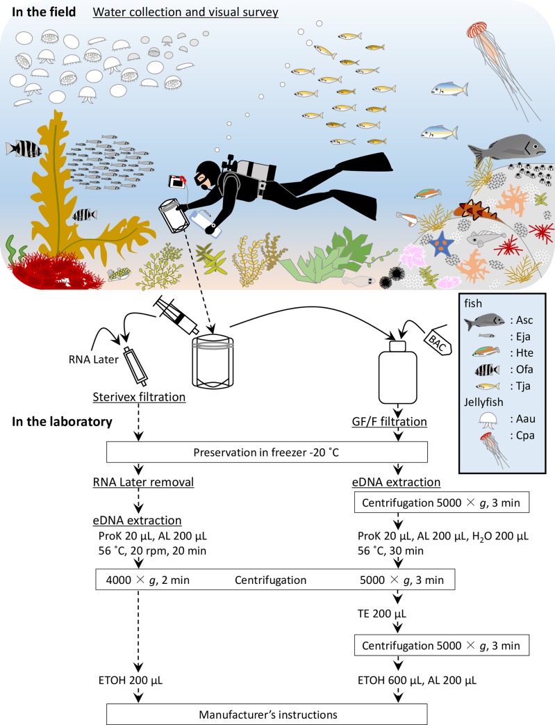
Fig 2. Schematic drawing of visual survey, and outline flowchart of water collection, extraction, and comparison of filtration using GF/F and Sterivex.

Fig 2

The illustration depicts two species of jellyfish (Aau and Cpa), pelagic fish (Eja and Tja) and demersal fish (Ofa, Hte and Asc) and highlights a visual representation of the position in the water column and schooling behavior of some of these species assemblages during a typical transect. Seawater samples, collected during visual survey, were filtrated using Sterivex on site, while samples for GF/F filtration were transferred to the laboratory. BAC: benzalkonium chloride, ProK: Proteinase K, AL: buffer AL, TE: buffer TE, ETOH: ethanol, Asc: Acanthopagrus schlegelii, Eja: Engraulis japonicus, Hte: Halichoeres tenuispinnis, Ofa: Oplegnathus fasciatus, Tja: Trachurus japonicus, Aau: Aurelia aurita, Cpa: Chrysaora pacifica.

The eDNA analysis process was conducted based on the methods previously described in the Environmental DNA Sampling and Experiment Manual (Version 2.1) [45], with a slight modification. One liter of each sample was poured into a prewashed plastic bottle, and 1 mL of benzalkonium chloride (BAC, Nihon Pharmaceutical, Co., Ltd., Tokyo, Japan) was added. Another 1 L of each sample was filtered through Sterivex (0.45 μm-pore size, Merck Millipore, Darmstadt, Germany) using a 50-mL syringe, and 2 mL RNAlater (Thermo Fisher Scientific, Waltham, MA, USA) was added for DNA preservation. As a blank control, 500 mL purified water (Kenei Pharmaceutical, Osaka, Japan) was filtered in the same way using a measuring cup bleached with 0.1% sodium hypochlorite and washed with purified water. Bottles and Sterivex filters were transported on ice in a cooler box to the laboratory. It took less than 60 min from sampling to Sterivex filtration.

Each 1 L sample bottle and 500 mL of distilled water, was filtered through an aspirator using glass fiber filters (GF/F, 0.7 μm-pore size, Whatman, Maidstone, UK) in the laboratory. Filtering devices were bleached after every filtration with 0.1% sodium hypochlorite for 5 min, washed with tap water, and rinsed with distilled water. Filters were wrapped in aluminum foil, placed in plastic bags, and preserved at -20°C until DNA extraction. The process from sampling to preservation was conducted within seven hours. Nitrile gloves were worn both during filtration and the procedures that followed.

Water sampling and filtration in Nagahama

Seawater samples (3 L each) were collected at 8–10 min intervals at 1 m off the bottom at six locations (three at each of two piers in the Maizuru Fisheries Research Station of Kyoto University) [46] (Nagahama, Maizuru, Kyoto, Japan; 35°29ˈN, 135°22ˈE; Figs 1A, 1C and 2) on September 19, 2018. Environmental data collection and the process from water sampling to preservation was performed in the same manner as at the Otomi site. The water temperature, salinity and depth near pier 1 and pier 2 was 26.4°C, 30.0 and 3.7 m, and 26.2°C, 28.7 and 2.7 m, respectively. The visibility was approximately 2 m near the surface and 5 m around the sampling points in both piers. It took less than 10 min from sampling to Sterivex filtration.

Water samples were also collected at the same locations on December 18, 2018. Water temperature around sampling points of pier 1 and pier 2 were 16.2°C and 15.6°C, respectively. Salinity near pier 2 was 28.0, and visibility was about 1 m near the surface and 4 m around the sampling points. Except for Sterivex filtration, the process from water sampling to preservation was performed in the same manner. The process from sampling to preservation was performed within three hours.

For the Sterivex filter, we checked for a potential bias between the upper and lower layers of the Lamizip sampling bag. During the Sterivex water filtration in December, we first filtered the upper layer, and then the lower layer by using a vinyl tube. The eDNA concentrations of these two layers were compared.

Biomass estimation based on underwater visual censuses

Underwater visual censuses by SCUBA were conducted at six locations in Otomi (Fig 1D) and six locations in Nagahama (three locations × two piers) (Fig 1C) at the time of water sampling, above. The number of individuals, body length of fishes, and umbrella diameter of jellyfish was recorded on an underwater slate in an area of approximately 100 m2 (50 m by 2 m) around each water sampling point [46]. In this survey, the modified transect method termed “fin-kick transect” was applied, in which the distance traveled was estimated by the number of fin kicks made [47, 48]. Fish and jellyfish with the minimum size of 1 cm were recorded on an underwater slate in our routine survey, although the smallest individuals recorded in the present study were 3 cm. The length (L cm) of each species was converted to biomass (W g) using the length-weight relationship reported in previous studies [4952].

W=aLb

a: a parameter describing body shape and condition

b: a parameter for the allometric growth in body proportion

eDNA extraction from Sterivex filters

Extraction of eDNA from Sterivex filters was performed using a DNeasy Blood & Tissue Kit (Qiagen, Hilden, Germany) based on the method of Miya et al. [31] and the Environmental DNA Sampling and Experiment Manual (Version 2.1) [45], with a slight modification (Fig 2). Each filter cartridge was centrifuged for 2 min at 4,000 × g. After addition of 1 mL of Bottled Water for Molecular Biology (Merck Millipore, Darmstadt, Germany), the cartridge was centrifuged again. Then, a 220 μL solution composed of 20 μL Proteinase K and 200 μL lysis buffer (AL buffer) was added into the cartridge. The cartridge was incubated at 56°C, 20 rpm for 20 min, and the lysed DNA solution was collected after centrifugation. After 200 μL of ethanol was added to the collected liquid, the mixture was transported to a spin column and centrifuged for 1 min at 6,000 × g. Subsequently, we followed the manufacturer’s instructions and eluted in a 100 μL elution buffer (AE buffer) before preserving at -20°C.

eDNA extraction from GF/F

Extraction of eDNA from GF/F was performed using a DNeasy Blood & Tissue Kit according to a previous study [20] (Fig 2). Each filter was placed in a Salivette tube (Sarstedt, Nümbrecht, Germany) and centrifuged for 3 min at 5,000 × g. Then, a 420 μL solution composed of 20 μL Proteinase K, 200 μL AL buffer and 200 μL H2O was put on the filter. The tube was incubated at 56°C for 30 min, and the lysed DNA solution was collected by centrifugation. After adding 200 μL of tris-ethylenediaminetetraacetic buffer (TE buffer) to the filter, the liquid was again collected by centrifugation. After a 200 μL AL buffer and 600 μL ethanol were added to the collected liquid, the mixture was transported to a spin column and centrifuged for 1 min at 6,000 × g. Subsequently, we followed the manufacturer’s instructions and eluted in a 100 μL AE buffer before preserving at -20°C.

PCR analysis

Five fish species and two cnidarian species with a high frequency of occurrence in the study sites were selected for PCR analysis; blackhead seabream Acanthopagrus schlegelii, Japanese anchovy Engraulis japonicus, wrasse Halichoeres tenuispinnis, striped knifejaw Oplegnathus fasciatus, Japanese jack mackerel Trachurus japonicus, moon jellyfish Aurelia aurita, and Japanese sea nettle Chrysaora pacifica. The eDNA concentrations were quantified by quantitative PCR (qPCR) using a LightCycler 96 System (Roche Diagnostics, Mannheim, Germany). DNA from each target species was amplified by using the species-specific designed primers and probe sets from mitochondrial cytochrome b gene and partial mitochondrial cytochrome oxidase I gene (COI) region (Table 1). Primers/probe sets were confirmed to amplify each specific target; E. japonicus [53], T. japonicus [20 and Suppl. material 3], C. pacifica [18], and A. aurita (Yoden et al., unpublished data). The sequences of the three target species A. schlegelii, H. tenuispinnis, O. fasciatus, and related species known to inhabit Maizuru and Wakasa Bay [46], were collected from GenBank (see S1 Table for details of related species considered). Primer sets with more than two substitutions between the target and related species, within five bases from the 3ʹ end, were designed for each fish species; base pair mismatches in the 3ʹ end are important for primer specificity [54]. The probes were designed using Primer Express 3.0 software (Thermo Fisher Scientific, Waltham, MA, USA). The specificity of the primer sets was checked using primer-BLAST (NCBI nucleotide database) with default settings.

Table 1. Sequences of primers and probes for detecting eDNA of five fish and two cnidarian species targeted in this study.

Target species Name of Sequence (5' —> 3') Amplicon Reference
primers/probe length
Acanthopagrus Asc_CytB_F CTGTCTGCCGTCCCCTACA 129 This study
schlegelii Asc_CytB_R TATGGCGGCTACGATAAAAGGA
Asc_CytB_P FAM-TCAGTTGACAACGCAACCCTAACCCG-TAMRA
Engraulis Eja_CytB_F GAAAAACCCACCCCCTACTCA 115 Ushio et al. [51]
japonicus Eja_CytB_R GTGGCCAAGCATAGTCCTAAAAG
Eja_CytB_P FAM-CGCAGTAGTAGACCTCCCAGCACCATCC-TAMRA
Halichoeres Hte_CytB_F CGCAGACGTTGTAGTCCTCACA 113 This study
tenuispinnis Hte_CytB_R GTGAGAAGACTAGGAATAGTATAAAGTAGATGATG
Hte_CytB_P FAM-CCGTACGTAATTATTGGCCAAATCGCG-TAMRA
Oplegnathus Ofa_COI_F GAAACTGACTCATCCCCCTCA 166 This study
fasciatus Ofa_COI_R CCTGCGAGAGGCGGAT
Ofa_COI_P FAM-TAACATGAGCTTTTGACTGCTCCCACCCTC-TAMRA
Trachurus Tja_CytB_F CAGATATCGCAACCGCCTTT 127 Yamamoto et al. [20]
japonicus Tja_CytB_R CCGATGTGAAGGTAAATGCAAA ; Probe was this study
Tja_CytB_P FAM-CCGTAGCACACATCTGCCGGGA-TAMRA
Aurelia aurita Aau_COI_F TTACTACCCCCAGCTCTGCTTT 120 Yoden et al., unpublished data
Aau_COI_R TACTGAACCACCGGAATGG
Aau_COI_P FAM-ATGAACAATTTATCCCCCCCTAAGCGCA-TAMRA
Chrysaora Cpa_COI_F CCCAGATATGGCTTTTCCTAGA 231 Minamoto et al. [18]
pacifica Cpa_COI_R TGAGTGAGCTTGTATAGCTGATA
Cpa_COI_P FAM-TAGGATCCTCCCTAATTG-NFQ-MGB

To confirm the specificity of the primers/probe sets, the DNA of the most closely related fish species was tested with the established real-time PCR for each species. The red seabream (Pagrus major), the multicolorfin rainbowfish (Halichoeres poecilopterus), and the rudderfish (Girella punctata) are the most closely related fish species to A. schlegelii, H. tenuispinnis, and O. fasciatus, respectively, and inhabit the same survey area [46]. Tissue samples of these fishes were mostly obtained from the Fish Collection of Kyoto University (FAKU), in which fish specimens are routinely provided from a local fish market. Two species, G. punctata (n = 4) and O. fasciatus (n = 3), were additionally collected by hook-and-line fishing or a hand net in Wakasa Bay, Sea of Japan for the present study. They were euthanized by an overdose of 2-phenoxy ethanol prior to dissection to obtain tissue samples. The total DNA of the related species was extracted using a DNeasy Blood & Tissue Kit according to the protocol for tissue samples, and 10 or 100 pg of the total DNA of related species was applied as a template.

Based on a previous study [20], each 20 μL reaction mixture contained 900 nM primers (forward and reverse; F/R) and 125 nM TaqMan Probe in 10 μL TaqMan Environmental Master Mix 2.0 (Thermo Fisher Scientific, Waltham, MA, USA) and 0.1 μL AmpErase Uracil N-Glycosylase (Thermo Fisher Scientific) or 10 μL FastStart Essential DNA Probes Master (Roche), and 2 μL DNA sample. Dilution series containing 3 × 101–3 × 10⁴ copies per PCR tube were prepared and used as quantification standards. The qPCR conditions were as follows: 2 min at 50°C, 10 min at 95°C, 55 or 60 cycles of 15 s at 95°C and 60 s at 60°C, or 10 min at 95°C, 50 cycles of 10 s at 95°C and 30 s at 60°C. Three replicates were used for each sample, and three replicate negative controls containing PCR Grade Water (Roche) instead of template DNA were included in all PCR plates. The average of the triplicates was taken to represent eDNA concentration. For each species, PCR of standard and samples obtained by GF/F and Sterivex were performed in one plate. In all the runs, R2 values of calibration curves were more than 0.98, the range of slopes, Y-intercept, and PCR efficiency were between -3.94 and -3.44, 37.38 and 40.55, and 0.79 and 0.95, respectively (S2 Table). None of the PCR-negative controls or field blank controls were PCR-amplified. To reduce the risk of carry-over contamination, the pre- and post-PCR experiments were performed in independent rooms.

Data analysis

Biomass based on visual census and eDNA concentration was log10 (x+1) transformed to improve homogeneity of variance. The average of three replications was used as eDNA concentration, and all possible combinations of the sample species, sampling date and sample locations yielded 90 fish datasets and 36 jellyfish datasets. We used one observation result corresponding to each eDNA dataset for biomass data. Correlations between eDNA concentrations obtained by the two different filtration methods (GF/F and Sterivex), and between biomass based on visual census and eDNA concentration detected by GF/F and Sterivex, were analyzed by a linear mixed effect model, in which species was treated as a random effect using the “lmer” functionality of R statistical software (ver. 3.4.3) [55] for all species. Parameters were estimated by linear regression analysis (the 95% confidence and prediction limits, and analysis of intercepts and slopes of these lines) using “lm” of R for fish and jellyfish, and for each species. The choice of the most suitable model was made by using Akaike's Information Criteria (AIC) from the “ANOVA” in R. Values were removed from the linear regression analysis of biomass and eDNA concentration when either of them was 0. Differences in eDNA detected in the upper and lower layers of collection bags were evaluated using a paired Student’s t-test in the R software.

Results

The specificity of the primers

For three targeted species, A. schlegelii, H. tenuispinnis, and O. fasciatus, real-time PCRs with 10 or 100 pg of the total DNA of the most related sympatric species as a template, showed no amplification in any of the three replicates. Thus, primers designed in this study had enough specificity for detecting the targeted species in our survey area.

Comparison of eDNA concentrations obtained by GF/F and Sterivex

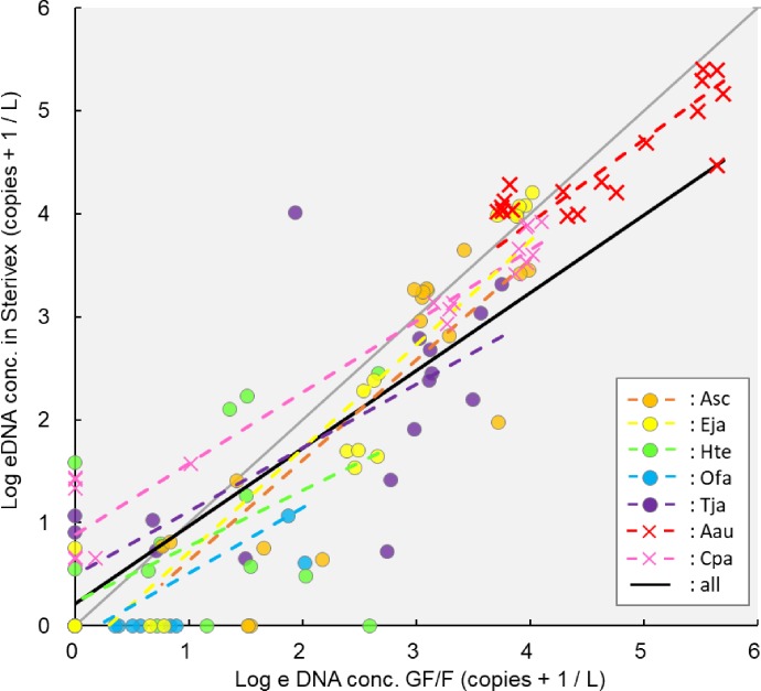
A linear regression equation of eDNA concentrations obtained by the two different filtration methods (GF/F and Sterivex) was calculated for all species (YSterivex = 0.75 XGF/F + 0.22; Fig 3). There was a strong correlation between them in both fish and jellyfish (R2 = 0.74 and 0.95, respectively; Fig 4). The intercept of the linear regression line of fish was not significantly different from 0 (p = 0.54), whereas that of jellyfish was significantly higher than 0 (p < 0.01). The 95% confidence limits were lower than y = x when the eDNA amount was high in both taxa. The eDNA amount detected using the Sterivex filtration method was higher than that detected using the GF/F filtration method when the eDNA concentration of jellyfish was low (Fig 4). There were several cases where the eDNA concentration, in both fish and jellyfish, obtained using GF/F was 0, whereas that obtained using Sterivex was higher than 0. There were some cases where the eDNA concentration, only in fish, obtained using Sterivex was 0, but that obtained using GF/F was higher than 0. Intercepts and slopes of these lines were slightly different depending on the different fish and jellyfish taxa using “lmer” analysis (Fig 3). There was a significant difference in intercept between A. aurita, C. pacifica, H. tenuispinnis, T. japonicus and A. schlegelii, E. japonicus, O fasciatus. There was a difference in slopes between A. aurita, C. pacifica, H. tenuispinnis, O fasciatus, T. japonicus and A. schlegelii, E. japonicus using “lm” analysis (p = 0.05; S1 Fig). There was no significant difference in eDNA concentration between the upper and lower layer of a stable standing Lamizip (p = 0.92).

Fig 3. Correlation of eDNA concentration estimated by two different filtration methods (GF/F and Sterivex) in all species.

Fig 3

Linear regression equation including all species: YSterivex = 0.75 XGF/F + 0.22, gray lines: y = x, Asc: Acanthopagrus schlegelii, Eja: Engraulis japonicus, Hte: Halichoeres tenuispinnis, Ofa: Oplegnathus fasciatus, Tja: Trachurus japonicus, Aau: Aurelia aurita, Cpa: Chrysaora pacifica.

Fig 4.
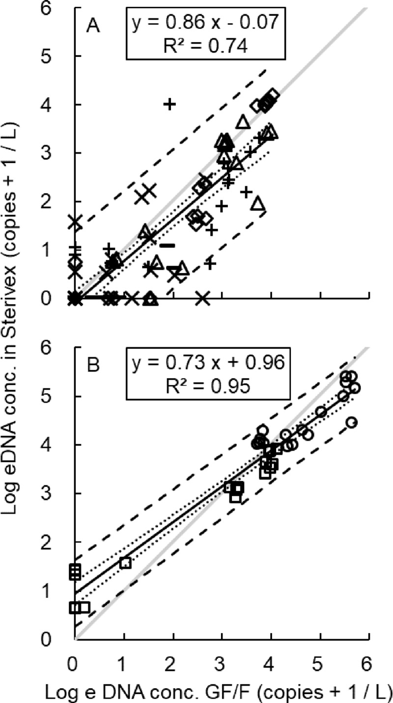
Fig 4

Correlation of eDNA concentration estimated by two different filtration methods (GF/F and Sterivex) in fish (A) and jellyfish (B). (A) fish, △: Acanthopagrus schlegelii, ◇: Engraulis japonicus, ×: Halichoeres tenuispinnis, −: Oplegnathus fasciatus, +: Trachurus japonicus, (B) jellyfish, ○: Aurelia aurita, □: Chrysaora pacifica, dotted lines: the 95% confidence limits, dashed lines: the 95% prediction limits, gray lines: y = x.

Comparison between eDNA concentration and biomass

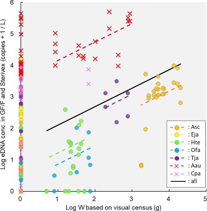
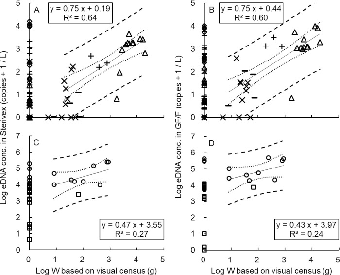
A linear regression equation of eDNA concentrations (obtained by GF/F and Sterivex), and biomass (based on underwater visual census) was calculated for all species (YeDNAconc = 0.58 XW + 1.52; Fig 5). There was a strong positive correlation between eDNA and fish biomass (R2 = 0.64; Fig 6A). A similar but weaker correlation was observed between eDNA and jellyfish (R2 = 0.27; Fig 6C). There was no significant difference between the slopes of these two lines (p = 0.26), whereas the intercepts were significantly higher than 0 (p < 0.01), and those of jellyfish were significantly higher than those of fish (p < 0.01). The Log10 (x+1) transformed copy numbers of eDNA concentration per biomass in jellyfish (intercept: 3.55) were much greater than those in fish (intercept: 0.19), representing an eDNA emission rate which was about 700 times (344–991 times) higher in jellyfish than in fish. The correlation between biomass and eDNA concentration showed no difference between GF/F (S4 Fig) and Sterivex (S2 Fig) either in fish (R2 = 0.64; Fig 6A and R2 = 0.60; Fig 6B) or in jellyfish (R2 = 0.27; Fig 6C and R2 = 0.24; Fig 6D). Intercepts of regression lines were different depending on species.

Fig 5. Correlation between biomass (W) and eDNA concentration estimated by GF/F and Sterivex.

Fig 5

Linear regression equation including all species: YeDNAconc = 0.58 XW + 1.52, Asc: Acanthopagrus schlegelii, Eja: Engraulis japonicus, Hte: Halichoeres tenuispinnis, Ofa: Oplegnathus fasciatus, Tja: Trachurus japonicus, Aau: Aurelia aurita, Cpa: Chrysaora pacifica.

Fig 6.

Fig 6

Correlation between biomass (W) and eDNA concentration estimated by Sterivex (A, C) and GF/F (B, D) in fish (A, B) and jellyfish (C, D). (A, B) fish, △: Acanthopagrus schlegelii, ◇: Engraulis japonicus, ×: Halichoeres tenuispinnis, −: Oplegnathus fasciatus, +: Trachurus japonicus, (C, D) jellyfish, ○: Aurelia aurita, □: Chrysaora pacifica; dotted lines: the 95% confidence limits, dashed lines: the 95% prediction limits.

Out of all the 126 sampling stations, 33 (26%) were positive in both the eDNA analysis and the visual census, 26 (21%) were negative in both, 61 (48%) were positive only in the eDNA analysis, and 6 (5%) were positive only in the visual census. Each species was detected by eDNA analysis but not by visual census at least once, whereas the opposite was true only in H. tenuispinnis and O. fasciatus (Fig 6).

A large amount of C. pacifica eDNA was detected at the point where one individual of this species was observed underwater, and smaller amounts were detected at other points (S3 Table; S3 and S5 Figs). Aurelia aurita eDNA was abundant at every point (S3 and S5 Figs). Although live individuals were sparse (S3 Table; S3 and S5 Figs), many dead individuals were found on the seafloor, especially in Otomi in May (Fig 1B). Amounts of eDNA in A. schlegelii, H. tenuispinnis, O. fasciatus and T. japonicus were larger at the points where biomass was greater, and smaller where biomass was less, in Nagahama in September (S3 Table; S3 and S5 Figs).

Detection of eDNA and visually detected abundance in jellyfish, H. tenuispinnis, O. fasciatus and T. japonicus was larger in September than in December (S3 Table; S3 and S5 Figs). Between the two species of jellyfish, the eDNA of A. aurita was consistently larger than that of C. pacifica (S3 and S5 Figs). Detection rates of eDNA in A. schlegelii, E. japonicus and T. japonicus were higher than those in H. tenuispinnis and O. fasciatus (S3 Table; S3 and S5 Figs).

Discussion

Comparison of performance between GF/F and Sterivex

There was a clear correlation between the eDNA concentrations obtained by GF/F and Sterivex, and the linear regression equation including all the species was YSterivex = 0.75 XGFF + 0.22 (Fig 3). In the present study, the amount of eDNA detected using the Sterivex filtration method was higher than that detected using the GF/F filtration method when the eDNA concentration of jellyfish was relatively low (Fig 4B). These results suggest that Sterivex may have superior sensitivity for eDNA. In previous studies on freshwater fish, Sterivex yielded higher amounts of eDNA, represented by lower cycle quantification (Cq) values, than did GF/F [39]. In addition, for eDNA metabarcoding, the extracted amount of eDNA was lower and the detection rate of fish species was higher using Sterivex than by using open filtration methods [38]; the number of species detected by Sterivex was approximately 1.5 times higher than those detected by GF/F [31]. On the other hand, the amount of eDNA detected by GF/F was higher than that detected by Sterivex when the eDNA concentration was higher, both in fish and jellyfish (Figs 3 and 4). These results suggest that GF/F may be superior when large amounts of eDNA need to be extracted. As the pore size of Sterivex (0.45 μm-pore size) was smaller than that of the GF/F (0.7 μm-pore size), the eDNA amount captured by Sterivex was expected to be greater than that captured by the GF/F; nevertheless, this was not the case in this study. There may be four possible explanations for this: i) the difference of eDNA concentration between the upper and lower layers in water sampling bags, ii) filtration processes, iii) extraction losses, and iv) species differences.

Regarding explanation i), it was reported that eDNA concentrations of fish [14] and jellyfish [18] were higher near the bottom than near the surface; another study [56] reports that the detection rate of eDNA was not significantly different between the near-surface and subsurface. Urine, slimy coatings, saliva, dead carcasses, and predator and prey feces [2, 25] have all been suggested as possible eDNA origins; the eDNA state in the water may be free, cellular or particle-bound [9]. The amount of eDNA in the lower layer is suspected to be higher than that in the upper layer because larger particles sink faster than smaller ones [57]. However, in the present study there was no significant difference in eDNA concentrations between the two layers in the sampling bags (p = 0.92). In the method of this study, Sterivex samples were filtered from the upper layer and GF/F from lower layer of water sampling bags. The difference between the layers is not likely to have caused any systematic difference between the two filtration methods. Thus, the difference of detection tendency between the two methods should be attributed to filtration and/or extraction processes.

Regarding explanation ii), it is possible that the amount of eDNA detected depends on how one applies pressure during the filtration process. There are many variables one needs to consider when applying a filtration method [25, 26]. The choice of the filter paper types can substantially affect DNA yield, depending on eDNA binding capacity [30]. Finer filters tend to clog, and therefore either require a longer time, or are unable to filter a sufficient volume of water [28, 38, 40]. Too much filtration power in relation to filter hole size causes samples to leak from the filter funnels during filtration [30]. In this study, GF/F filtration was conducted with negative pressure using an aspirator, while Sterivex filtration was conducted with positive pressure using a syringe. The difference between negative and positive pressure may cause differences in the amount of detected eDNA.

Regarding explanation iii), extraction losses may offer a possible explanation as to why the eDNA concentrations obtained by the two filtration methods were different. It is known that the size distribution of eDNA particles varies with environmental conditions [40]. The amount of detected eDNA depends on different eDNA characteristics (size, spatial structure, extra- and intracellular, and particle-bound and free) [30], indicating that the optimal filter choice varies for different extraction methods and there are different protocol combinations suitable for different organisms [28, 58]. At the time of extraction, it is necessary for DNA to flow out of cells, and be removed from filter paper by elution and centrifugation, so that it does not remain on the walls of the tube and column, resulting in eDNA loss. Furthermore, the procedure of eDNA extraction using the GF/F and Sterivex filtration method was similar, but not exactly the same, during the experiments presented here, which was a necessity to compare the two methods (Fig 2). Therefore, it is possible that these differences in methods may contribute to differences detected in the results. Specifically, BAC was used for DNA preservation of the GF/F samples, and RNAlater was used for DNA preservation of the Sterivex filtrations. BAC is cationic surfactant and reduces microbial activity [59], whereas RNAlater is a stable reagent which deactivates nucleases [60]. Water samples with BAC are reported to retain more than 92% of fish DNA for 8-h at ambient temperature [59], and those with RNAlater are reported to successfully preserve the same amount of planktonic DNA for over 1 month at ambient temperature as frozen samples [60]. This study conducted the process from sampling to preservation in a freezer in less than seven hours, and the samples were transported on ice immediately after the addition of BAC or RNAlater. Therefore, we assume that there was not much difference in the result due to the preservation step. However, it is also reported that fish species detected by metabarcoding analysis using BAC were lower than those stored on ice [61], whereas RNAlater yielded a substantial precipitate that inhibited qPCR amplification of fish [62]. RNAlater has been shown to store good quality DNA, but not always in high enough quantities for metabarcoding analysis of marine organisms [63]. Therefore, the difference between BAC and RNAlater preservation may be a subject that requires further investigation.

Regarding explanation iv), species differences, intercepts and slopes of linear regression lines, representing the correlation of eDNA concentrations to the two filtration methods, were slightly different depending on fish and jellyfish species (S1 Fig). A few fish species had different parameters for intercepts and slopes compared to the other fish species, but remained closer to the parameters of jellyfish (Fig 3). Such differences may be related to the “ecology” of eDNA, i.e., myriad interactions between extraorganismal genetic material and its environment [2], which would be variable among species.

Accuracy of estimating biomass of marine species by eDNA

A positive correlation between eDNA concentration and biomass has been reported in freshwater amphibians [9, 10] and fish [11, 12, 19, 21], and marine fish [4, 6, 20] and jellyfish [18]. It is possible to quantify temporal variation, seasonal changes and annual fluctuation of marine species based on underwater visual census [46, 48, 64]. In the present study, there was also a positive correlation between eDNA concentration and biomass estimation based on visual observation in all species (YeDNAconc = 0.58 XW + 1.52; Fig 5), and in both fish and jellyfish (R2 = 0.64, R2 = 0.27; Fig 6A and 6C). Furthermore, eDNA concentration per biomass in jellyfish was approximately 700 times greater than it was in fish. Our results indicate that the detection or release rate of eDNA may be different depending on the target species.

Focusing on the differences in the detection or release rate of eDNA for each target species, the following factors can be considered. Minamoto et al. [18] reported that the eDNA concentration of C. pacifica was ~13 times higher near the bottom than on the surface. In the present study, the eDNA concentration was higher at the point where C. pacifica was visually detected, and the eDNA of this species was also detected at other points (S3 Table; S2 Fig). We collected samples near the bottom; therefore, eDNA released from individuals who passed through before our visual survey was likely to be detected. The long tentacles of this species are easily torn and can, therefore, be a major source of eDNA. For A. aurita, the eDNA concentration was high at every point (S2 Fig). Especially in Otomi in May, the eDNA was positive at the points where live individuals were not observed (S3 Table; S3 and S5 Figs) whereas many dead individuals were found on the bottom (Fig 1B). Since A. aurita is often eaten by fishes [65], many dead and torn individuals drift in the sea. Merkes et al. [66] reported that eDNA released from dead fish was detected for more than one month. Thus, eDNA may over-estimate the abundance of species when high mortality occurs nearby. Furthermore, various life stage of jellyfish other than medusae, i.e., eggs, planulae and polyps, can be a source of eDNA but would have been missed in visual census. Generally, a high correlation between eDNA concentration and biomass exists in fishes (Fig 6A). Larger amounts of eDNA in A. schlegelii, H. tenuispinnis, O. fasciatus, and T. japonicus were detected at points where visually evaluated biomass was greater (S3 Table; S2 Fig). This may be related to lifestyle issues discussed below.

It has been reported that the presence/absence of aquatic species can be monitored using eDNA analysis, even for non-native species [3], threatened species [37, 41], and fish in mountainous rivers during the winter [42]. It is also known that eDNA detection does not necessarily correlate with the presence of organisms [22]. This was also the case in 53% of the samples examined in the present study. Potential causes of this could be currents, seasonality, and animal activity, including their life stage.

A positive correlation between biomass and eDNA concentration has been reported in rivers [10, 21] and the sea when there are currents [6, 1820], although it has been pointed out that currents may influence eDNA quantification [4, 6]. When examining the eDNA at a certain location and time, the outcome may depend on whether the source is up or down the current. For instance, in a river, eDNA has been detected in a range of 240 m to 12 km downstream [43, 6769]. Sansom and Sassoubre [69] reported that, in theory, eDNA could be transported for 4.3–36.7 km downstream. In the sea, eDNA has been detected in a range of 10 m to 150 m from its source [20, 70, 71]. It is likely that eDNA, released from dead or injured A. aurita and C. pacifica in adjacent waters, or from T. japonicus schooling and migrating just before observation (e.g., Fig 2), or from large amounts of small size individuals (such as gametes or larvae), drifted in the range and was collected. In one instance, H. tenuispinnis and O. fasciatus were detected by visual census but not by eDNA analysis (Fig 6A). Both species are demersal (Fig 2), and water was sampled at 1 m above the sea bottom. A relatively small size and abundance, as well as complex water movement, may have hindered eDNA detection in this case.

Seasonal fluctuations of eDNA amounts have been reported, and they are consistent with seasonal variations of biomass in freshwater fishes [14, 19, 21] and marine jellyfish [18]. The seasonal change of fish species in an estuary can also be detected by eDNA metabarcoding analysis [72]. In the present study, detection of eDNA and individuals of H. tenuispinnis, O. fasciatus, T. japonicus, and C. pacifica, which occur in the surveyed area from spring to summer, was larger in September than in December (S3 Table; S4 and S5 Figs). A dense patch of A. aurita is often found north, off the coast of the survey point in Nagahama, and it occasionally comes close to the shore (Masuda R, pers. obs.). This suggests that biomass quantification of A. aurita using eDNA may be more feasible in a larger spatial scale (such as off coast where the area may be less affected by many dead individuals or non-medusa stages, as would happen closer to the coast) and/or with temporal change. eDNA is more likely to capture such a seasonal fluctuation of biomass than the spatial variation.

The activity variation of organisms may also influence the seasonal fluctuation of eDNA. eDNA emission increases by feeding [12, 73] and reproduction [24, 74, 75], and is dependent on the lifestyle of the target species [69]. In the present study, the detection rate of eDNA was different between fish species having different lifestyles (e.g., Fig 2). The detection rate of eDNA may be high in E. japonicus and T. japonicus, of which many, small sized individuals form schools of several hundreds and migrate near the survey point. The eDNA detection rate may also be high in A. schlegelii, of which a small number of large sized individuals are consistently found in the shallow reef area. eDNA detection rates may be low in H. tenuispinnis and O. fasciatus, as they are small in size, few in numbers and demersal (S3 Table; S2 Fig). There is a possibility that the difference in activity of each species and difference in behavioral traits (such as bold-shy behavior [76]), could affect eDNA release, and can be a subject of future study.

Furthermore, because eDNA could not be detected when the population density is very low [3] and eDNA concentration is very low in seawater samples, it may be predicted that some samples will be negative in eDNA. Possible factors of mismatch between detection in observation and non-detection in eDNA are PCR amplification inhibition, interference of non-target species [25, 77], and mixing of different haplotypes in the same waters (Takahashi et al., unpublished data). Unlike in aquaria or ponds, eDNA may not be detected in marine environments even if the target species is found in them.

eDNA detection does not necessarily correlate with the presence of certain organisms in complex oceanic environmental conditions. Therefore, when it comes to estimating biomass, it is better to consider data obtained from several samplings, than to just rely on visual observation at the time of each water sampling. At the time of the underwater visual census in Otomi, there was a point where some H. tenuispinnis individuals were found in the return path and not in the forward path where water was sampled (Fig 1A and 1D; S3 Table). In Nagahama in December, one A. aurita with a 10-cm umbrella diameter was observed near the pier 1 at about 1 h before the census of this study (Fig 1A and 1C). By combining several observations, it is expected that the correlation between biomass and eDNA amount will be improved. Furthermore, by knowing the behavior patterns and physiology of target species as well as the characteristics of the habitat, it is possible to estimate the biomass of marine organisms with a higher accuracy when performing eDNA analysis by the GF/F or Sterivex filtration methods.

Conclusion

This study evaluated the eDNA concentration obtained by two different filtration methods (GF/F and Sterivex). A comparison with an underwater visual census also showed a positive relationship between the eDNA concentration and the fish and jellyfish biomass. It is possible to convert data obtained by GF/F to that obtained by Sterivex. We found that some species are detected more easily by eDNA, while a small number of other species showed an opposite trend. This was likely to be due to the size of the target species and their lifestyle (such as pelagic or demersal), seasonality and behavior, as well as physical factors such as water currents. Therefore, we suggest that the eDNA method can be particularly effective in combination with knowledge of the ecology, behavior and life history of the target species. Such an approach is expected to give us further ecological insights that would be valuable for the conservation of the ocean environment and the management of fisheries resources [72, 78].

Supporting information

S1 Table. List of related species used in the in silico specificity test and further checking with real-time PCR.

The most closely related species within the same order were checked since no fish species belonging to the same family are present in the surveyed area. *Asc: Acanthopagrus schlegelii, Hte: Halichoeres tenuispinnis, Ofa: Oplegnathus fasciatus. ** X indicates specificity was checked.

(XLSX)

S2 Table. Parameters of the standard curve for each species at each sampling site and date.

*Asc: Acanthopagrus schlegelii, Eja: Engraulis japonicus, Hte: Halichoeres tenuispinnis, Ofa: Oplegnathus fasciatus, Tja: Trachurus japonicus, Aau: Aurelia aurita, Cpa: Chrysaora pacifica.

(XLSX)

S3 Table. Body length (L cm) and estimated biomass (W g) of target organisms encountered in underwater observation.

*Asc: Acanthopagrus schlegelii, Eja: Engraulis japonicus, Hte: Halichoeres tenuispinnis, Ofa: Oplegnathus fasciatus, Tja: Trachurus japonicus, Aau: Aurelia aurita, Cpa: Chrysaora pacifica. **W = aLb was used as weight-length relationships based on the FishBase website [49]. The values of Ofa were based on those of the same genus, O. woodwardi, and Aau based on Aoki et al. [51] and Cpa based on Yasuda [50]. ***L: length, N: number, W: weight. ****Points and lines were shown in Fig 1. *****Tentacles of Cpa were torn.

(XLSX)

S4 Table. Raw data used in Figs 3, 4, 5 and 6 and S1 and S2 Figs.

(XLSX)

S1 Fig. Correlation of eDNA concentration between GF/F and Sterivex for each species in water samples collected on May 15 in Otomi, on September 19 and December 18 in Nagahama.

Asc: Acanthopagrus schlegelii, Eja: Engraulis japonicus, Hte: Halichoeres tenuispinnis, Ofa: Oplegnathus fasciatus, Tja: Trachurus japonicus, Aau: Aurelia aurita, Cpa: Chrysaora pacifica, gray lines: y = x.

(TIF)

S2 Fig. Correlation between biomass (W) and eDNA concentration estimated by Sterivex.

Linear regression equation including all species: YeDNAconc = 0.63 XW + 1.21, Asc: Acanthopagrus schlegelii, Eja: Engraulis japonicus, Hte: Halichoeres tenuispinnis, Ofa: Oplegnathus fasciatus, Tja: Trachurus japonicus, Aau: Aurelia aurita, Cpa: Chrysaora pacifica.

(TIF)

S3 Fig. Correlation between estimated biomass (W) and eDNA concentration obtained by Sterivex for each species in water samples and biomass data collected on May 15 in Otomi, on September 19 and December 18 in Nagahama.

Abbreviations are the same as in S2 Fig.

(TIF)

S4 Fig. Correlation between biomass (W) and eDNA concentration estimated by GF/F.

Linear regression equation including all species: YeDNAconc = 0.55 XW + 1.77, abbreviations are the same as in S2 Fig.

(TIF)

S5 Fig. Correlation between estimated biomass (W) and eDNA concentration obtained by GF/F for each species in water samples and biomass data collected on May 15 in Otomi, on September 19 and December 18 in Nagahama.

Abbreviations are the same as in S2 Fig.

(TIF)

Acknowledgments

We would like to thank Dr. Hiroki Yamanaka (Ryukoku University) for his advice on the eDNA extraction technique from Sterivex and Mr. Takaya Yoden, Mr. Tatsuki Toya, and Ms. Misaki Shiomi (Kyoto University) for their help in water sampling and filtration. The fish tissue samples archived in the Fish Collection of Kyoto University (FAKU) were kindly provided by Drs. Yoshiaki Kai and Fumihito Tashiro at Maizuru Fisheries Research Station, Kyoto University. We would also like to thank Dr. Ruslan Kalendar (University of Helsinki) and three anonymous reviewers for their constructive comments that helped us to substantially improve the quality of the manuscript.

Data Availability

All relevant data are within the manuscript and its Supporting Information files.

Funding Statement

This study was partly supported by the CREST program from the Japan Science and Technology Agency (grant number: JPMJCR13A2; http://www.jst.go.jp/kisoken/crest/en/project/33/e33_13.html) and JSPS Grant-in-Aid for Scientific Research (B) 19H03031, Japan. The funders had no role in study design, data collection and analysis, decision to publish, or preparation of the manuscript.

References

  • 1.Kelly RP, Port JA, Yamahara KM, Martone RG, Lowell N, Thomsen PF, et al. Harnessing DNA to improve environmental management. Science. 2014;344: 1455–1456. 10.1126/science.1251156 [DOI] [PubMed] [Google Scholar]
  • 2.Barnes MA, Turner CR. The ecology of environmental DNA and implications for conservation genetics. Conserv Genet. 2016;17: 1–17. [Google Scholar]
  • 3.Ficetola GF, Miaud C, Pompanon F, Taberlet P. Species detection using environmental DNA from water samples. Biol Lett. 2008;4: 423–425. 10.1098/rsbl.2008.0118 [DOI] [PMC free article] [PubMed] [Google Scholar]
  • 4.Thomsen PF, Kielgast J, Iversen LL, Møller PR, Rasmussen M, Willerslev E. Detection of a diverse marine fish fauna using environmental DNA from seawater samples. PLoS One. 2012;7: e41732 10.1371/journal.pone.0041732 [DOI] [PMC free article] [PubMed] [Google Scholar]
  • 5.Yamamoto S, Masuda R, Sato Y, Sado T, Araki H, Kondoh M, et al. Environmental DNA metabarcoding reveals local fish communities in a species-rich coastal sea. Sci Rep. 2017;7: 40368 10.1038/srep40368 [DOI] [PMC free article] [PubMed] [Google Scholar]
  • 6.Thomsen PF, Møller PR, Sigsgaard EE, Knudsen SW, Jørgensen OA, Willerslev E. Environmental DNA from seawater samples correlate with trawl catches of subarctic, deepwater fishes. PLoS One. 2016;11: e0165252 10.1371/journal.pone.0165252 [DOI] [PMC free article] [PubMed] [Google Scholar]
  • 7.Evans NT, Li Y, Renshaw MA, Olds BP, Deiner K, Turner CR, et al. Fish community assessment with eDNA metabarcoding: effects of sampling design and bioinformatic filtering. Can J Fish Aquat Sci. 2017;74: 1362–1374. [Google Scholar]
  • 8.Miya M, Sato Y, Fukunaga T, Sado T, Poulsen JY, Sato K, et al. MiFish, a set of universal PCR primers for metabarcoding environmental DNA from fishes: detection of more than 230 subtropical marine species. R Soc Open Sci. 2015;2: 150088 10.1098/rsos.150088 [DOI] [PMC free article] [PubMed] [Google Scholar]
  • 9.Thomsen PF, Kielgast J, Iversen LL, Wiuf C, Rasmussen M, Gilbert MTP, et al. Monitoring endangered freshwater biodiversity using environmental DNA. Mol Ecol. 2012;21: 2565–2573. 10.1111/j.1365-294X.2011.05418.x [DOI] [PubMed] [Google Scholar]
  • 10.Pilliod DS, Goldberg CS, Arkle RS, Waits LP. Estimating occupancy and abundance of stream amphibians using environmental DNA from filtered water samples. Can J Fish Aquat Sci. 2013;70: 1123–1130. [Google Scholar]
  • 11.Takahara T, Minamoto T, Yamanaka H, Doi H, Kawabata Z. Estimation of fish biomass using environmental DNA. PLoS One. 2012;7: e35868 10.1371/journal.pone.0035868 [DOI] [PMC free article] [PubMed] [Google Scholar]
  • 12.Klymus KE, Richter CA, Chapman DC, Paukert C. Quantification of eDNA shedding rates from invasive bighead carp Hypophthalmichthys nobilis and silver carp Hypophthalmichthys molitrix. Biol Conserv. 2015;183: 77–84. [Google Scholar]
  • 13.Maruyama A, Nakamura K, Yamanaka H, Kondoh M, Minamoto T. The release rate of environmental DNA from juvenile and adult fish. PLoS One. 2014;9: e114639 10.1371/journal.pone.0114639 [DOI] [PMC free article] [PubMed] [Google Scholar]
  • 14.Klobucar SL, Rodgers TW, Budy P. At the forefront: evidence of the applicability of using environmental DNA to quantify the abundance of fish populations in natural lentic waters with additional sampling considerations. Can J Fish Aquat Sci. 2017;74: 2030–2034. [Google Scholar]
  • 15.Mizumoto H, Urabe H, Kanbe T, Fukushima M, Araki H. Establishing an environmental DNA method to detect and estimate the biomass of Sakhalin taimen, a critically endangered Asian salmonid. Limnology. 2018;19: 219–227. [Google Scholar]
  • 16.Kakuda A, Doi H, Souma R, Nagano M, Minamoto T, Katano I. Detection of environmental DNA of the invasive red-eared slider in ponds for evaluating their distribution with comparison to water quality. PeerJ. 2019;7: e27505v1. [DOI] [PMC free article] [PubMed] [Google Scholar]
  • 17.Horiuchi T, Masuda R, Murakami H, Yamamoto S, Minamoto T. Biomass-dependent emission of environmental DNA in jack mackerel Trachurus japonicus juveniles. J Fish Biol. 2019;95: 979–981. 10.1111/jfb.14095 [DOI] [PubMed] [Google Scholar]
  • 18.Minamoto T, Fukuda M, Katsuhara KR, Fujiwara A, Hidaka S, Yamamoto S, et al. Environmental DNA reflects spatial and temporal jellyfish distribution. PLoS One. 2017;12: e0173073 10.1371/journal.pone.0173073 [DOI] [PMC free article] [PubMed] [Google Scholar]
  • 19.Uchii K, Doi H, Yamanaka H, Minamoto T. Distinct seasonal migration patterns of Japanese native and non-native genotypes of common carp estimated by environmental DNA. Ecol Evol. 2017;7: 8515–8522. 10.1002/ece3.3346 [DOI] [PMC free article] [PubMed] [Google Scholar]
  • 20.Yamamoto S, Minami K, Fukaya K, Takahashi K, Sawada H, Murakami H, et al. Environmental DNA as a ‘snapshot’ of fish distribution: A case study of Japanese jack mackerel in Maizuru Bay, Sea of Japan. PLoS One. 2016;11: e0149786 10.1371/journal.pone.0149786 [DOI] [PMC free article] [PubMed] [Google Scholar]
  • 21.Doi H, Inui R, Akamatsu Y, Kanno K, Yamanaka H, Takahara T, Minamoto T. Environmental DNA analysis for estimating the abundance and biomass of stream fish. Freshw Biol. 2017;62: 30–39. [Google Scholar]
  • 22.Kelly RP, Port JA, Yamahara KM, Crowder LB. Using environmental DNA to census marine fishes in a large mesocosm. PLoS One. 2014;9: e86175 10.1371/journal.pone.0086175 [DOI] [PMC free article] [PubMed] [Google Scholar]
  • 23.Roussel J-M, Paillisson J-M, Trèguier A, Petit E. The downside of eDNA as a survey tool in water bodies. J Appl Ecol. 2015;52: 823–826. [Google Scholar]
  • 24.Spear SF, Groves JD, Williams LA, Waits LP. Using environmental DNA methods to improve detectability in a hellbender (Cryptobranchus alleganiensis) monitoring program. Biol Conserv. 2015;183: 38–45. [Google Scholar]
  • 25.Rees HC, Maddison BC, Middleditch DJ, Patmore JRM, Gough KC. The detection of aquatic animal species using environmental DNA–a review of eDNA as a survey tool in ecology. J Appl Ecol. 2014;51: 1450–1459. [Google Scholar]
  • 26.Tsuji S, Takahara T, Doi H, Shibata N, Yamanaka H. The detection of aquatic macroorganisms using environmental DNA analysis—A review of methods for collection, extraction, and detection. Environ DNA. 2019;1: 99–108. [Google Scholar]
  • 27.Eichmiller JJ, Miller LM, Sorensen PW. Optimizing techniques to capture and extract environmental DNA for detection and quantification of fish. Mol Ecol Resour. 2016;16: 56–68. 10.1111/1755-0998.12421 [DOI] [PubMed] [Google Scholar]
  • 28.Minamoto T, Naka T, Moji K, Maruyama A. Techniques for the practical collection of environmental DNA: filter selection, preservation, and extraction. Limnology. 2016;17: 23–32. [Google Scholar]
  • 29.Piggott MP. Evaluating the effects of laboratory protocols on eDNA detection probability for an endangered freshwater fish. Ecol Evol. 2016;6: 2739–2750. 10.1002/ece3.2083 [DOI] [PMC free article] [PubMed] [Google Scholar]
  • 30.Hinlo R, Gleeson D, Lintermans M, Furlan E. Methods to maximise recovery of environmental DNA from water samples. PLoS One 2017;12: e0179251 10.1371/journal.pone.0179251 [DOI] [PMC free article] [PubMed] [Google Scholar]
  • 31.Miya M, Minamoto T, Yamanaka H, Oka S, Sato K, Yamamoto S, et al. Use of a filter cartridge for filtration of water samples and extraction of environmental DNA. J Vis Exp. 2016;117: e54741. [DOI] [PMC free article] [PubMed] [Google Scholar]
  • 32.Djurhuus A, Port J, Closek CJ, Yamahara KM, Romero-Maraccini O, Walz KR, et al. Evaluation of filtration and DNA extraction methods for environmental DNA biodiversity assessments across multiple trophic levels. Front Mar Sci. 2017;4: 314. [Google Scholar]
  • 33.Deiner K, Lopez J, Bourne S, Holman LE, Seymour M, Grey EK, et al. Optimising the detection of marine taxonomic richness using environmental DNA metabarcoding: the effects of filter material, pore size and extraction method. Metabarcoding Metagenom. 2018;2: e28963. [Google Scholar]
  • 34.Majaneva M, Diserud OH, Eagle SHC, Boström E, Hajibabaei M, Ekrem T. Environmental DNA filtration techniques affect recovered biodiversity. Sci Rep. 2018;8: 4682 10.1038/s41598-018-23052-8 [DOI] [PMC free article] [PubMed] [Google Scholar]
  • 35.Yamanaka H, Motozawa H, Tsuji S, Miyazawa RC, Takahara T, Minamoto T. On-site filtration of water samples for environmental DNA analysis to avoid DNA degradation during transportation. Ecol Res. 2016;31: 963–967. [Google Scholar]
  • 36.Keskin E. Detection of invasive freshwater fish species using environmental DNA survey. Biochem Syst Ecol. 2014;56: 68–74. [Google Scholar]
  • 37.Bergman PS, Schumer G, Blankenship S, Campbell E. Detection of adult green sturgeon using environmental DNA analysis. PLoS One. 2016;11: e0153500 10.1371/journal.pone.0153500 [DOI] [PMC free article] [PubMed] [Google Scholar]
  • 38.Li J, Lawson Handley LJ, Read DS, Hänfling B. The effect of filtration method on the efficiency of environmental DNA capture and quantification via metabarcoding. Mol Ecol Resour. 2018;18: 1102–1114. [DOI] [PubMed] [Google Scholar]
  • 39.Spens J, Evans AR, Halfmaerten D, Knudsen SW, Sengupta ME, Mak SST, et al. Comparison of capture and storage methods for aqueous macrobial eDNA using an optimized extraction protocol: advantage of enclosed filter. Methods Ecol Evol. 2017;8: 635–645. [Google Scholar]
  • 40.Turner CR, Barnes MA, Xu CCY, Jones SE, Jerde CL, Lodge DM. Particle size distribution and optimal capture of aqueous macrobial eDNA. Methods Ecol Evol. 2014;5: 676–684. [Google Scholar]
  • 41.Yamanaka H, Takao D, Maruyama A, Imamura A. Species-specific detection of the endangered piscivorous cyprinid fish Opsariichthys uncirostris uncirostris, three-lips, using environmental DNA analysis. Ecol Res. 2018;33: 1075–1078. [Google Scholar]
  • 42.Minamoto T, Hayami K, Sakata MK, Imamura A. Real-time polymerase chain reaction assays for environmental DNA detection of three salmonid fish in Hokkaido, Japan: Application to winter surveys. Ecol Res. 2019;34: 237–242. [Google Scholar]
  • 43.Nukazawa K, Hamasuna Y, Suzuki Y. Simulating the advection and degradation of the environmental DNA of common carp along a River. Environ Sci Technol. 2018;52: 10562–10570. 10.1021/acs.est.8b02293 [DOI] [PubMed] [Google Scholar]
  • 44.Masuda R, Matsuda K, Tanaka M. Laboratory video recordings and underwater visual observations combined to reveal activity rhythm of red-spotted grouper and banded wrasse, and their natural assemblages. Environ Biol Fishes. 2012;95: 335–346. [Google Scholar]
  • 45.The eDNA Society. Environmental DNA Sampling and Experiment Manual. Version 2.1. Otsu, Japan; 2019. Available from: http://ednasociety.org/eDNA_manual_Eng_v2_1_3b.pdf.
  • 46.Masuda R. Seasonal and interannual variation of subtidal fish assemblages in Wakasa Bay with reference to the warming trend in the Sea of Japan. Environ Biol Fishes. 2008;82: 387–399. [Google Scholar]
  • 47.Masuda R. Ontogenetic changes in the ecological function of the association behavior between jack mackerel Trachurus japonicus and jellyfish. Hydrobiologia. 2009;616: 269–277. [Google Scholar]
  • 48.Masuda R, Hatakeyama M, Yokoyama K, Tanaka M. Recovery of coastal fauna after the 2011 tsunami in Japan as determined by bimonthly underwater visual censuses conducted over five years. PLoS One. 2016;11: e0168261 10.1371/journal.pone.0168261 [DOI] [PMC free article] [PubMed] [Google Scholar]
  • 49.FishBase website [Internet]. [cited 2018 Nov 9]. Available from: http://www.fishbase.org/search.php.
  • 50.Yasuda T. UFO jellyfish of sea-generation and ecology and countermeasures. Kouseisha-kouseikaku Corporation; 2003. (in Japanese). [Google Scholar]
  • 51.Aoki K, Yamada S, Toyokawa M, Yasuda A, Kikuchi T. Horizontal distribution and growth of jellyfish, Aurelia aurita (Linnaeus 1758) sensu lato, in Mikawa Bay, Japan. Coast Mar Sci. 2012;35: 103–111. [Google Scholar]
  • 52.Froese R, Thorson JT, Reyes RB Jr. A Bayesian approach for estimating length-weight relationships in fishes. J Appl Ichthyol. 2014;30: 78–85. [Google Scholar]
  • 53.Ushio M, Murakami H, Masuda R, Sado T, Miya M, Sakurai S, et al. Quantitative monitoring of multispecies fish environmental DNA using high-throughput sequencing. Metabarcoding Metagenom. 2018;2: e23297. [Google Scholar]
  • 54.Wilcox TM, McKelvey KS, Young MK, Jane SF, Lowe WH, Whiteley AR, et al. Robust detection of rare species using environmental DNA: The importance of primer specificity. PLoS One. 2013;8: e59520 10.1371/journal.pone.0059520 [DOI] [PMC free article] [PubMed] [Google Scholar]
  • 55.R Development Core Team: R Statistical Software. Version 3.4.3 [software]. A language and environment for statistical computing. Vienna, Austria: R Foundation for Statistical Computing; 2018. [Google Scholar]
  • 56.Eichmiller JJ, Bajer PG, Sorensen PW. The relationship between the distribution of common carp and their environmental DNA in a small lake. PLoS One. 2014;9: e112611 10.1371/journal.pone.0112611 [DOI] [PMC free article] [PubMed] [Google Scholar]
  • 57.Maggi F. The settling velocity of mineral, biomineral, and biological particles and aggregates in water. J Geophys Res Oceans. 2013;118: 2118–2132. [Google Scholar]
  • 58.Deiner K, Walser J-C, Mächler E, Altermatt F. Choice of capture and extraction methods affect detection of freshwater biodiversity from environmental DNA. Biol Conserv. 2015;183: 53–63. [Google Scholar]
  • 59.Yamanaka H, Minamoto T, Matsuura J, Sakurai S, Tsuji S, Motozawa H, et al. A simple method for preserving environmental DNA in water samples at ambient temperature by addition of cationic surfactant. Limnology. 2017;18: 233–241. [Google Scholar]
  • 60.Gorokhova E. Effects of preservation and storage of microcrustaceans in RNAlater on RNA and DNA degradation. Limnol Oceanogr Methods. 2005;3: 143–148. [Google Scholar]
  • 61.Sales NG, Wangensteen OS, Carvalho DC, Mariani S. Influence of preservation methods, sample medium and sampling time on eDNA recovery in a neotropical river. Environ DNA. 2019;1: 119–130. [Google Scholar]
  • 62.Renshaw MA, Olds BP, Jerde CL, McVeigh MM, Lodge DM. The room temperature preservation of filtered environmental DNA samples and assimilation into a phenol–chloroform–isoamyl alcohol DNA extraction. Mol Ecol Resour. 2015;15: 168–176. 10.1111/1755-0998.12281 [DOI] [PMC free article] [PubMed] [Google Scholar]
  • 63.Ransome E, Geller JB, Timmers M, Leray M, Mahardini A, Sembiring A, et al. The importance of standardization for biodiversity comparisons: A case study using autonomous reef monitoring structures (ARMS) and metabarcoding to measure cryptic diversity on Mo’orea coral reefs, French Polynesia. PLoS One. 2017;12: e0175066 10.1371/journal.pone.0175066 [DOI] [PMC free article] [PubMed] [Google Scholar]
  • 64.Ushio M, Hsieh C-h, Masuda R, Deyle ER, Ye H, Chang C-W, et al. Fluctuating interaction network and time-varying stability of a natural fish community. Nature. 2018;554: 360–363. 10.1038/nature25504 [DOI] [PubMed] [Google Scholar]
  • 65.Masuda R, Yamashita Y, Matsuyama M. Jack mackerel Trachurus japonicus juveniles use jellyfish for predator avoidance and as a prey collector. Fish Sci. 2008;74: 276–284. [Google Scholar]
  • 66.Merkes CM, McCalla SG, Jensen NR, Gaikowski MP, Amberg JJ. Persistence of DNA in carcasses, slime and avian feces may affect interpretation of environmental DNA data. PLoS One. 2014;9: e113346 10.1371/journal.pone.0113346 [DOI] [PMC free article] [PubMed] [Google Scholar]
  • 67.Deiner K, Altermatt F. Transport distance of invertebrate environmental DNA in a natural river. PLoS One. 2014;9: e88786 10.1371/journal.pone.0088786 [DOI] [PMC free article] [PubMed] [Google Scholar]
  • 68.Jane SF, Wilcox TM, McKelvey KS, Young MK, Schwartz MK, Lowe WH, et al. Distance, flow and PCR inhibition: eDNA dynamics in two headwater streams. Mol Ecol Resour. 2015;15: 216–227. 10.1111/1755-0998.12285 [DOI] [PubMed] [Google Scholar]
  • 69.Sansom BJ, Sassoubre LM. Environmental DNA (eDNA) shedding and decay rates to model freshwater mussel eDNA transport in a river. Environ Sci Technol. 2017;51: 14244–14253. 10.1021/acs.est.7b05199 [DOI] [PubMed] [Google Scholar]
  • 70.O’Donnell JL, Kelly RP, Shelton AO, Samhouri JF, Lowell NC, Williams GD. Spatial distribution of environmental DNA in a nearshore marine habitat. PeerJ. 2017;5: e3044 10.7717/peerj.3044 [DOI] [PMC free article] [PubMed] [Google Scholar]
  • 71.Murakami H, Yoon S, Kasai A, Minamoto T, Yamamoto S, Sakata MK, et al. Dispersion and degradation of environmental DNA from caged fish in a marine environment. Fish Sci. 2019;85: 327–337. [Google Scholar]
  • 72.Stoeckle MY, Soboleva L, Charlop-Powers Z. Aquatic environmental DNA detects seasonal fish abundance and habitat preference in an urban estuary. PLoS One. 2017;12: e0175186 10.1371/journal.pone.0175186 [DOI] [PMC free article] [PubMed] [Google Scholar]
  • 73.Ghosal R, Eichmiller JJ, Witthuhn BA, Sorensen PW. Attracting Common Carp to a bait site with food reveals strong positive relationships between fish density, feeding activity, environmental DNA, and sex pheromone release that could be used in invasive fish management. Ecol Evol. 2018;8: 6714–6727. [DOI] [PMC free article] [PubMed] [Google Scholar]
  • 74.Bylemans J, Furlan EM, Hardy CM, McGuffie P, Lintermans M, Gleeson DM. An environmental DNA-based method for monitoring spawning activity: a case study, using the endangered Macquarie perch (Macquaria australasica). Methods Ecol Evol. 2017;8: 646–655. [Google Scholar]
  • 75.Takahashi MK, Meyer MJ, Mcphee C, Gaston JR, Venesky MD, Case BF. Seasonal and diel signature of eastern hellbender environmental DNA. J Wildl Manage. 2018;82: 217–225. [Google Scholar]
  • 76.Sneddon LU. The bold and the shy: individual differences in rainbow trout. J Fish Biol. 2003;62: 971–975. [Google Scholar]
  • 77.Schrader C, Schielke A, Ellerbroek L, Johne R. PCR inhibitors-occurrence, properties and removal. J Appl Microbiol. 2012;113: 1014–1026. 10.1111/j.1365-2672.2012.05384.x [DOI] [PubMed] [Google Scholar]
  • 78.Lacoursière‐Roussel A, Côté G, Leclerc V, Bernatchez L. Quantifying relative fish abundance with eDNA: a promising tool for fisheries management. J Appl Ecol. 2016;53: 1148–1157. [Google Scholar]

Decision Letter 0

Ruslan Kalendar

31 Dec 2019

PONE-D-19-32313

Comparing the efficiency of open and enclosed filtering systems in fish and jellyfish environmental DNA quantification

PLOS ONE

Dear Dr. Takahashi,

Thank you for submitting your manuscript to PLOS ONE. After careful consideration, we feel that it has merit but does not fully meet PLOS ONE’s publication criteria as it currently stands. Therefore, we invite you to submit a revised version of the manuscript that addresses the points raised during the review process.

We would appreciate receiving your revised manuscript by Feb 14 2020 11:59PM. When you are ready to submit your revision, log on to https://www.editorialmanager.com/pone/ and select the 'Submissions Needing Revision' folder to locate your manuscript file.

If you would like to make changes to your financial disclosure, please include your updated statement in your cover letter.

To enhance the reproducibility of your results, we recommend that if applicable you deposit your laboratory protocols in protocols.io, where a protocol can be assigned its own identifier (DOI) such that it can be cited independently in the future. For instructions see: http://journals.plos.org/plosone/s/submission-guidelines#loc-laboratory-protocols

Please include the following items when submitting your revised manuscript:

  • A rebuttal letter that responds to each point raised by the academic editor and reviewer(s). This letter should be uploaded as separate file and labeled 'Response to Reviewers'.

  • A marked-up copy of your manuscript that highlights changes made to the original version. This file should be uploaded as separate file and labeled 'Revised Manuscript with Track Changes'.

  • An unmarked version of your revised paper without tracked changes. This file should be uploaded as separate file and labeled 'Manuscript'.

Please note while forming your response, if your article is accepted, you may have the opportunity to make the peer review history publicly available. The record will include editor decision letters (with reviews) and your responses to reviewer comments. If eligible, we will contact you to opt in or out.

We look forward to receiving your revised manuscript.

Kind regards,

Ruslan Kalendar, PhD

Academic Editor

PLOS ONE

Journal Requirements:

1. When submitting your revision, we need you to address these additional requirements.

Please ensure that your manuscript meets PLOS ONE's style requirements, including those for file naming. The PLOS ONE style templates can be found at

http://www.journals.plos.org/plosone/s/file?id=wjVg/PLOSOne_formatting_sample_main_body.pdf and http://www.journals.plos.org/plosone/s/file?id=ba62/PLOSOne_formatting_sample_title_authors_affiliations.pdf

2. We note that you are reporting an analysis of a microarray, next-generation sequencing, or deep sequencing data set. PLOS requires that authors comply with field-specific standards for preparation, recording, and deposition of data in repositories appropriate to their field. Please upload these data to a stable, public repository (such as ArrayExpress, Gene Expression Omnibus (GEO), DNA Data Bank of Japan (DDBJ), NCBI GenBank, NCBI Sequence Read Archive, or EMBL Nucleotide Sequence Database (ENA)). In your revised cover letter, please provide the relevant accession numbers that may be used to access these data. For a full list of recommended repositories, see http://journals.plos.org/plosone/s/data-availability#loc-omics or http://journals.plos.org/plosone/s/data-availability#loc-sequencing.

Reviewers' comments:

Reviewer's Responses to Questions

Comments to the Author

1. Is the manuscript technically sound, and do the data support the conclusions?

Reviewer #1: No

Reviewer #2: Yes

Reviewer #3: Yes

**********

2. Has the statistical analysis been performed appropriately and rigorously?

Reviewer #1: No

Reviewer #2: Yes

Reviewer #3: Yes

**********

3. Have the authors made all data underlying the findings in their manuscript fully available?

Reviewer #1: Yes

Reviewer #2: Yes

Reviewer #3: Yes

**********

4. Is the manuscript presented in an intelligible fashion and written in standard English?

Reviewer #1: Yes

Reviewer #2: Yes

Reviewer #3: Yes

**********

5. Review Comments to the Author

Reviewer #1:

Takahashi and colleagues present the results of an experiment comparing the effects of two different extraction methods — Sterivex and Glass-Fiber Filters — on an eDNA study of fish and jellyfish from the field. In particular, they evaluate the suitability of these two filtering methods in studies comparing species-specific qPCR results to visual counts of biomass. Reassuringly, they show the two filtering methods generally agree with respect to species-specific DNA concentration, and they include some speculation about the benefits of each filtering method. This kind of study is important as eDNA methods work themselves out, and the work is generally applicable beyond the specific case of fish/jellyfish assays. However, my main concerns are statistical: given the data in hand, the authors need to ensure they have a good statistical grounding for their analyses, and that their results are presented in a rigorous way. By pooling across species and engaging in simple linear regression when the data seem to call for some kind of hierarchical model, the authors have smoothed over important difficulties in their dataset.

Line 77-79: distinguish between open and closed filters; define terms

Line 88 and elsewhere: lower-case Greek letter (phi); perhaps accidental?

Lines 134-136: Might the treatment with BAC vs. RNA Later make any difference in the results?

Lines 292-293: Here begin the statistical issues I have questions about. The analysis and results shown in Figure 3 have several problems, I think:

1. The authors are pooling across species in their linear regression. This alone would seem to violate the assumption of independence among data points, but in addition, they have pooled selectively: even if pooling were acceptable, what would be the justification for splitting apart fish and jellyfish? (Individual species are shown in the supplement, but no statistics are shown, and the un-pooled results are not nearly as strong as the pooled results suggest).

2. The hypothesis being tested here is that, for a given species, the concentration of eDNA detected from samples with GFF and Sterivex are correlated. Accordingly, each species must be treated separately (which will then show, for example, that Halichoeres seems to behave quite differently than the other species tested, etc). Or, if not separately, the authors could do a mixed-effects model in which species is treated as a random effect.

3. It is also not clear that the assumption of normality is met, even after log-transformation, but this is probably a much more minor issue.

Lines 311-315: if the authors are testing the hypothesis that the eDNA concentrations of the upper and lower parts of the same bag are different, they should do that explicitly. Moreover, by my math, there is a HIGHLY significant correlation between upper and lower samples (treating each species as a single data point of (x,y) with x being upper concentration and y being lower concentration, the slope is 0.98 and r-squared 0.98, or a nearly perfect relationship). And in any case, later in the manuscript the authors determine upper and lower not to matter at all, so why report these results?

Lines 321 - 332 and Figure 4: Same major pooling problem as before, plus:

1. Does the regression not count the zero values? That is what the lines in Figure 4 imply.

2. The plots indicate substantial Sterivex eDNA with no accompanying visual counts. This may be due EITHER to a) much visually-unobserved biomass, OR b) a high false-positive rate. Given the qPCR assay tests, what is the observed false-positive rate? And then, the authors should discuss the observation in this light.

The equivalent GFF result figure is in the supplement (S2, disaggregated into species, as it should be), but this is a core result of the paper, so it should be in the main piece.

Lines 368 - 370: the authors show no data on *total* eDNA collected, so this hypothesis is untestable. The raw qbit (or similar) results would allow them to test the idea that a larger total amount of eDNA results from a smaller pore size.

Overall, why not model the concentration of eDNA as a function of biomass (fixed effect) given species and extraction method (random effects)? This would combine all data into a single, clear and more powerful analysis.

Reviewer #2:

The authors compared two extraction methods for eDNA for jellyfish and fish in two sites. The manuscript is well written.

The experimental design and data analysis were both solid.

Discussion is detailed and very organized.

The conclusion is generally solid. Overall this is a well-conducted study.

There are several areas of improvement for the manuscript.

Four possible explanations on method differences were provided. One of the main differences if the addition of BAC in GF/F. The authors relied on one reference in the literature, which clearly showed that BAC significantly affected eDNA extraction efficiency. Therefore it is likely that the observed result difference was from BAC. However, no control experiments were conducted to compare the effects of the addition of BAC in Strerivex or effects of different concentration of BAC on GF/F. At the minimum, the authors should discuss the mechanisms of BAC of DNA preservation on effects on DNA extraction efficiency.

Another possibility on eDNA extraction efficiency is the difference eDNA extraction procedure as shown in Figure 2. They are similar but not the same ans therefore could still contribute to different results. This should be discussed as well.

Please provide melting curve and amplification efficiency of PCR analyses.

Reviewer #3:

This manuscript quantifies the efficiency and capacity of two eDNA analysis techniques and compares them to existing observational biomass census techniques. Their results indicate that there is a positive correlation between the two tested eDNA sampling methods, GF/F and Sterivex, and these two methods were also positively correlated with an underwater visual census. This information is extremely valuable in this new era eDNA research and method development. Overall I found this manuscript very well written and well presented, and I believe this manuscript has strong merit for publication with some reasonable minor changes/edits and some inclusions of finer detail in places. I have provided specific comments and suggestions below, however, please do not feel discouraged by the number of them or how they may be worded, this is in my opinion a very good manuscript.

The number refers to the line number in the original manuscript, followed by the comment.

18 Strange font

41-42 “more simple” reword. Possibly “more efficient” or similar

42-43 “After Ficetola [3] applied this method to detect bullfrogs in ponds” consider rewording so that ‘this’ is not the subject of the sentence. “After Ficetola [3] applied a newly developed eDNA method to detect bullfrogs in ponds” or similar.

52 “and tanks [11-12]. eDNA” usually try not to use abbreviations at the beginning of a sentence

57 “visual observation on shore or board [18],” reword this, suggest “visual observations detected via land or vessel based surveys”

60 “and mark-recapture techniques” not quite the right word, try “and during mark-recapture experiments”

67 “species, and this is problematic.” Change to “species, which is problematic.”

67-70 Reword this sentence so that it does not include “we”. Typically write in the third person and not with “we” as the subject of the sentence. Try something like “These studies elucidate the necessity to choose suitable filters”

86 Similar comment as above, instead of “and we also used” try “which were also used”

86 Where possible, try to avoid starting a sentence with an abbreviation (eDNA)

90-92 It is uncommon to begin a paragraph with a question. Consider changing to “To determine if there are any differences” and edit the sentence to suit.

94 change to third person rather than use “us” as the subject. Try “will facilitate the ability to estimate” or similar.

96-100 There was the additional aim whereby the upper layer and the lower layer of a sample was compared. I suggest adding it into the aims.

109 a space is required at the end of the sentence

121 change “Seawater samples, of volume 3 L each” to “Seawater samples (3 L each)”

121 please explain what a Lamizip is on the first mention, also, is this an adaptation of an existing method of water sample collection? If so please site it.

124 how were these water quality quantified. For example, was the salinity measured with a probe or refractometer, also, it is more common to report salinity in parts per thousand (ppt or ‰), practical salinity units (psu). Also, how was visibility measured; visually estimated, or with a secchi disk?

127 This figure and/or figure legend requires more detail and I am not sure how relevant the picture of dead jellyfish is to the sample site. If it is retained, explain the importance of the black arrow, add a scale bar, and label the jellyfish as most people will not know what they are looking at. Please label each of the sample sites to match the text and supplementary material (Otomi and Nagahama), explain what P1 to P6 are, explain the grey curved arrow, describe what L1-L12 are. Each figure should be able to stand alone, I mean this in the nicest way. These suggestions apply to line 127 and to figure 1.

130 This figure legend could also use a bit more detail. Consider adding text to clarify the abbreviations, and consider briefly describing each step in the legend. Each figure should be able to stand alone. I am also not sure what the top half of the diagram is there for either, other than looking nice, there is no other information than a diver conducted a visual survey during sample collection.

133-137 I would imagine that many of these steps are part of a previously published method. Please state this and site the relevant sources. This will give the methods more credibility. If these are new methods, please explain the importance of each step and/or why it was included.

155 “salinity in those piers” change to “salinity near each pier”

156-157 explain which value was associated with which pier (1or2). Explain how the values were measured (as previously suggested) (see comment line 124)

158 change to “in the same manner as the Otomi site presented above.”

161 similar comment for salinity units, and which pier was associated with these measurements, as above there were individual values for each pier.

171-177 I think the limits of the visual censuses should be included so that the minimum animal size that was detected. For example, no fish were detected under 40 mm in length or jellyfish under 50 mm, even though small specimens may be present.

205-206 Try to avoid “The procedure that followed was the same as the above.” and briefly explain the steps as there are a lot of procedures above.

218 “as being able to amplify specifically each target” suggest reword to amplify each specific target”

235 “provided from local fish market.” Reword “provided from a local fish market.”

243 consider including more information in the table legend that briefly describes the contents of the table

252-262 are any of these existing and/or published methods? If so, I suggest the relevant sources are cited which will also add credibility to performing the PCR analysis.

273 Excel is not considered the best program for statistical analysis and it may be worth checking the results in another program, such as R.

275 consider rewording the sentence so that it does not start with “S1”

285 consider writing in the third person and avoid “our primers” as the sentence subject.

295 reword to “higher than 0”

314 please add labels to these values similar to what is above on line 312-313

325 “these two lines (p = 0.26). Intercepts of them were” reword “these two lines (p = 0.26), the intercepts were”

329-330 “higher in the former than in the latter.” Reword “higher in jellyfish than in fish.”

334 Figure 4 legend requires the same amount of detail as Figure 3 legend.

345 spell out species name when at the start of a sentence

359 It is uncommon to head a section with a question, consider rewording, and line 420.

512 psychology is not the right word, I think its physiology.

358-515 This discussion is well written and well thought out, however I would like to suggest another point as to the positive results of the eDNA compared with the visual census, which is the possibility of polyp stage jellyfish, ephyra, planula, and larval fish stages. This would add to your argument near line 440, 453, 463, 479 and flesh out your conclusion on life stage line 524. I think it would be well worth considering these small stages throughout this discussion.

545 references. I recommend spelling out the journal names in full throughout the reference list, this applies to almost every reference throughout. Also, PLOS ONE in all capitals.

545 also, put all species names in italics, lines 577, 586, 592, 611, 660, 683, 709, 738.

774-778 and S1 and S2 Figures. Add more information to the legend or to the figure including sampling locations and dates in the figure legends and species names so that these figures can stand alone.

**********

6. PLOS authors have the option to publish the peer review history of their article (what does this mean?). If published, this will include your full peer review and any attached files.

If you choose “no”, your identity will remain anonymous but your review may still be made public.

Do you want your identity to be public for this peer review? For information about this choice, including consent withdrawal, please see our Privacy Policy.

Reviewer #1: No

Reviewer #2: No

Reviewer #3: No

[NOTE: If reviewer comments were submitted as an attachment file, they will be attached to this email and accessible via the submission site. Please log into your account, locate the manuscript record, and check for the action link "View Attachments". If this link does not appear, there are no attachment files to be viewed.]

While revising your submission, please upload your figure files to the Preflight Analysis and Conversion Engine (PACE) digital diagnostic tool, https://pacev2.apexcovantage.com/. PACE helps ensure that figures meet PLOS requirements. To use PACE, you must first register as a user. Registration is free. Then, login and navigate to the UPLOAD tab, where you will find detailed instructions on how to use the tool. If you encounter any issues or have any questions when using PACE, please email us at figures@plos.org. Please note that Supporting Information files do not need this step.

PLoS One. 2020 Apr 20;15(4):e0231718. doi: 10.1371/journal.pone.0231718.r002

Author response to Decision Letter 0


9 Mar 2020

March 2, 2020

Academic Editor, PhD. Ruslan Kalendar,

PLOS ONE

RE: Revised version of the manuscript for PLOS ONE (PONE-D-19-32313R1)

Dear Editor:

I, along with my co-authors would like to re-submit the attached manuscript entitled newly “Comparing the efficiency of open and enclosed filtration systems in environmental DNA quantification for fish and jellyfish” by English editor (entitled formerly “Comparing the efficiency of open and enclosed filtering systems in fish and jellyfish environmental DNA quantification”) as a research article. (‘Response to Reviewers’, ‘Revised Manuscript with Track Changes’, ‘Manuscript’ figures 6; table 1; Supporting Information 6). The paper was co-authored by Masayuki K. Sakata, Toshifumi Minamoto and Reiji Masuda.

The manuscript has been carefully rechecked and appropriate changes have been made according to the reviewer’s suggestion. The responses to their comments have been prepared and are attached herewith.

We thank you and the reviewers for your thoughtful suggestions and insights, which have enriched the manuscript and produced a more balanced and better account of the research. We hope that the revised manuscript is now suitable for publication in your journal.

We would like to make changes to our financial disclosure; JST CREST Grant Number JPMJCR13A2, Japan and JSPS Grant-in-Aid for Scientific Research (B) 19H03031, Japan.

References were cited in all our laboratory protocols of “Materials and Methods”.

We did not perform the sequence in this study. We described all accession numbers of the sequences of related species for specificity test in S1 Table.

I look forward to your reply.

Sincerely,

Sayaka Takahashi

Faculty of Life and Environmental Science, Shimane University

Nishikawatsu-cho 1060, Matsue, Shimane 6908504, Japan

Phone / Fax: +81-852-32-6513, e-mail: tsayaka@life.shimane-u.ac.jp

Responses to the reviewers’ comments and suggestions

Reviewer #1:

Takahashi and colleagues present the results of an experiment comparing the effects of two different extraction methods — Sterivex and Glass-Fiber Filters — on an eDNA study of fish and jellyfish from the field. In particular, they evaluate the suitability of these two filtering methods in studies comparing species-specific qPCR results to visual counts of biomass. Reassuringly, they show the two filtering methods generally agree with respect to species-specific DNA concentration, and they include some speculation about the benefits of each filtering method. This kind of study is important as eDNA methods work themselves out, and the work is generally applicable beyond the specific case of fish/jellyfish assays. However, my main concerns are statistical: given the data in hand, the authors need to ensure they have a good statistical grounding for their analyses, and that their results are presented in a rigorous way. By pooling across species and engaging in simple linear regression when the data seem to call for some kind of hierarchical model, the authors have smoothed over important difficulties in their dataset.

(Response)

Thank you for your constructive comments. We extensively revised the Materials and methods, Results, and Discussion sections, especially we added revised statistical analysis and discussion of treatment with BAC vs. RNAlater.

#R1-1: Line 77-79: distinguish between open and closed filters; define terms

Our reply: As per your suggestion, we added definition of ’open filter’ (requiring handling, a filter funnel and a vacuum pump) and ‘enclosed filter’ (enclosed in a capsule during filtration and DNA extraction) (L 75-77).

#R1-2: Line 88 and elsewhere: lower-case Greek letter (phi); perhaps accidental?

Our reply: To avoid any confusion, we revised “φ” as “pore size” (L 91-92, 156-157, 165, 421).

#R1-3: Lines 134-136: Might the treatment with BAC vs. RNAlater make any difference in the results?

Our reply: We thank the reviewer for raising this important question. Possible effect of difference between BAC and RNAlater has been discussed in the revise manuscript as follows (L 460-474):

“Especially, BAC and RNAlater was used for DNA preservation of the GF/F and Sterivex filtrations, respectively. BAC is cationic surfactant and reduces microbial activity (Yamanaka et al. 2017), whereas RNAlater is a stable reagent which deactivates nucleases (Gorokhova 2005). Water samples with BAC are reported to retain more than 92% of fish DNA for 8-h at ambient temperature (Yamanaka et al. 2017), and those with RNAlater are reported to successfully preserve as the same amount of planktonic DNA for over 1 month as freezing preservation (Gorokhova 2005). This study conducted the process from sampling to preservation in a freezer in short time, and samples were transported on ice immediately after the addition of BAC or RNAlater. Therefore, we assume that there was not much difference in the result due to this step. However, it is also reported that fish species detected by metabarcoding analysis using BAC were lower than those stored on ice (Sales et al. 2019), whereas RNAlater yielded a substantial precipitate that inhibited qPCR amplification of fish (Renshaw et al. 2015). RNAlater stored good quality DNA, but not always in high quantities for metabarcoding analysis of marine organisms (Ransome et al. 2017). The difference between BAC and RNAlater can thus be a subject of future study.”

#R1-4: Lines 292-293: Here begin the statistical issues I have questions about. The analysis and results shown in Figure 3 have several problems, I think:

1. The authors are pooling across species in their linear regression. This alone would seem to violate the assumption of independence among data points, but in addition, they have pooled selectively: even if pooling were acceptable, what would be the justification for splitting apart fish and jellyfish? (Individual species are shown in the supplement, but no statistics are shown, and the un-pooled results are not nearly as strong as the pooled results suggest).

2. The hypothesis being tested here is that, for a given species, the concentration of eDNA detected from samples with GFF and Sterivex are correlated. Accordingly, each species must be treated separately (which will then show, for example, that Halichoeres seems to behave quite differently than the other species tested, etc). Or, if not separately, the authors could do a mixed-effects model in which species is treated as a random effect.

3. It is also not clear that the assumption of normality is met, even after log-transformation, but this is probably a much more minor issue.

Our reply: Thank you for your constructive comments.

1. We split apart fish and jellyfish, because ecological and physiological characteristics are substantially different between these groups of animals, and we had expected that such differences would affect eDNA detection. Previous study in our research group also suggested that emission of eDNA in sea nettles [18] is much more than that of jack mackerel [20], although this comparison was not as straightforward as the present study.

2. We revised statistical analyses according to the suggestion. We have used linear mixed effect model (lmer) analysis with a mixed-effects model in which species was treated as a random effect using R (new Fig 3). The analysis including all the species (new Fig 3, L 322-324) was first presented, followed by the comparison between fish and jellyfish (new Fig 4, L 324-335). We also analyzed each species separately (S1 Fig, L 335-339).

3. Actually, we obtained the same results using “glm” and “glmer” as those using “lm” and “lmer”. We wanted the value of R2, so we used “lm”. Normality was not fully met, yet we applied linear mixed effect model assuming its robustness.

#R1-5: Lines 311-315: if the authors are testing the hypothesis that the eDNA concentrations of the upper and lower parts of the same bag are different, they should do that explicitly. Moreover, by my math, there is a HIGHLY significant correlation between upper and lower samples (treating each species as a single data point of (x,y) with x being upper concentration and y being lower concentration, the slope is 0.98 and r-squared 0.98, or a nearly perfect relationship). And in any case, later in the manuscript the authors determine upper and lower not to matter at all, so why report these results?

Our reply: As you mentioned, there is a HIGHLY significant correlation between upper and lower samples. We tried another statistical analysis using “paired t-test” in R, and it turned out to be the same result as the previous analysis. We reported these results because we wanted to discuss the reason why the eDNA amount captured by Sterivex was different from that captured by the GF/F (L 419-426). The result suggested that a potential bias between the upper and lower layers of water sampling bag would be negligible to affect the difference in result of Sterivex (we sampled from the upper layer in a method of this study) and GF/F (we sampled from the lower layer in a method of this study) (L 427-440). We added the following sentence in the revised Discussion (L 435-437): ”In the method of this study, Sterivex samples were filtered from the upper layer and GF/F from lower layer of water sampling bags.”

#R1-6: Lines 321 - 332 and Figure 4: Same major pooling problem as before, plus:

1. Does the regression not count the zero values? That is what the lines in Figure 4 imply.

2. The plots indicate substantial Sterivex eDNA with no accompanying visual counts. This may be due EITHER to a) much visually-unobserved biomass, OR b) a high false-positive rate. Given the qPCR assay tests, what is the observed false-positive rate? And then, the authors should discuss the observation in this light.

Our reply: Thank you for your constructive comments and valuable suggestion. We have revised the statistical analyses to use linear mixed effect models (lmer) in which species is treated as a random effect (new Fig 5).

For the tendency that Sterivex detect fish more often than visual census, we generally agree with the view-point provided by the reviewer. This is exactly the issue of “biomass estimation by eDNA from seawater” that we are facing. Regarding the explanation for this phenomenon a), there was 48% of visually-unobserved biomass from our results (there was 53% mismatch case including positive only in the visual census) (L 385-389, 521-522). We showed “Potential causes of this could be currents, seasonality, and animal activity, including their life stage” in the Discussion section (L 522-523). This is actually not surprising, since eDNA survey tends to include a wider spatiotemporal information than visual census as was suggested by previous studies [ex. Ref 5]. Regarding explanation b), We took a great care during the experiment and assume that there were essentially no false-positives with qPCR assay about the experiment. For example, the specificity of the primer sets was checked in each species (L 244-270, 314-318), none of the PCR-negative controls or field blank controls were PCR-amplified (L 290-291), and carry-over contamination was effectively avoided (L 166-167, 170, 291-292).

#R1-7: The equivalent GFF result figure is in the supplement (S2, disaggregated into species, as it should be), but this is a core result of the paper, so it should be in the main piece.

Our reply: As per this suggestion and #R1-9, we have included “GF/F results” and “Sterivex results” in new Fig 5 (L 357-359). The equivalent GF/F result figure (new Fig 6B, D) has also been added in the main piece of this study (L 367-370). The other GF/F results have been added in the supplement (S2-3, S2-4 Figs).

#R1-8: Lines 368 - 370: the authors show no data on *total* eDNA collected, so this hypothesis is untestable. The raw qbit (or similar) results would allow them to test the idea that a larger total amount of eDNA results from a smaller pore size.

Our reply: According to this suggestion we tried an additional experiment to measure total eDNA obtained in each method using Qubit (3.0 dsDNA High Sensitivity). The result was consistent with our result in qPCR having higher eDNA concentration in GF/F compared to Sterivex when the total eDNA was ample. There was however a problem such that four of GF/F samples were undetectably low in concentration, most likely due to the damage during the preservation. Therefore, we consider that the data of Qubit analysis might better not to be included in the present manuscript. (L 417-419).

#R1-9: Overall, why not model the concentration of eDNA as a function of biomass (fixed effect) given species and extraction method (random effects)? This would combine all data into a single, clear and more powerful analysis.

Our reply: As per the suggestion, we have revised the statistical analyses to use linear mixed effect model (lmer) in which species was treated as a random effect using R (new Fig 5). We did not use extraction method as a random effect, because the model did not improve when extraction method added to the model (we compared the models by AIC of “anova” in R) (L 301-304, 357-359, 490-493).

Reviewer #2:

The authors compared two extraction methods for eDNA for jellyfish and fish in two sites. The manuscript is well written.

The experimental design and data analysis were both solid.

Discussion is detailed and very organized.

The conclusion is generally solid. Overall this is a well-conducted study.

There are several areas of improvement for the manuscript.

(Response)

Thank you for your constructive comments.

#R2-1: Four possible explanations on method differences were provided. One of the main differences if the addition of BAC in GF/F. The authors relied on one reference in the literature, which clearly showed that BAC significantly affected eDNA extraction efficiency. Therefore it is likely that the observed result difference was from BAC. However, no control experiments were conducted to compare the effects of the addition of BAC in Strerivex or effects of different concentration of BAC on GF/F. At the minimum, the authors should discuss the mechanisms of BAC of DNA preservation on effects on DNA extraction efficiency.

Our reply: We thank the reviewer for raising this important question. Possible effect of difference between BAC and RNAlater has been discussed in the revise manuscript as follows (L 460-474):

“Especially, BAC and RNAlater was used for DNA preservation of the GF/F and Sterivex filtrations, respectively. BAC is cationic surfactant and reduces microbial activity (Yamanaka et al. 2017), whereas RNAlater is a stable reagent which deactivates nucleases (Gorokhova 2005). Water samples with BAC are reported to retain more than 92% of fish DNA for 8-h at ambient temperature (Yamanaka et al. 2017), and those with RNAlater are reported to successfully preserve as the same amount of planktonic DNA for over 1 month as freezing preservation (Gorokhova 2005). This study conducted the process from sampling to preservation in a freezer in short time, and samples were transported on ice immediately after the addition of BAC or RNAlater. Therefore, we assume that there was not much difference in the result due to this step. However, it is also reported that fish species detected by metabarcoding analysis using BAC were lower than those stored on ice (Sales et al. 2019), whereas RNAlater yielded a substantial precipitate that inhibited qPCR amplification of fish (Renshaw et al. 2015). RNAlater stored good quality DNA, but not always in high quantities for metabarcoding analysis of marine organisms (Ransome et al. 2017). The difference between BAC and RNAlater can thus be a subject of future study.”

#R2-2: Another possibility on eDNA extraction efficiency is the difference eDNA extraction procedure as shown in Figure 2. They are similar but not the same ans therefore could still contribute to different results. This should be discussed as well.

Our reply: Thank you for your insightful thoughts. We had pointed out the difference of eDNA extraction procedure in the Discussion sentence (one of four possible explanations: iii) extraction losses) in the previous manuscript, but it seemed to be insufficient. We added a sentence to explain in more detail as follows (L 458-460): “Procedure of eDNA extraction using the GF/F and Sterivex filtration method was similar but not exactly the same (Fig 2), therefore it could still contribute to different results.”

#R2-3: Please provide melting curve and amplification efficiency of PCR analyses.

Our reply: We used TaqMan probe method, in which the specificity was confirmed. Melting curve could not be created in TaqMan method. Amplification efficiencies of PCR analyses were shown in S2 Table.

Reviewer #3:

This manuscript quantifies the efficiency and capacity of two eDNA analysis techniques and compares them to existing observational biomass census techniques. Their results indicate that there is a positive correlation between the two tested eDNA sampling methods, GF/F and Sterivex, and these two methods were also positively correlated with an underwater visual census. This information is extremely valuable in this new era eDNA research and method development. Overall I found this manuscript very well written and well presented, and I believe this manuscript has strong merit for publication with some reasonable minor changes/edits and some inclusions of finer detail in places. I have provided specific comments and suggestions below, however, please do not feel discouraged by the number of them or how they may be worded, this is in my opinion a very good manuscript.

The number refers to the line number in the original manuscript, followed by the comment.

(Response)

Thank you for your constructive comments. We are very glad that our manuscript improved by lots of your comments and suggestions. We extensively revised the manuscript.

#R3-1: 18 Strange font

Our reply: Thank you for bringing this to our attention. We revised this font (L 18).

#R3-2: 41-42 “more simple” reword. Possibly “more efficient” or similar

Our reply: As per your suggestion, we revised “more simple” as “more efficient” (L 42-43).

#R3-3: 42-43 “After Ficetola [3] applied this method to detect bullfrogs in ponds” consider rewording so that ‘this’ is not the subject of the sentence. “After Ficetola [3] applied a newly developed eDNA method to detect bullfrogs in ponds” or similar.

Our reply: As per your suggestion, we revised “After Ficetola [3] applied this method to detect bullfrogs in ponds” as “After Ficetola [3] applied a newly developed eDNA method to detect bullfrogs in ponds” (L 43-44)

#R3-4: 52 “and tanks [11-12]. eDNA” usually try not to use abbreviations at the beginning of a sentence

Our reply: As per your suggestion, we revised “and tanks [11-12]. eDNA concentration” as “and tanks [11-12]. Concentration of eDNA” (L 54-55).

#R3-5: 57 “visual observation on shore or board [18],” reword this, suggest “visual observations detected via land or vessel based surveys”

Our reply: As per your suggestion, we revised “visual observation on shore or board [18],” as “visual observations via land or vessel based surveys” (L 58-59).

#R3-6: 60 “and mark-recapture techniques” not quite the right word, try “and during mark-recapture experiments”

Our reply: As per your suggestion, we revised “and mark-recapture techniques” as “and during mark-recapture experiments” (L 62).

#R3-7: 67 “species, and this is problematic.” Change to “species, which is problematic.”

Our reply: As per your suggestion, we revised “species, and this is problematic.” as “species, which is problematic.” (L 69).

#R3-8: 67-70 Reword this sentence so that it does not include “we”. Typically write in the third person and not with “we” as the subject of the sentence. Try something like “These studies elucidate the necessity to choose suitable filters”

Our reply: As per your suggestion, we deleted “we” and revised this sentence as “These studies elucidate the necessity to choose suitable filters” (L 69-70).

#R3-9: 86 Similar comment as above, instead of “and we also used” try “which were also used”

Our reply: We used the pore size of 0.7 μm. This was clarified in the revised manuscript as follows: “the most generally used pore sizes were 0.45 μm and 0.7 μm [26], the latter being used in this study.” (L 88-89).

#R3-10: 86 Where possible, try to avoid starting a sentence with an abbreviation (eDNA)

Our reply: As per your suggestion, we moved “in the water” to start of the sentence to avoid starting a sentence with an abbreviation (eDNA) (L 89).

#R3-11: 90-92 It is uncommon to begin a paragraph with a question. Consider changing to “To determine if there are any differences” and edit the sentence to suit.

Our reply: To avoid beginning a paragraph with a question, we changed this paragraph to simpler one (L 94-96).

#R3-12: 94 change to third person rather than use “us” as the subject. Try “will facilitate the ability to estimate” or similar.

Our reply: To avoid using “us”, we changed this paragraph to simpler one (L 97-99).

#R3-13: 96-100 There was the additional aim whereby the upper layer and the lower layer of a sample was compared. I suggest adding it into the aims.

Our reply: Thank you for your valuable suggestion. We added the additional aim (To check for a potential bias between the upper and lower layers of sampling bags, the eDNA concentrations of these two layers were also compared.) (L 102-103).

#R3-14: 109 a space is required at the end of the sentence

Our reply: Thank you for bringing this to our attention. We added a space at the end of the sentence (L 114).

#R3-15: 121 change “Seawater samples, of volume 3 L each” to “Seawater samples (3 L each)”

Our reply: As per your suggestion, we revised “Seawater samples, of volume 3 L each” as “Seawater samples (3 L each)” (L 125).

#R3-16: 121 please explain what a Lamizip is on the first mention, also, is this an adaptation of an existing method of water sample collection? If so please site it.

Our reply: Thank you for your kind suggestion. We added the explanation and model number (Standup Nylon Bag with Zipper and LZ-14) (L 125-126).

#R3-17: 124 how were these water quality quantified. For example, was the salinity measured with a probe or refractometer, also, it is more common to report salinity in parts per thousand (ppt or ‰), practical salinity units (psu). Also, how was visibility measured; visually estimated, or with a secchi disk?

Our reply: Salinity was measured using a conductivity meter with a probe (ES-71, Horiba). The salinity unit with which we measured was so called psu (practical salinity unit) but in our understanding measurement by this apparatus has no unit. Visibility was estimated by a diver who sampled water (Masuda), depth was measured by a SUUNTO D6 diving computer. These descriptions have been included in the revised manuscript (L 128-133, 178-180, 183-185).

#R3-18: 127 This figure and/or figure legend requires more detail and I am not sure how relevant the picture of dead jellyfish is to the sample site. If it is retained, explain the importance of the black arrow, add a scale bar, and label the jellyfish as most people will not know what they are looking at. Please label each of the sample sites to match the text and supplementary material (Otomi and Nagahama), explain what P1 to P6 are, explain the grey curved arrow, describe what L1-L12 are. Each figure should be able to stand alone, I mean this in the nicest way. These suggestions apply to line 127 and to figure 1.

Our reply: Thank you for bringing this to our attention. We added a scale bar and labels of jellyfish in Fig 1(B). We also added explanation of the black arrow, and of the sample points (P1-P6) and visual census lines (L1-12) in figure legend, to match the text and supplementary material (Fig 1, L 135-140).

#R3-19: 130 This figure legend could also use a bit more detail. Consider adding text to clarify the abbreviations, and consider briefly describing each step in the legend. Each figure should be able to stand alone. I am also not sure what the top half of the diagram is there for either, other than looking nice, there is no other information than a diver conducted a visual survey during sample collection.

Our reply: Thank you for your valuable suggestion. We added more detailed description in the procedure, including abbreviations in the legend, and revised this figure (Fig 2, L 142-150). The top half of the diagram is not only to show a visual image of survey during sample collection, but also to show how each species of fish and jellyfish behave in the sea. To obtain these, I took a SCUBA diving license, dived the survey sites, and drew as I have recognized. During the preliminary survey by diving myself, I realized lifestyles (body size, schooling size, activity, pelagic or demersal and so on) of target species are substantially different and considered that eDNA concentration could be affected by lifestyle of each species. I would like to show it visually, and it is the top half of the diagram. We added three “Fig 2” in the Discussion section (L 533, 536, 555) to make use of this figure. Potential difference of eDNA detection depending on life style such as pelagic or demersal is also included referring this figure in the revised manuscript.

#R3-20: 133-137 I would imagine that many of these steps are part of a previously published method. Please state this and site the relevant sources. This will give the methods more credibility. If these are new methods, please explain the importance of each step and/or why it was included.

Our reply: As per your suggestion, we cited the relevant sources “the Environmental DNA Sampling and Experiment Manual (Version 2.1) [45]” (L 153, 212-213).

#R3-21: 155 “salinity in those piers” change to “salinity near each pier”

Our reply: As per your suggestion and next suggestion, we revised “salinity in those piers” as “salinity and depth near pier 1 and pier 2” (L 178).

#R3-22: 156-157 explain which value was associated with which pier (1or2). Explain how the values were measured (as previously suggested) (see comment line 124)

Our reply: We revised explanation of piers in which the water temperature, salinity and depth were measured. The visibility was low near the surface and high near the bottom (or around the sampling points) in both sites. This has been clarified in the revised manuscript. These values were measured in the same manner as the Otomi site presented above (L 128-133) (L 178-180).

#R3-23: 158 change to “in the same manner as the Otomi site presented above.”

Our reply: As per your suggestion, we change this sentence to “in the same manner as at the Otomi site” (L 177).

#R3-24: 161 similar comment for salinity units, and which pier was associated with these measurements, as above there were individual values for each pier.

Our reply: We revised explanation of piers in which the water temperature, salinity was measured. The visibility measurement has been described in more detail (L 183-185).

#R3-25: 171-177 I think the limits of the visual censuses should be included so that the minimum animal size that was detected. For example, no fish were detected under 40 mm in length or jellyfish under 50 mm, even though small specimens may be present.

Our reply: In our routine visual survey the minimum size recorded is 1 cm, yet in the present study the smallest individual recorded were 3 cm. This has been clarified in the revised manuscript (L 200-202).

#R3-26: 205-206 Try to avoid “The procedure that followed was the same as the above.” and briefly explain the steps as there are a lot of procedures above.

Our reply: Thank you for bringing this to our attention. We revised “The procedure that followed was the same as the above” as “Subsequently, we followed the manufacturer’s instructions and eluted in a 100 μL AE buffer before preserving at -20 °C” (L 233-235).

#R3-27: 218 “as being able to amplify specifically each target” suggest reword to amplify each specific target”

Our reply: As per your suggestion, we revised “as being able to amplify specifically each target” as “amplify each specific target” (L 247).

#R3-28: 235 “provided from local fish market.” Reword “provided from a local fish market.”

Our reply: As per your suggestion, we revised “provided from local fish market.” as “provided from a local fish market.” (L 264-265).

#R3-29: 243 consider including more information in the table legend that briefly describes the contents of the table

Our reply: As per your suggestion, we revised the title of Table 1 as “Sequences of primers and probes used for detecting eDNA of five fish and two cnidarian species targeted in this study” (L 272-273).

#R3-30: 252-262 are any of these existing and/or published methods? If so, I suggest the relevant sources are cited which will also add credibility to performing the PCR analysis.

Our reply: As per your suggestion, we added the reference [20] (L 276).

#R3-31: 273 Excel is not considered the best program for statistical analysis and it may be worth checking the results in another program, such as R.

Our reply: Thank you for your valuable suggestion. We checked the results by R. We found slight difference of jellyfish (new Fig 4) and Eja (new S1 Fig) results between Excel and R (concerning rounding off), and we revised them (L 306, 325).

#R3-32: 275 consider rewording the sentence so that it does not start with “S1”

Our reply: In the revised manuscript, this sentence has been deleted.

#R3-33: 285 consider writing in the third person and avoid “our primers” as the sentence subject.

Our reply: As per your suggestion, we changed “our primers” to “primers” (L 317).

#R3-34: 295 reword to “higher than 0”

Our reply: As per your suggestion, we revised “higher from 0” as “higher than 0” (L 327).

#R3-35: 314 please add labels to these values similar to what is above on line 312-313

Our reply: In the revised manuscript, this sentence has been deleted.

#R3-36: 325 “these two lines (p = 0.26). Intercepts of them were” reword “these two lines (p = 0.26), the intercepts were”

Our reply: As per your suggestion, we revised “these two lines (p = 0.26). Intercepts of them were” as “these two lines (p = 0.26), whereas the intercepts were” (L 362).

#R3-37: 329-330 “higher in the former than in the latter.” Reword “higher in jellyfish than in fish.”

Our reply: As per your suggestion, we revised “higher in the former than in the latter.” as “higher in jellyfish than in fish.” (L 366-367).

#R3-38: 334 Figure 4 legend requires the same amount of detail as Figure 3 legend.

Our reply: We revised Figure 4 as new “Fig 6” and added the same amount of detail as Figure 3 (new ”Fig 4”) legend (L 378-383).

#R3-39: 345 spell out species name when at the start of a sentence

Our reply: As per your suggestion, we spelled out “Aurelia” (L 392).

#R3-40: 359 It is uncommon to head a section with a question, consider rewording, and line 420.

Our reply: As per your suggestion, we changed “Which is superior, Sterivex or GF/F?” to “Comparison of performance between GF/F and Sterivex” (L 406).

We also changed “Is it possible Accuracy to estimate biomass of marine species by eDNA?” to “Accuracy of estimating biomass of marine species by eDNA” (L 486).

#R3-41: 512 psychology is not the right word, I think its physiology.

Our reply: We revised the term according to the suggestion (L 581).

#R3-42: 358-515 This discussion is well written and well thought out, however I would like to suggest another point as to the positive results of the eDNA compared with the visual census, which is the possibility of polyp stage jellyfish, ephyra, planula, and larval fish stages. This would add to your argument near line 440, 453, 463, 479 and flesh out your conclusion on life stage line 524. I think it would be well worth considering these small stages throughout this discussion.

Our reply: Thank you for your kind comment and valuable suggestion. We are in agreement with your view. We have added the possibility of eDNA emission from various stage of fish and jellyfish that would not have been visually detected in our argument (L 511-513, 522-523, 533-535, 548-550, 591-592).

#R3-43: 545 references. I recommend spelling out the journal names in full throughout the reference list, this applies to almost every reference throughout. Also, PLOS ONE in all capitals.

Our reply: According to the Submission Guidelines of PLOS ONE, the journal names should be abbreviated, and journal name abbreviations should be those found in the National Center for Biotechnology Information (NCBI) databases.

https://journals.plos.org/plosone/s/submission-guidelines#loc-references.

In the databases, NLM Title Abbreviation of PLOS ONE is “PLoS One”.

#R3-44: 545 also, put all species names in italics, lines 577, 586, 592, 611, 660, 683, 709, 738.

Our reply: As per your suggestion, we put all species names in italics (L 646-647, 661, 680, 729, 756, 796, 824). “Sakhalin taimen” is not a species name in italics (L 655).

#R3-45: 774-778 and S1 and S2 Figures. Add more information to the legend or to the figure including sampling locations and dates in the figure legends and species names so that these figures can stand alone.

Our reply: As per your suggestion, we showed sampling locations, dates and species names in the figure or the figure legends in S1 and S2 Figures (new “S1 and S2 Figs”) (L 860-864, 866-870, 872-875, 881-884).

Attachment

Submitted filename: Response to Reviewers.docx

Decision Letter 1

Ruslan Kalendar

19 Mar 2020

PONE-D-19-32313R1

Comparing the efficiency of open and enclosed filtration systems in environmental DNA quantification for fish and jellyfish

PLOS ONE

Dear Dr. Takahashi,

Thank you for submitting your manuscript to PLOS ONE. After careful consideration, we feel that it has merit but does not fully meet PLOS ONE’s publication criteria as it currently stands. Therefore, we invite you to submit a revised version of the manuscript that addresses the points raised during the review process.

Authors need to make minor corrections in accordance with the comments of reviewer #3 before the manuscript is accepted.

We would appreciate receiving your revised manuscript by May 03 2020 11:59PM. When you are ready to submit your revision, log on to https://www.editorialmanager.com/pone/ and select the 'Submissions Needing Revision' folder to locate your manuscript file.

If you would like to make changes to your financial disclosure, please include your updated statement in your cover letter.

To enhance the reproducibility of your results, we recommend that if applicable you deposit your laboratory protocols in protocols.io, where a protocol can be assigned its own identifier (DOI) such that it can be cited independently in the future. For instructions see: http://journals.plos.org/plosone/s/submission-guidelines#loc-laboratory-protocols

Please include the following items when submitting your revised manuscript:

  • A rebuttal letter that responds to each point raised by the academic editor and reviewer(s). This letter should be uploaded as separate file and labeled 'Response to Reviewers'.

  • A marked-up copy of your manuscript that highlights changes made to the original version. This file should be uploaded as separate file and labeled 'Revised Manuscript with Track Changes'.

  • An unmarked version of your revised paper without tracked changes. This file should be uploaded as separate file and labeled 'Manuscript'.

Please note while forming your response, if your article is accepted, you may have the opportunity to make the peer review history publicly available. The record will include editor decision letters (with reviews) and your responses to reviewer comments. If eligible, we will contact you to opt in or out.

We look forward to receiving your revised manuscript.

Kind regards,

Ruslan Kalendar, PhD

Academic Editor

PLOS ONE

Reviewers' comments:

Reviewer's Responses to Questions

Comments to the Author

1. If the authors have adequately addressed your comments raised in a previous round of review and you feel that this manuscript is now acceptable for publication, you may indicate that here to bypass the “Comments to the Author” section, enter your conflict of interest statement in the “Confidential to Editor” section, and submit your "Accept" recommendation.

Reviewer #2: All comments have been addressed

Reviewer #3: All comments have been addressed

**********

2. Is the manuscript technically sound, and do the data support the conclusions?

 Reviewer #2: Yes

Reviewer #3: Yes

**********

3. Has the statistical analysis been performed appropriately and rigorously?

Reviewer #2: Yes

Reviewer #3: Yes

**********

4. Have the authors made all data underlying the findings in their manuscript fully available?

Reviewer #2: Yes

Reviewer #3: Yes

**********

5. Is the manuscript presented in an intelligible fashion and written in standard English?

Reviewer #2: Yes

Reviewer #3: Yes

**********

6. Review Comments to the Author

Reviewer #3: 

Thank you for allowing me to re-review your manuscript, I feel that it is much improved. I have compiled a list of suggestions and comments below, however I will leave these suggestions up to the authors and/or editor to implement at your discretion as most of the comments are wording/style suggestions and small mistakes, all of which should be very easily remedied. The line numbers presented below refer to the line numbers on the revised manuscript.

Line 34 “was relatively low. Concentration of eDNA correlated” insert ‘The’ at the beginning of the sentence “was relatively low. The concentration of eDNA correlated”

Line 54-55 “and tanks [11, 12]. Concentration of eDNA is also positively correlated with” reword by inserting The and change is to was. “and tanks [11, 12]. The concentration of eDNA was also positively correlated with”

Line 81-82 “when using an enclosed, as opposed to an open, filter to detect fish species in ponds [39].” Reword and repunctuate to “when using an enclosed filter, as opposed to an open filter, to detect fish species in ponds [39].”

Line 85-86 “Especially in the eDNA metabarcoding method that utilizes MiFish PCR primers, the number of species” Reword “This may be particularly true when using the eDNA metabarcoding method that utilizes MiFish PCR primers, whereby the number of species”

Line 97-98 “filtration methods will clarify such differences between them. Based on such knowledge, adaptive usage” suggested reword so that “such” is not used twice in a row.

Line 131 If you are going to go with the argument of reporting salinity as unit-less, and you are using a conductivity probe, you still need to state what you are reporting in the methods. I suggest “salinity (measured by a water quality meter with a conductivity probe and reported in practical salinity units (psu): LAQUAact ES-71, Horiba” and then the values of salinity reported throughout the document do not require units.

Line 143-145 I suggest adding a little more detail to explain the inclusion of the picture and I agree with the response you provided in your rebuttal that it should be included. I suggest rewording this “filtration using GF/F and Sterivex. Illustration depicted two species of jellyfish (Aau and Cpa), pelagic fish (Eja and Tja) and demersal fish (Ofa, Hte and Asc). Seawater samples,” and adding this information “filtration using GF/F and Sterivex. The illustration depicts two species of jellyfish (Aau and Cpa), pelagic fish (Eja and Tja) and demersal fish (Ofa, Hte and Asc) and highlights a visual representation of the position in the water column and schooling behaviour of some of these species assemblages during a typical transect. Seawater samples,” or any similar information as this will point out the importance of this illustration.

Line 184 Check this salinity value (2.80) as this would be freshwater “respectively. Salinity near pier 2 was 2.80, and visibility was about 1 m near the surface” I think it is 28.0.

Line 370 change “ware” to “were”

Paragraph Line 452-477 is not presented correctly, has many minor wording mistakes, and the section that was added Line 458-474 does not fit well. I will try and rework a suggestion below, however this is only a suggestion.

Regarding explanation iii), extraction losses may offer a possible explanation as to why the eDNA concentrations obtained by the two filtration methods were different. It is known that the size distribution of eDNA particles varies with environmental conditions [40]. The amount of detected eDNA depends on different eDNA characteristics (size, spatial structure, extra- and intracellular, and particle-bound and free) [30], indicating that the optimal filter choice varies for different extraction methods and there are different protocol combinations suitable for different organisms [28, 58]. At the time of extraction, it is necessary for DNA to flow out of cells, and be removed from filter paper by elution and centrifugation, so that it does not remain on the walls of the tube and column, resulting in eDNA loss.

Furthermore, the procedure of eDNA extraction using the GF/F and Sterivex filtration method was similar, but not exactly the same, during the experiments presented here, which was a necessity to compare the two methods (Fig 2). Therefore it possible that these differences in methods may contribute to differences detected in the results. Specifically, BAC was used for DNA preservation of the GF/F samples, and RNAlater was used for DNA preservation of the Sterivex filtrations. BAC is cationic surfactant and reduces microbial activity [59], whereas RNAlater is a stable reagent which deactivates 463 nucleases [60]. Water samples with BAC are reported to retain more than 92% of fish DNA for 8-h at ambient temperature [59], and those with RNAlater are reported to successfully preserve the same amount of planktonic DNA for over 1 month at ambient temperature as frozen samples [60]. This study conducted the process from sampling to preservation in a freezer in less than seven hours, and the samples were transported on ice immediately after the addition of BAC or RNAlater. Therefore, we assume that there was not much difference in the result due to the preservation step. However, it is also reported that fish species detected by metabarcoding analysis using BAC were lower than those stored on ice [61], whereas RNAlater yielded a substantial precipitate that inhibited qPCR amplification of fish [62]. RNAlater has been shown to store good quality DNA, but not always in high enough quantities for metabarcoding analysis of marine organisms [63]. Therefore, the difference between BAC and RNAlater preservation may be a subject that requires further investigation.

Line 488 “seawater fish” should be changed to “marine fish”

I sincerely hope these suggestions are helpful, and please implement them at your discretion, especially the suggested paragraph edit above.

**********

7. PLOS authors have the option to publish the peer review history of their article (what does this mean?). If published, this will include your full peer review and any attached files.

If you choose “no”, your identity will remain anonymous but your review may still be made public.

Do you want your identity to be public for this peer review? For information about this choice, including consent withdrawal, please see our Privacy Policy.

Reviewer #2: No

Reviewer #3: No

[NOTE: If reviewer comments were submitted as an attachment file, they will be attached to this email and accessible via the submission site. Please log into your account, locate the manuscript record, and check for the action link "View Attachments". If this link does not appear, there are no attachment files to be viewed.]

While revising your submission, please upload your figure files to the Preflight Analysis and Conversion Engine (PACE) digital diagnostic tool, https://pacev2.apexcovantage.com/. PACE helps ensure that figures meet PLOS requirements. To use PACE, you must first register as a user. Registration is free. Then, login and navigate to the UPLOAD tab, where you will find detailed instructions on how to use the tool. If you encounter any issues or have any questions when using PACE, please email us at figures@plos.org. Please note that Supporting Information files do not need this step.

PLoS One. 2020 Apr 20;15(4):e0231718. doi: 10.1371/journal.pone.0231718.r004

Author response to Decision Letter 1


23 Mar 2020

March 23, 2020

Academic Editor, PhD. Ruslan Kalendar,

PLOS ONE

Dear Editor:

I, along with my co-authors would like to re-submit the attached manuscript entitled “Comparing the efficiency of open and enclosed filtration systems in environmental DNA quantification for fish and jellyfish” as a research article. (‘Response to Reviewers’, ‘Revised Manuscript with Track Changes’, ‘Manuscript’ figures 6; table 1; Supporting Information 6). The paper was co-authored by Masayuki K. Sakata, Toshifumi Minamoto and Reiji Masuda.

The manuscript has been carefully rechecked and revised in accordance with the comments of reviewer #3. The responses to the comments have been prepared and are attached herewith.

We thank you and the reviewer for your kind suggestions. The manuscript has much more improved. We hope that the revised manuscript is now suitable for publication in your journal.

I look forward to your reply.

Sincerely,

Sayaka Takahashi

Faculty of Life and Environmental Science, Shimane University

Nishikawatsu-cho 1060, Matsue, Shimane 6908504, Japan

Phone / Fax: +81-852-32-6513, e-mail: tsayaka@life.shimane-u.ac.jp

6. Review Comments to the Author

Reviewer #3:

Thank you for allowing me to re-review your manuscript, I feel that it is much improved. I have compiled a list of suggestions and comments below, however I will leave these suggestions up to the authors and/or editor to implement at your discretion as most of the comments are wording/style suggestions and small mistakes, all of which should be very easily remedied. The line numbers presented below refer to the line numbers on the revised manuscript.

(Response)

Thank you for your constructive comments. We revised our manuscript as per your suggestions. We consider that it was substantially improved.

Line 34 “was relatively low. Concentration of eDNA correlated” insert ‘The’ at the beginning of the sentence “was relatively low. The concentration of eDNA correlated”

Our reply: As per your suggestion, we inserted ‘The’ at the beginning of the sentence (Line 34).

Line 54-55 “and tanks [11, 12]. Concentration of eDNA is also positively correlated with” reword by inserting The and change is to was. “and tanks [11, 12]. The concentration of eDNA was also positively correlated with”

Our reply: As per your suggestion, we revised “and tanks [11, 12]. Concentration of eDNA is also positively correlated with” as “and tanks [11, 12]. The concentration of eDNA was also positively correlated with” (Line 54-55).

Line 81-82 “when using an enclosed, as opposed to an open, filter to detect fish species in ponds [39].” Reword and repunctuate to “when using an enclosed filter, as opposed to an open filter, to detect fish species in ponds [39].”

Our reply: As per your suggestion, we revised “when using an enclosed, as opposed to an open, filter to detect fish species in ponds [39].” as “when using an enclosed filter, as opposed to an open filter, to detect fish species in ponds [39].” (Line 81-82).

Line 85-86 “Especially in the eDNA metabarcoding method that utilizes MiFish PCR primers, the number of species” Reword “This may be particularly true when using the eDNA metabarcoding method that utilizes MiFish PCR primers, whereby the number of species”

Our reply: As per your suggestion, we revised “Especially in the eDNA metabarcoding method that utilizes MiFish PCR primers, the number of species” as “This may be particularly true when using the eDNA metabarcoding method that utilizes MiFish PCR primers, whereby the number of species” (Line 82-84).

Line 97-98 “filtration methods will clarify such differences between them. Based on such knowledge, adaptive usage” suggested reword so that “such” is not used twice in a row.

Our reply: As per your suggestion, we removed the first “such” and revised “such differences” as “the differences” (Line 97).

Line 131 If you are going to go with the argument of reporting salinity as unit-less, and you are using a conductivity probe, you still need to state what you are reporting in the methods. I suggest “salinity (measured by a water quality meter with a conductivity probe and reported in practical salinity units (psu): LAQUAact ES-71, Horiba” and then the values of salinity reported throughout the document do not require units.

Our reply: As per your suggestion, we revised “salinity (measured by a water conductivity meter with a probe: LAQUAact ES-71, Horiba” as “salinity (measured by a water quality meter with a conductivity probe and reported in practical salinity units (psu): LAQUAact ES-71, Horiba” (Line 131-132).

Line 143-145 I suggest adding a little more detail to explain the inclusion of the picture and I agree with the response you provided in your rebuttal that it should be included. I suggest rewording this “filtration using GF/F and Sterivex. Illustration depicted two species of jellyfish (Aau and Cpa), pelagic fish (Eja and Tja) and demersal fish (Ofa, Hte and Asc). Seawater samples,” and adding this information “filtration using GF/F and Sterivex. The illustration depicts two species of jellyfish (Aau and Cpa), pelagic fish (Eja and Tja) and demersal fish (Ofa, Hte and Asc) and highlights a visual representation of the position in the water column and schooling behaviour of some of these species assemblages during a typical transect. Seawater samples,” or any similar information as this will point out the importance of this illustration.

Our reply: Thank you for agreeing with us on that. As per your suggestion, we added a little more detail. We revised “filtration using GF/F and Sterivex. Illustration depicted two species of jellyfish (Aau and Cpa), pelagic fish (Eja and Tja) and demersal fish (Ofa, Hte and Asc). Seawater samples,” as “filtration using GF/F and Sterivex. The illustration depicts two species of jellyfish (Aau and Cpa), pelagic fish (Eja and Tja) and demersal fish (Ofa, Hte and Asc) and highlights a visual representation of the position in the water column and schooling behaviour of some of these species assemblages during a typical transect. Seawater samples,” (Line 144-148).

Line 184 Check this salinity value (2.80) as this would be freshwater “respectively. Salinity near pier 2 was 2.80, and visibility was about 1 m near the surface” I think it is 28.0.

Our reply: Thank you for finding our mistake. We revised “2.80” as “28.0” (Line 187).

Line 370 change “ware” to “were”

Our reply: It is also our mistake. Thank you for finding it. We revised “ware” as “were” (Line 373).

Paragraph Line 452-477 is not presented correctly, has many minor wording mistakes, and the section that was added Line 458-474 does not fit well. I will try and rework a suggestion below, however this is only a suggestion.

Regarding explanation iii), extraction losses may offer a possible explanation as to why the eDNA concentrations obtained by the two filtration methods were different. It is known that the size distribution of eDNA particles varies with environmental conditions [40]. The amount of detected eDNA depends on different eDNA characteristics (size, spatial structure, extra- and intracellular, and particle-bound and free) [30], indicating that the optimal filter choice varies for different extraction methods and there are different protocol combinations suitable for different organisms [28, 58]. At the time of extraction, it is necessary for DNA to flow out of cells, and be removed from filter paper by elution and centrifugation, so that it does not remain on the walls of the tube and column, resulting in eDNA loss.

Furthermore, the procedure of eDNA extraction using the GF/F and Sterivex filtration method was similar, but not exactly the same, during the experiments presented here, which was a necessity to compare the two methods (Fig 2). Therefore it possible that these differences in methods may contribute to differences detected in the results. Specifically, BAC was used for DNA preservation of the GF/F samples, and RNAlater was used for DNA preservation of the Sterivex filtrations. BAC is cationic surfactant and reduces microbial activity [59], whereas RNAlater is a stable reagent which deactivates 463 nucleases [60]. Water samples with BAC are reported to retain more than 92% of fish DNA for 8-h at ambient temperature [59], and those with RNAlater are reported to successfully preserve the same amount of planktonic DNA for over 1 month at ambient temperature as frozen samples [60]. This study conducted the process from sampling to preservation in a freezer in less than seven hours, and the samples were transported on ice immediately after the addition of BAC or RNAlater. Therefore, we assume that there was not much difference in the result due to the preservation step. However, it is also reported that fish species detected by metabarcoding analysis using BAC were lower than those stored on ice [61], whereas RNAlater yielded a substantial precipitate that inhibited qPCR amplification of fish [62]. RNAlater has been shown to store good quality DNA, but not always in high enough quantities for metabarcoding analysis of marine organisms [63]. Therefore, the difference between BAC and RNAlater preservation may be a subject that requires further investigation.

Our reply: We are in agreement with your view. We moved the last sentence to the original place, and revised as per your suggestion (Line 455-483).

Line 488 “seawater fish” should be changed to “marine fish”

Our reply: As per your suggestion, we revised “seawater fish” as “marine fish” (Line 494).

I sincerely hope these suggestions are helpful, and please implement them at your discretion, especially the suggested paragraph edit above.

(Response)

Your suggestions were really helpful for us. Thank you so much.

Attachment

Submitted filename: Response to Reviewers_2.docx

Decision Letter 2

Ruslan Kalendar

31 Mar 2020

Comparing the efficiency of open and enclosed filtration systems in environmental DNA quantification for fish and jellyfish

PONE-D-19-32313R2

Dear Dr. Takahashi,

We are pleased to inform you that your manuscript has been judged scientifically suitable for publication and will be formally accepted for publication once it complies with all outstanding technical requirements.

Within one week, you will receive an e-mail containing information on the amendments required prior to publication. When all required modifications have been addressed, you will receive a formal acceptance letter and your manuscript will proceed to our production department and be scheduled for publication.

Shortly after the formal acceptance letter is sent, an invoice for payment will follow. To ensure an efficient production and billing process, please log into Editorial Manager at https://www.editorialmanager.com/pone/, click the "Update My Information" link at the top of the page, and update your user information. If you have any billing related questions, please contact our Author Billing department directly at authorbilling@plos.org.

If your institution or institutions have a press office, please notify them about your upcoming paper to enable them to help maximize its impact. If they will be preparing press materials for this manuscript, you must inform our press team as soon as possible and no later than 48 hours after receiving the formal acceptance. Your manuscript will remain under strict press embargo until 2 pm Eastern Time on the date of publication. For more information, please contact onepress@plos.org.

With kind regards,

Ruslan Kalendar, PhD

Academic Editor

PLOS ONE

Acceptance letter

Ruslan Kalendar

6 Apr 2020

PONE-D-19-32313R2

Comparing the efficiency of open and enclosed filtration systems in environmental DNA quantification for fish and jellyfish

Dear Dr. Takahashi:

I am pleased to inform you that your manuscript has been deemed suitable for publication in PLOS ONE. Congratulations! Your manuscript is now with our production department.

If your institution or institutions have a press office, please notify them about your upcoming paper at this point, to enable them to help maximize its impact. If they will be preparing press materials for this manuscript, please inform our press team within the next 48 hours. Your manuscript will remain under strict press embargo until 2 pm Eastern Time on the date of publication. For more information please contact onepress@plos.org.

For any other questions or concerns, please email plosone@plos.org.

Thank you for submitting your work to PLOS ONE.

With kind regards,

PLOS ONE Editorial Office Staff

on behalf of

Dr. Ruslan Kalendar

Academic Editor

PLOS ONE

Associated Data

    This section collects any data citations, data availability statements, or supplementary materials included in this article.

    Supplementary Materials

    S1 Table. List of related species used in the in silico specificity test and further checking with real-time PCR.

    The most closely related species within the same order were checked since no fish species belonging to the same family are present in the surveyed area. *Asc: Acanthopagrus schlegelii, Hte: Halichoeres tenuispinnis, Ofa: Oplegnathus fasciatus. ** X indicates specificity was checked.

    (XLSX)

    S2 Table. Parameters of the standard curve for each species at each sampling site and date.

    *Asc: Acanthopagrus schlegelii, Eja: Engraulis japonicus, Hte: Halichoeres tenuispinnis, Ofa: Oplegnathus fasciatus, Tja: Trachurus japonicus, Aau: Aurelia aurita, Cpa: Chrysaora pacifica.

    (XLSX)

    S3 Table. Body length (L cm) and estimated biomass (W g) of target organisms encountered in underwater observation.

    *Asc: Acanthopagrus schlegelii, Eja: Engraulis japonicus, Hte: Halichoeres tenuispinnis, Ofa: Oplegnathus fasciatus, Tja: Trachurus japonicus, Aau: Aurelia aurita, Cpa: Chrysaora pacifica. **W = aLb was used as weight-length relationships based on the FishBase website [49]. The values of Ofa were based on those of the same genus, O. woodwardi, and Aau based on Aoki et al. [51] and Cpa based on Yasuda [50]. ***L: length, N: number, W: weight. ****Points and lines were shown in Fig 1. *****Tentacles of Cpa were torn.

    (XLSX)

    S4 Table. Raw data used in Figs 3, 4, 5 and 6 and S1 and S2 Figs.

    (XLSX)

    S1 Fig. Correlation of eDNA concentration between GF/F and Sterivex for each species in water samples collected on May 15 in Otomi, on September 19 and December 18 in Nagahama.

    Asc: Acanthopagrus schlegelii, Eja: Engraulis japonicus, Hte: Halichoeres tenuispinnis, Ofa: Oplegnathus fasciatus, Tja: Trachurus japonicus, Aau: Aurelia aurita, Cpa: Chrysaora pacifica, gray lines: y = x.

    (TIF)

    S2 Fig. Correlation between biomass (W) and eDNA concentration estimated by Sterivex.

    Linear regression equation including all species: YeDNAconc = 0.63 XW + 1.21, Asc: Acanthopagrus schlegelii, Eja: Engraulis japonicus, Hte: Halichoeres tenuispinnis, Ofa: Oplegnathus fasciatus, Tja: Trachurus japonicus, Aau: Aurelia aurita, Cpa: Chrysaora pacifica.

    (TIF)

    S3 Fig. Correlation between estimated biomass (W) and eDNA concentration obtained by Sterivex for each species in water samples and biomass data collected on May 15 in Otomi, on September 19 and December 18 in Nagahama.

    Abbreviations are the same as in S2 Fig.

    (TIF)

    S4 Fig. Correlation between biomass (W) and eDNA concentration estimated by GF/F.

    Linear regression equation including all species: YeDNAconc = 0.55 XW + 1.77, abbreviations are the same as in S2 Fig.

    (TIF)

    S5 Fig. Correlation between estimated biomass (W) and eDNA concentration obtained by GF/F for each species in water samples and biomass data collected on May 15 in Otomi, on September 19 and December 18 in Nagahama.

    Abbreviations are the same as in S2 Fig.

    (TIF)

    Attachment

    Submitted filename: Response to Reviewers.docx

    Attachment

    Submitted filename: Response to Reviewers_2.docx

    Data Availability Statement

    All relevant data are within the manuscript and its Supporting Information files.


    Articles from PLoS ONE are provided here courtesy of PLOS

    RESOURCES