Abstract
Diagnostic testing to identify persons infected with severe acute respiratory syndrome–related coronavirus-2 (SARS–CoV-2) infection is central to control the global pandemic of COVID-19 that began in late 2019. In a few countries, the use of diagnostic testing on a massive scale has been a cornerstone of successful containment strategies. In contrast, the United States, hampered by limited testing capacity, has prioritized testing for specific groups of persons. Real-time reverse transcriptase polymerase chain reaction–based assays performed in a laboratory on respiratory specimens are the reference standard for COVID-19 diagnostics. However, point-of-care technologies and serologic immunoassays are rapidly emerging. Although excellent tools exist for the diagnosis of symptomatic patients in well-equipped laboratories, important gaps remain in screening asymptomatic persons in the incubation phase, as well as in the accurate determination of live viral shedding during convalescence to inform decisions to end isolation. Many affluent countries have encountered challenges in test delivery and specimen collection that have inhibited rapid increases in testing capacity. These challenges may be even greater in low-resource settings. Urgent clinical and public health needs currently drive an unprecedented global effort to increase testing capacity for SARS–CoV-2 infection. Here, the authors review the current array of tests for SARS–CoV-2, highlight gaps in current diagnostic capacity, and propose potential solutions.
Diagnostic testing to identify infected persons is central to efforts to control the COVID-19 pandemic. This review synthesizes current knowledge of diagnostic options available to clinicians, highlights key gaps in current diagnostic capacity, and discusses potential solutions.
Key Summary Points
The COVID-19 pandemic demonstrates the essential role of diagnostics in the control of communicable diseases.
Laboratory-based molecular assays for detecting SARS–CoV-2 in respiratory specimens are the current reference standard for COVID-19 diagnosis, but point-of-care technologies and serologic immunoassays are rapidly emerging.
Early, massive deployment of SARS–CoV-2 diagnostics for case finding helped curb the epidemic in several countries.
Urgent clinical and public health needs now drive an unprecedented global effort to increase testing capacity.
In December 2019, a cluster of patients with pneumonia of unknown cause was reported in Wuhan, China (1). The causative pathogen was subsequently identified as severe acute respiratory syndrome–related coronavirus-2 (SARS–CoV-2) (2), a newly described betacoronavirus. This virus, now recognized as the etiologic agent of COVID-19 disease, is the seventh known coronavirus to infect humans (1). Since the recognition of COVID-19, there has been an exponential rise in the number of cases worldwide. As of 1 April 2020, the World Health Organization reported more than 926 000 cases in more than 195 countries, areas, or territories (3). Reasons for the rapid spread include high transmissibility of the virus (4, 5), especially among asymptomatic or minimally symptomatic carriers (6, 7); the apparent absence of any cross-protective immunity from related viral infections; and delayed public health response measures (8–10).
Age and the presence of comorbid illnesses increase the risk for death among persons with COVID-19 (11, 12). The clinical manifestations of COVID-19 in children are less severe compared with adults, yet age younger than 1 year seems to increase the risk for critical illness (13). Current case-fatality rate estimates range from 0.6% to 7.2% by region and seem to be substantially higher than the 0.1% mortality rate of seasonal influenza (12, 14, 15). However, current estimates of COVID-19 case-fatality rates are probably inflated because of preferential testing in many countries of persons with severe manifestations, who are at risk for death (12, 16). In Germany and South Korea, the case-fatality rates are less than 0.5%, probably because extensive testing revealed a large denominator of mild illness (17).
It has been estimated that before the wide-scale travel restrictions in China, undiagnosed SARS–CoV-2 represented the infection source for 79% of documented cases (7). These observations underscore the critical importance of ample, accurate diagnostic testing in this pandemic. Here, we review the current array of tests for SARS–CoV-2, highlight gaps in current diagnostic capacity, and propose potential solutions.
Methods
We searched the PubMed database for articles on SARS–CoV-2 and diagnostics. The Medical Subject Headings (MeSH) search terms used were “Coronavirus”[MeSH]; “Coronavirus Infections”[MeSH]; “Severe Acute Respiratory Syndrome”[MeSH]; “Betacoronavirus”[MeSH]; “SARS Virus”[MeSH]; “Polymerase Chain Reaction”[MeSH]; “Reverse Transcriptase Polymerase Chain Reaction”[MeSH]; “High-Throughput Nucleotide Sequencing”[MeSH]; “Sensitivity and Specificity”[MeSH]; “Point-of-Care Testing”[MeSH]; “Antigens”[MeSH]; “Serology”[MeSH]; “Immunoglobulin G”[MeSH]; “Immunoglobulin M”[MeSH]; “Clustered Regularly Interspaced Short Palindromic Repeats”[MeSH]; “CRISPR-Cas Systems”[MESH]; and “Diagnosis, Differential”[MESH]. Non-MeSH search terms used were covid, SARS, SARS-CoV, pcr, digital droplet PCR, next generation sequencing, point-of-care test, antigen, analyte, serology, Immunoglobulin, CRISPR-CAS, Diagnos, and turn around time. Only articles including human subjects and those published from 2003 to the present were included. Articles in languages other than English or French were excluded. We screened the results on title and abstract for relevant information. Starting from the articles found in this search, we used a snowball search strategy, scanning useful references and similar articles and retrieving those that were considered relevant. Furthermore, experts were consulted for additional literature. Guidelines and resources from international organizations were used where appropriate. This search was last updated on 1 April 2020.
The Role of Diagnostic Testing in the SARS–CoV-2 Pandemic
The primary goal of epidemic containment is to reduce disease transmission by reducing the number of susceptible persons in the population or by reducing the basic reproductive number (R0). This number is modulated by such factors as the duration of viral shedding, the infectiousness of the organism, and the contact matrix between infected and susceptible persons (18). Given the lack of effective vaccines or treatments (19), the only currently available lever to reduce SARS–CoV-2 transmission is to identify and isolate persons who are contagious.
Deployment of SARS–CoV-2 testing has varied widely across the globe. A few Asian countries have illustrated the power of preparedness, flexible isolation systems, and intensive case finding. South Korea dramatically slowed the epidemic by implementing an unprecedented testing effort (20). Using innovative measures, South Korea performed more than 300 000 tests (5828.6 tests per million persons) in the 9 weeks after the first case was identified (20, 21). Singapore used a broad case definition, aggressive contact tracing, and isolation (10). Moreover, to identify infected persons not meeting the case definition, Singapore screened patients with pneumonia and influenza-like illnesses in hospitals and primary care settings, severely ill patients in intensive care, and deaths with a possible infectious cause (10). Taiwan and Hong Kong used similar approaches (22). These countries rapidly deployed resource-intensive strategies that prioritized aggressive testing and isolation to interrupt transmission (20, 22).
In the face of widespread transmission, the role of diagnostic testing is contingent on the type of testing available, the resources required for testing, and time to obtain results. For example, rapidly identifying cases among hospitalized patients remains a high priority to properly allocate personal protective equipment and to prevent nosocomial spread with subsequent community transmission (23, 24). Likewise, specific treatment decisions and enrollment in ongoing clinical trials require prompt diagnosis.
Diagnostic Testing: Defining Key Use Cases
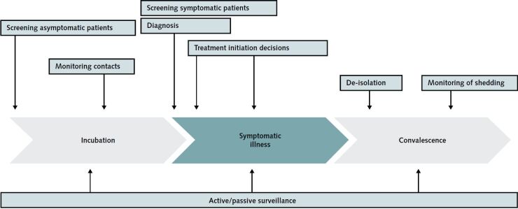
Despite the remarkable speed with which accurate diagnostic tests have been developed and made available for SARS–CoV-2 (25), current tools only partially meet several clinically relevant needs. Figure 1 illustrates different indications for diagnostic testing among persons with proven or suspected COVID-19. For each of these, the most important consideration is the clinical decision a test result will help to inform. Test designs must account for several parameters, such as whether the test detects infection directly (such as the virus itself) or indirectly (such as host antibodies), test turnaround time, the ability to perform many tests at the same time (that is, throughput), the need to have a minimum number of specimens before testing (that is, batching), and the ability to perform the test in low-infrastructure settings (such as on cruise ships or in remote communities). The potential for use at the point of care depends on test complexity. The U.S. Food and Drug Administration (FDA) categorizes diagnostic tests by their complexity: Waived tests are available for use at the point of care, whereas moderate- and high-complexity tests must be performed in a laboratory. The intended use also determines which specimen types are ideal or feasible. Finally, it is important to recognize that the acceptable diagnostic accuracy of a test may vary according to use case. For example, sensitivity and specificity requirements of an assay used to confirm results of a screening test need not be as stringent as those of a method used for standalone diagnosis, because the pool of persons being tested is already enriched with true infections. The Foundation for Innovative New Diagnostics has published a detailed assessment of priority use cases to be considered by test developers and policymakers (26).
Figure 1.
Examples of use cases for diagnostic testing among persons with proven or suspected COVID-19.
A test well suited for one use case (such as epidemiologic surveillance) may be completely inadequate for another (such as rapid screening of symptomatic patients for allocation of personal protective equipment). For test results to enable a specific clinical decision, test developers, policymakers, and clinicians need to consider each of these with respect to the intention of testing and the population being tested as specifically as possible. For the moment, most use cases placed above the green and gray bar are best met by nucleic acid amplification tests, whereas detection of host-derived antibodies directed against SARS–CoV-2 will be crucial for surveillance, epidemic forecasting, and determination of SARS–CoV-2 immunity. SARS–CoV-2 = severe acute respiratory syndrome–related coronavirus-2.
Who to Test: Current Diagnostic Recommendations in the United States
In response to the rapidly evolving COVID-19 pandemic, countries have used different testing approaches depending on testing capacity, public health resources, and the spread of the virus in the community. In the United States, diagnostic testing indications and capacity were limited at the beginning of the outbreak, largely because of regulatory hurdles for the use of new tests. To expand access to testing, the FDA released policies to allow laboratories to use their validated assays in a more timely manner (27). On 4 March, the Centers for Disease Control and Prevention (CDC) removed restrictive testing criteria, recommending that clinicians use their judgment to determine whether a test should be performed (28). Because testing capacity remains suboptimal (27), the implementation of this recommendation remains a challenge. The CDC still recommends priority for testing 3 groups: hospitalized patients with presentations compatible with COVID-19, other symptomatic persons at risk for poor outcomes, and persons who had close contact with someone with suspected or confirmed COVID-19 within 14 days of illness onset or have a history of travel in an affected area (28). These patients should be evaluated with a molecular diagnostic test, as described later. The CDC does not recommend testing asymptomatic persons.
How to Test: Diagnostic Tests in Use or Under Evaluation
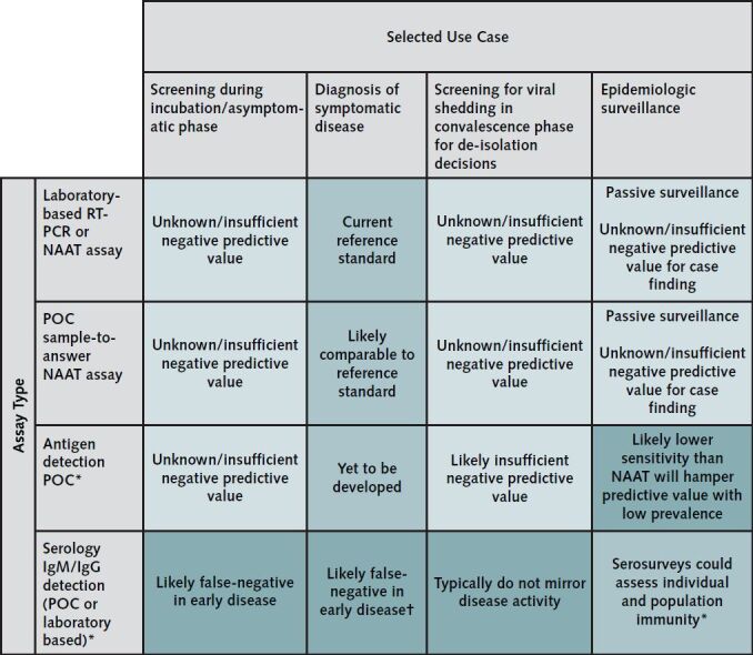
Although real-time reverse transcriptase polymerase chain reaction (RT-PCR)–based assays performed in the laboratory on respiratory specimens are the cornerstone of COVID-19 diagnostic testing, several novel or complementary diagnostic methods are being developed and evaluated (16). Figure 2 depicts the adequacy of the principal assay types used or proposed for COVID-19 for 4 key use cases. Among patients diagnosed with COVID-19, the occurrence of concomitant viral infections has been reported to range from below 6% (29) to greater than 60% (30). As a result, it is not possible to rule out SARS–CoV-2 infection merely by detecting another respiratory pathogen.
Figure 2.

Heat map showing the adequacy of principal assay types (rows) for 4 key use cases.
* This assumes that assays in development or currently undergoing regulatory evaluation prove to be accurate.
† The utility of antibody detection assays for diagnosing acute infections is probably very limited around the time of symptom onset, when viral shedding and transmission risk seem to be highest. Thus, although such tests may have a role among persons presenting late in the course of their infection, the potential for misuse is high.
Laboratory-Based Molecular Testing
The current diagnostic strategy recommended by the CDC to identify patients with COVID-19 is to test samples taken from the respiratory tract to assess for the presence of 1 or several nucleic acid targets specific to SARS–CoV-2 (25). A nasopharyngeal specimen is the preferred choice for swab-based SARS–CoV-2 testing, but oropharyngeal, mid-turbinate, or anterior nares samples also are acceptable (31, 32). Samples should be obtained by using a flocked swab, if available, to enhance the collection and release of cellular material. Swabs with an aluminum or plastic shaft are preferred. Swabs that contain calcium alginate, wood, or cotton should be avoided, because they may contain substances that inhibit PCR testing. Ideally, swabs should be transferred into universal transport medium immediately after sample collection to preserve viral nucleic acid. Samples taken from sputum, endotracheal aspirates, and bronchoalveolar lavage also may be sent directly to the microbiology laboratory for processing, and may have greater sensitivity than upper respiratory tract specimens (33). Inadequate sample collection may result in a false-negative test. After specimen collection, samples undergo RNA extraction followed by qualitative RT-PCR for target detection.
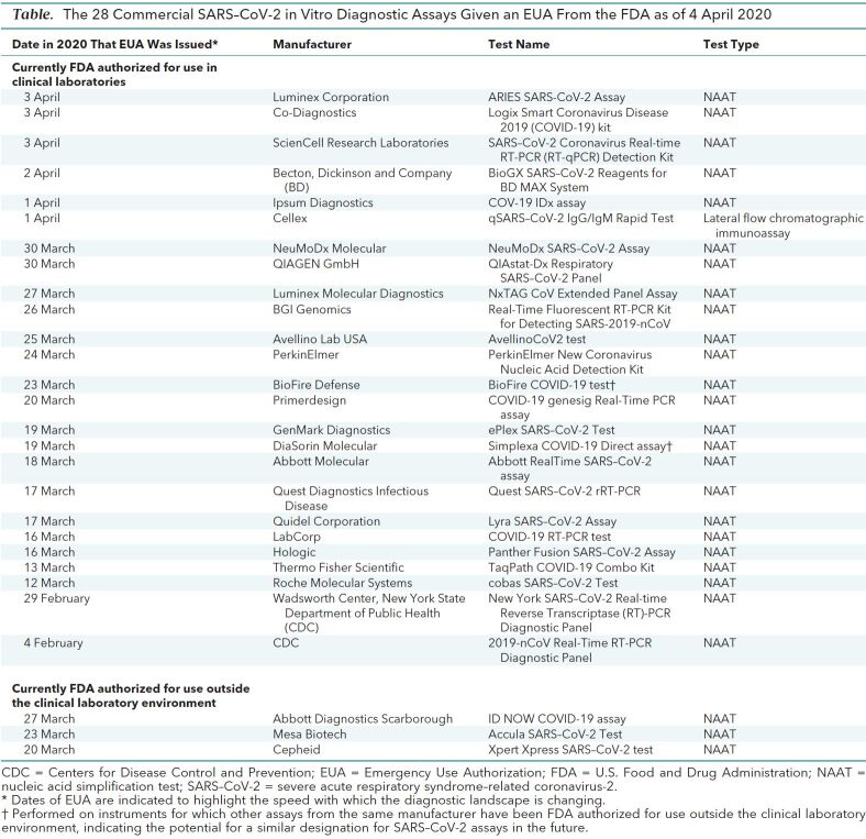
In the United States, the CDC has developed the most widely used SARS–CoV-2 assay. The kit contains PCR primer–probe sets for 2 regions of the viral nucleocapsid gene (N1 and N2), and for the human RNase P gene to ensure the RNA extraction was successful. This assay differs from the World Health Organization primer–probe sets, which target the SARS–CoV-2 RNA-dependent RNA polymerase (RdRP) and envelope (E) genes (25). Both assays have high analytic sensitivity and specificity for SARS–CoV-2, with minimal cross-reactivity with other circulating strains of coronaviruses, and both use a cycle threshold of less than 40 as the criterion for positivity. The CDC kit may be used by state public health laboratories, other laboratories determined by the state to be qualified, and clinical laboratories that meet the regulatory requirements of the Clinical Laboratories Improvement Amendment (CLIA) to perform high-complexity testing (27). Dozens of laboratories have applied for Emergency Use Authorization (EUA) from the FDA for their own laboratory-developed assays (34). The FDA also has granted an EUA for several commercial assays (35), further expanding the ability of clinical laboratories to use these platforms (Table).
Table. The 28 Commercial SARS–CoV-2 in Vitro Diagnostic Assays Given an EUA From the FDA as of 4 April 2020.
The lack of an established reference standard, use of differing sample collection and preparation methods, and an incomplete understanding of viral dynamics across the time course of infection hamper rigorous assessment of the diagnostic accuracy of the many newly introduced SARS–CoV-2 assays (36). Serum and urine are usually negative for the presence of viral nucleic acid, regardless of illness severity (33). Of importance, the ability of RT-PCR assays to rule out COVID-19 on the basis of upper respiratory tract samples obtained at a single time point remains unclear. Conversely, after a patient has had a positive test result, several authorities have recommended obtaining at least 2 negative upper respiratory tract samples, collected at intervals of 24 hours or longer, to document SARS–CoV-2 clearance (37, 38).
Point-of-Care Molecular Diagnostics
Low-complexity, rapid (results within 1 hour) molecular diagnostic tests for respiratory viral infections that are CLIA waived (FDA approved for use outside the laboratory by nonlaboratory personnel) include cartridge-based assays on platforms that include the Abbott ID NOW (Abbott Laboratories), BioFire FilmArray (bioMérieux), cobas Liat (Roche Diagnostics), and GeneXpert (Cepheid) (39).
Rapid point-of-care assays for SARS–CoV-2 on instruments such as these will be critical to expand point-of-care testing. The Xpert Xpress SARS–CoV-2 test (Cepheid) has received an FDA EUA and is performed on the GeneXpert platform, which is already widely used for tuberculosis and HIV testing, especially in low- and middle-income countries. This capacity might be useful to scale up testing across the world as well as in settings where rapid results at the point of care would enable clinical decisions, although testing throughput may be a limiting factor.
Antigen Detection Tests
Tests that detect respiratory syncytial virus or influenza virus antigens by immunoassay directly from clinical specimens have been commercially available for decades, are of low complexity, and may provide results within minutes at the point of care (40). Current tools for influenza and respiratory syncytial virus suffer from suboptimal sensitivity to rule out disease (41, 42); the same challenge would probably exist for SARS–CoV-2, and tests would need to be implemented with clear guidance on correct interpretation. Prototypes of such tests for other novel coronaviruses have not received regulatory approval (43, 44) but are under development (45). Monoclonal antibodies against the nucleocapsid protein of SARS–CoV-2 have been generated, which might form the basis of a future rapid antigen detection test (20).
Serology
Serologic tests that identify antibodies (such as IgA, IgM, and IgG) to SARS–CoV-2 from clinical specimens (such as blood or saliva), such as enzyme-linked immunosorbent assays, may be less complex than molecular tests and have the potential to be used for diagnosis in certain situations (46). However, their utility for diagnosing acute infections is probably limited around the time of symptom onset, when viral shedding and transmission risk seem to be highest (32). Antibody responses to infection take days to weeks to be reliably detectable (46). Negative results would not exclude SARS–CoV-2 infection, particularly among those with recent exposure to the virus. Cross-reactivity of antibody to non–SARS–CoV-2 coronavirus proteins is also a potential problem, whereby positive results may be the result of past or present infection with other human coronaviruses (47). Serologic assays might be more relevant in scenarios in which patients present to medical care with late complications of disease, when RT-PCR may be falsely negative, because viral shedding drops over time (48).
The development of serologic assays that accurately assess prior infection and immunity to SARS–CoV-2 will be essential for epidemiologic studies, ongoing surveillance, vaccine studies, and potentially for risk assessment of health care workers. Immunoassays are already on the market in some countries, but their diagnostic accuracy and optimal use remain undefined.
Ancillary Diagnostic Tests
The optimal use of diagnostic imaging, biomarkers, and other nonmicrobiologic tests is rapidly evolving.
Radiographic Tests
Many centers have evaluated the utility of chest imaging for diagnosis. On chest radiography, bilateral pneumonia is the most frequently reported feature (range, 11.8% to 100%) and is more common than a unilateral focus (49, 50). Computed tomography is regarded as more sensitive than radiography, with several cohort studies reporting that most patients (77.8% to 100%) had ground glass opacities. Other features commonly reported with COVID-19 on chest computed tomography include a peripheral distribution, fine reticular opacities, and vascular thickening (51). Compared with serial nasopharyngeal sampling, chest computed tomography may be more sensitive than an RT-PCR test at a single time point for the diagnosis of COVID-19 (52, 53). In addition, artificial intelligence may help distinguish COVID-19 from other etiologic agents of community-acquired pneumonia (54). However, these findings are not completely specific to COVID-19 and do not exclude a co-infection or an alternative diagnosis (55).
Biomarkers Associated With COVID-19 Patients
The most common laboratory features reported in patients with COVID-19 include decreased albumin (75.8% [95% CI, 30.5% to 100%]), elevated C-reactive protein (58.3% [CI, 21.8% to 94.7%]), and elevated lactate dehydrogenase levels (57.0% [CI, 38.0% to 76.0%]), and lymphopenia (43.1% [CI, 18.9% to 67.3%]) (56). Other biomarkers that have been reported include increased erythrocyte sedimentation rates; elevated aspartate aminotransferase, alanine aminotransferase, and creatinine kinase levels; leukopenia; leukocytosis; and increased bilirubin and creatinine levels (57–59). Such findings are not surprising, because these biomarkers represent an inflammatory host response to SARS–CoV-2 or are early markers of end-organ dysfunction, similar to that seen in patients with sepsis (60). No biomarker or combination of biomarkers currently exists that is sensitive or specific enough to establish a diagnosis of COVID-19, or to pragmatically predict its clinical course.
Unmet Needs and the Diagnostic Test Pipeline
Scaling Up Access to Diagnostic Testing
In the face of a public health emergency, important first steps to expand testing capacity include relaxing and streamlining regulatory requirements and procedures. Local public health laboratories and academic diagnostic laboratories in the United States are being rapidly enabled to perform EUA-granted commercial assays and laboratory-developed tests using research use–only reagents (61). University research laboratories could also add capacity, although concerns exist regarding quality control and the absence of protocols for managing clinical specimens. Flexibility regarding nucleic acid extraction methods and amplification instruments when using CDC protocols is being introduced (34). National agencies are expeditiously making materials for test development and validation available to clinical laboratories and diagnostic test manufacturers.
Safely evaluating clinically stable persons for COVID-19 at traditional health care access points is resource intensive and slow, and risks exposing staff to infection. Many jurisdictions are enabling innovative testing venues, such as external tents or drive-through or “phone booth” testing, as well as home assessment teams to expedite specimen collection while limiting potential exposures (62). Telemedicine combined with at-home nasal swab self-testing also has been proposed (63). Of importance, in jurisdictions without universal health care coverage, policy solutions must be introduced to eliminate financial barriers to testing for uninsured and underinsured patients. Efforts to increase accessibility of testing for multiple use cases need to be coupled to appropriate public health interventions to isolate infected persons and their contacts.
Alternatives to Usual Specimen Types, Collection Devices, and Transport Media
Nasopharyngeal swabs are the recommended specimen for molecular analysis. The sudden demand for flocked nasopharyngeal swabs and viral transport medium generated by the pandemic has put enormous pressures on supply chain capacities for these products. As of 19 March 2020 the CDC made oropharyngeal, mid-turbinate, and nasal swabs acceptable specimen types if nasopharyngeal swabs are not available (31). Early-morning posterior oropharyngeal saliva samples (coughed up by clearing the throat) also have been assessed as useful specimen types and would not require use of a swab (48). The CDC has released a standard operating procedure for laboratories to create their own viral transport medium (64); other solutions also may be used if viral transport medium is unavailable, including phosphate-buffered saline, liquid Amies, and normal saline (65). The FDA has provided guidance on its Web site for alternative materials to collect and transport samples for RT-PCR SARS–CoV-2 assays (34). The diagnostic value of molecular testing of nonrespiratory specimens currently is unclear.
Diagnostics Pipeline in the Short and Medium Term
Although excellent tools exist for the diagnosis of symptomatic patients in well-equipped laboratories, important gaps remain in screening asymptomatic persons in the incubation phase, as well as for the accurate determination of live viral shedding among patients in the convalescence phase to inform de-isolation decisions (Figure 2). Further, it is critical to advance solutions that require less well-equipped laboratories to curb the pandemic globally. The Foundation for Innovative New Diagnostics (FIND) and others have created online resources to collate the rapidly evolving set of assays at various stages of development, from proof of concept to full regulatory approval (20, 53). Simple antigen-based tests, if sensitive enough, might be useful in lower-resource and home settings to inform quarantine and spatial distancing measures for patients without severe illness and their contacts. Novel technologies, such as Clustered Regularly Interspersed Short Palindromic Repeats (CRISPR)-based diagnostics are being used to develop rapid, simple, low-cost, portable, temperature-stable assays for deployment in the field in nontraditional and resource-limited settings, such as airports and border crossings (20, 51, 54). Other technologies might be deployed to lower-resource settings if they can be standardized. For example, it might be possible to leverage existing loop-mediated isothermal amplification testing networks established for other diseases, such as human African trypanosomiasis surveillance (66).
Other Considerations
Critical considerations for diagnostics used for epidemic diseases of public health importance include the quality assurance and regulatory frameworks surrounding testing. Mature regulatory agencies have developed mechanisms to account for emergencies, such as the FDA's EUA stream, but pragmatic solutions must be found to facilitate wide-scale, independent evaluation of emerging tests.
Initially, the need for elaborate biosafety precautions and inconsistent recommendations for their application across regions severely hampered COVID-19 testing. Although these continue to evolve, current recommendations in Canada and the United States acknowledge that nonpropagative work for molecular testing may be performed in containment level 2 conditions found in routine diagnostic laboratories and provide specific guidance on diagnostic testing of specimens conducted outside a biosafety level 2 laboratory, such as rapid respiratory testing performed at the point of care (67) .
Conclusion
The COVID-19 pandemic has dramatically highlighted the essential role of diagnostics in the control of communicable diseases. Intensive diagnostics deployment probably contributed to the success of a few countries in controlling transmission. Urgent clinical and public health needs now drive an unprecedented global effort to increase SARS–CoV-2 testing capacity. Finally, the blinding speed with which COVID-19 has spread illustrates the need for preparedness and long-term investments in diagnostic testing.
Biography
Acknowledgment: The authors thank Professor Ellen Jo Baron for insightful comments on earlier versions of this manuscript and Ms. Chelsea Caya for assistance with literature searches and assembling figures.
Grant Support: Drs. Papenburg, Quach, and Yansouni have received career awards from the Québec Health Research Fund.
Disclosures: Dr. Cheng reports grants from the Canadian Institute of Health Research and McGill Interdisciplinary Initiative in Infection and Immunity during the conduct of the study; after the manuscript was written and submitted, he was offered a position on the scientific advisory board of GEn1E Lifesciences, but this position is unrelated to the submitted work. Dr. Papenburg reports grants and personal fees from BD Diagnostics, Seegene, and AbbVie; personal fees from Cepheid; and grants from MedImmune, Sanofi Pasteur, Janssen Pharmaceuticals, and Hoffman–La Roche outside the submitted work. Dr. Kanjilal reports grants from PhAST Diagnostics outside the submitted work. Dr. Libman reports grants from GeoSentinel Network outside the submitted work. Dr. Yansouni reports nonfinancial support from bioMérieux outside the submitted work. Authors not named here have disclosed no conflicts of interest. Disclosures can also be viewed at www.acponline.org/authors/icmje/ConflictOfInterestForms.do?msNum=M20-1301.
Editors' Disclosures: Christine Laine, MD, MPH, Editor in Chief, reports that her spouse has stock options/holdings with Targeted Diagnostics and Therapeutics. Darren B. Taichman, MD, PhD, Executive Editor, reports that he has no financial relationships or interests to disclose. Cynthia D. Mulrow, MD, MSc, Senior Deputy Editor, reports that she has no relationships or interests to disclose. Jaya K. Rao, MD, MHS, Deputy Editor, reports that she has stock holdings/options in Eli Lilly and Pfizer. Christina C. Wee, MD, MPH, Deputy Editor, reports employment with Beth Israel Deaconess Medical Center. Sankey V. Williams, MD, Deputy Editor, reports that he has no financial relationships or interests to disclose. Yu-Xiao Yang, MD, MSCE, Deputy Editor, reports that he has no financial relationships or interest to disclose.
Corresponding Author: Matthew P. Cheng, MDCM, Division of Infectious Diseases, McGill University Health Centre, 1001 Decarie Boulevard, E05.1709, Montreal, Quebec, Canada H4A 3J1; e-mail, matthew.cheng@mcgill.ca.
Current author addresses and author contributions are available at Annals.org.
Current Author Addresses: Dr. Cheng, MDCM, Division of Infectious Diseases, McGill University Health Centre, 1001 Decarie Boulevard, E05.1709, Montreal, Quebec, Canada H4A 3J1.
Dr. Papenburg: Division of Infectious Diseases, Montreal Children's Hospital, 1001 Decarie Boulevard, E05.1905, Montreal, Quebec, Canada H4A 3J1.
Dr. Desjardins: Division of Transplant Infectious Disease, Brigham and Women's Hospital, 75 Francis Street, PBB-A4, Boston, MA 02115.
Dr. Kanjilal: Department of Population Medicine, Harvard Medical School and Harvard Pilgrim Health Care Institute, 401 Park Drive, Suite 401, East Boston, MA 02115.
Dr. Quach: Divison of Infectious Diseases, CHU Sainte-Justine, 3175, ch. Côte Ste-Catherine, B.17.102, Montreal, Quebec, Canada H3T 1C5.
Dr. Libman: Division of Infectious Diseases, McGill University Health Centre, 1001 Decarie Boulevard, E05.1830, Montreal, Quebec, Canada H4A 3J1.
Dr. Dittrich: Foundation of Innovative New Diagnostics (FIND), Campus Biotech, Chemin des Mines 91 202, Geneva, Switzerland.
Dr. Yansouni: Division of Infectious Diseases, McGill University Health Centre, 1001 Decarie Boulevard, EM3.3242, Montreal, Quebec, Canada H4A 3J1.
Author Contributions: Conception and design: M.P. Cheng, J. Papenburg, M. Desjardins, M. Libman, S. Dittrich, C.P. Yansouni.
Analysis and interpretation of the data: M.P. Cheng, J. Papenburg, S. Dittrich, C.P. Yansouni.
Drafting of the article: M.P. Cheng, J. Papenburg, M. Desjardins, S. Kanjilal, M. Libman, S. Dittrich, C.P. Yansouni.
Critical revision for important intellectual content: M.P. Cheng, J. Papenburg, M. Desjardins, S. Kanjilal, C. Quach, M. Libman, C.P. Yansouni.
Final approval of the article: M.P. Cheng, J. Papenburg, M. Desjardins, S. Kanjilal, C. Quach, M. Libman, S. Dittrich, C.P. Yansouni.
Obtaining of funding: C.P. Yansouni.
Administrative, technical, or logistic support: C.P. Yansouni.
Collection and assembly of data: M.P. Cheng, J. Papenburg, C.P. Yansouni.
Footnotes
This article was published at Annals.org on 13 April 2020.
References
- 1. doi: 10.1056/NEJMoa2001017. Zhu N, Zhang D, Wang W, et al; China Novel Coronavirus Investigating and Research Team. A novel coronavirus from patients with pneumonia in China, 2019. N Engl J Med. 2020;382:727-733. [PMID: 31978945] doi:10.1056/NEJMoa2001017. [DOI] [PMC free article] [PubMed]
- 2. doi: 10.1038/s41564-020-0695-z. Coronaviridae Study Group of the International Committee on Taxonomy of Viruses. The species Severe acute respiratory syndrome-related coronavirus: classifying 2019-nCoV and naming it SARS-CoV-2. Nat Microbiol. 2020;5:536-544. [DOI] [PMC free article] [PubMed]
- 3. World Health Organization. Novel coronavirus (COVID-19) situation. Accessed at https://experience.arcgis.com/experience/685d0ace521648f8a5beeeee1b9125cd. on 24 March 2020.
- 4. doi: 10.1056/NEJMoa2001316. Li Q, Guan X, Wu P, et al. Early transmission dynamics in Wuhan, China, of novel coronavirus-infected pneumonia. N Engl J Med. 2020;382:1199-1207. [PMID: 31995857] doi:10.1056/NEJMoa2001316. [DOI] [PMC free article] [PubMed]
- 5. Mizumoto K, Kagaya K, Chowell G. Early epidemiological assessment of the transmission potential and virulence of coronavirus disease 2019 (COVID-19) in Wuhan City: China, January-February, 2020. Accessed at https://www.medrxiv.org/content/10.1101/2020.02.12.20022434v2. on 24 March 2020.
- 6. doi: 10.1001/jama.2020.2565. Bai Y, Yao L, Wei T, et al. Presumed asymptomatic carrier transmission of COVID-19. JAMA. 2020. [PMID: 32083643] doi:10.1001/jama.2020.2565. [DOI] [PMC free article] [PubMed]
- 7. doi: 10.1126/science.abb3221. Li R, Pei S, Chen B, et al. Substantial undocumented infection facilitates the rapid dissemination of novel coronavirus (SARS-CoV2). Science. 2020. [PMID: 32179701] doi:10.1126/science.abb3221. [DOI] [PMC free article] [PubMed]
- 8. doi: 10.1073/pnas.2002616117. Wells CR, Sah P, Moghadas SM, et al. Impact of international travel and border control measures on the global spread of the novel 2019 coronavirus outbreak. Proc Natl Acad Sci U S A. 2020;117:7504-7509. [PMID: 32170017] doi:10.1073/pnas.2002616117. [DOI] [PMC free article] [PubMed]
- 9. doi: 10.7326/M20-0696. Tuite AR, Bogoch II, Sherbo R, et al. Estimation of coronavirus disease 2019 (COVID-19) burden and potential for international dissemination of infection from Iran. Ann Intern Med. 2020. [PMID: 32176272] doi:10.7326/M20-0696. [DOI] [PMC free article] [PubMed]
- 10. doi: 10.1093/jtm/taaa039. Lee VJ, Chiew CJ, Khong WX. Interrupting transmission of COVID-19: lessons from containment efforts in Singapore. J Travel Med. 2020. [PMID: 32167146] doi:10.1093/jtm/taaa039. [DOI] [PMC free article] [PubMed]
- 11. doi: 10.1016/S0140-6736(20)30566-3. Zhou F, Yu T, Du R, et al. Clinical course and risk factors for mortality of adult inpatients with COVID-19 in Wuhan, China: a retrospective cohort study. Lancet. 2020;395:1054-1062. [PMID: 32171076] doi:10.1016/S0140-6736(20)30566-3. [DOI] [PMC free article] [PubMed]
- 12. doi: 10.1001/jama.2020.4683. Onder G, Rezza G, Brusaferro S. Case-fatality rate and characteristics of patients dying in relation to COVID-19 in Italy. JAMA. 2020. [PMID: 32203977] doi:10.1001/jama.2020.4683. [DOI] [PubMed]
- 13. Dong Y, Mo X, Hu Y, et al. Epidemiological characteristics of 2143 pediatric patients with 2019 coronavirus disease in China. Pediatrics. 2020. [PMID: 32179660] doi:10.1542/peds.2020-0702.
- 14. Centers of Disease Control and Prevention. Disease burden of influenza. Accessed at www.cdc.gov/flu/about/burden/index.html. on 4 April 2020.
- 15. doi: 10.3201/eid2606.200320. Wilson N, Kvalsvig A, Barnard LT, et al. Case-fatality risk estimates for COVID-19 calculated by using a lag time for fatality. Emerg Infect Dis. 2020;26. [PMID: 32168463] doi:10.3201/eid2606.200320. [DOI] [PMC free article] [PubMed]
- 16. FIND. COVID-19 Diagnostics Resource Centre. Accessed at www.finddx.org/covid-19. on March 21 2020.
- 17. doi: 10.1016/S1473-3099(20)30120-1. Dong E, Du H, Gardner L. An interactive web-based dashboard to track COVID-19 in real time [Letter]. Lancet Infect Dis. 2020. [PMID: 32087114] doi:10.1016/S1473-3099(20)30120-1. [DOI] [PMC free article] [PubMed]
- 18. doi: 10.3201/eid2501.171901. Delamater PL, Street EJ, Leslie TF, et al. Complexity of the basic reproduction number (R0). Emerg Infect Dis. 2019;25:1-4. [PMID: 30560777] doi:10.3201/eid2501.171901. [DOI] [PMC free article] [PubMed]
- 19. doi: 10.1503/cmaj.200438. Cheng MP, Lee TC, Han DHS, et al. Generating randomized trial evidence to optimize treatment in the COVID-19 pandemic. CMAJ. 2020. doi: 10.1503/cmaj.200438. [DOI] [PMC free article] [PubMed]
- 20. doi: 10.1038/d41587-020-00010-2. Sheridan C. Fast, portable tests come online to curb coronavirus pandemic. Nat Biotechnol. 2020. [PMID: 32203294] doi:10.1038/d41587-020-00010-2. [DOI] [PubMed]
- 21. doi: 10.3346/jkms.2020.35.e112. Korean Society of Infectious Diseases. Report on the epidemiological features of coronavirus disease 2019 (COVID-19) outbreak in the Republic of Korea from January 19 to March 2, 2020. J Korean Med Sci. 2020;35:e112. [PMID: 32174069] doi:10.3346/jkms.2020.35.e112. [DOI] [PMC free article] [PubMed]
- 22. doi: 10.1001/jama.2020.3151. Wang CJ, Ng CY, Brook RH. Response to COVID-19 in Taiwan: big data analytics, new technology, and proactive testing. JAMA. 2020. [PMID: 32125371] doi:10.1001/jama.2020.3151. [DOI] [PubMed]
- 23. doi: 10.3934/mbe.2004.1.1. Webb GF, Blaser MJ, Zhu H, et al. Critical role of nosocomial transmission in the Toronto SARS outbreak. Math Biosci Eng. 2004;1:1-13. [PMID: 20369956] [DOI] [PubMed]
- 24. doi: 10.3201/eid1102.040607. Wong T, Wallington T, McDonald LC, et al. Late recognition of SARS in nosocomial outbreak, Toronto. Emerg Infect Dis. 2005;11:322-5. [PMID: 15752456] [DOI] [PMC free article] [PubMed]
- 25. doi: 10.2807/1560-7917.ES.2020.25.3.2000045. Corman VM, Landt O, Kaiser M, et al. Detection of 2019 novel coronavirus (2019-nCoV) by real-time RT-PCR. Euro Surveill. 2020;25. [PMID: 31992387] doi:10.2807/1560-7917.ES.2020.25.3.2000045. [DOI] [PMC free article] [PubMed]
- 26. FIND. SARS-COV-2 Diagnostic Use Cases. Accessed at www.finddx.org/covid-19/dx-use-cases/ on 27 March 2020.
- 27. U.S. Food and Drug Administration. Guidance Document: Policy for Diagnostic Tests for Coronavirus Disease-2019 during the Public Health Emergency. Accessed at www.fda.gov/regulatory-information/search-fda-guidance-documents/policy-diagnostic-tests-coronavirus-disease-2019-during-public-health-emergency. on 21 March 2020.
- 28. Centers for Disease Control and Prevention. Evaluating and Testing Persons for Coronavirus Disease 2019 (COVID-19). Accessed at www.cdc.gov/coronavirus/2019-nCoV/hcp/clinical-criteria.html. on 21 March 2020.
- 29. Wang M, Wu Q, Xu W, et al. Clinical diagnosis of 8274 samples with 2019-novel coronavirus in Wuhan. Accessed at www.medrxiv.org/content/10.1101/2020.02.12.20022327v2. on 21 March 2020.
- 30. Xing Q, Li G, Xing Y, et al. Precautions are needed for COVID-19 patients with coinfection of common respiratory pathogens. Accessed at https://ssrn.com/abstract=3550013. on 21 March 2020.
- 31. Centers for Disease Control and Pevention. Interim Guidelines for Collecting, Handling, and Testing Clinical Specimens from Persons for Coronavirus Disease 2019 (COVID-19). Accessed at www.cdc.gov/coronavirus/2019-nCoV/lab/guidelines-clinical-specimens.html. on 27 March 2020.
- 32. doi: 10.1056/NEJMc2001737. Zou L, Ruan F, Huang M, et al. SARS-CoV-2 viral load in upper respiratory specimens of infected patients [Letter]. N Engl J Med. 2020;382:1177-1179. [PMID: 32074444] doi:10.1056/NEJMc2001737. [DOI] [PMC free article] [PubMed]
- 33. doi: 10.1001/jama.2020.3786. Wang W, Xu Y, Gao R, et al. Detection of SARS-CoV-2 in different types of clinical specimens. JAMA. 2020. [PMID: 32159775] doi:10.1001/jama.2020.3786. [DOI] [PMC free article] [PubMed]
- 34. U.S. Food and Drug Administration. FAQs on Diagnostic Testing for SARS-CoV-2. Accessed at www.fda.gov/medical-devices/emergency-situations-medical-devices/faqs-diagnostic-testing-sars-cov-2. on 22 March 2020.
- 35. U.S. Food and Drug Administration. Emergency Use Authorizations. Accessed at www.fda.gov/medical-devices/emergency-situations-medical-devices/emergency-use-authorizations#covid19ivd. on 22 March 2020.
- 36. doi: 10.1016/S1473-3099(20)30232-2. Liu Y, Yan LM, Wan L, et al. Viral dynamics in mild and severe cases of COVID-19 [Letter]. Lancet Infect Dis. 2020. [PMID: 32199493] doi:10.1016/S1473-3099(20)30232-2. [DOI] [PMC free article] [PubMed]
- 37. European Centre for Disease Prevention and Control. Novel coronavirus (SARS-CoV-2). Accessed at www.ecdc.europa.eu/sites/default/files/documents/COVID-19-Discharge-criteria.pdf. on 4 April 2020.
- 38. Centers for Disease Control and Prevention. Discontinuation of Transmission-Based Precautions and Disposition of Patients with COVID-19 in Healthcare Settings (Interim Guidance). Accessed at www.cdc.gov/coronavirus/2019-ncov/hcp/disposition-hospitalized-patients.html. on 4 April 2020.
- 39. doi: 10.1080/14737159.2018.1487293. Hogan CA, Caya C, Papenburg J. Rapid and simple molecular tests for the detection of respiratory syncytial virus: a review. Expert Rev Mol Diagn. 2018;18:617-629. [PMID: 29890085] doi:10.1080/14737159.2018.1487293. [DOI] [PubMed]
- 40. doi: 10.2217/fmb.13.9. Prendergast C, Papenburg J. Rapid antigen-based testing for respiratory syncytial virus: moving diagnostics from bench to bedside? Future Microbiol. 2013;8:435-44. [PMID: 23534357] doi:10.2217/fmb.13.9. [DOI] [PubMed]
- 41. doi: 10.1128/JCM.01816-15. Chartrand C, Tremblay N, Renaud C, et al. Diagnostic accuracy of rapid antigen detection tests for respiratory syncytial virus infection: systematic review and meta-analysis. J Clin Microbiol. 2015;53:3738-49. [PMID: 26354816] doi:10.1128/JCM.01816-15. [DOI] [PMC free article] [PubMed]
- 42. doi: 10.7326/M17-0848. Merckx J, Wali R, Schiller I, et al. Diagnostic accuracy of novel and traditional rapid tests for influenza infection compared with reverse transcriptase polymerase chain reaction: a systematic review and meta-analysis. Ann Intern Med. 2017;167:394-409. [PMID: 28869986] doi:10.7326/M17-0848. [DOI] [PubMed]
- 43. doi: 10.1128/JCM.42.7.2884-2889.2004. Lau SK, Woo PC, Wong BH, et al. Detection of severe acute respiratory syndrome (SARS) coronavirus nucleocapsid protein in SARS patients by enzyme-linked immunosorbent assay. J Clin Microbiol. 2004;42:2884-9. [PMID: 15243033] [DOI] [PMC free article] [PubMed]
- 44. doi: 10.1038/emi.2015.26. Chen Y, Chan KH, Kang Y, et al. A sensitive and specific antigen detection assay for Middle East respiratory syndrome coronavirus. Emerg Microbes Infect. 2015;4:e26. [PMID: 26421268] doi:10.1038/emi.2015.26. [DOI] [PMC free article] [PubMed]
- 45. FIND. SARS-CoV-2 Diagnostic Pipeline. Accessed at www.finddx.org/covid-19/pipeline. on 23 March 2020.
- 46. doi: 10.1093/cid/ciaa310. Guo L, Ren L, Yang S, et al. Profiling early humoral response to diagnose novel coronavirus disease (COVID-19). Clin Infect Dis. 2020. [PMID: 32198501] doi:10.1093/cid/ciaa310. [DOI] [PMC free article] [PubMed]
- 47. doi: 10.1155/2006/152612. Patrick DM, Petric M, Skowronski DM, et al. An outbreak of human coronavirus OC43 infection and serological cross-reactivity with SARS coronavirus. Can J Infect Dis Med Microbiol. 2006;17:330-6. [PMID: 18382647] [DOI] [PMC free article] [PubMed]
- 48. doi: 10.1016/S1473-3099(20)30196-1. To KK, Tsang OT, Leung WS, et al. Temporal profiles of viral load in posterior oropharyngeal saliva samples and serum antibody responses during infection by SARS-CoV-2: an observational cohort study. Lancet Infect Dis. 2020. [PMID: 32213337] doi:10.1016/S1473-3099(20)30196-1. [DOI] [PMC free article] [PubMed]
- 49. doi: 10.1148/radiol.2020200230. Chung M, Bernheim A, Mei X, et al. CT imaging features of 2019 novel coronavirus (2019-nCoV). Radiology. 2020;295:202-207. [PMID: 32017661] doi:10.1148/radiol.2020200230. [DOI] [PMC free article] [PubMed]
- 50. doi: 10.1001/jama.2020.1585. Wang D, Hu B, Hu C, et al. Clinical characteristics of 138 hospitalized patients with 2019 novel coronavirus-infected pneumonia in Wuhan, China. JAMA. 2020. [PMID: 32031570] doi:10.1001/jama.2020.1585. [DOI] [PMC free article] [PubMed]
- 51. doi: 10.1148/radiol.2020200823. Bai HX, Hsieh B, Xiong Z, et al. Performance of radiologists in differentiating COVID-19 from viral pneumonia on chest CT. Radiology. 2020:200823. [PMID: 32155105] doi:10.1148/radiol.2020200823. [DOI] [PMC free article] [PubMed]
- 52. doi: 10.1148/radiol.2020200642. Ai T, Yang Z, Hou H, et al. Correlation of chest CT and RT-PCR testing in coronavirus disease 2019 (COVID-19) in China: a report of 1014 cases. Radiology. 2020:200642. [PMID: 32101510] doi:10.1148/radiol.2020200642. [DOI] [PMC free article] [PubMed]
- 53. doi: 10.1148/radiol.2020200432. Fang Y, Zhang H, Xie J, et al. Sensitivity of chest CT for COVID-19: comparison to RT-PCR. Radiology. 2020:200432. [PMID: 32073353] doi:10.1148/radiol.2020200432. [DOI] [PMC free article] [PubMed]
- 54. Li L, Qin L, Xu Z, et al. Artificial intelligence distinguishes COVID-19 from community acquired pneumonia on chest CT. Radiology. 2020:200905. [PMID: 32191588] doi:10.1148/radiol.2020200905.
- 55. doi: 10.3201/eid2606.200299. Wu X, Cai Y, Huang X, et al. Co-infection with SARS-CoV-2 and influenza A virus in patient with pneumonia, China. Emerg Infect Dis. 2020;26. [PMID: 32160148] doi:10.3201/eid2606.200299. [DOI] [PMC free article] [PubMed]
- 56. doi: 10.1016/j.tmaid.2020.101623. Rodriguez-Morales AJ, Cardona-Ospina JA, Gutiérrez-Ocampo E, et al; Latin American Network of Coronavirus Disease 2019-COVID-19 Research (LANCOVID-19). Electronic address: https://www.lancovid.org . Clinical, laboratory and imaging features of COVID-19: a systematic review and meta-analysis. Travel Med Infect Dis. 2020:101623. [PMID: 32179124] doi:10.1016/j.tmaid.2020.101623. [DOI] [PMC free article] [PubMed]
- 57. doi: 10.3760/cma.j.issn.1001-0939.2020.03.013. Chen L, Liu HG, Liu W, et al. [Analysis of clinical features of 29 patients with 2019 novel coronavirus pneumonia]. Zhonghua Jie He He Hu Xi Za Zhi. 2020;43:203-208. [PMID: 32164089] doi:10.3760/cma.j.issn.1001-0939.2020.03.013. [DOI] [PubMed]
- 58. doi: 10.1016/S0140-6736(20)30211-7. Chen N, Zhou M, Dong X, et al. Epidemiological and clinical characteristics of 99 cases of 2019 novel coronavirus pneumonia in Wuhan, China: a descriptive study. Lancet. 2020;395:507-513. [PMID: 32007143] doi:10.1016/S0140-6736(20)30211-7. [DOI] [PMC free article] [PubMed]
- 59. doi: 10.1016/S0140-6736(20)30183-5. Huang C, Wang Y, Li X, et al. Clinical features of patients infected with 2019 novel coronavirus in Wuhan, China. Lancet. 2020;395:497-506. [PMID: 31986264] doi:10.1016/S0140-6736(20)30183-5. [DOI] [PMC free article] [PubMed]
- 60. doi: 10.1093/cid/ciaa248. Qin C, Zhou L, Hu Z, et al. Dysregulation of immune response in patients with COVID-19 in Wuhan, China. Clin Infect Dis. 2020. [PMID: 32161940] doi:10.1093/cid/ciaa248. [DOI] [PMC free article] [PubMed]
- 61. Infectious Diseases Society of America. COVID-19 Diagnostic Testing Policy Recommendations. 2020. Accessed at: www.idsociety.org/globalassets/idsa/public-health/covid-19-diagnostic-testing-policy-recommendations.pdf. on 4 April 4 2020.
- 62. doi: 10.1093/cid/ciaa256. Bryson-Cahn C, Duchin J, Makarewicz VA, et al. A novel approach for a novel pathogen: using a home assessment team to evaluate patients for 2019 novel coronavirus (SARS-CoV-2). Clin Infect Dis. 2020. [PMID: 32166310] doi:10.1093/cid/ciaa256. [DOI] [PMC free article] [PubMed]
- 63. Nundy S, Patel KK. Self-service diagnosis of COVID-19—ready for prime time? Accessed at https://jamanetwork.com/channels/health-forum/fullarticle/2763264. on 22 March 2020.
- 64. Centers for Disease Control and Prevention. Preparation of Viral Transport Medium. Accessed at www.cdc.gov/coronavirus/2019-ncov/downloads/Viral-Transport-Medium.pdf. on 22 March 2020.
- 65. doi: 10.1128/JCM.00590-20. Rodino KG, Espy MJ, Buckwalter SP, et al. Evaluation of saline, phosphate buffered saline and minimum essential medium as potential alternatives to viral transport media for SARS-CoV-2 testing [Letter]. J Clin Microbiol. 2020. [PMID: 32229604] doi:10.1128/JCM.00590-20. [DOI] [PMC free article] [PubMed]
- 66. doi: 10.1371/journal.pntd.0002504. Mitashi P, Hasker E, Ngoyi DM, et al. Diagnostic accuracy of loopamp Trypanosoma brucei detection kit for diagnosis of human African trypanosomiasis in clinical samples. PLoS Negl Trop Dis. 2013;7:e2504. [PMID: 24147176] doi:10.1371/journal.pntd.0002504. [DOI] [PMC free article] [PubMed]
- 67. Government of Canada. SARS-CoV-2 (severe acute respiratory syndrome-related coronavirus 2). Biosafety advisory. Accessed at www.canada.ca/en/public-health/services/laboratory-biosafety-biosecurity/biosafety-directives-advisories-notifications/novel-coronavirus-january-27.html. on 21 March 2020.


