

Abstract
Background
This is an update of the review on 'Percutaneous transluminal rotational atherectomy for coronary artery disease' first published in The Cochrane Library Issue 4, 2003. Percutaneous transluminal coronary rotational atherectomy (PTCRA) debulks atherosclerotic plaque from coronary arteries using an abrasive burr. On rotation, the burr selectively removes hard tissue. PTCRA has been used both as an alternative to and in conjunction with balloon angioplasty to open up blocked coronary arteries. Its ongoing effectiveness and safety compared with other modes of removing atherosclerotic plaques is reviewed.
Objectives
To assess the effects of PTCRA for coronary artery disease in patients with non‐complex and complex lesions (e.g. ostial, long or diffuse lesions or those arising from in‐stent re‐stenosis) of the coronary arteries.
Search methods
For the original review, we searched the Heart Group Specialised Register; The Cochrane Library to Issue 2, 2001; and MEDLINE, CINAHL, EMBASE and Current Contents to December 2002 and reviewed reference lists for relevant articles. For the current review, we searched the same registries from 2002 to 2012 and reviewed reference lists for relevant articles.
Selection criteria
We included randomised and quasi‐randomised controlled trials of PTCRA compared with placebo, no treatment or another intervention and excluded cross‐over trials.
Data collection and analysis
Two review authors independently extracted data and assessed the risk of bias of the studies identified. Data were extracted independently by two review authors. We asked authors of trials to provide information when missing data were encountered. Statistical summaries used risk ratios (RR) and weighted mean differences.
Main results
We included 12 trials enrolling 3474 patients. The overall risk of bias was unclear for the majority of articles due to a lack of reported data; however, the authors determined that this would be unlikely to impact negatively as most data outcomes were objective (e.g. death vs. no death). There was no evidence of the effectiveness in improving patient outcomes of PTCRA in non‐complex lesions. In complex lesions, there were no statistically significant differences in re‐stenosis rates at six months (RR 1.05; 95% confidence interval (CI) 0.83 to 1.33) and at one year (RR 1.21; 95% CI 0.95 to 1.55) in those receiving PTCRA with adjunctive balloon angioplasty (PTCA) (PTCRA/PTCA) compared to those receiving PTCA alone. Morphological characteristics distinguishing complex lesions have not been examined in parallel‐arm randomised controlled trials. The evidence for the effectiveness of PTCRA in in‐stent re‐stenosis is unclear
Compared to angioplasty alone, PTCRA/PTCA did not result in a statistically significant increase in the risk of major adverse cardiac events (myocardial infarction (MI), emergency cardiac surgery or death) during the in‐hospital period (RR 1.27; 95% CI 0.86 to 1.90). Compared to angioplasty, PTCRA was associated with nine times the risk of an angiographically detectable vascular spasm (RR 9.23; 95% CI 4.61 to 18.47), four times the risk of perforation (RR 4.28; 95% CI 0.92 to 19.83) and about twice the risk of transient vessel occlusions (RR 2.49; 95% CI 1.25 to 4.99) while angiographic dissections (RR 0.48; 95% CI 0.34 to 0.68) and stents used as a bailout procedure (RR 0.29; 95% CI 0.09 to 0.87) were less common.
Authors' conclusions
When conventional PTCA is feasible, PTCRA appears to confer no additional benefits. There is limited published evidence and no long‐term data to support the routine use of PTCRA in in‐stent re‐stenosis. Compared to angioplasty alone, PTCRA/PTCA did not result in a higher incidence of major adverse cardiac events, but patients were more likely to experience vascular spasm, perforation and transient vessel occlusion. In certain circumstances (e.g. patients ineligible for cardiac surgery, those with architecturally complex lesions, or those with lesions that fail PTCA), PTCRA may achieve satisfactory re‐vascularisation in subsequent procedures.
Plain language summary
Percutaneous transluminal rotational atherectomy for coronary artery disease
Atherosclerosis is the build‐up of fat and other substances within blood vessels. Several methods are used to remove this build‐up including a procedure known as percutaneous transluminalcoronary rotational atherectomy (PTCRA). PTCRA utilises small rotating devices to selectively remove the build‐up of atherosclerotic plaques from within coronary vessels. This review sought to determine whether PTCRA leads to improved patient outcomes compared to balloon angioplasty. It was important to do this review as it is not known whether or not PTCRA provides greater benefits to patients compared to balloon angioplasty. The review analysed data from 12 studies, which showed that there is limited evidence to support the routine use of PTCRA for in‐stent re‐stenosis; however, only for those people who were not suitable for surgery. For those with complex lesions, PTCRA may provide some benefit in comparison to balloon angioplasty. The review also showed that patients receiving PTCRA were more likely to have perforations during the procedure compared to patients receiving balloon angioplasty. This review was limited by the small number of studies and deficiency of data reported in some of the studies.
Background
Description of the condition
Atherosclerosis is the presence of fat and other connective tissue elements and debris built‐up within blood vessels. It affects a large number people and causes a blockage of blood flow through vessels, leading to angina, MI or stroke (Hansson 2005). Over time, several methods have been devised to remove atherosclerotic plaques, which include balloon angioplasty and rotational atherectomy.
In 1977, the performance of the first percutaneous transluminal coronary angioplasty (PTCA) by Andreas Gruentzig (Gruentzig 1981) paved the way for the recognition that less invasive methods may be employed in the management of obstructive coronary artery disease. Although first indicated for patients with lesions of the proximal coronary arteries that were concentric, short and non‐calcified (Ryan 1988), the increase in operator skill and technological developments have allowed PTCA to be used in more complicated lesions with some success. However, there remains a distinct subset of lesions that are treated sub‐optimally by PTCA (Reisman 1994) including lesions that are calcified, longer than 10 mm, angulated, total occlusions or those found in the ostial areas. New devices have been developed in an attempt to improve management of patients with complicated lesions.
Description of the intervention
Percutaneous transluminal coronary rotational atherectomy (PTCRA) is one of the cardiac interventional devices that was introduced to relieve the burden of coronary artery stenoses by myocardial re‐vascularisation and was devised to improve upon existing percutaneous coronary re‐vascularisation procedures. Like all coronary procedures, the technology has the ability to relieve or remove some of the symptoms, but cannot actually cure the underlying disease that has caused the formation of these plaques. Rather than increasing luminal diameter by arterial stretching and plaque fracture as with balloon angioplasty, PTCRA debulks atherosclerotic plaques with an abrasive, diamond‐coated burr similar to the action of 'sanding' (Reisman 1994; Dill 1997; Morii 2000).
How the intervention might work
Rotational atherectomy works by debulking plaque and calcified lesions into small particles (approximately 5 μm), which pass into the capillary circulation, where they are thought to be scavenged by the reticuloendothelial system. The device itself consists of a brass burr coated with diamond chips measuring 30 to 120 μm in diameter. Available in various sizes from 1.25 to 2.50 mm, the burr is selected to match the diameter of the vessel being treated. The burr is welded to a drive shaft. On rotation, the burr selectively removes hard tissue, soft tissue being deflected by the elastic recoil of normal segments of vessel. The device was developed in the late 1980s (Ritchie 1987; Ahn 1988; Hansen 1988a; Hansen 1988b) and first used in humans shortly thereafter (Erbel 1989a; Erbel 1989b; Fourrier 1989). Its techniques have since undergone refinement to its present usage (Morii 2000).
Following local anaesthesia, sheaths are inserted into the femoral artery and vein. An appropriately shaped, large‐lumen guiding catheter with side holes is positioned in the ostium of the coronary artery. If the lesion to be treated is located in the right coronary or a dominant left circumflex coronary artery, a temporary pacing electrode is positioned in the right ventricular apex. Under fluoroscopy, the steerable guidewire is advanced through the stenosis and directed into the distal part of the coronary artery. The device is then advanced along the guidewire and placed just above the stenosis. If resistance is encountered, shown by a fall in rotational speed (as measured in revolutions per minute), the tip is withdrawn slightly and then advanced again in order to maintain the high‐speed rotation (Ramsdale 1997Morii 2000).
Like other interventional cardiology procedures, rotational atherectomy is associated with a number of complications. These complications may include vasospasm of the coronary arteries, which may manifest as transient ischaemic chest pain. More particularly, if the procedure involves the right coronary, circumflex and ostial left anterior descending arteries, the presence of arrhythmias such as bradycardia, atrioventricular block or asystole may be apparent (Reisman 1994; Ramsdale 1997).
Why it is important to do this review
Rotational atherectomy is intended for patients suffering from complex cardiac lesions requiring re‐vascularisation (Guerin 1996). This evaluation examines its use in patients with complex lesions of the coronary arteries, defined using the modified joint American Heart Association (AHA) and the American College of Cardiology (ACC) classification of Type B1, B2 or C lesions (Table 1; Ellis 1990). In this AHA/ACC modification, Type B lesions with only one adverse characteristic were classified further as Type B1 lesions, while those with more than one adverse characteristic were classified as Type B2 lesions (Ellis 1990).
1. Joint AHA/ACC Task Force stenosis characteristic classification.
| Lesion type | Characteristics | Note |
| Type A | Discrete (< 10 mm), concentric, readily accessible, non‐angulated segment (< 45°), smooth contour, little or no calcification, less than totally occlusive, not ostial in location, no major branch involvement, absence of thrombus | |
| Type B | Tubular (10 to 20 mm), eccentric, moderate tortuosity of proximal segment, moderately angulated segment (between 45° and 90°), irregular contour, moderate to heavy calcification, total occlusion < 3 months old, ostial in location, bifurcation lesions requiring double guidewires, some thrombus present | Type B lesions with only 1 adverse characteristic were classified further as Type B1 lesions, while those with more than 1 adverse characteristic were classified as Type B2 lesions (Ellis 1990) |
| Type C | Diffuse (> 20 mm), excessive tortuosity of proximal segment, extremely angulated segments (> 90°), total occlusion > 3 months old, Inability to protect major side branches, degenerated vein grafts with friable lesions |
ACC: American College of Cardiology; AHA: American Heart Association.
It is important that this review be conducted, as there are as yet no large‐scale studies reporting the risks and benefits of PTCRA compared to PTCA and whether or not PTCRA produces better outcomes for patients.
Objectives
The objective of this review was to assess the safety, effectiveness and cost of PTCRA compared to PTCA or other therapies in patients with non‐complex and complex lesions (bifurcation or ostial lesions, long or diffuse lesions, lesions arising from in‐stent re‐stenosis, chronic total occlusions) of the coronary arteries.
Lesion complexity was defined according to the modified AHA/ACC criteria (Table 1). Type A lesions were classified as non‐complex lesions. Type B1, B2 or C lesions were classified as complex lesions. Chronic total occlusions (complete blockage of flow in an artery for longer than three months) are Type C lesions.
Methods
Criteria for considering studies for this review
Types of studies
We included all randomised controlled trials (RCTs) and quasi‐RCTs that focus on the use of PTCRA in specific populations. Cross‐over trials were not included.
Types of participants
We focused on participants with clinically or angiographically documented coronary artery disease eligible for interventional cardiology procedures.
Types of interventions
We compared PTCRA to no therapy, placebo or another intervention.
Types of outcome measures
Outcomes that addressed clinical, physiological, patient‐relevant outcomes and costs attributable to PTCRA were assessed. Primary outcomes measured were 1) in‐stent re‐stenosis and 2) major adverse cardiac events (MACE) (Q‐wave MI, emergency coronary artery bypass graft (CABG) surgery, death) and secondary outcomes were 1) perforation, 2) angiographic dissection, 3) bailout stenting, 4) vessel spasm, 5) transient vessel occlusion and 6) slow flow.
Search methods for identification of studies
We conducted searches of the following databases:
the Cochrane Central Register of Controlled Trials (CENTRAL) ‐ The Cochrane Library Issue 3 of 12, 2012;
Ovid MEDLINE ‐ 2008 to April Week 2, 2012;
Ovid EMBASE ‐ 2008 to 2012 Week 16;
EBSCO CINAHL – 2008 to April 2012.
The search strategies used can be found in Appendix 1.
The Ovid MEDLINE search was combined with the Cochrane Highly Sensitive Search Strategy for identifying RCTs in MEDLINE: sensitivity‐ and precision‐maximising version (2008 revision); Ovid format (Lefebvre 2011). The EMBASE and CINAHL searches were combined with the trial filters developed by the Scottish Intercollegiate Guidelines Network (SIGN) (SIGN 2010).
No date or language restrictions were applied.
Data collection and analysis
Selection of studies
In the original review, two review authors reviewed titles and abstracts to identify potentially relevant studies using the selection criteria. Trials that clearly failed to meet the inclusion criteria were not reviewed. Those that could not be excluded were retrieved and reviewed in full‐text by two independent review authors. In this update, three independent review authors (JW, JL and PW) retrieved, scanned and reviewed records in a similar manner.
Data extraction and management
In the original study, data were extracted independently by at least two review authors using standardised forms. Primary authors were contacted to provide information when missing data was encountered. In this update, three review authors (JW, JL and PW) independently extracted data as per the original review. Data extracted included: study characteristics, intervention and comparison details plus outcome measures and results.
Assessment of risk of bias in included studies
In the original review, study quality was assessed using the method outlined in Schulz 1995. In this update, we assessed the risk of bias for each study according to the recommendations described in the Cochrane Handbook for Systematic Reviews of Interventions (Higgins 2011). The 'Risk of bias' tool incorporates assessment of randomisation (sequence generation and allocation concealment), blinding (of participants, treatment providers and outcome assessors), completeness of outcome data, selection of outcomes reported and other sources of bias.
Measures of treatment effect
If possible, we expressed dichotomous data as risk ratios (RR) and 95% confidence intervals (CIs). We planned all analyses to be made on data reported for intention‐to‐treat (ITT) results.
Assessment of heterogeneity
If possible, we tested statistical heterogeneity using the Chi2 test with significance at P < 0.10 and a quantification of the degree of heterogeneity using the I2 statistic, and planned to carry out further exploration using subgroup analysis.
Subgroup analysis and investigation of heterogeneity
No specific subgroup analyses were pre‐specified.
Results
Description of studies
Results of the search
In the original review, 586 references to articles were identified. Independent scrutiny of the titles and abstracts by two review authors identified 367 (62.6%) potentially relevant articles. Of the 367 articles assessed in full‐text form, 351 (95.6%) were excluded. Of the remaining 16 articles, 13 reports of nine RCTs met the criteria for inclusion in this review (Guerin 1996; DART 1997; EDRES 1997; Eltchaninoff 1997; ERBAC 1997; COBRA 2000; ROSTER 2000; SPORT 2000; ARTIST 2001). The BAROCCO 1995 study was a randomised cross‐over trial and was excluded, as were two studies that examined variations in PTCRA technique (CARAT 2001; STRATAS 2001).
For this update, two further searches were performed: a search in December 2008 retrieved 189 references, and a search in April 2012 identified a further 125 references. From these two searches, four studies were examined more closely, of which we identified three articles to be included in this update (Mehran 2000; Kwon 2003; Lee 2005). Tsuchikane 2008 was put into Characteristics of studies awaiting classification despite being an RCT as individual trial data has been requested by the author team. The current article by Jacksch 1996 is in German and requires translation.
Therefore, 12 studies now meet the inclusion criteria for this review. Further details can be found in the Characteristics of included studies. Ten studies were available for review in full text and data from two RCTs were available in abstract form only (EDRES 1997; SPORT 2000). Of the full texts identified, there were three studies (DART 1997; COBRA 2000; ARTIST 2001) for which more than one paper was published. The most recent publication for each was used for the purposes of data extraction and analysis.
Included studies
Of the 12 included studies, five were conducted in Europe, two in Korea, one in Saudi Arabia and the remaining four in the US. Of those studies reporting dates of enrolment, patients were recruited from the early 1990s to early 2000s. Sample sizes were wide ranging, from 41 to 685 patients. Participants had a mean age of 55 to 65 years, and all studies reported a preponderance of male patients. One study described a recruitment process that was suspended for four months (September to December 1992) due to the "market withdrawal of the Rotablator system" (ERBAC 1997). No further information was given about the reasons for withdrawal and recruitment proceeded ostensibly after the recall was rescinded. The COBRA 2000 trial reported an analysis of interim findings that provided enough information (based on statistical advice) that brought the study to a close.
Medication given to patients prior to the procedures usually consisted of acetylsalicylic acid (aspirin) and heparin (Characteristics of included studies). Other studies administered nitroglycerin (DART 1997; Eltchaninoff 1997; ERBAC 1997; COBRA 2000; Kwon 2003) and other adjuncts, although through different routes, dosages or timing. The procedures used in the performance of PTCRA were varied. Ten studies aimed for a burr‐to‐artery ratio of about 0.6 to 0.7. Six studies (DART 1997; ERBAC 1997; COBRA 2000; ROSTER 2000; ARTIST 2001; Kwon 2003) reported using rotational speeds of at least 160,000 revolutions per minute (rpm). The administration of pharmaceutical agents in saline to flush the equipment and affect distal haemodynamic physiology was reported by the COBRA 2000 and ERBAC 1997 studies. All studies applied adjunctive PTCA to those undergoing PTCRA. This meant that a patient underwent angioplasty following the completion of rotational atherectomy. Most studies allowed the operator to decide how to perform adjunctive PTCA to attain optimal post‐procedural results. This dependence on individual operator technique was also used to describe the comparison interventions in all studies. The ERBAC 1997 study also included an extra comparison group that received debulking of the atheromatous plaque using two different xenon chloride excimer laser systems followed by adjunctive PTCA. In addition to PTCRA, the Lee 2005 study also used β‐radiation with a rhenium 188‐mercaptoacetyltriglycine‐filled balloon delivering a radiation dose of 18 Gy.
Risk of bias in included studies
In this update, we have used the new 'Risk of bias' tool. The commentary on the individual study is reported in the 'Risk of bias' section of the Characteristics of included studies and in Table 2; Figure 1 and Figure 2.
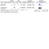
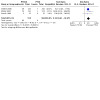
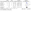
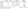
2. Descriptive characteristics of randomised controlled trials.
| Study ID | Location | Dates of enrolment | Sample size | Age (years) mean (SD)† | Males (%) † | Follow‐up |
| ARTIST | Europe; multicentre | Unknown | 298 | 61 (11) | 97.9 | 6 months |
| COBRA | Germany; multicentre | May 1992 to May 1996 | 502 | PTCRA = 62 (9); PTCA = 61 (9) |
PTCRA = 74; PTCA = 73 |
6 months |
| DART | USA; multicentre | Jun 1995 to Jun 1996 | 447 | PTCRA = 61 (10); PTCA = 61 (11) |
PTCRA = 60; PTCA = 70.0 |
6 months to 1 year |
| EDRES | Saudi Arabia; single centre | To Feb 1997 | 150 | Unknown | PTCRA = 86.7; PTCA = 88.0 |
6 months |
| Eltchaninoff | France; single centre | Unknown | 50 | PTCRA = 61 (11); PTCA = 56 (11) |
PTCRA = 81.8; PTCA = 91.7 |
In‐hospital |
| ERBAC | Germany; single centre | Oct 1991 to Aug 1991; Jan 1993 to Dec 1993 |
685 | PTCRA = 61.6 (10); PTCA = 62.5 (9.5); ELCA = 61.7 (8.8) |
PTCRA = 79.6; PTCA = 72.0; ELCA = 77.6 |
6 months to 1 year |
| Guerin | France; single centre | Apr 1992 to Sep 1993 | 64 | PTCRA = 64.6 (10.8); PTCA = 63.3 (10.4) |
PTCRA = 78.1; PTCA = 71.9 |
6 months |
| Kwon | Korea; single centre | Apr 1999 to Jan 2001 | 41 | PTCRA = 62.5 (8.6); PTCA = 61.6 (10.4) |
PTCRA = 61.6; PTCA = 70.0 |
In‐hospital 1 month 3 months 6 months |
| Lee | Korea; single centre | Jun 2001 to Jan 2003 | 113 | PTCRA = 58 (9); PTCA = 59 (10) |
PTCRA = 36.0; PTCA = 37.0 |
In‐hospital 6 months |
| Mehran | Unknown | Unknown | 249 | PTCRA = 62 (13); ELCA = 63 (11) |
PTCRA = 68.1; ELCA = 67.7 |
‐ |
| ROSTER | USA; single centre | Unknown | 200 | Unknown | Unknown | In‐hospital |
| SPORT | USA | ‐ | 675 | PTCRA = 63.6; PTCA = 64.4 |
PTCRA = 68.0; PTCA = 69.9 |
In‐hospital |
ELCA: excimer laser coronary angioplasty; PTCA: percutaneous transluminal coronary angioplasty; SD: standard deviation; PTCRA: percutaneous transluminal coronary rotational atherectomy. † Information is given for intervention and comparison groups, where available. In one case (ARTIST), total population figures are given.
1.
Risk of bias graph: review authors' judgements about each risk of bias item presented as percentages across all included studies.
2.
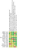
Risk of bias summary: review authors' judgements about each risk of bias item for each included study.
Allocation
The process of randomisation was described in four studies (Guerin 1996; ERBAC 1997; COBRA 2000; Kwon 2003), all of which used computer‐generated random sequences of numbers. However, only ERBAC 1997 described the concealment process used. Overall, there were insufficient data to indicate whether studies were high or low risk for selection bias.
Patient baseline characteristics
Patient criteria for enrolment differed among the trials (Table 3). Most specified that a certain degree of occlusion of the target vessel had to be present. ARTIST 2001 required symptomatic diffuse in stent re‐stenosis at least three months after stent implantation. ROSTER 2000 and SPORT 2000 did not report any specific entry criteria. Eltchaninoff 1997, Kwon 2003 and Lee 2005 required a reduction in luminal area of more than 50%, Guerin 1996 set the lower limit at 60%, and COBRA 2000 specified a range from 70% to 99%. Other lesion characteristics that differed among the studies were presence of ostial or bifurcational lesions (included in COBRA 2000 but excluded by Guerin 1996, Eltchaninoff 1997 and ERBAC 1997) and different angulation and size criteria. Patients' demographics were relatively similar between studies, with similar proportions of male patients.
3. Patient criteria in randomised controlled trials.
| Study | Patient criteria |
| ARTIST | Symptomatic, diffuse in‐stent re‐stenosis (10 to 50 mm in length) at least 3 months after stent implantation |
| COBRA | Patients aged 20 to 80 years with angiographically documented coronary artery disease and clinical symptoms of angina or anginal equivalents. The target coronary stenosis was considered haemodynamically significant and eligible for the study if there was a reduction in luminal area of 70% to 99% and absolute stenosis diameters were < 1 mm for a length of at least 5 mm as visually estimated by the operator. In addition, 1 secondary criterion had to be fulfilled, such as a heavily calcified, ostial or bifurcation location, or 1 that was eccentric, diffuse or within an angulated (> 45°) segment. Exclusions: unstable angina, MI within the previous 4 weeks, previous coronary angioplasty of the target vessel within the last 2 months, poor left ventricular function (ejection fraction ≤ 30%), or any other condition that will limit long‐term prognosis |
| DART | No specific entry criteria reported. Tested effectiveness of rotational atherectomy versus PTCA in vessels < 3 mm |
| EDRES | No specific entry criteria reported |
| Eltchaninoff | Patients were eligible for the study if they had stable or unstable angina with at least 1 lesion (> 50% stenosis) in a native vessel suitable for angioplasty. Additional inclusion criteria for angioscopy were coronary artery lumen diameter between 2.5 and 3.5 mm; location of the target lesion in a straight segment of the artery; location of the lesion at least 20 mm away from the coronary ostium; absence of left main coronary artery disease. Exclusions: acute MI within 24 hours before the procedure, a re‐stenotic lesion, a total occlusion or a vein graft lesion |
| ERBAC | Patients were included if they had target lesions and vessels suitable for all 3 techniques. Patients with multi‐vessel coronary disease were also eligible, but the culprit lesion was specified as the target before coronary intervention began. Exclusions: lesion characteristics (stenosis angulation > 60°, bend stenosis with an outwardly eccentric lumen, and bifurcational lesions requiring double guidewires) and vessel (extreme proximal vessel tortuosity, saphenous bypass graft or presence of intraluminal thrombus (filling defect), and total occlusion deemed not transferable with guidewires). Patients with acute MI and those who had undergone PTCA of any other vessel within the last 4 months were also excluded |
| Guerin | Patients presenting with a significant stenosis (defined as > 60% reduction of the lumen diameter as assessed by quantitative computed angiography) in ≥ 1 major coronary vessels, a clinical indication for re‐vascularisation, and a left ventricular ejection fraction > 40%. Exclusions: MI within the last month, re‐stenosis, bypass graft lesions, presence of intraluminal defect, ostial lesions and total occlusions |
| Kwon | Patients with long, 'denovo lesions'. Lesion length > 20 mm, stenosis diameter > 50% in a native left anterior descending artery between 2 and 2.9 mm in size. Exclusions: contraindication to antiplatelet therapy, total occlusion, infarct‐related artery, left ventricular dysfunction (ejection fraction < 40%) or an inability to follow the protocol |
| Lee | Diffuse in‐stent re‐stenosis (lesion length > 10 mm, diameter stenosis N50%) in a native coronary artery with angina, demonstrable myocardial ischaemia |
| ROSTER | No specific entry criteria reported. Tested effectiveness of PTCRA versus PTCA in diffuse in‐stent re‐stenosis |
| SPORT | No specific entry criteria reported |
MI: myocardial infarction; PTCA: percutaneous transluminal coronary angioplasty; PTCRA: percutaneous transluminal coronary rotational atherectomy.
Blinding
Most of the studies did not provide enough information to determine the strategies used to mask patients or investigators or to determine whether analysis was conducted according to originally assigned groups, although four studies (Guerin 1996; DART 1997; Eltchaninoff 1997; Kwon 2003) mentioned that assessors of angiographic outcomes were unaware of treatment allocation. The ERBAC 1997 study reported that all analyses were conducted using the "intention‐to‐treat principle". The ARTIST 2001 and Mehran 2000 trials mentioned that researchers were not blinded to the treatments arms that participants were allocated to. However, these two studies have been classified as low risk as the outcome measures were objective measurements and would not be influenced by the lack of blinding.
Incomplete outcome data
More than half of the studies available in full text reported minimal or no losses to follow‐up. There were six exceptions: DART 1997; COBRA 2000; ROSTER 2000; SPORT 2000; ARTIST 2001; Lee 2005. These trials had up to 40% of participants lost to follow‐up without explanation for the cause of loss.
Selective reporting
Overall, all studies had a low risk of reporting bias, except for EDRES 1997 and SPORT 2000, which were only available for data extraction in abstract form.
Other potential sources of bias
Only three studies (DART 1997; Eltchaninoff 1997; ERBAC 1997) reported details about the type of lesion according to ACC/AHA criteria.
Effects of interventions
Effectiveness in non‐complex coronary artery lesions
There is a general lack of available evidence examining the effectiveness of PTCRA on non‐complex lesions of the coronary arteries due largely to operator preference for lesions with more complex morphological characteristics (Zaacks 1998), and only one study was included in this review.
DART 1997 reported target re‐stenosis rates at one year of 25% and 23% for each of the rotational atherectomy and balloon angioplasty arms, respectively, indicating that re‐vascularisation in non‐complex coronary artery lesions are similar between treatments.
Effectiveness in complex coronary artery lesions
Six studies (Guerin 1996; ERBAC 1997; COBRA 2000; Mehran 2000; Kwon 2003; Lee 2005) restricted enrolment to patients with complex lesions. While using entry criteria that allowed the inclusion of patients with non‐complex (Type A) lesions, two studies (Eltchaninoff 1997; ERBAC 1997) were included in this analysis because neither reported separate results according to lesion type. Moreover, the proportion of patients with such lesions was small. In the study by Eltchaninoff 1997, five out of 50 patients (10%) had Type A lesions while ERBAC 1997 reported that 21 of 685 participants (3%) had non‐complex lesions. If lesion type is associated with specific outcomes according to treatment received, the magnitude or direction of systematic deviation of effect estimates cannot be measured without additional information, although small proportions may attenuate this potential bias.
Participants in the PTCRA groups had a mean age of 61.7 (standard deviation (SD) 9.7) years, although the groups enrolled in Eltchaninoff 1997 and Lee 2005 were younger than the other groups. About 20% of the total PTCRA group reported suffering from unstable angina, although the proportion of such patients from the Lee 2005, Kwon 2003 and Guerin 1996 trials were greater than this. There were no differences in the distributions of males, pre‐existing diabetes, previous MI and previous CABG surgery. The group receiving PTCA alone did not show significant differences in the distributions of age, sex, pre‐existing diabetes or previous CABG surgery. Lee 2005 and Mehran 2000 reported different proportions of patients with MI in both arms of treatment, with Lee 2005 reporting much lower rates of previous MI and Mehran 2000 reporting nearly double the number of participants with previous MI compared to other trials.
Angiographic baseline characteristics were relatively more heterogeneous than clinical characteristics. Overall, lesions were more commonly located in the left anterior descending artery (in about 45% of cases), followed by the right coronary artery (about 25% of cases) and the left circumflex artery (in about 20% of cases). However, in the trial by Lee 2005, they observed stenosis in the left anterior descending artery in about 70% of all participants, about 17% in the right coronary artery followed by about 10% in the left circumflex.
The morphology of vascular lesions was described in three studies (Guerin 1996; ERBAC 1997; COBRA 2000), although the definitions used to determine whether lesions met certain criteria were not. For the most part, the COBRA 2000 and ERBAC 1997 studies enrolled participants with similar morphological features: about two in five had calcified lesions; four in five, eccentric lesions; three in five, lesions of less than 10 mm; and about 15% were angulated beyond 45 degrees. The patients enrolled in Guerin 1996 showed different characteristics. Statistically significant differences in the diameter, length and per cent stenosis of the arteries were also apparent.
Five studies reported re‐stenosis rates at follow‐up (Guerin 1996; ERBAC 1997; COBRA 2000; Kwon 2003; Lee 2005). The outcome was defined similarly across all five studies: at least 50% stenosis determined angiographically at follow‐up. The COBRA 2000, Guerin 1996, Kwon 2003 and Lee 2005 studies provided data at six months of follow‐up while the ERBAC 1997 study presented information up to one year.
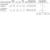
Overall, there were no statistically significant differences in the re‐stenosis rates at six months in the group receiving PTCRA with adjunctive PTCA compared to the group receiving PTCA alone (RR 1.05; 95% CI 0.83 to 1.33) with no substantial statistical heterogeneity apparent (Chi2 0.78; degrees of freedom (df) = 4; P = 0.94). At one year, PTCRA was associated with a 21% increase in re‐stenosis rate compared to PTCA, although the result was not statistically significant (RR 1.21; 95% CI 0.95 to 1.55). The results of one trial were not included due to the lack of primary data (EDRES 1997).
Particular morphological characteristics that distinguish specific complex lesions (lesions that are calcified, ostial, angulated or diffuse) have not been examined in parallel‐arm RCTs. The BAROCCO 1995 trial focused on chronic total occlusions, but the study was excluded from consideration because it was a cross‐over trial.
Effectiveness in in‐stent re‐stenosis
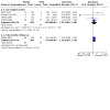
The ROSTER 2000 trial reported the results of PTCRA versus PTCA in the treatment of diffuse in‐stent re‐stenosis. The trial enrolled 200 patients assigned in equal numbers to the two procedures. The results show that mean pre‐ and post‐procedural minimum luminal diameters (MLDs) and the gain in luminal diameter following the placement of the stent were comparable between the groups. Prior to the treatment for in‐stent re‐stenosis, the mean MLD between the groups was similar. Rotational atherectomy produced a smaller residual intimal hyperplasia area compared to PTCA. The trial also reported 12‐month follow‐up results indicating the rates of clinical re‐stenosis following PTCRA were statistically significantly reduced. Clinical re‐stenosis was reported in 32% of patients receiving PTCRA compared to 45% receiving PTCA (P < 0.05). The authors did not provide enough information to calculate 95% CIs.
The results for the re‐stenosis rate of the ROSTER 2000 trial contrast with those from the ARTIST 2001 trial. The latter study enrolled 298 patients with symptomatic, diffuse in‐stent re‐stenosis. Participants were assigned to PTCRA with low‐pressure PTCA (N = 152) or PTCA alone (N = 146). Post‐procedural angiographic success (defined as residual stenosis of < 50%) was 94% for PTCRA and 95% for PTCA. MLD and luminal gain were likewise similar between the groups (MLD: PTCRA 1.9 mm (SD 0.4); PTCA 1.9 mm (SD 0.3)). After six months of follow‐up, those receiving PTCA alone were noted to have more favourable outcomes in terms of event‐free survival (91.1% vs. 79.6%; P = 0.005), MLD (1.2 mm (SD 0.6) vs. 1.0 mm (SD 0.6); P = 0.008), residual stenosis (56% (SD 20%) vs. 64% (SD 22%); P = 0.005), re‐stenosis rate (51.2% vs. 64.8%; P = 0.04) and rates of target lesion re‐vascularisation (36.2% vs. 47.8%; P = 0.06).
The results of these RCTs should be interpreted with care given that important information (e.g. the patient population, quality domains, etc.) was not available. Moreover, the distributions of potential confounding factors were undescribed. Although the common expectation is that measured and unmeasured characteristics will be equally distributed between the groups, no information is presented to support this position. Procedural characteristics also differ between the two studies. Given the potential for significant clinical heterogeneity to be present, it was inappropriate to present pooled results.
MACE
MACE defined as MI, emergency CABG or death were reported by all studies as in‐hospital events and by a subset during follow‐up.
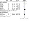
Composite MACE
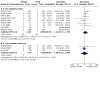
As a composite end point, MACE was reported by four studies (DART 1997; Eltchaninoff 1997; ERBAC 1997; SPORT 2000). Rotational atherectomy with adjunctive angioplasty was not associated with a statistically significant increase in the risk of in‐hospital MACE compared to angioplasty alone (RR 1.27; 95% CI 0.86 to 1.90). No substantial statistical heterogeneity was detected (Chi2 2.31; df = 3; P = 0.51). Data from ERBAC 1997 indicated that rotational atherectomy with adjunctive PTCA was associated with a 25% increase in the risk of MACE at six months. The increase was of borderline statistical significance (RR 1.25; 95% CI 0.99 to 1.59). In determining the composite end point MACE, ERBAC 1997 included the number of repeat surgical and non‐surgical interventions. It is unclear whether the investigators counted all interventions (which could conceivably occur more than once in a single patient) towards the total or whether an indicator variable was used (to specify the presence of any intervention regardless of the total number received).
Aspects of MACE were separated in order to determine whether the technology was preferentially affecting a particular outcome.
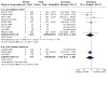
Myocardial infarction
The definition of MI used in each of the studies is shown in Table 4. All six studies (ERBAC 1997; COBRA 2000; Mehran 2000; ARTIST 2001; Kwon 2003; Lee 2005) that defined the event used a definition with at least two components: serum creatine kinase and electrocardiographic findings. Both ARTIST 2001 and ERBAC 1997 required a rise in creatine kinase of twice the normal level, COBRA 2000, Lee 2005 and Kwon 2003 used a level that was three times the normal limit, while Mehran 2000 used a level that was five times the normal limit. Eltchaninoff 1997 and Guerin 1996 failed to specify the definition of MI used.
4. Definitions of myocardial infarction (MI) used in the RCTs.
| Study | Definition of MI |
| ARTIST | New Q waves or creatine kinase and creatine kinase MB greater than twice normal, or both |
| COBRA | Rise in creatine kinase of more than 3 times the normal limit in the presence of Q waves |
| Eltchaninoff | Not stated. Standard 12‐lead electrocardiogram and serial measurement of total and MB fraction of creatine kinase was performed while in hospital |
| ERBAC | New Q waves in ≥ 2 contiguous leads and a creatine kinase elevation of 2 or more times the upper limit of normal and/or elevated creatine kinase‐MB fraction to at least twice the upper limit of normal |
| Guerin | Not stated. Electrocardiographic descriptors used |
Assuming there is little clinical dissimilarity in the variations in the definitions used, the estimate of the pooled effect showed a non‐statistically significant 42% increase in the risk of MI during the in‐hospital period in those receiving PTCA (RR 1.42; 95% CI 0.75 to 2.70) with no statistically significant heterogeneity present (Chi2 2.95; df = 6; P = 0.81). The risk of MI six months after the procedure did not show a statistically significant difference between the two groups (RR 1.08; 95% CI 0.35 to 3.40).
Emergency CABG
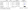
Rotational atherectomy with adjunctive PTCA was associated with a non‐statistically significant 21% increase in the risk of emergency CABG during the in‐hospital period compared to PTCA alone (RR 1.21; 95% CI 0.43 to 3.40). While statistical heterogeneity was not present (Chi2 5.94; df = 5; P = 0.31) the Guerin 1996 and Mehran 2000 studies seemed to indicate the presence of a protective effect induced by the PTCRA‐PTCA combination (although, admittedly, the study was small). Results from Eltchaninoff 1997, Lee 2005 and Kwon 2003 were excluded because no outcomes were observed. The risk of emergency CABG six months after the procedure did not show a statistically significant difference between the two groups (RR 0.93; 95% CI 0.54 to 1.61).
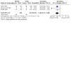
Death
The risk of mortality while in hospital was reduced by nearly half in the group receiving PTCRA‐PTCA compared to those receiving PTCA alone, although the result failed to reach statistical significance (RR 0.67; 95% CI 0.22 to 2.05). This trend was also present at six months of follow‐up (RR 0.67; 95% CI 0.21 to 2.06) in which out‐of‐hospital deaths were included.
Safety and adverse events
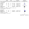
Patients undergoing PTCRA were nine times as likely to experience an angiographically detectable vascular spasm (RR 9.23; 95% CI 4.61 to 18.47), four times as likely to experience a perforation (RR 1.287; 95% CI 0.92 to 19.83) and about twice as likely to have transient vessel occlusions (RR 2.49; 95% CI 1.25 to 4.99) as those receiving PTCA alone. Both angiographic dissections (RR 0.48; 95% CI 0.34 to 0.68) and the use of stents as a bailout procedure (RR 0.29; 95% CI 0.09 to 0.87) were less common in the group receiving PTCRA. Admittedly, for some outcomes, the 95% CIs of the point estimates include 1.0. However, these same intervals include clinically relevant risks that would benefit from further investigation.
'Slow flow' or 'no flow' is an adverse outcome that is recognised by the reduction or absence of antegrade blood flow distal to a specific segment not attributable to abrupt closure, high‐grade stenosis or spasm of the target lesion (Abbo 1995). Reisman suggested that this might be due to a large plaque burden being delivered to the distal vascular bed by the ablative action of the device (Reisman 1994). Patients undergoing PTCRA experienced this outcome nearly three times more often than those undergoing PTCA.
Discussion
There is little evidence to support the routine use of PTCRA for non‐complex lesions when PTCA, with or without stenting. There is a lack of evidence from RCTs about the effectiveness of PTCRA in complex lesions. Moreover, the evidence for its use in in‐stent re‐stenosis is equivocal.
The refinement of technology, the continued development of technique and the use of adjunctive medications have made the presence of vascular spasms, slow/no flow phenomena and transient vessel occlusions manageable or avoidable. Although beyond the scope of this evaluation, results from published studies that have examined procedural aspects of the device (i.e. burr speed, adjunctive drugs, procedural time, etc.) have led to reductions in the severity and incidence of these events. In addition, the advent of newer agents that modify platelet function (glycoprotein IIb/IIIa and adenosine diphosphate (ADP) antagonists) may contribute to better outcomes over time (Presbitero 2002; Tsubokawa 2002).
Regarding whether the technology has a place in the current medical armamentarium, the answer is equally equivocal. For instance, mechanistically the action of the rotating burr may have benefits in calcified lesions. These are difficult to treat and represent both a therapeutic and an operator challenge due to their morphology and anatomical placement within coronary arteries (e.g. at ostial sites). Re‐vascularisation procedures involving these lesions must be carefully managed for several reasons:
it may be difficult to pass a guidewire across such lesions and deploy an interventional catheter;
assuming a guidewire can be passed, PTCA (with or without stenting) is often unsuccessful because lesion architecture does not allow sufficient balloon expansion to permit a sustained increase in lumen diameter;
if balloon expansion is possible with standard PTCA (with or without stenting), uneven deployment, as a result of anatomy and architecture, may result in parts of the lesions breaking off and embolising downstream and/or exposing underlying atheromatous material to the blood stream resulting in thrombosis.
Moreover, technical failures may arise during balloon angioplasty due to a failure to cross the lesion with the balloon catheter or to inflate the balloon due to rigidity of the lesion. In some lesions, PTCA may be avoided altogether because of unfavourable findings on coronary angiography. PTCRA may be considered in the management of lesions refractory to PTCA or for those patients where the only feasible alternative is CABG. Such patients may not cope well with the rigours of open‐chest surgery, particularly those with underlying comorbidities that would be contraindicated for such a procedure. It is these patients who may be best‐served with PTCRA as an alternative. By partial ablation of the lesion, PTCRA may result in a substantial decrease in plaque burden so that other techniques are unnecessary or may alter plaque morphology such that other adjunctive techniques may be used.
Needless to say, there are currently no trials supporting this view. It is unlikely that high‐level evidence will be collected to examine the effectiveness of PTCRA on such lesions (and in other circumstances) given that it might not be feasible to construct a clinical trial that compares PTCRA of these lesions with an appropriate comparator. PTCA is not appropriate (as per above), while CABG surgery may be. However, it may be unlikely that CABG surgery will be undertaken for single vessel disease, particularly if a patient has significant underlying comorbidities that are, of themselves, contraindicated for CABG surgery (e.g. advanced age, severe respiratory or cardiac disease, renal failure, dialysis, diabetes).
Limitations of this meta‐analysis include population differences, study quality and lack of information. Evidence exists that the RCTs enrolled groups with clinically and angiographically dissimilar characteristics (at least within statistically relevant bounds). The interpretation of effect size summaries, therefore, must take into account the presence of potentially sizeable systematic deviations compared to similar effect sizes derived from studies without such dissimilar groups. To some extent, the use of random‐effects models to obtain summary effect size estimates will generally produce more conservative results, but this is not always the case (Poole 1999) and it is prudent to moderate inferences drawn from such methods with the knowledge that the potential for bias is present. This is especially relevant to the re‐stenosis end points, since there is evidence of between‐study differences in specific baseline characteristics. An ITT analysis was planned for this review; however, a number of papers had significant loss to follow‐up, which raises issues about the veracity of this analysis. First, it raises the issue of whether the data are reliable – if there is a significant proportion of participants lost, one can question whether the result demonstrates an indication of the true effect. Second, a high drop‐out rate may be indicative of a poor‐quality study. Even though the data from these papers were extracted and analysed for this review, outcomes from this data should be interpreted with an awareness of the possibility that the values may not reflect a true result.
The range of outcomes available for extraction was limited. Important end points that enhance the capacity to use the information in decision‐making, such as cost or quality of life, were unavailable.
Authors' conclusions
Implications for practice.
When conventional PTCA, with or without stent placement, is feasible, PTCRA appears to confer no additional benefit to the patient. In cases of in‐stent re‐stenosis, there is limited and conflicting published evidence and no long‐term data to support the routine use of rotational atherectomy. In certain circumstances, PTCRA may be a useful adjunctive procedure to increase the success of subsequent angioplasty in achieving satisfactory re‐vascularisation in complicated or calcified lesions.
Implications for research.
In specific cases where conventional angioplasty and stenting cannot successfully be undertaken or is associated with a poor clinical or angiographic outcome, PTCRA appears to be an effective adjunctive procedure to increase the likelihood of successful re‐vascularisation. Further information is needed to verify this position. In addition, the range of outcomes studied in future trials needs to be broadened in order to make the findings directly useful in decision‐making.
What's new
| Date | Event | Description |
|---|---|---|
| 10 March 2021 | Review declared as stable | The authors of the review have identified two additional studies (ROTAXUS; PROTECT II) for inclusion (up to date to September 2019) but concluded that they would not change the findings of the review. There is a large amount of non‐Cochrane evidence published and newer techniques are being assessed. |
History
Protocol first published: Issue 4, 2001 Review first published: Issue 4, 2003
| Date | Event | Description |
|---|---|---|
| 12 November 2012 | New citation required but conclusions have not changed | New search led to inclusion of 3 new studies |
| 21 August 2012 | New search has been performed | Three new studies (Kwon 2003; Lee 2005; Mehran 2000) have been included in this review update. Updated publications were also found for ARTIST and DART. Although the relative risks have changed slightly for some outcomes, the conclusions have not changed. Background section has been updated. The search strategies used were the dates over which the databases were searched were updated. In this update, we have used a new risk of bias tool to assess the quality of publications. The conclusion of this updated review have not changed from the original review. |
| 9 September 2008 | Amended | Converted to new review format. |
| 1 August 2003 | New citation required and conclusions have changed | Substantive amendment |
Acknowledgements
The authors would like to thank Elmer Villaneuva and Emily Petherick for their work on the original review as well as Joanne Abbott, Trials Search Co‐ordinator, for her assistance with respect to the search strategy utilised in this review.
Appendices
Appendix 1. Search strategies
CENTRAL (The Cochrane Library)
#1 MeSH descriptor Atherectomy, Coronary, this term only
#2 (rotablat*)
#3 (rotat* near/5 atherectom*)
#4 (ptcra)
#5 (#1 OR #2 OR #3 OR #4)
MEDLINE (OVID)
1 Atherectomy, Coronary/
2 rotablat$.tw.
3 (rotat$ adj5 atherectom$).tw.
4 ptcra.tw.
5 or/1‐4
6 randomized controlled trial.pt.
7 controlled clinical trial.pt.
8 randomized.ab.
9 placebo.ab.
10 drug therapy.fs.
11 randomly.ab.
12 trial.ab.
13 groups.ab.
14 6 or 7 or 8 or 9 or 10 or 11 or 12 or 13
15 exp animals/ not humans.sh.
16 14 not 15
17 5 and 16
EMBASE (OVID)
1 Atherectomy, Coronary/
2 rotablat$.tw.
3 (rotat$ adj5 atherectom$).tw.
4 ptcra.tw.
5 or/1‐4
6 random$.tw.
7 factorial$.tw.
8 crossover$.tw.
9 cross over$.tw.
10 cross‐over$.tw.
11 placebo$.tw.
12 (doubl$ adj blind$).tw.
13 (singl$ adj blind$).tw.
14 assign$.tw.
15 allocat$.tw.
16 volunteer$.tw.
17 crossover procedure/
18 double blind procedure/
19 randomized controlled trial/
20 single blind procedure/
21 6 or 7 or 8 or 9 or 10 or 11 or 12 or 13 or 14 or 15 or 16 or 17 or 18 or 19 or 20
22 (animal/ or nonhuman/) not human/
23 21 not 22
24 5 and 23
CINAHL (EBSCO)
#1 Atherectomy, Coronary,
#2 (rotablat*)
#3 (rotat* N5 atherectom*)
#4 (ptcra)
#5 (#1 OR #2 OR #3 OR #4)
Data and analyses
Comparison 1. 'Non‐complex' coronary lesions.
| Outcome or subgroup title | No. of studies | No. of participants | Statistical method | Effect size |
|---|---|---|---|---|
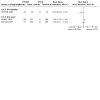
| 1.1 Procedural success | 1 | Risk Ratio (M‐H, Random, 95% CI) | Subtotals only |
1.1. Analysis.
Comparison 1: 'Non‐complex' coronary lesions, Outcome 1: Procedural success
Comparison 2. Complex coronary lesions.
| Outcome or subgroup title | No. of studies | No. of participants | Statistical method | Effect size |
|---|---|---|---|---|
| 2.1 Re‐stenosis rates | 5 | Risk Ratio (M‐H, Random, 95% CI) | Subtotals only | |
| 2.1.1 Six months | 5 | 855 | Risk Ratio (M‐H, Random, 95% CI) | 1.05 [0.83, 1.33] |
| 2.1.2 One year | 1 | 254 | Risk Ratio (M‐H, Random, 95% CI) | 1.21 [0.95, 1.55] |
2.1. Analysis.
Comparison 2: Complex coronary lesions, Outcome 1: Re‐stenosis rates
Comparison 3. In‐stent re‐stenosis.
| Outcome or subgroup title | No. of studies | No. of participants | Statistical method | Effect size |
|---|---|---|---|---|
| 3.1 Re‐stenosis rates | 3 | Risk Ratio (M‐H, Random, 95% CI) | Totals not selected | |
| 3.1.1 Six months | 1 | Risk Ratio (M‐H, Random, 95% CI) | Totals not selected | |
| 3.1.2 One year | 2 | Risk Ratio (M‐H, Random, 95% CI) | Totals not selected | |
| 3.2 Minimum luminal diameter | 4 | Mean Difference (IV, Random, 95% CI) | Totals not selected | |
| 3.2.1 Six months | 2 | Mean Difference (IV, Random, 95% CI) | Totals not selected | |
| 3.2.2 One year | 2 | Mean Difference (IV, Random, 95% CI) | Totals not selected |
3.1. Analysis.
Comparison 3: In‐stent re‐stenosis, Outcome 1: Re‐stenosis rates
3.2. Analysis.
Comparison 3: In‐stent re‐stenosis, Outcome 2: Minimum luminal diameter
Comparison 4. Major adverse cardiac events.
| Outcome or subgroup title | No. of studies | No. of participants | Statistical method | Effect size |
|---|---|---|---|---|
| 4.1 MACE as a composite event | 4 | Risk Ratio (M‐H, Random, 95% CI) | Subtotals only | |
| 4.1.1 In‐hospital period | 4 | 1315 | Risk Ratio (M‐H, Random, 95% CI) | 1.27 [0.86, 1.90] |
| 4.1.2 Six months' follow‐up | 1 | 396 | Risk Ratio (M‐H, Random, 95% CI) | 1.25 [0.99, 1.59] |
| 4.2 Myocardial infarction | 9 | Risk Ratio (M‐H, Random, 95% CI) | Subtotals only | |
| 4.2.1 In‐hospital period | 9 | 2218 | Risk Ratio (M‐H, Random, 95% CI) | 1.42 [0.75, 2.70] |
| 4.2.2 Six months' follow‐up | 3 | 932 | Risk Ratio (M‐H, Random, 95% CI) | 1.08 [0.35, 3.40] |
| 4.3 Emergency CABG | 9 | Risk Ratio (M‐H, Random, 95% CI) | Subtotals only | |
| 4.3.1 In‐hospital period | 9 | 2218 | Risk Ratio (M‐H, Random, 95% CI) | 1.21 [0.43, 3.40] |
| 4.3.2 Six months' follow‐up | 2 | 819 | Risk Ratio (M‐H, Random, 95% CI) | 0.93 [0.54, 1.61] |
| 4.4 Death | 9 | Risk Ratio (M‐H, Random, 95% CI) | Subtotals only | |
| 4.4.1 In‐hospital period | 9 | 2218 | Risk Ratio (M‐H, Random, 95% CI) | 0.67 [0.22, 2.05] |
| 4.4.2 Six months' follow‐up | 3 | 932 | Risk Ratio (M‐H, Random, 95% CI) | 0.67 [0.21, 2.06] |
4.1. Analysis.
Comparison 4: Major adverse cardiac events, Outcome 1: MACE as a composite event
4.2. Analysis.
Comparison 4: Major adverse cardiac events, Outcome 2: Myocardial infarction
4.3. Analysis.
Comparison 4: Major adverse cardiac events, Outcome 3: Emergency CABG
4.4. Analysis.
Comparison 4: Major adverse cardiac events, Outcome 4: Death
Comparison 5. Adverse events.
| Outcome or subgroup title | No. of studies | No. of participants | Statistical method | Effect size |
|---|---|---|---|---|
| 5.1 Perforation | 5 | 1948 | Risk Ratio (M‐H, Random, 95% CI) | 4.28 [0.92, 19.83] |
| 5.2 Angiographic dissection | 3 | 949 | Risk Ratio (M‐H, Random, 95% CI) | 0.48 [0.34, 0.68] |
| 5.3 Bailout stenting | 2 | 955 | Risk Ratio (M‐H, Random, 95% CI) | 0.29 [0.09, 0.87] |
| 5.4 Spasm | 3 | 1019 | Risk Ratio (M‐H, Random, 95% CI) | 9.23 [4.61, 18.47] |
| 5.5 Transient vessel occlusion | 5 | 1700 | Risk Ratio (M‐H, Random, 95% CI) | 2.49 [1.25, 4.99] |
| 5.6 'Slow flow' | 4 | 1442 | Risk Ratio (M‐H, Random, 95% CI) | 2.68 [1.28, 5.59] |
5.1. Analysis.
Comparison 5: Adverse events, Outcome 1: Perforation
5.2. Analysis.
Comparison 5: Adverse events, Outcome 2: Angiographic dissection
5.3. Analysis.
Comparison 5: Adverse events, Outcome 3: Bailout stenting
5.4. Analysis.
Comparison 5: Adverse events, Outcome 4: Spasm
5.5. Analysis.
Comparison 5: Adverse events, Outcome 5: Transient vessel occlusion
5.6. Analysis.
Comparison 5: Adverse events, Outcome 6: 'Slow flow'
Characteristics of studies
Characteristics of included studies [ordered by study ID]
ARTIST 2001.
| Study characteristics | ||
| Methods | RCT | |
| Participants | Group 1 PTCRA: N = 152 Group 2 PTCA: N = 146 Inclusions: angina or objective evidence of target vessel‐related ischaemia, or both; documented ISR > 70% by visual assessment within a stent ± 5 mm of the stent edges, stent diameter ≥ 2.5 mm (balloon during implantation), ISR as the only lesion for treatment, length of ISR of 10 to 50 mm by visual assessment, and lesion accessible for rotablation Exclusions: acute MI within the previous month, left ventricular ejection fraction < 30%, evidence of intraluminal thrombus or dissection, unprotected ostial stenoses, missing visualisation of the distal lumen after crossing with a guidewire, stents obviously not fully expanded, stents at or directly distal to a bend > 45°, stents implanted within the previous 3 months, and stents with a classic coil design that might impair QCA |
|
| Interventions | Group 1: the final burr‐to‐artery (stent) ratio was ≥ 0.7. Adjunctive PTCA was performed with a balloon 0.25 to 0.5 mm larger than during stent implantation with a pressure of ≤ 6 atm. If the investigator was not satisfied with the angiographic result, higher pressures in 2‐atm steps were allowed. The use of ≥ 160,000 rpm was initially proposed, during the study, this was changed to ≥ 140,000 rpm. Operators were urged to avoid drops ≥ 5000 rpm Group 2: balloon angioplasty was performed with locally customised balloon catheters. The balloon‐to‐artery ratio should be ≥ 1.0; the inflation pressure used was at the discretion of the investigator to achieve a final diameter stenosis of < 30% Pre‐medication: patients received a bolus of heparin 10,000 to 15,000 IU before the intervention. Supplementary heparin was used under activated clotting time monitoring (≥ 250 s). All patients were continuously treated with of acetylsalicylic acid (aspirin) 100 mg for 6 months and ticlopidine 250 mg twice daily, or clopidogrel 75 mg once daily for ≥ 2 weeks after the procedure |
|
| Outcomes | Primary study end point: minimum lumen diameter assessed from quantitative angiography at 6 months after treatment Secondary end point: safety and efficacy (short‐term success), event‐free survival and re‐stenosis (> 50% diameter reduction) of the target lesion after 6 months |
|
| Notes | ‐ | |
| Risk of bias | ||
| Bias | Authors' judgement | Support for judgement |
| Random sequence generation (selection bias) | Unclear risk | "Balance between treatment arms within the centers was achieved by randomizing in blocks." Insufficient information |
| Allocation concealment (selection bias) | Unclear risk | Insufficient information |
| Blinding of participants and personnel (performance bias) All outcomes | Low risk | Not blinded |
| Blinding of outcome assessment (detection bias) All outcomes | Low risk | Not blinded |
| Incomplete outcome data (attrition bias) All outcomes | Unclear risk | Incomplete outcome data for both primary and secondary end points |
| Selective reporting (reporting bias) | Unclear risk | The study protocol is available and all of the study's pre‐specified (primary and secondary) outcomes that are of interest in the review have been reported in the pre‐specified way |
COBRA 2000.
| Study characteristics | ||
| Methods | RCT | |
| Participants | Group 1 PTCRA: N = 252 Group 2 PTCA: N = 250 Inclusions: patients aged 20 to 80 years with angiographically documented coronary artery disease and clinical symptoms of angina. Angiographic inclusion criteria: stenosis was considered haemodynamically significant and eligible for the study, if there was a reduction in luminal area of 70% to 99% and diameters were < 1 mm for a length of at least 5 mm. In addition at least one secondary criterion had to be fulfilled, such as heavily calcified, ostial, bifurcational location, eccentric, diffuse, within an angulated (> 45°) segment Exclusions: patients with unstable angina, MI within the previous 4 weeks, previous coronary angioplasty of the target vessel within the last 2 months, poor left ventricular function (ejection fraction ≤ 30%) or any other condition that will limit long‐term prognosis were excluded from the study |
|
| Interventions | Group 1: PTCRA was performed with burr sizes from 1.25 to 2.5 mm, recommended burr speed was 160,000 to 190,000 rpm with each sequence being < 30 s; intracoronary nitroglycerin 100 to 200 µg was administered after each sequence. It was advised to use incremental burr sizes to achieve a burr‐to‐artery ratio of at least 0.7. The decision about performing an adjunctive PTCA at low pressure (< 4 atm) was left to the operator Group 2: PTCA was performed with balloon lengths of 20 to 40 mm. The technique to achieve an optimal angiographic result was left to the operator. The use of stents for bail out (flow‐limiting dissections, severe recoil, vessel closure) or unsatisfactory results (residual diameter stenosis > 50%) was allowed, but was explicitly not encouraged Pre‐medication: included acetylsalicylic acid (aspirin). In both treatment arms IV vasodilating pre‐treatment consisted of nitroglycerin 2 to 4 mg/hour and nifedipine 0.5 to 1.5 mg/hour at least 2 hours before the intervention, accompanied by a 500‐mL saline infusion. In the catheterisation laboratory heparin was administered as a bolus of 15,000 to 20,000 units to maintain the activated clotting time above 350 s during the procedure. Intracoronary nitroglycerin was allowed according to operator judgement |
|
| Outcomes | Primary study end points: 1. procedural success, defined as angiographically confirmed residual stenoses < 50% and stenosis reduction of at least 20% in the absence of new MI, emergency CABG and death 2. 6 months' re‐stenosis in the treated segment defined as (a) > 50% diameter stenosis or (b) > 50% diameter stenosis and > 50% late loss of the acute luminal gain 3. major cardiac events during the follow‐up period Secondary end point: clinical outcome as assessed by the angina grading of the Canadian Cardiovascular Society Classification and the exercise tolerance scale of the modified Bruce protocol |
|
| Notes | ‐ | |
| Risk of bias | ||
| Bias | Authors' judgement | Support for judgement |
| Random sequence generation (selection bias) | Low risk | "Randomization was carried out with computer generated permutated blocks for each participating centre" |
| Allocation concealment (selection bias) | Unclear risk | Insufficient information |
| Blinding of participants and personnel (performance bias) All outcomes | Low risk | Insufficient information, but the review authors judge that the outcome is not likely to be influenced by lack of blinding |
| Blinding of outcome assessment (detection bias) All outcomes | Low risk | Insufficient information, but the review authors judge that the outcome is not likely to be influenced by lack of blinding |
| Incomplete outcome data (attrition bias) All outcomes | High risk | "Complete follow‐up over 6 months was available from 423/497 patients". 15% loss to follow‐up |
| Selective reporting (reporting bias) | Low risk | The study protocol is available and all of the study's pre‐specified outcomes that are of interest in the review have been reported in the pre‐specified way |
DART 1997.
| Study characteristics | ||
| Methods | RCT | |
| Participants | Group 1 PTCRA: N = 227 Group 2 PTCA: N = 219 Inclusions: target lesion on angiography of ≥ 70% diameter stenosis by careful visual estimate and evidence of myocardial ischaemia caused by the target lesion, defined as either symptoms of angina or positive results of a functional study. The lesion had to be no more than 20 mm in length within a native coronary artery with a reference diameter of 2.0 to 3.0 mm, without other haemodynamically significant stenoses in that vessel that warranted re‐vascularisation Exclusions: lesions with severe calcification, past history of 3 prior procedures involving the target lesion, contraindication to emergent CABG surgery, MI within 7 days before the procedure, rest angina unrelieved by medical therapy within the preceding 48 hours The angiographic criteria for patient exclusion were: total occlusion presence of thrombus, aorto‐ostial lesions, vessel angulation > 60°, bifurcation lesions requiring intervention on both involved vessels, prior stent re‐vascularisation proximal to the target lesion |
|
| Interventions | Group 1: a stepped burr approach was used to achieve a final burr‐to‐artery ratio of 0.70 to 0.85. A platform speed of approximately 180,000 rpm was used for burrs ≤ 2.0 mm in diameter, and 160,000 was used for burrs > 2.0 mm. It was recommended that there be no burr decelerations of > 5000 rpm. After rotational arthrectomy, in cases where a suboptimal result had been achieved, low‐pressure adjunctive balloon angioplasty with inflation pressures not exceeding 1 atm was permitted Group 2: the final balloon size was selected to provide a balloon‐to‐artery ratio of between 0.9 and 1.1. The final pressure and duration of balloon inflation were left to the discretion of the operator Pre‐medication: all patients received acetylsalicylic acid (aspirin) 325 mg orally, a calcium channel blocker and IV heparin before the start of the procedure. Intracoronary nitroglycerin was administered before baseline and after intervention angiography |
|
| Outcomes | Primary study end point: target vessel failure as defined as the composite end point of death, Q‐wave MI, and clinically driven repeat re‐vascularisation of the target vessel Secondary study end points: acute procedural success that was a composite of attainment of a < 50% diameter stenosis in the absence of in hospital major adverse cardiac events; acute device success, defined as the achievement < 50% stenosis of the target lesion without cross‐over treatment or unplanned coronary stenting; binary angiographic re‐stenosis, defined as per cent diameter stenosis > 50% at the 8‐month follow‐up; target lesion re‐vascularisation, defined as clinically driven re‐vascularisation of the target lesion; target vessel re‐vascularisation, defined as clinically driven re‐vascularisation of any lesion of the target vessel; and MI, defined as: 1) Q‐wave, the development of new, pathological Q‐waves in ≥ 2 contiguous leads with post‐procedure CK‐MB levels higher than normal, or 2) non‐Q‐wave, elevation of post‐procedure CK levels to > 2 times normal with CK‐MB levels higher than normal in the absence of new, pathological Q waves |
|
| Notes | ‐ | |
| Risk of bias | ||
| Bias | Authors' judgement | Support for judgement |
| Random sequence generation (selection bias) | Unclear risk | "Patients were randomised" |
| Allocation concealment (selection bias) | Unclear risk | Insufficient information |
| Blinding of participants and personnel (performance bias) All outcomes | Low risk | Incomplete description of blinding, but outcomes are not likely to be influenced by lack of blinding |
| Blinding of outcome assessment (detection bias) All outcomes | Low risk | "An independent clinical events committee that was unaware of each patient's treatment assignment adjudicated all major adverse cardiac events" |
| Incomplete outcome data (attrition bias) All outcomes | Unclear risk | Patient loss to follow‐up similar in both groups. Large number lost |
| Selective reporting (reporting bias) | Low risk | The study protocol is available and all of the study's pre‐specified (primary and secondary) outcomes that are of interest in the review have been reported in the pre‐specified way |
EDRES 1997.
| Study characteristics | ||
| Methods | RCT | |
| Participants | No specific entry criteria reported | |
| Interventions | Pre‐medication not stated Group 1: PTCRA with adjunctive PTCA and stent placement Group 2: PTCA with stent placement |
|
| Outcomes | ‐ | |
| Notes | Insufficient information reported, unable to obtain full text for review | |
| Risk of bias | ||
| Bias | Authors' judgement | Support for judgement |
| Random sequence generation (selection bias) | Unclear risk | Insufficient information |
| Allocation concealment (selection bias) | Unclear risk | Insufficient information |
| Blinding of participants and personnel (performance bias) All outcomes | Unclear risk | Insufficient information |
| Blinding of outcome assessment (detection bias) All outcomes | Unclear risk | Insufficient information |
| Incomplete outcome data (attrition bias) All outcomes | Unclear risk | Insufficient information |
| Selective reporting (reporting bias) | Unclear risk | Insufficient information |
Eltchaninoff 1997.
| Study characteristics | ||
| Methods | RCT | |
| Participants | Group 1 PTCRA: N = 24 Group 2 PTCA: N = 26 Inclusions: patients with stable or unstable angina with at least 1 lesion (> 50% stenosis) in a native vessel suitable for angioplasty. Additional inclusion criteria for angioscopy were coronary artery lumen diameter between 2.5 and 3.5 mm; target lesion in a straight segment of artery; lesion at least 20 mm away from the coronary ostium; absence of left main coronary artery disease Exclusions: acute MI within 24 hours before the procedure, a re‐stenotic lesion, a total occlusion, or a vein graft lesion |
|
| Interventions | Group 1: PTCRA using 8F‐9F sheath. 1 burr used per lesion with size chosen to obtain a burr‐to‐artery ratio of 0.7. Adjunctive PTCA performed after PTCRA with inflation pressure < 6 atm Group 2: PTCA using "standard techniques". 8F sheath and balloon size chosen to obtain a balloon‐to‐artery ratio of approximately 1.0 Pre‐medication: acetylsalicylic acid (aspirin); IV heparin 10,000 IU bolus and intracoronary nitroglycerin 150 mg |
|
| Outcomes | Angioscopic findings defined as: 1. Flaps, graded 1 to 3 2. Thrombi, graded 1 to 3 3. Subintimal haemorrhage 4. Longitudinal dissection Angiographic success defined as residual stenosis ≤ 50% in the absence of severe coronary artery dissection (grade D1 or higher) Clinical success defined as angiographic success in the absence of major complications, such as death, MI and bypass surgery |
|
| Notes | ‐ | |
| Risk of bias | ||
| Bias | Authors' judgement | Support for judgement |
| Random sequence generation (selection bias) | Unclear risk | "After initial angiography each patient was randomized". Insufficient information |
| Allocation concealment (selection bias) | Unclear risk | Insufficient information |
| Blinding of participants and personnel (performance bias) All outcomes | Low risk | Insufficient information, but the review authors judge that the outcome is not likely to be influenced by lack of blinding |
| Blinding of outcome assessment (detection bias) All outcomes | Low risk | Angioscopic and angiographic assessors were blinded, unclear if clinical assessors were blinded, but the review authors judge that the outcome is not likely to be influenced by lack of blinding |
| Incomplete outcome data (attrition bias) All outcomes | Low risk | No missing outcome data |
| Selective reporting (reporting bias) | Low risk | The study protocol is available and all of the study's pre‐specified outcomes that are of interest in the review have been reported in the pre‐specified way |
ERBAC 1997.
| Study characteristics | ||
| Methods | RCT | |
| Participants | Group 1 PTCRA: N = 231 Group 2 ELCA: N = 232 Group 3 PTCA: N = 222 Inclusions: target lesions and vessels suitable for all techniques. Patients with multi‐vessel coronary disease were also eligible, but the culprit lesion was specified as the target before coronary intervention began Exclusions: lesion characteristics (stenosis angulation > 60°, bend stenosis with an outwardly eccentric lumen, and bifurcational lesions requiring double guidewires) and vessel (extreme proximal vessel tortuosity, saphenous bypass graft or presence of intraluminal thrombus (filling defect), and total occlusion deemed not transferable with guidewires), acute MI or PTCA of any other vessel within the last 4 months |
|
| Interventions | Group 1: PTCRA used burr sizes from 1.25 to 2.25 mm rotating at 160,000 to 180,000 rpm with each sequence lasting from 10 to 15 seconds with extended pauses to allow for washout of debris. Teflon sheath over the drive shaft flushed with solution containing a cocktail of heparin 10,000 IU, nitroglycerin 2 mg and verapamil 5 mg in 500 mL saline. Target burr‐to‐artery ratio was 0.67. Adjunctive PTCA used to obtain < 50% residual stenosis. Inflation pressures used were at most 4 atm Group 2: ELCA used 2 different 308‐nm xenon chloride excimer lasers. The first system used a pulse duration of 210 ns, a pulse repetition rate of 20 to 30 Hz, and energy to 45 to 70 mJ/mm3. The second used a pulse duration of 135 ns, a pulse repetition rate of 25 Hz and energy of 45 to 60 mJ/mm3. No saline infusion protocol used. Adjunctive PTCA used to obtain < 50% residual stenosis. Inflation pressures were at most 4 atm Group 3: PTCA used any approved rapid exchange balloon dilation system of length 20, 30, 35 and 40 mm. Specific protocols used to achieve optimal angiographic results left to the operator. Recommendations include a balloon‐to‐artery ratio of 1 and incremental increase of pressure by 1 atm per 10 to 15 seconds until full expansion Pre‐medication: 1 day prior to procedure, acetylsalicylic acid (aspirin) > 160 mg and oral nitrates. Heparin 25,000 IU bolus restricted to patients with long, spiral dissections |
|
| Outcomes | ‐ | |
| Notes | ‐ | |
| Risk of bias | ||
| Bias | Authors' judgement | Support for judgement |
| Random sequence generation (selection bias) | Low risk | "Treatment assignments were cross‐checked against a computer‐generated randomization sequence" |
| Allocation concealment (selection bias) | Low risk | "Randomization to one of the three treatment arms was carried out by means of sealed envelopes on the day of admission" |
| Blinding of participants and personnel (performance bias) All outcomes | High risk | "This is an unblinded study" |
| Blinding of outcome assessment (detection bias) All outcomes | High risk | "This is an unblinded study" |
| Incomplete outcome data (attrition bias) All outcomes | Low risk | "Primary analysis of procedural angiographic and clinical outcomes was based on the intention‐to‐treat principle and involved all randomized patients" |
| Selective reporting (reporting bias) | Low risk | Authors addressed sources of bias |
Guerin 1996.
| Study characteristics | ||
| Methods | RCT | |
| Participants | Group 1 PTCRA: N = 32 Group 2 PTCA: N = 32 Inclusion: patients with a significant stenosis (defined as > 60% reduction of the lumen diameter as assessed by quantitative computed angiography) in 1 or more major coronary vessels, a clinical indication for re‐vascularisation, and a left ventricular ejection fraction > 40% Exclusions: Ms within the last month, re‐stenosis, bypass graft lesions, presence of intraluminal defect, ostial lesions and total occlusions |
|
| Interventions | Group 1: several burr passes of < 15 seconds. Burr‐to‐artery ratio 50% to 70% "Medium sized bur" passed for 15 seconds followed by adjunctive balloon angioplasty. Intracoronary injection of isosorbide dinitrate 2 mg Group 2: balloon‐artery ratio of 1:1. Use of a 7F or 8F catheter Pre‐medication: acetylsalicylic acid (aspirin) 250 mg/day for 3 days prior to intervention. 10,000 units of heparin given IV before procedure |
|
| Outcomes | Primary end point: primary success rate defined as lesion stenosis reduction > 20% with residual stenosis < 50% in the absence of death, emergency CABG, or Q‐wave MI Secondary end point: re‐stenosis rate |
|
| Notes | ‐ | |
| Risk of bias | ||
| Bias | Authors' judgement | Support for judgement |
| Random sequence generation (selection bias) | Unclear risk | A centralised randomisation was done with blocking in groups of 4 assigned patients to 2 treatment groups |
| Allocation concealment (selection bias) | Unclear risk | Insufficient information |
| Blinding of participants and personnel (performance bias) All outcomes | Unclear risk | Insufficient information |
| Blinding of outcome assessment (detection bias) All outcomes | Low risk | "A technician and a cardiologist unaware of the protocol" |
| Incomplete outcome data (attrition bias) All outcomes | High risk | 10 patients have no follow‐up data. Loss to follow‐up not explained |
| Selective reporting (reporting bias) | Low risk | All outcomes reported from available sample patients |
Kwon 2003.
| Study characteristics | ||
| Methods | RCT | |
| Participants | Group 1 PTCRA: N = 21 Group 2 PTCA: N = 20 Inclusion: patients with long, 'denovo lesions'. Lesion length > 20 mm, stenosis diameter > 50% in a native left anterior descending artery between 2 and 2.9 mm in size. Exclusions: contraindication to antiplatelet therapy, total occlusion, infarct‐related artery, left ventricular dysfunction (ejection fraction < 40%), or an inability to follow the protocol |
|
| Interventions | Group 1: single burr. Burr‐to‐artery ratio 0.6. Either 1.5 to 2.0 mm burr at 160,000 rpm or > 2.0 burr at 140,000 rpm Group 2: non‐compliant balloon. Balloon‐artery ratio of 1.1:1. Pressures < 4 atm Tubular stents used in both patients Pre‐medication: ticlopidine twice daily for 3 days prior and acetylsalicylic acid (aspirin) 200 mg for both groups |
|
| Outcomes | Primary end point: incidence of angiographic re‐stenosis at follow‐up Secondary end point: adverse clinical events such as MI, stroke or target vessel re‐vascularisation |
|
| Notes | ‐ | |
| Risk of bias | ||
| Bias | Authors' judgement | Support for judgement |
| Random sequence generation (selection bias) | Low risk | "Computer generated randomization lists" Comment: adequate sequence generation |
| Allocation concealment (selection bias) | Unclear risk | Insufficient information |
| Blinding of participants and personnel (performance bias) All outcomes | Unclear risk | Insufficient information |
| Blinding of outcome assessment (detection bias) All outcomes | Low risk | "Two experienced angiographers unaware of the study purpose performed the angiographic measurements" |
| Incomplete outcome data (attrition bias) All outcomes | Low risk | All patients have clinical follow‐up for at least 12 months |
| Selective reporting (reporting bias) | Low risk | All outcomes reported from sample patients |
Lee 2005.
| Study characteristics | ||
| Methods | RCT | |
| Participants | Group 1 PTCRA: N = 58 Group 2 CBA: N = 55 Inclusion criteria: diffuse ISR (lesion length N10 mm, diameter stenosis N50%) in a native coronary artery with angina, demonstrable MI Exclusion criteria: acute MI < 72 hours before treatment, poor renal function (serum creatinine N3.0 mg/dL), pregnancy, contraindication to antiplatelet therapy and concomitant serious disease with expected survival of < 2 years |
|
| Interventions | Group 1: single burr approach. Burr‐to‐artery ratio 0.7 Group 2: single of multi‐balloon approach dependent on size of the lesion. Pressures up to 14 atm used Brachytherapy was then used to deliver a 18‐Gy dose of B radiation 1.0 mm deep into vessel wall. Fractionation was allowed in patients with severe angina or significant haemodynamic instability Pre‐medication: all participants were given with acetylsalicylic acid (aspirin) 200 mg/day, clopidogrel 75 mg/day and cilostazol 200 mg/day for 2 days prior to intervention |
|
| Outcomes | Primary end point: angiographic re‐stenosis at 6 months Secondary end point: major adverse cardiac event, MI, death, target lesion re‐vascularisation) at 9 months |
|
| Notes | ||
| Risk of bias | ||
| Bias | Authors' judgement | Support for judgement |
| Random sequence generation (selection bias) | Unclear risk | "Patients were randomly assigned" |
| Allocation concealment (selection bias) | Unclear risk | Insufficient information |
| Blinding of participants and personnel (performance bias) All outcomes | Unclear risk | Insufficient information |
| Blinding of outcome assessment (detection bias) All outcomes | Unclear risk | "Experienced angiographers used an online quantitative… system" Comment: no discussion as to whether assessors were separate from study or aware of groups |
| Incomplete outcome data (attrition bias) All outcomes | Unclear risk | "We obtained angiographic follow‐up in 90 patients (80%), 47 in group 1 and 43 in group 2" Comment: 20% of patients lost to angiographic follow‐up with no discussion regarding reasons why the patients were not reviewed. 9‐month clinical data were available for all patients, but not at 6 months. No discussion by the authors |
| Selective reporting (reporting bias) | Low risk | All obtained data is reported |
Mehran 2000.
| Study characteristics | ||
| Methods | Retrospective analysis of clinical trial | |
| Participants | Group 1 RA + PTCA: N = 130 Group 2 ELCA + PTCA: N = 119 Inclusions: diffuse (> 10 mm in length) ISR in tubular slotted or multi‐cellular stents Exclusions: limited to patients participating in brachytherapy protocols, focal ISR, and saphenous vein graft ISR |
|
| Interventions | Group 1: mean value of the largest laser fibre catheter used was 1.81 ± 0.18 mm in diameter; the maximum catheter size was 1.4 mm in 19 (12%), 1.7 mm in 59 (37%), and 2.0 mm in 80 (51%) cases. The laser catheter–to–artery ratio was 0.72 ± 0.21. A single‐pass technique was used in 52%. Energy densities were 35 to 55 mJ/mm2 (mean 45.7 ± 5.5 mJ/mm2). A 'saline flush' technique was used in all cases. Adjunct PTCA was performed with nominal balloon size 3.6 ± 0.6 mm, balloon‐to‐artery ratio 1.3 ± 0.3, and maximum inflation pressure 17 ± 3 atm. Additional stents were placed in 41 (25.6%) lesions Group 2: the mean value of the largest laser fibre catheter used was 1.81 ± 0.18 mm in diameter; the maximum catheter size was 1.4 mm in 19 (12%), 1.7 mm in 59 (37%), and 2.0 mm in 80 (51%) cases. The laser catheter–to–artery ratio was 0.72 ± 0.21. A single‐pass technique was used in 52%. Energy densities were 35 to 55 mJ/mm2 (mean 45.7 ± 5.5 mJ/mm2). A 'saline flush' technique was used in all cases. Adjunct PTCA was performed with nominal balloon size 3.6 ± 0.6 mm, balloon‐to‐artery ratio 1.3 ± 0.3, and maximum inflation pressure 17 ± 3 atm. Additional stents were placed in 41 (25.6%) lesions Pre‐medication: not stated |
|
| Outcomes | Primary end point: minimal lumen diameter, reference diameter, and per cent diameter stenosis were measured by: coronary angiography, quantative planar IVUS and volumetric IVUS Secondary end point: major late clinical events were source documented and adjudicated (death, Q‐wave MI, and ischaemia‐driven target lesion site re‐vascularisation) |
|
| Notes | ‐ | |
| Risk of bias | ||
| Bias | Authors' judgement | Support for judgement |
| Random sequence generation (selection bias) | High risk | "Lesions were not randomized to the 2 treatment strategies" |
| Allocation concealment (selection bias) | High risk | "Operators were not blinded to any of the IVUS imaging runs" |
| Blinding of participants and personnel (performance bias) All outcomes | Unclear risk | Insufficient information |
| Blinding of outcome assessment (detection bias) All outcomes | Unclear risk | Insufficient information |
| Incomplete outcome data (attrition bias) All outcomes | Low risk | No missing outcome measures |
| Selective reporting (reporting bias) | Low risk | "No significant between‐group differences in preintervention or final postintervention quantitative coronary angiographic or planar IVUS measurements of luminal dimensions" |
ROSTER 2000.
| Study characteristics | ||
| Methods | RCT | |
| Participants | Group 1 PTCRA: N = 100 Group 2 PTCA: N = 100 Inclusions: diffuse ISR who were lesion length of ≥ 10 mm in a high‐pressure deployed stent (3.0 to 3.5 mm in size) at least 8 weeks before current PCI Exclusion criteria: patients with acute MI, underdeployed stents (stents cross‐sectional area < 60% of average of proximal and distal vessel area), inability to cross the lesion with the guidewire, possible or definite thrombus within the stent, unprotected left main coronary artery disease of > 50% obviating the use of 8F guiding catheter or failure to obtain informed consent |
|
| Interventions | Group 1: RA using an 8F to 10F guide catheter and a step‐burr approach to achieve a > 0.70 burr‐to‐artery ratio. Rotablator burr was activated at 160,000 to 180,000 rpm and a pecking motion used to avoid decelerations of > 5000 rpm. Post‐dilation with a semi‐compliant long (20 mm) balloon with a balloon‐to‐artery ratio of 1.1 to 1.2:1 was performed at 3 to 6 atm PTCA group: a semi‐compliant or non‐compliant long balloon with balloon‐to‐artery ratio of 1.1 to 1.2:1 was used for high‐pressure inflation (12 to 16 atm). Decision to use a coronary stent was left to the discretion of the operator and its use was advised only in those cases with > 30% residual diameter stenosis or significant intimal dissections Pre‐medication: all patients received acetylsalicylic acid (aspirin) 325 mg orally and heparin 70 units/kg IV bolus. Subsequently, IV heparin boluses were given periodically to maintain the activated clotting time between 250 and 300 s throughout the procedure with a trend towards lower values (225 to 250 s) if glycoprotein IIb/IIIa inhibitor was used. The IIb/IIIa inhibitors were administered only if clinically necessary due to procedural events and were rarely started before procedure |
|
| Outcomes | Primary end point: incidence of repeat target lesion re‐vascularisation at 9 months Secondary end points: MACE defined as death, MI or repeat target lesion intervention (PCI or bypass surgery) |
|
| Notes | ‐ | |
| Risk of bias | ||
| Bias | Authors' judgement | Support for judgement |
| Random sequence generation (selection bias) | Unclear risk | "A total of 200 patients were randomized, 100 to PRCA and 100 to PTCA". Method unclear |
| Allocation concealment (selection bias) | Unclear risk | Insufficient information |
| Blinding of participants and personnel (performance bias) All outcomes | Unclear risk | Insufficient information |
| Blinding of outcome assessment (detection bias) All outcomes | Unclear risk | Insufficient information |
| Incomplete outcome data (attrition bias) All outcomes | Unclear risk | Missing data not reporting in text |
| Selective reporting (reporting bias) | Low risk | All relevant outcomes reported |
SPORT 2000.
| Study characteristics | ||
| Methods | RCT | |
| Participants | Group 1 PCTRA: N = 328 Group 2 PTCA: N = 342 No specific entry criteria mentioned |
|
| Interventions | Intervention: rotational ablation prior to stenting Comparison: balloon dilation prior to stenting |
|
| Outcomes | Post‐treatment minimal luminal diameter, angiographic success, clinical success, MACE (in hospital), non‐Q MI, re‐intervention | |
| Notes | ‐ | |
| Risk of bias | ||
| Bias | Authors' judgement | Support for judgement |
| Random sequence generation (selection bias) | Unclear risk | Insufficient information |
| Allocation concealment (selection bias) | Unclear risk | Insufficient information |
| Blinding of participants and personnel (performance bias) All outcomes | Unclear risk | Insufficient information |
| Blinding of outcome assessment (detection bias) All outcomes | Unclear risk | Insufficient information |
| Incomplete outcome data (attrition bias) All outcomes | High risk | Data only available for 194/370 PTCA and 170/328 PTCRA participants |
| Selective reporting (reporting bias) | Unclear risk | Insufficient information |
atm: atmosphere; CABG: coronary artery bypass graft; CBA: cutting balloon angioplasty; CK‐MB: creatine kinase MB; ELCA: excimer laser coronary angioplasty; ISR: in‐stent re‐stenosis; IU: international units; IV: intravenous; IVUS: intravascular ultrasound; MI: myocardial infarction; PCI: percutaneous coronary intervention; PTCA: percutaneous transluminal coronary angioplasty; PTCRA: percutaneous transluminal coronary rotational atherectomy; QCA: quantitative coronary angiography; RA: rotational atherectomy; RCT: randomised controlled trial; rpm: revolutions per minute.
Characteristics of excluded studies [ordered by study ID]
| Study | Reason for exclusion |
|---|---|
| Aboufakher 2009 | Not an RCT |
| BAROCCO 1995 | Cross‐over study |
| Ben‐Dor 2011 | Case‐series study |
| CARAT 2001 | Both groups received PTCRA |
| Egred 2008 | Case series, not an RCT |
| Fang 2010 | Non‐randomised retrospective trial |
| Feldman 2009 | Retrospective study |
| Ito 2009 | Case series, not an RCT |
| Mezilis 2010 | All patients received PTCRA |
| Okamura 2009 | Case series, not an RCT |
| Parikh 2009 | Earlier paper on Parikh 2011 ‐ all patients received orbital artherectomy |
| Parikh 2011 | All patients received orbital artherectomy |
| Rathore 2010 | All patients received PTCRA |
| Schwartz 2011 | Case series, not an RCT |
| Shah 2010 | All patients received PTCRA |
| Shenoy 2010 | Not an RCT |
| STRATAS 2001 | Both groups received PTCRA |
| Vaquerizo 2010 | Observational study |
| Watt 2009 | Both groups received PTCRA |
| Wilentz 2011 | Narrative review, not an RCT |
PTCRA: percutaneous transluminal coronary rotational atherectomy; RCT: randomised controlled trial.
Characteristics of studies awaiting classification [ordered by study ID]
Jacksch 1996.
| Methods | |
| Participants | |
| Interventions | |
| Outcomes | |
| Notes | Awaiting translation |
Tsuchikane 2008.
| Methods | Randomised control trial |
| Participants | |
| Interventions | |
| Outcomes | |
| Notes | Have contacted authors |
Contributions of authors
JW and AS drafted and revised the systematic review along with PW and JL extracting data. All authors extensively reviewed and commented upon the draft.
Sources of support
Internal sources
The authors received no sources of support, Other
External sources
No sources of support supplied
Declarations of interest
None known.
Stable (no update expected for reasons given in 'What's new')
References
References to studies included in this review
ARTIST 2001 {published data only}
- vom Dahl J, Dietz U, Haager PK, Silber S, Niccoli E, Buettner HJ, et al. Rotational atherectomy does not reduce in-stent restenosis: results of the angioplasty versus rotational atherectomy for treatment of diffuse in-stent restenosis trial (ARTIST). Circulation 2002;105:583-8. [DOI] [PubMed] [Google Scholar]
- vom Dahl J, Dietz U, Silber S, Niccoli E, Buettner H, Schiele F, et al. Angioplasty versus rotational atherectomy for treatment of diffuse in-stent restenosis: clinical and angiographic results from a randomized multicenter trial (ARTIST Study). Journal of the American College of Cardiology 2000;35:7A-8A. [Google Scholar]
- vom Dahl J, Silber S, Buettner H, Gaul G, Niccoli G, Schiele F, et al. Rotational atherectomy versus balloon angioplasty for diffuse in-stent restenosis: preliminary results of a randomized multicenter trial (ARTIST Trial). American Journal of Cardiology 1999;84:88P. [Google Scholar]
COBRA 2000 {published data only}
- Dill T, Dietz U, Hamm CW, Kuchler R, Rupprecht HJ, Haude M, et al. A randomized comparison of balloon angioplasty versus rotational atherectomy in complex coronary lesions (COBRA study). European Heart Journal 2000;21:1759-66. [DOI] [PubMed] [Google Scholar]
- Erbel R, Dill T, Dietz U, Weber P, Liu F, Kochler R, et al. The Comparison of Balloon versus Rotational Angioplasty (COBRA) study protocol: a prospective randomized study. Journal of Interventional Cardiology 1997;10:271-5. [Google Scholar]
DART 1997 {published data only}
- Mauri L, Reisman M, Buchbinder M, Popma J, Sharma S, Cutlip D, et al. Comparison of rotational atherectomy with conventional balloon angioplasty in the prevention of restenosis of small coronary arteries: results of the Dilatation vs Ablation Revascularization Trial Targeting Restenosis (DART). American Hearth Journal 2003;145:847-54. [DOI] [PubMed] [Google Scholar]
- Reisman M, Buchbinder M, Sharma SK, Bailey SR, Applegate RJ, Ho KK, et al. A multicentre randomized trial of rotational atherectomy vs PTCA: DART. Circulation 1997;96 (Suppl):I-467. [Google Scholar]
EDRES 1997 {published data only}
- Niazi K, Patel A, Nohza F, Alburaiki J, Fawzy M, Bakhshi M, et al. A prospective randomized study of the effects of debulking on restenosis (EDRES Trial). Circulation 1997;98:I-709. [Google Scholar]
Eltchaninoff 1997 {published data only}
- Eltchaninoff H, Cribier A, Koning R, Chan C, Sicard V, Tan A, et al. Angioscopic evaluation of rotational atherectomy followed by additional balloon angioplasty versus balloon angioplasty alone in coronary artery disease: a prospective, randomized study. Journal of the American College of Cardiology 1997;30:888-93. [DOI] [PubMed] [Google Scholar]
ERBAC 1997 {published data only}
- Reifart N, Vandormael M, Krajcar M, Gohring S, Preusler W, Schwarz F, et al. Randomized comparison of angioplasty of complex coronary lesions at a single center. Excimer Laser, Rotational Atherectomy, and Balloon Angioplasty Comparison (ERBAC) Study. Circulation 1997;96 (Suppl):91-8. [DOI] [PubMed] [Google Scholar]
Guerin 1996 {published data only}
- Guerin Y, Spaulding C, Desnos M, Funck F, Rahal S, Py A, et al. Rotational atherectomy with adjunctive balloon angioplasty versus conventional percutaneous transluminal coronary angioplasty in type B2 lesions: results of a randomized study. American Heart Journal 1996;131:879-83. [DOI] [PubMed] [Google Scholar]
Kwon 2003 {published data only}
- Kwon K, Choi D, Choi SH, Koo BK, Jang Y, Shim WH, et al. Coronary stenting after rotational atherectomy in diffuse lesions of the small coronary artery: comparison with balloon angioplasty before stenting. Angiology 2003;54(4):9. [DOI] [PubMed] [Google Scholar]
Lee 2005 {published data only}
- Lee SW, Park SW, Hong MK, Kim YH, Han KH, Moon DH, et al. Comparison of angiographic and clinical outcomes between rotational atherectomy and cutting balloon angioplasty followed by radiation therapy with a rhenium 188-mercaptoacetyltriglycine-filled balloon in the treatment of diffuse in-stent restenosis. American Heart Journal 2005;150(3):6. [DOI] [PubMed] [Google Scholar]
Mehran 2000 {published data only}
- Mehran R, Dangas G, Mintz G, Waksman R, Abizaid A, Satler L, et al. Treatment of in-stent restenosis with excimer laser coronary angioplasty versus rotational atherectomy: comparative mechanisms and results. Circulation 2000;101:2584. [DOI] [PubMed] [Google Scholar]
ROSTER 2000 {published data only}
- Sharma SK, Kini A, King T, Reich D, Marmur JD. Randomized trial of rotational atherectomy versus balloon angioplasty for diffuse in-stent restenosis (ROSTER): final results. Circulation 2000;102:II-730. [DOI] [PubMed] [Google Scholar]
- Sharma SK, Kini A, Shalouh E, King T, Garapati A, Santiago M, et al. Rotational atherectomy achieves a higher acute luminal gain vs. PTCA in the treatment of diffuse in-stent restenosis: insight from the randomized ROSTER trial. Journal of the American College of Cardiology 1999;33:49A-50A. [Google Scholar]
SPORT 2000 {published data only}
- Buchbinder M, Fortuna R, Sharma SK, Bass T, Kipperman R, Greenberg J, et al. Debulking prior to stenting improves acute outcomes: early results from the SPORT trial. Journal of the American College of Cardiology 2000;35:8A. [Google Scholar]
References to studies excluded from this review
Aboufakher 2009 {published data only}
- Aboufakher R, Torey J, Szpunar S, Davis T. Peripheral plaque volume changes pre- and post-rotational atherectomy followed by directional plaque excision: assessment by intravascular ultrasound and virtual histology. Journal of Invasive Cardiology 2009;22(12):206-8. [PubMed] [Google Scholar]
BAROCCO 1995 {published data only}
- Danchin N, Cassagnes J, Juilliere Y, Machecourt J, Bassand JP, LaBlanche JM, et al. Balloon angioplasty versus rotational angioplasty in chronic coronary occlusions (the BAROCCO study). American Journal of Cardiology 1995;75:330-4. [DOI] [PubMed] [Google Scholar]
Ben‐Dor 2011 {published data only}
- Ben-Dor I, Maluenda G, Pichard AD, Satler LF, Gallino R, Lindsay J, et al. The use of excimer laser for complex coronary artery lesions. Cardiovascular Revascularization Medicine 2011;12(1):61-8. [DOI] [PubMed] [Google Scholar]
CARAT 2001 {published data only}
- Safian RD, Feldman T, Muller DW, Mason D, Schreiber T, Haik B, et al. Coronary angioplasty and Rotablator atherectomy trial (CARAT): immediate and late results of a prospective multicenter randomized trial. Catheterization and Cardiovascular Interventions 2001;53:213-20. [DOI] [PubMed] [Google Scholar]
Egred 2008 {published data only}
- Egred M, Andron M, Alahmar A, Albouaini K. High-speed rotational atherectomy during transradial percutaneous coronary intervention. Journal of Invasive Cardiology 2008;20(5):219-21. [PubMed] [Google Scholar]
Fang 2010 {published data only}
- Fang HY, Fang CY, Hussein H, Hsueh SK, Yang CH, Chen CJ, et al. Can a penetration catheter (Tornus) substitute traditional rotational atherectomy for recanalizing chronic total occlusions? International Heart Journal 2010;51(3):144-52. [DOI] [PubMed] [Google Scholar]
Feldman 2009 {published data only}
- Feldma, DN, Minutello RM, Bergman G, Moussa I, Wong SC. Short- and long-term outcomes in patients undergoing rotational atherectomy for complex coronary lesions in the current era of drug-eluting stents. Journal of the American College of Cardiology 2009;53(10):65. [Google Scholar]
Ito 2009 {published data only}
- Ito H, Piel S, Das P, Chhokar V, Khadim G, Nierzwicki R, et al. Long-term outcomes of plaque debulking with rotational atherectomy in side-branch ostial lesions to treat bifurcation coronary disease. Journal of Invasive Cardiology 2009;28(11):598-601. [PubMed] [Google Scholar]
Mezilis 2010 {published data only}
- Mezilis N, Dardas P, Ninios V, Tsikaderis D. Rotablation in the drug eluting era: immediate and long-term results from a single center experience. Journal of Interventional Cardiology 2010;23(3):249-53. [DOI] [PubMed] [Google Scholar]
Okamura 2009 {published data only}
- Okamura A, Ito H, Fujii K. Rotational atherectomy is useful to treat restenosis lesions due to crushing of a sirolimus-eluting stent implanted in severely calcified lesions: experimental study and initial clinical experience. Journal of Invasive Cardiology 2009;21(10):191-6. [PubMed] [Google Scholar]
Parikh 2009 {published data only}
- Parikh K, Seth A, Baxi H, Chandarana A, Gupta S, Shah U, et al. First in man assessment of Orbital Atherectomy System in treating de novo calcified coronary lesions (ORBIT I). Journal of the American College of Cardiology 2009;53(10):20. [Google Scholar]
Parikh 2011 {published data only}
- Parikh K, Chandrana P, Patel A, Chag M, Chambers J. Six month follow up data of orbital atherectomy system for the treatment of de novo calcified coronary lesions (ORBIT I Trial). Journal of the American College of Cardiology 2011;58(20):B172-3. [Google Scholar]
Rathore 2010 {published data only}
- Rathore S, Matsuo H, Terashima M, Kinoshita Y, Kimura M, Tsuchikane E, et al. Rotational atherectomy for fibro-calcific coronary artery disease in drug eluting stent era: procedural outcomes and angiographic follow-up results. Catheterization and Cardiovascular Interventions 2010;75(6):919-27. [DOI] [PubMed] [Google Scholar]
Schwartz 2011 {published data only}
- Schwartz BG, Mayeda GS, Economides C, Kloner RA, Shavelle DM, Burstein S. Rotational atherectomy in the drug-eluting stent era: a single-center experience. Journal of Invasive Cardiology 2011;23(4):133-9. [PubMed] [Google Scholar]
Shah 2010 {published data only}
- Shah U, Kurian D, Staniloae JCC. Radial-rota: safe and feasible. Catheterization and Cardiovascular Interventions 2010;75:71-2. [Google Scholar]
Shenoy 2010 {published data only}
- Shenoy C, Harjai KJ. Bivalirudin for mechanical rotational atherectomy: the quest for better outcomes. Journal of Interventional Cardiology 2010;23(3):230-2. [DOI] [PubMed] [Google Scholar]
STRATAS 2001 {published data only}
- Whitlow PL, Bass TA, Kipperman RM, Sharaf BL, Ho KK, Cutlip DE, et al. Results of the study to determine rotablator and transluminal angioplasty strategy (STRATAS). American Journal of Cardiology 2001;87:699-705. [DOI] [PubMed] [Google Scholar]
Vaquerizo 2010 {published data only}
- Vaquerizo B, Serra A, Miranda F, Triano JL, Sierra G, Delgado G, et al. Aggressive plaque modification with rotational atherectomy and/or cutting balloon before drug-eluting stent implantation for the treatment of calcified coronary lesions. Journal of Interventional Cardiology 2010;23(3):240-8. [DOI] [PubMed] [Google Scholar]
Watt 2009 {published data only}
- Watt J, Oldroyd KG. Radial versus femoral approach for high-speed rotational atherectomy. Catheterization and Cardiovascular Interventions 2009;74(4):550-4. [DOI] [PubMed] [Google Scholar]
Wilentz 2011 {published data only}
- Wilentz JR. Rotational atherectomy in the des era - away go troubles down the drain. Journal of Invasive Cardiology 2011;23(1):33-4. [PubMed] [Google Scholar]
References to studies awaiting assessment
Jacksch 1996 {published data only}
- Jacksch R, Niehues R, Bockenforde J. Importance of high-speed rotational angioplasty in reopening chronic coronary occlusion. Zeitschrift Fur Kardiologie 1996;85:25-31. [PubMed] [Google Scholar]
Tsuchikane 2008 {published data only}
- Tsuchikane E, Suzuki T, Asakura Y, Oda H, Ueda K, Tanaka T, et al. Debulking of chronic coronary total occlusions with rotational or directional atherectomy before stenting: final results of DOCTORS Study. International Journal of Cardiology 2008;125:397-403. [DOI] [PubMed] [Google Scholar]
Additional references
Abbo 1995
- Abbo KM, Dooris M, Glazier S, O'Neill WW, Byrd D, Grines CL, et al. Features and outcome of no-reflow after percutaneous coronary intervention. American Journal of Cardiology 1995;75:778-82. [DOI] [PubMed] [Google Scholar]
Ahn 1988
- Ahn SS, Auth D, Marcus DR, Moore WS. Removal of focal atheromatous lesions by angioscopically guided high-speed rotary atherectomy. Preliminary experimental observations. Journal of Vascular Surgery 1988;7:292-300. [DOI] [PubMed] [Google Scholar]
Dill 1997
- Dill T, Hamm CW. Rotational atherectomy: technique, indications, results. Herz 1997;22:291-8. [DOI] [PubMed] [Google Scholar]
Ellis 1990
- Ellis SG, Vandormael MG, Cowley MJ, DiSciascio G, Deligonul U, Topol EJ, et al. Coronary morphologic and clinical determinants of procedural outcome with angioplasty for multivessel coronary disease. Implications for patient selection. Circulation 1990;82:1193-202. [DOI] [PubMed] [Google Scholar]
Erbel 1989a
- Erbel R, O'Neill W, Auth D, Haude N, Nixdorf U, Rupprecht HJ, et al. High-frequency rotablation of occluded coronary artery during heart catheterization. Catheterization and Cardiovascular Diagnosis 1989;17:56-8. [DOI] [PubMed] [Google Scholar]
Erbel 1989b
- Erbel R, O'Neil W, Auth D, Haude M, Nixdorff U, Dietz U, et al. High-frequency rotational atherectomy in coronary heart disease. Deutsche Medizinische Wochenschrift 1989;114:487-495. [DOI] [PubMed] [Google Scholar]
Fourrier 1989
- Fourrier JL, Bertrand ME, Auth DC, Lablanche JM, Gommeaux A, Brunetaud JM. Percutaneous coronary rotational angioplasty in humans: preliminary report. Journal of the American College of Cardiology 1989;14:1278-82. [DOI] [PubMed] [Google Scholar]
Gruentzig 1981
- Gruentzig AR. Percutaneous transluminal coronary angioplasty. Seminars in Roentgenology 1981;16:152-3. [DOI] [PubMed] [Google Scholar]
Hansen 1988a
- Hansen DD, Auth DC, Vracko R, Ritchie JL. Rotational atherectomy in atherosclerotic rabbit iliac arteries. American Heart Journal 1988;115:160-5. [DOI] [PubMed] [Google Scholar]
Hansen 1988b
- Hansen DD, Auth DC, Hall M, Ritchie JL. Rotational endarterectomy in normal canine coronary arteries: preliminary report. Journal of the American College of Cardiology 1988;11:1973-7. [DOI] [PubMed] [Google Scholar]
Hansson 2005
- Hansson GK. Inflammation, atherosclerosis and coronary artery disease. New England Journal of Medicine 2005;352:1685-95. [DOI] [PubMed] [Google Scholar]
Higgins 2011
- Higgins JPT, Green S (editors). Cochrane Handbook for Systematic Reviews of Interventions Version 5.1.0 [updated March 2011]. The Cochrane Collaboration, 2011. Available from www.cochrane-handbook.org.
Lefebvre 2011
- Lefebvre C, Manheimer E, Glanville J. Chapter 6: Searching for studies. In: In: Higgins JPT, Green S (editors). Cochrane Handbook for Systematic Reviews of Interventions Version 5.1.0 [updated March 2011]. The Cochrane Collaboration, 2011. Available from www.cochrane-handbook.org. [Google Scholar]
Morii 2000
- Morii I, Miyazaki S. Current overview of rotational atherectomy. Does Rotablator make sense? Emodinamica 2000;22:2-9. [Google Scholar]
Poole 1999
- Poole C, Greenland S. Random-effects meta-analyses are not always conservative. American Journal of Epidemiology 1999;150:469-75. [DOI] [PubMed] [Google Scholar]
Presbitero 2002
- Presbitero P, Asioli M. Drug-eluting stents: do they make the difference? Minerva Cardioangiologica 2002;50:431-42. [PubMed] [Google Scholar]
Ramsdale 1997
- Ramsdale DR, Morris JL. If Rotablator is useful, why don't we use it? Heart 1997;78:36-7. [DOI] [PMC free article] [PubMed] [Google Scholar]
Reisman 1994
- Reisman M, Buchbinder M. Rotational ablation. The Rotablator catheter. Cardiology Clinics 1994;12(4):595-610. [PubMed] [Google Scholar]
Ritchie 1987
- Ritchie JL, Hansen DD, Intlekofer MJ, Hall M, Auth DC. Rotational approaches to atherectomy and thrombectomy. Zeitschrift fur Kardiologie 1987;76(Suppl 6):59-65. [PubMed] [Google Scholar]
Ryan 1988
- Ryan TJ, Faxon DP, Gunnar RM, Kennedy JW, King SB, 3rd, Loop FD, et al. Guidelines for percutaneous transluminal coronary angioplasty. A report of the American College of Cardiology/American Heart Association Task Force on Assessment of Diagnostic and Therapeutic Cardiovascular Procedures (Subcommittee on Percutaneous Transluminal Coronary Angioplasty). Circulation 1988;78(2):486-502. [DOI] [PubMed] [Google Scholar]
Schulz 1995
- Schulz KF, Chalmers I, Hayes RJ, Altman DG. Empirical evidence of bias. Dimensions of methodological quality associated with estimates of treatment effects in controlled trials. JAMA 1995;273(5):408-12. [DOI] [PubMed] [Google Scholar]
SIGN 2010
- SIGN - Search filters - Randomised controlled trials. http://www.sign.ac.uk/methodology/filters.html#random (Accessed 1 April 2010).
Tsubokawa 2002
- Tsubokawa A, Ueda K, Sakamoto H, Iwase T, Tamaki S. Effect of intracoronary nicorandil administration on preventing no-reflow/slow flow phenomenon during rotational atherectomy. Circulation Journal 2002;66:1119-23. [DOI] [PubMed] [Google Scholar]
Zaacks 1998
- Zaacks SM, Allen JE, Calvin JE, Schaer GL, Palvas BW, Parrillo JE, et al. Value of the American College of Cardiology/American Heart Association stenosis morphology classification for coronary interventions in the late 1990s. American Journal of Cardiology 1998;82:43-9. [DOI] [PubMed] [Google Scholar]