Abstract
To achieve productive binding, enzymes and substrates must align their geometries to complement each other along an entire substrate binding site, which may require enzyme flexibility. In pursuit of novel drug targets for the human pathogen S. aureus, we studied peptidoglycan N-acetylglucosaminidases, whose structures are composed of two domains forming a V-shaped active site cleft. Combined insights from crystal structures supported by site-directed mutagenesis, modeling, and molecular dynamics enabled us to elucidate the substrate binding mechanism of SagB and AtlA-gl. This mechanism requires domain sliding from the open form observed in their crystal structures, leading to polysaccharide substrate binding in the closed form, which can enzymatically process the bound substrate. We suggest that these two hydrolases must exhibit unusual extents of flexibility to cleave the rigid structure of a bacterial cell wall.
Subject terms: X-ray crystallography, Hydrolases, Pathogens
Pintar et al. identify a substrate-binding mechanism of bacterial peptidoglycan N-acetylglucosaminidases. They find that a domain sliding from an open form of these enzymes, which can bind substrates, to their closed form triggers substrate catalysis. This study highlights the structural flexibility of enzymes, which accommodates the structure to process their substrates.
Introduction
The mechanistic understanding of enzymatic reactions has been pursued since the beginnings of biochemistry. It started with the rigid lock and key model of Fischer in 18941. However, over time, the dynamic nature of proteins became increasingly important. When studying protein synthesis in 1958, Koshland proposed the induced fit model, which posits that substrate binding can cause an enzyme conformational change, thereby bringing the catalytic groups into the precise position required for the reaction to occur2. In recent decades, the importance of the dynamic nature of proteins for their physiological and biochemical properties has gained momentum3,4. To overcome limitations associated with the induced fit model, protein flexibility in solution has been accounted for through the conformational selection mechanism, which suggests that enzymes adopt a range of conformations that can bind a substrate and that binding then shifts the equilibrium in favor of the bound conformation5. It has been suggested that both theories are not mutually exclusive, as there have been an increasing number of cases where conformational selection was shown to be followed by conformational adjustment6.
Experimentally determined structures and computational approaches have been used to gain insights into the conformational landscape of biological macromolecules. The majority of protein structures at various atomic resolutions have been determined by protein crystallography, yet crystal structures provide very limited insights into protein flexibility, and was very aptly pointed out by Gutteridge and Thornton, there is an inherent bias towards identifying small motions due to the restraints that this method imposes7. Taylor et al. found the most common rotation angle was 15° with angles above 30° rarely observed8. When analyzing the entire PDB for multiple structures of the same protein before and after ligand binding, Amemiya et al. considered large motions as the displacement of Cα atoms by more than 1.0 Å9. In contrast to crystallography, NMR and cryo-EM provide insights into conformational dynamics within a single data set; however, their accuracy often defies unambiguous interpretation of conformational ensembles. Contrary to experimental methods, computational approaches are not restrained by a data set; however, they suffer from restrictions imposed by the unavoidable simplifications of molecular models coupled with limited computational resources. This suggests that combination of experimental and computational approaches appears most suitable for these studies10.
In spite of the limited insights into molecular flexibility, trapping conformational states in a crystal has still enabled us to obtain insights into the dynamic properties of enzymes. In our quest for potential drug targets, we analyzed crystal structures of the glycoside hydrolase 73 family (GH73)11 of enzymes from the human pathogen S. aureus and came across a case of exceptional mobility within a pair of enzymes that extended our understanding of conformational flexibility and shed light on their biological function. GH73 enzymes have been shown to be involved in cell division12–14 and flagellar rod formation15. Their substrate is a component of the bacterial cell wall, a carbohydrate polymer composed of alternating N-acetylglucosamine (NAG) and N-acetylmuramic acid (NAM). We recently determined the crystal structures of a homologous protein, autolysin E (AtlE) in complex with substrate fragments, which we used together with other available structural data on lysozymes to build a model of (NAG-NAM)3 substrate binding to AtlE16. We have also analyzed the amino acids that are important for the reaction to transpire and identified E138 as the catalytic general acid and Y224-A225-S226-D227 as the motif responsible for substrate binding17.
Here, we present two crystal structures of GH73 enzymes from S. aureus, including glucosaminidase B (SagB) and the glucosaminidase part of major bifunctional autolysin (AtlA-gl). Inactivation of SagB instigates slower culture growth, cell morphological defects as well as the perturbed excretion of cytoplasmic proteins18,19. AtlA is a two domain protein that is post-translationally cleaved into two proteins, each with its own enzymatic specificity, N-acetylglucosaminidase and amidase20. AtlA has been shown to be localized at the septal region and to be involved in cell division14,21. We combined the understanding of substrate binding obtained from our previous AtlE studies16,17 with insights obtained from the SagB and AtlA-gl structures. The analysis of substrate binding sites revealed that for the active site residues to enable the enzymatic reaction to proceed, a substantial conformational change has to take place. This was corroborated by point mutations and molecular dynamics simulations.
Results
Description of AtlA-gl and SagB crystal structures
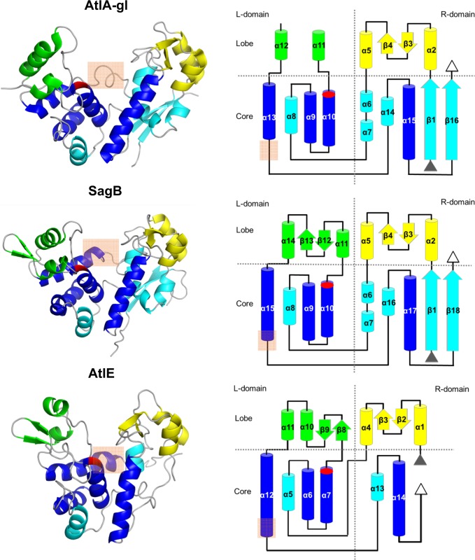
SagB crystallized in the P6122 space group with two molecules per asymmetric unit (Table 1). The structure of SagB is well-resolved based on electron density maps along the entire chain with the exception of the N36 to A41 region in both molecules, as indicated by the Ramachandran outliers and higher atomic B-values, as well as a few other side chains. The crystal structure also contains eight PEG molecules (PG4), two chloride ions and one sodium ion. The latter is positioned at the crystallographic two-fold axis. The two molecules superimpose with a root-mean-square-deviation (RMSD) of 0.46 Å over all Cα atoms. The superimposition parameters slightly deviate from a proper noncrystallographic symmetry dimer. The structure of SagB shares the topological layout and heart-like shape of AtlE (4PIA)16. it is composed of right (R) and left (L) domains divided into core and lobe regions (Fig. 1). The catalytic residue E121 is positioned at the C-terminus of helix α10. The R-domain comprises residues from A1 to D71 and S187 to K250, whereas the L-domain comprises residues from L72 to S186. The two domains in molecule A superimpose onto their counterparts in molecule B with similar RMSDs (0.46 and 0.38 Å) indicating equivalent domain packing.
Table 1.
Data collection and refinement statistics.
| AtlA-gl | SagB | SagB_SeMet_remote | |
|---|---|---|---|
| Data collection | |||
| Space group | P 64 2 2 (181) | P 61 2 2 (178) | P 61 2 2 (178) |
| Cell dimensions | |||
| a, b, c (Å) | 110.17, 110.17, 92.39 | 151.77, 151.77, 122.51 | 152.78, 152.78, 123.83 |
| α, β, γ (°) | 90, 90, 120 | 90, 90, 120 | 90, 90, 120 |
| Wavelength (Å) | 0.9184 | 0.8943 | 0.9770 |
| Resolution (Å) | 42.39–2.28 (2.41–2.28) | 47.66–2.03 (2.15–2.03) | 48.10–2.19 (2.32–2.19) |
| Rmeasured (%) | 9.1 (162.6) | 9.1 (105.0) | 17.5 (192.7) |
| I/σI | 25.31 (1.96) | 27.07 (2.02) | 17.06 (1.83) |
| Completness (%) | 99.8 (99.3) | 99.7 (98.5) | 99.8 (99.0) |
| Multiciplicity | 21.1 | 18.3 | 21.2 |
| CC(1/2) | 100.0 (63.1) | 99.9 (70.4) | 99.9 (62.8) |
| ISa | 24.95 | 31.33 | 24.85 |
| Refinement | |||
| Resolution (Å) | 36.06–2.50 (2.54–2.50) | 19.95–2.03 (2.07–2.03) | |
| No. reflections | 11943 (578) | 53814 (2544) | |
| Rwork/Rkick1 | 0.295/0.334 | 0.212/0.220 | |
| No. atoms | |||
| Protein | 2261 | 4046 | |
| Ligand/ion | 3 | 107 | |
| Water | 162 | 548 | |
| B-factors | |||
| Protein | 68.1 | 40.2 | |
| Ligand/ion | 68.1 | 66.6 | |
| Water | 66.6 | 66.2 | |
| R.m.s. deviations | |||
| Bond lengths (Å) | 0.79 | 0.78 | |
| Bond angles (°) | 1.08 | 0.78 |
1The idea behind Rkick is similar to the Rfree, however, it is calculated from the structure factors of the kicked model against WORK set of data and not from structure factors of unperturbed model against TEST part of data. This corrects the conceptual problem of Rfree. Namely, the claim that TEST set used in cross validation maximum likelihood refinement target function as source of structure independent information is false because errors in models under refinement are not randomly distributed55.
Fig. 1. Structures of AtlA-gl, SagB and AtlE.
The left column shows the ribbon presentation of the structures, whereas the right column shows topology diagrams. The highly conserved helices in the lysozyme-type fold enzymes are depicted in blue and the other core secondary structure elements are shown in cyan. The L-lobe secondary structure elements are shown in green and the R-lobe elements are in yellow. The active site glutamate is marked in red. An orange rectangle marks the region of differences between helices α13 in AtlA-gl, α15 in SagB, and α12 in AtlE.
AtlA-gl crystallized in the P6422 space group with one molecule per asymmetric unit (Table 1). In spite of the weak diffraction data, its structure is well-resolved based on the electron density maps with the exception of the two N-terminal amino acid residues and the A128-N140 loop. The crystallographic R-factor of 0.29 reflects the irreproducible crystal, its small size and the low intensity of the diffraction data. Similar to SagB, the AtlA-gl structure also shares the topological layout of AtlE (Fig. 1). Its R-domain comprises the region from A1 to D68 and A190 to K244, whereas the L-domain comprises from Q69 to K176. In contrast to the R-lobe, the L-lobe is smaller and partially disordered.
Comparison of the AtlA-gl, SagB, and AtlE structures
The failure of molecular replacement was the first indication that the SagB structure must differ from the AtlE structure in spite of their high sequence similarity. When the structure was determined, it became evident that the two structures share the same fold; however the L-domains and R-domains are positioned differently relative to one another (Fig. 1). To quantify the differences, the structures were superimposed as a whole and in parts with the FatCat22 interface imbedded in MAIN23. Superimposition of the complete structures yielded an RMSD of 3.4 Å over 195 residues with 28% sequence identity; in contrast, superimposition of the two parts corresponding to the R-domains and L-domains yielded an RMSD of 2.2 Å over 104 matched residues with 28% identity and an RMSD of 1.7 Å over 98 matching residues with the 36% identity for the R-domain and L-domain, respectively. Moreover, the angle of rotation of the SagB structure superimposed on the AtlE R-domain with the SagB structure superimposed onto the AtlE L-domain is 31° with a 3.5 Å screw translational component. Thus, the packing of the R-domains and L-domains in the SagB structure differs from their packing in the AtlE structure.
The AtlA-gl structure was solved by molecular replacement using the SagB structure as the search model. A similar comparison with the AtlE structure as described above for the SagB structure yielded an RMSD of 4.0 Å over 199 matching residues with 14% identity, whereas the superimposition of the R-domains and L-domains yielded RMSDs of 2.2 and 1.7 Å over 104 and 99 matching residues, respectively. The angle of rotation of the AtlA-gl structure superimposed on the R-domains and L-domains is 39° and resulted in a screw translational component of 4.6 Å. Thus, the packing of the R-domains and L-domains in the AtlA-gl structure differs from their packing in the AtlE structure even more than in the SagB structure.
The positioning of the conserved core helices is equivalent to that of AtlE in seven other known GH73 crystal structures16, the only two structures that substantially deviate are SagB and Atl-gl (Supplementary Fig. 1). Apart from the R-domain and L-domain packing, the major difference among the three structures is in the helix region connecting the R-lobes and L-lobes marked with a rectangle in Figs. 1, 2. In the AtlA-gl structure, the 4th turn of helix α13 is stretched out compared to the slightly longer equivalent helix α15 in SagB and the even longer helix α12 in AtlE. The equivalent helix in the AtlE structure is distorted after the fourth turn with the aromatic residue Y201 slightly exposed. The α15 helix in SagB is almost a whole turn shorter and shares the same minor widening in the last turn of the helix at position F185.
Fig. 2. Conserved amino acid residues in the AtlA-gl, SagB, and AtlE substrate binding sites.
In the white ribbon structure, the catalytic glutamates (E116, E121, and E138) are shown as red sticks, the YA(S/T)D regions as blue sticks, and the conserved aromatic residues (Y191, F185, and Y201) as pink sticks. Note that S226 has a double conformation in the AtlE crystal.
Substrate binding
As a consequence of the different positioning of the L-domain and R-domain substrate binding clefts in SagB and AtlA-gl, structures are much wider in comparison to the AtlE structure. This is reflected in the distances between the catalytic glutamate residue Cα atoms (E121 in SagB, E116 in AtlA, and E138 in AtlE) from the L-domain and the Cα atoms from the conserved YA(S/T)D region (Y209-D212 in SagB, Y214-D217 in AtlA-gl, and Y224-D227 in AtlE) positioned in the opposite side in the R-domain (Fig. 2). The shortest distances between the catalytic glutamates and YA(S/T)D regions were measured in the AtlE structure and are in the range from 10.8 to 12.8 Å, followed by the distances from the SagB structure in the range from 17.6 to 20.5 Å, and the distances from AtlA-gl structure in the range from 17.7 to 21.8 Å. Hence, the substrate-binding clefts of AtlA-gl and SagB, with an 8 Å difference, are wider than their equivalent in the AtlE structure.
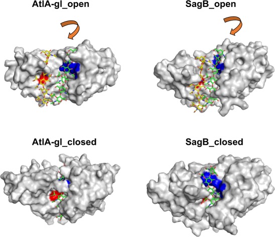
To gain insights into the possible (NAG-NAM)3 substrate binding geometry in the AtlA-gl and SagB structures, we used the binding geometry previously established for the AtlE structure16. When bound to AtlE, the substrate was in contact with the catalytic glutamate residue in the L-domain and the YA(S/T)D region in the R-domain in the active site cleft. The only way to conserve these previously identified contacts in the L-domains and R-domains was to independently superimpose the (NAG-NAM)3 AtlE-substrate complex to each domain independently. This resulted in two putative substrate binding geometries (Fig. 3). When the L-domain pairs were superimposed, the (NAG-NAM)3 substrate retained its position over the catalytic glutamates, and when the R-domains were superimposed, the substrate retained the contacts with the YA(S/T)D binding region. This suggests that a conformational change has to occur to bring those two regions together. We have previously shown that AtlA-gl can cleave (NAG-NAM)2 tetrasaccharide16, similar to AtlE, which indicates that despite the large distance between the substrate binding region and the catalytic site, a single glycan strand is a substrate for this enzyme. This suggests that the AtlA-gl and SagB structures were observed in the open form compared to the AtlE structure, which was observed in the closed form.
Fig. 3. The (NAG-NAM)3 substrate binding models of the AtlA-gl and SagB open and closed forms.

The color coding of the surface is the same as used in Fig. 2. The top two structures include (NAG-NAM)3 shown as a stick model with carbon bound to the L-domains and R-domains colored yellow and green, respectively. The bottom images represent the closed structures with bound (NAG-NAM)3 substrate. The NAM(−2) and NAG(−1) residues in the pair of substrate models attached to the L-domain exhibit clashes with the features in the SagB (A124 and K127) and AtlA-gl (Y152) structures, which can be resolved with minor conformational adjustments. The arrows indicate the direction of domain rotations.
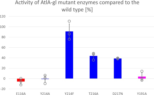
To identify the equivalent residues in AtlA-gl involved in substrate binding as in AtlE17, we introduced point mutations that either targeted the catalytic Glu directly or the substrate binding YA(S/T)D region (Fig. 4). An analogous substrate degradation assay was not functional for SagB. All produced AtlA-gl mutants were correctly folded as confirmed by CD spectra. Mutation of the catalytic glutamate E116 to alanine rendered enzymes inactive, as has been shown previously for several enzymes in this family, including AtlE17, AtlWM24, AcmA25, LytB26,27, SpFlgJ28, StFlgJ29, Auto30, TM063331, and AcpCD32. The mutation of the tyrosine residue Y214 in the YATD region to alanine severely affected enzyme activity, whereas mutation of Y214 to phenylalanine completely restored the substrate degrading ability. Similar observations were previously established for AtlE17, as well as for four other GH73 enzymes24,25,28,32,33, demonstrating that an aromatic residue is required at this position. The T216A and D217N mutants exhibited a marked decrease in activity. Similar observations were made for the equivalent S226A and D227A mutants in AtlE17. We were unable to obtain a soluble D217A mutant from AtlA-gl, indicating that D217 is important for the structural stability of the region and protein folding. Mutation of the conserved aromatic residue Y191A positioned within the helix connecting the L-domains and R-domains resulted in an inactive AtlA-gl, which was equivalent to mutation of the equivalent residue Y201A in AtlE, which also resulted in an inactive enzyme. From these results, we concluded that the catalytic glutamate and the YA(S/T)D region are both involved in substrate binding during hydrolysis. Hence, the structures must adopt closed forms to perform the reaction.
Fig. 4. The effects of the point mutations on the AtlA-gl substrate binding site residues on the peptidoglycan-degrading ability.

The AtlE mutations were described by Borišek et al.17.
To gain insights into the structural flexibility of the two proteins closed form models were constructed. Both were modeled using the AtlE structure as the template. Simple models were created by superimposition of each domain of SagB and AtlA-gl to corresponding domain of AtlE followed by minimization of the combined models (Fig. 1). The models obtained by superimposition resulted only in minor side chain clashes: 24 non-hydrogen atoms from four residues of the L-domain and 23 non-hydrogen atoms from five residues of D-domains of AtlA-gl were closer than 2.2 Å (the distance used by PDB structure validation tools to indicate possible false covalent bond interactions), whereas for the SagB 30 non-hydrogen atoms from five residues of the L-domain and 34 non-hydrogen atoms form five residues of the D-domain were closer than 2.2 Å. These clashes were resolved by minimization. These simple models were further adjusted to match the chain trace of the α12 helix in the AtlE template (marked with an orange rectangle in Fig. 1, see Methods section for further details).
According to DynDom34,35 analyses the transitions between open and closed forms of AtlA-gl and SagB were classified as hinge motion with a 35.3° rotation angle, −0.7 Å translation and 97.3% closure for AtlA-gl (Supplementary Movie 1) and 29.4° rotation angle, −1.1 Å translation and 83.3% closure for SagB (Supplementary Movie 2). According to the structural flexibility analysis, structures with such high rotation angles are rare8. Due to the specifics of the structural change, it seems more appropriate to describe the domain movement as sliding domains along their interface than rotation. The extent of changes during transition between the open and closed forms was analyzed by counting the unique contacts between residues and their RMSDs (for details see Methods section). Visualization of the changes in inter-residue contacts revealed their location (shown in red in Supplementary Fig. 2) along the two domain interfaces of the AtlA-gl and SagB structures. The AtlA-gl pair of open and closed forms has 46 and 76 inter-residue specific interactions, respectively, and an RMSD of 4.5 Å, whereas the SagB pair of open and closed forms has 96 and 100 inter-residue specific interactions, respectively, and an RMSD of 3.6 Å. In comparison, the pair of SagB structures related by non-crystallographic symmetry has 70 and 66 inter-residue specific interactions and an RMSD of 0.6 Å, whereas the pair of T and R states of allosterically regulated l-Lactate dehydrogenase has 204 and 228 unique interactions, respectively, and an RMSD of 1.9 Å. The differences in the RMSD deviations between the pairs of structures in different forms and the similarity of the number of differences between the inter-residue specific interactions suggest that the differences between the open and closed forms of the AtlA-gl and SagB structures are likely larger than modeled. To show that also the closed forms of both enzymes can accommodate peptidyl links attached to polysaccharides we built the models using the bound (NAG-NAM)3 substrate with added L-Ala-D-iso-Gln-L-Lys part of the peptide stem (Supplementary Fig. 3).
Molecular simulations of AtlA-gl and SagB
To investigate the stability of the crystal structures and the models of the closed forms of the AtlA-gl and SagB structures, crystal structures and models were subjected to several classical molecular dynamics (MD) simulations in the time span of 500 ns for each performed simulation.
First, we performed MD simulations on the AtlA-gl and SagB crystal structures. Their MD-generated conformations exhibited RMSD values of 2.56 ± 0.32 Å (AtlA-gl) and 1.99 ± 0.41 Å (SagB). The distances between the Cα atoms from the catalytic glutamate and alanine in the YATD region were in the range from 20.3 ± 0.9 Å and 14.9 ± 2.0 Å for AtlA-gl and SagB, respectively, which are close to the values observed in the starting structures. The residues from the lobe parts displayed substantial motion indicated by the larger thickness and yellow/red color of the chain trace (Supplementary Fig. 4; Supplementary Table 1). Their trajectories did not display any significant tendencies of transition towards their closed forms.
We then simulated the simple closed form models of both enzymes. The MD-generated conformations were highly unstable as indicated by the substantially higher structural RMSD values of 4.76 ± 0.71 Å (AtlA-gl) and 3.52 ± 0.94 Å (SagB) compared to the other MD simulations (Supplementary Fig. 5; Supplementary Table 1). There was an increase in the interdomain distance between the catalytic glutamate and the YATD region of approximately 4 Å in AtlA-gl and 3 Å in SagB when compared with the starting structure. This suggested that both simple closed forms exhibited a tendency for their transition towards the open form. Although considerably long, a 500-ns MD simulation was not long enough to observe the full conformational transition.
Next, we simulated the closed forms with the rebuilt helices. During the MD simulations, both models remained in their closed forms with the observed average distances were 12.2 ± 0.87 Å (AtlA-gl) and 12.9 ± 0.78 Å (SagB) (Supplementary Fig. 4B). This suggested that the adjustment of the length of the domain-connecting helix indeed contributed to the stability of the closed forms of AtlA-gl and SagB.
Further, we simulated the closed forms of AtlA-gl and SagB with rebuilt helices and the bound substrate (NAG-NAM)3. In both simulations, the geometries remained stable (Supplementary Fig. 4; Supplementary Table 1). Compared with the simulation of the closed forms without the bound substrate, the average interdomain distances were slightly shorter in the case of AtlA-gl (10.4 ± 0.32 Å) and remained about the same as in SagB (13.0 ± 0.78 Å) (Supplementary Fig. 4).
Taken together, the molecular simulations confirmed the stability of the rebuilt closed forms and their complexes with substrate models. Furthermore, our analysis suggested that the interdomain conformational flexibility of AtlA-gl and SagB is accompanied by a partial helix-folding/unfolding mechanism.
Discussion
SagB and AtlA-gl crystallized in hexagonal crystals in unit cells of different size and packing. SagB crystallized with two and AtlA-gl with one molecule per asymmetric unit. In contrast to the crystal structure of AtlE and related N-acetylglucosaminidases, which were all observed in the closed form, these two proteins crystallized in the open form. The two independent crystal structures of the two distinct proteins exclude the possibility that these results are both crystallization artifacts. Hence, the observed open forms must reflect the real structural dynamics of these two bacterial N-acetylglucosaminidases.
Comparison of the SagB, AtlA-gl, and AtlE crystal structures revealed that the fold of the L-domains and R-domains of all three proteins is the same; however, the relative position of the domains exhibit unusually large differences. With respect to the closed form of the AtlE structure, the L-domains and R-domains in the SagB and AtlA-gl structures are rotated away from each other, resulting in a very wide substrate binding grove. Analysis of the substrate binding geometry together with site-directed mutagenesis identified residues in the L-domain and R-domain that are essential for the reaction to transpire. Hence, for AtlA-gl and SagB to cleave a single glycan strand, both of their domains have to slide together into a closed conformation as observed in the AtlE structure. The suggested conformational changes were corroborated by MD simulations.
Analysis of the specific inter-residue contacts has shown that they are located along the two domain interfaces; in the open forms, the contacts are located bellow the substrate binding site, whereas in the closed forms, they were positioned higher in the substrate binding region. AtlE appears to be able to bind the substrate in its closed form with minimal adjustments17. In contrast, the closed forms of SagB and even more so that of AtlA-gl embrace the substrate (Fig. 3), meaning that the substrate cannot bind to the active site without the transition of the enzymes into their open forms, indicating that closing occurs after substrate binding. In Rnase A, for example, it was shown that both open and closed forms exist in solution36. Similarly, open and close conformations associated with loop and helix flexibility were found in the substrate-free enzyme structures of adenylate kinase37. These works showed that the open forms of the AtlA-gl and SagB structures are not an exception. However, the kind and extent of flexibility of N-acetylglucosaminidases associated with domain sliding and helix transition described here surpass the range of expectations. Even our 500-ns long molecular dynamic simulations could only confirm the stability of the observed and modeled open and closed forms, but not reproduce their transitions.
The question here is, why are these molecular gymnastics necessary? It is common knowledge that enzymes catalyze chemical reactions by aligning the reactive groups in an optimal position for efficient chemistry to take place. Fersht proposed that as the enzyme is more flexible than the substrate, its distortion can be larger38. The S. aureus cell wall has been found to be more rigid than that of E. coli or B. subtillis39, and even further, glycan strands were shown to be less flexible than the peptide portions of peptidoglycan40. Moreover, it was also demonstrated that S. aureus cells deficient in GH73 enzymes have increased surface stiffness, with SagB inactivation having the largest effect, as well as the important contributions of other enzymes, including AtlA-gl and AtlE18. These findings further indicate that the substrates of these enzymes are particularly rigid and therefore require corresponding flexibility by the enzymes processing them. There are reports of conformational flexibility in enzymes that synthesize, hydrolyze or bind peptidoglycan glycan strands, and they range from altered relative positioning of different domains connected by a linker region41–44 to loop flexibility45 and intradomain movement46. Analysis of PDB structures with and without ligands exposed that among enzymes, only transferases and hydrolases exhibit domain motion; however, the extent of motion observed for hydrolases was smaller (below 1 Å RMSD)47, suggesting that the chemical reaction of linked chemical groups favors solvent exclusion, whereas hydrolysis requires solvent, explaining the smaller structural changes in hydrolases. The extent and kinds of movement observed in our structures however hints in the other direction. Taken together, our structural analysis suggests that these two hydrolases must have evolved an unusual extent of flexibility to cleave the targeted glycosidic bonds in rigid bacterial cell walls. It is not chemistry alone that requires adaption of flexibility, but also the large and rigid substrate which needs to be torn. Bailey et al. have shown that the newly formed peptidoglycan at the septum is stiffer and proposed that the subsequent drop in rigidity during division process is likely due to the peptidoglycan hydrolase activity48. This coincides with AtlA localization at the septal region and its proposed role in cell division14,21 and their ability to cleave highly cross linked S. aureus peptidoglycan19. Superimposition of other eight structures from GH73 family deposited in PDB provided no evidence that any of them were crystallized in other than the closed form (Supplementary Fig. 1). Hence, the insight into AtlA and SagB may shed a light on synthesis of peptidoglycan in S. aureus, which is inserted only during cell division at the septum49. Taken together, we can speculate that SagB and AtlA-gl have a role in cell division which involves reducing the rigidity of the peptidoglycan.
Methods
Protein production and crystallization
Protein coding sequences were cloned into the pMCSG7 vector using LIC procedures50 and KOD Hot Start Polymerase (Invitrogen). S. aureus Mu50 genomic DNA (ATCC) was used as the template with the primers listed in Supplementary Table 2. Proteins were produced in E. coli BL21DE3 grown in ZYM-5052 autoinduction media for 5 h at 37 °C and 18 h at 20 °C. Cells were resuspended in buffer A (30 mM Tris-HCl, pH 7.5, and 400 mM NaCl) with 1 mg ml−1 lysozyme and lysed by sonication. The lysate was cleared by centrifugation. Proteins were purified by Ni-NTA using buffer A with 10 mM imidazole for binding and 300 mM imidazole for elution, followed by SEC (HiPrep 16/60 Sephacryl S-200 HR (GE Healthcare)) with buffer A. The His tag was cleaved off with TEV protease and removed by Ni-NTA. Mutant enzymes were produced by IPTG induction in LB medium at an OD600 of approximately 0.6 and purified by the same procedure as the wild type enzymes. CD spectra confirmed mutant proteins assumed the same fold as the wild type proteins.
For crystallization, SagB was dialyzed into 20 mM citrate, pH 5, with 100 mM NaCl and AtlA-gl into 20 mM HEPES, pH 7.5 with 100 mM NaCl and concentrated to 13 mg ml−1 and 38 mg ml−1, respectively. SagB was crystallized in 0.1 M Na3citrate, pH 4.75 with 47% PEG 200 and AtlA-glu in 2 M (NH4)2SO4. AtlA-gl crystallized in the form of several crystals that grew from a single point. Crystals were small and not reproducible. Both proteins were crystallized using the sitting drop at 20 °C.
Activity assay
Peptidoglycan was purified and dyed and the assay was conducted as described previously17. The buffers used in the activity assay were 100 mM Tris, pH 8, for AtlA-gl and 100 mM MES, pH 5.5, for AtlE. AtlA-gl and AtlE activity assays were performed with 5 μM enzymes at 25 °C and 600 rpm.
Protein structure determination
Diffraction data sets for SagB and AtlA-gl were collected at 100 K using synchrotron radiation at the BESSY 14.1. and BESSY 14.3. beamlines at Helmholtz-Zentrum Berlin51. Indexing, integrating and scaling were performed with the XDS software package52. Native SagB crystals diffracted to 2.0 Å resolution, whereas SeMet crystals diffracted to 2.6 Å. The SagB structure was solved using an SeMet derivative data set collected at a remote wavelength of 0.977031 Å. Initial chain trace was performed by ShelX53. The AtlA-gl crystal diffracted to 2.3 Å. The phase problem for AtlA-gl was solved by molecular replacement of the SagB structure using PHASER54. Model building, refinement, and structure validation for SagB and AtlA-gl were performed with MAIN23. During refinement, the Free Kick maximum likelihood target used all diffraction data for both working and testing simultaneously55.
Construction and analysis of closed structures
The closed form models of both structures were constructed in two steps. First, we created the models by superimposition of the R-domains and L-domains on the AtlE structure. L-domains contained residues from sequence ranges from Q69 to K176 and L72 to S186 from AtlA-gl and SagB, respectively, whereas the R-domains contained the omitted parts. Then the broken link and side chain clashes were adjusted by energy minimization. Visual inspection of the superimposed models indicated the position of the pivoting point proximal to residues I89 in AtlE, L67 in AtlA-gl, and L106 in SagB. We called the generated models simple models. These two models were further modified by extension of the domain-connecting helices. The α13 helix in the AtlA-gl closed form model from S175 to K191 and the α15 helix in the SagB closed form model from P169 to S186 were rebuilt to match their chain traces to the α12 helix in the AtlE structure.
The extent of change of the contacts was analyzed by packing analysis of the structures using graph nodes described recently56. Here we calculated graphs of intra residue interactions within a 4.6 Å radius using only nonhydrogen atoms and disregarding two neighboring residues in the chain. The connectivity graphs for each pair of molecules were then subtracted from one another so that all matching connections were removed. The resulting differential graphs contained only interactions specific for the form and molecule.
MD simulations
The MD calculations were performed using the NAMD molecular modeling suite57 at the Azman Computing Center at the National Institute of Chemistry, Slovenia. AtlA-gl loop residues 128–140 from the 6FXO crystal structure that were not resolved in the experimental electron density map were constructed using Modeller58. Enzymes and substrates were prepared according to our previously published protocol17. Briefly, the protonation states of all ionizable amino acid residues were assigned based on the pKa estimated by PROPKA version 3.059 at pH = 6.0. All aspartate and glutamate residues were deprotonated, with the exception of catalytic Glu116 and Glu121 from AtlA-gl and SagB, respectively.
The CHARMM-GUI environment60 was utilized for protein manipulation and construction of the solvated protein-substrate complexes. CHARMM parameter and topology files (version 27)61,62 were utilized to specify the force field parameters for the amino acid residues comprising the protein. The CHARMM General Force Field (CGenFF)63 was used to describe the atom types and substrate partial charges17. The final systems prepared for the MD simulation was composed of 39,264 and 45,160 atoms for AtlA-gl and SagB.
Both systems were minimized for 10,000 steps with the steepest descent method followed by 10,000 steps of a modified Adopted Basis Newton–Raphson method and a 1-ns MD equilibration run. The production MD trajectory was generated using a leapfrog integration scheme and a 2-fs simulation step using the SHAKE algorithm. A 500-ns MD simulation production run was performed for each model. Conformations were sampled every 1000th step resulting in 25,000 conformations for subsequent analysis.
Visualization and analysis of the geometric parameters for the production MD trajectory were performed using the VMD program64. Structures, measured distances and RMSD graphs for all performed systems (AtlA-gl and SagB) are available in Supplementary Figs. 4, 5; Supplementary Table 1.
Reporting summary
Further information on research design is available in the Nature Research Reporting Summary linked to this article.
Supplementary information
Description of Additional Supplementary Files
Acknowledgements
The authors thank Sandra Gabelli and Marcin Cymborowski for consultancy on AtlA-gl diffraction data processing and acknowledge financial support from the Slovenian Research Agency (research core funding no. P1–0048, P1–0017, P1–0012 and IO-0048, and project no. J1–8152). Data were collected at BMX14.1 and BMX 13.3 (BESSY, Berlin).
Author contributions
D.T. conceived the project. S.P. and A.U. performed the cloning, protein expression and purification. S.P. performed crystallization and collected the X-ray diffraction data. S.P. and D.T. determined the structures and analyzed them. S.P. performed the activity assays. D.T. constructed and analyzed the closed enzyme models. J.B. and A.P. performed MD simulations and analyzed the results. S.P. and D.T. wrote the manuscript. A.P., A.U., and J.B. edited the manuscript.
Data availability
The atomic coordinates and structure factors for AtlA-gl and SagB have been deposited in the Protein Data Bank under accession codes 6FXO and 6FXP, respectively. The source data underlying the plots shown in Fig. 4 are provided in Supplementary Data 1.
Competing interests
The authors declare no competing interests.
Footnotes
Publisher’s note Springer Nature remains neutral with regard to jurisdictional claims in published maps and institutional affiliations.
Supplementary information
Supplementary information is available for this paper at 10.1038/s42003-020-0911-7.
References
- 1.Fischer E. Einfluss der configuration auf die Wirkung der enzyme. Ber. der Dtsch. chemischen Ges. 1894;27:2985–2993. doi: 10.1002/cber.18940270364. [DOI] [Google Scholar]
- 2.Koshland DE. Application of a theory of enzyme specificity to protein synthesis. Proc. Natl Acad. Sci. USA. 1958;44:98–104. doi: 10.1073/pnas.44.2.98. [DOI] [PMC free article] [PubMed] [Google Scholar]
- 3.Frauenfelder H, Sligar SG, Wolynes PG. The energy landscapes and motions of proteins. Science. 1991;254:1598–1603. doi: 10.1126/science.1749933. [DOI] [PubMed] [Google Scholar]
- 4.Boehr DD, Nussinov R, Wright PE. The role of dynamic conformational ensembles in biomolecular recognition. Nat. Chem. Biol. 2009;5:789–796. doi: 10.1038/nchembio.232. [DOI] [PMC free article] [PubMed] [Google Scholar]
- 5.Ma B, Kumar S, Tsai CJ, Nussinov R. Folding funnels and binding mechanisms. Protein Eng. 1999;12:713–720. doi: 10.1093/protein/12.9.713. [DOI] [PubMed] [Google Scholar]
- 6.Csermely P, Palotai R, Nussinov R. Induced fit, conformational selection and independent dynamic segments: an extended view of binding events. Trends Biochem. Sci. 2010;35:539–546. doi: 10.1016/j.tibs.2010.04.009. [DOI] [PMC free article] [PubMed] [Google Scholar]
- 7.Gutteridge A, Thornton J. Conformational changes observed in enzyme crystal structures upon substrate binding. J. Mol. Biol. 2005;346:21–28. doi: 10.1016/j.jmb.2004.11.013. [DOI] [PubMed] [Google Scholar]
- 8.Taylor D, Cawley G, Hayward S. Quantitative method for the assignment of hinge and shear mechanism in protein domain movements. Bioinformatics. 2014;30:3189–3196. doi: 10.1093/bioinformatics/btu506. [DOI] [PMC free article] [PubMed] [Google Scholar]
- 9.Amemiya T, Koike R, Fuchigami S, Ikeguchi M, Kidera A. Classification and annotation of the relationship between protein structural change and ligand binding. J. Mol. Biol. 2011;408:568–584. doi: 10.1016/j.jmb.2011.02.058. [DOI] [PubMed] [Google Scholar]
- 10.Henzler-Wildman K, Kern D. Dynamic personalities of proteins. Nature. 2007;450:964–972. doi: 10.1038/nature06522. [DOI] [PubMed] [Google Scholar]
- 11.Lombard V, Golaconda Ramulu H, Drula E, Coutinho PM, Henrissat B. The carbohydrate-active enzymes database (CAZy) in 2013. Nucleic Acids Res. 2014;42:D490–D495. doi: 10.1093/nar/gkt1178. [DOI] [PMC free article] [PubMed] [Google Scholar]
- 12.García P, González MP, García E, López R, García JL. LytB, a novel pneumococcal murein hydrolase essential for cell separation. Mol. Microbiol. 1999;31:1275–1281. doi: 10.1046/j.1365-2958.1999.01238.x. [DOI] [PubMed] [Google Scholar]
- 13.Visweswaran GRR, et al. AcmD, a homolog of the major autolysin AcmA of Lactococcus lactis, binds to the cell wall and contributes to cell separation and autolysis. PLoS One. 2013;8:e72167. doi: 10.1371/journal.pone.0072167. [DOI] [PMC free article] [PubMed] [Google Scholar]
- 14.Yamada S, et al. An autolysin ring associated with cell separation of Staphylococcus aureus. J. Bacteriol. 1996;178:1565–1571. doi: 10.1128/JB.178.6.1565-1571.1996. [DOI] [PMC free article] [PubMed] [Google Scholar]
- 15.Nambu T, Minamino T, Macnab RM, Kutsukake K. Peptidoglycan-hydrolyzing activity of the FlgJ protein, essential for flagellar rod formation in Salmonella typhimurium. J. Bacteriol. 1999;181:1555–1561. doi: 10.1128/JB.181.5.1555-1561.1999. [DOI] [PMC free article] [PubMed] [Google Scholar]
- 16.Mihelič M, et al. The mechanism behind the selection of two different cleavage sites in NAG-NAM polymers. IUCrJ. 2017;4:185–196. doi: 10.1107/S2052252517000367. [DOI] [PMC free article] [PubMed] [Google Scholar]
- 17.Borišek J, et al. A water-assisted catalytic mechanism in glycoside hydrolases demonstrated on the Staphylococcus aureus autolysin E. ACS Catal. 2018;8:4334–4345. doi: 10.1021/acscatal.8b01064. [DOI] [Google Scholar]
- 18.Wheeler R, et al. Bacterial cell enlargement requires control of cell wall stiffness mediated by peptidoglycan hydrolases. mBio. 2015;6:e00660–15. doi: 10.1128/mBio.00660-15. [DOI] [PMC free article] [PubMed] [Google Scholar]
- 19.Chan YGY, Frankel MB, Missiakas D, Schneewind O. SagB glucosaminidase is a determinant of Staphylococcus aureus glycan chain length, antibiotic susceptibility, and protein secretion. J. Bacteriol. 2016;198:1123–1136. doi: 10.1128/JB.00983-15. [DOI] [PMC free article] [PubMed] [Google Scholar]
- 20.Oshida T, et al. A Staphylococcus aureus autolysin that has an N-acetylmuramoyl-L-alanine amidase domain and an endo-B-N-acetylglucosaminidase domain: cloning, sequence analysis, and characterization. Proc. Natl Acad. Sci. USA. 1995;92:285–289. doi: 10.1073/pnas.92.1.285. [DOI] [PMC free article] [PubMed] [Google Scholar]
- 21.Takahashi J, et al. Molecular characterization of an atl null mutant of Staphylococcus aureus. Microbiol. Immunol. 2002;46:601–612. doi: 10.1111/j.1348-0421.2002.tb02741.x. [DOI] [PubMed] [Google Scholar]
- 22.Ye Y, Godzik A. FATCAT: a web server for flexible structure comparison and structure similarity searching. Nucleic Acids Res. 2004;32:W582–W585. doi: 10.1093/nar/gkh430. [DOI] [PMC free article] [PubMed] [Google Scholar]
- 23.Turk D. MAIN software for density averaging, model building, structure refinement and validation. Acta Crystallogr. D. 2013;69:1342–57. doi: 10.1107/S0907444913008408. [DOI] [PMC free article] [PubMed] [Google Scholar]
- 24.Yokoi KJ, et al. Molecular properties of the putative autolysin AtIWM encoded by Staphylococcus warneri M: mutational and biochemical analyses of the amidase and glucosaminidase domains. Gene. 2008;416:66–76. doi: 10.1016/j.gene.2008.03.004. [DOI] [PubMed] [Google Scholar]
- 25.Inagaki N, et al. Molecular properties of the glucosaminidase AcmA from Lactococcus lactis MG1363: mutational and biochemical analyses. Gene. 2009;447:61–71. doi: 10.1016/j.gene.2009.08.004. [DOI] [PubMed] [Google Scholar]
- 26.Rico-Lastres P, et al. Substrate recognition and catalysis by LytB, a pneumococcal peptidoglycan hydrolase involved in virulence. Sci. Rep. 2015;5:16198. doi: 10.1038/srep16198. [DOI] [PMC free article] [PubMed] [Google Scholar]
- 27.Bai XH, et al. Structure of pneumococcal peptidoglycan hydrolase LytB reveals insights into the bacterial cell wall remodeling and pathogenesis. J. Biol. Chem. 2014;289:23403–23416. doi: 10.1074/jbc.M114.579714. [DOI] [PMC free article] [PubMed] [Google Scholar]
- 28.Maruyama Y, et al. Mutational studies of the peptidoglycan hydrolase FlgJ of Sphingomonas sp. strain A1. J. Basic Microbiol. 2010;50:311–317. doi: 10.1002/jobm.200900249. [DOI] [PubMed] [Google Scholar]
- 29.Herlihey FA, Moynihan PJ, Clarke AJ. The essential protein for bacterial flagella formation FlgJ functions as a beta-N-acetylglucosaminidase. J. Biol. Chem. 2014;289:31029–31042. doi: 10.1074/jbc.M114.603944. [DOI] [PMC free article] [PubMed] [Google Scholar]
- 30.Bublitz M, et al. Structural basis for autoinhibition and activation of auto, a virulence-associated peptidoglycan hydrolase of Listeria monocytogenes. Mol. Microbiol. 2009;71:1509–1522. doi: 10.1111/j.1365-2958.2009.06619.x. [DOI] [PubMed] [Google Scholar]
- 31.Lipski A, et al. Structural and biochemical characterization of the β-N-acetylglucosaminidase from Thermotoga maritima: toward rationalization of mechanistic knowledge in the GH73 family. Glycobiology. 2015;25:319–330. doi: 10.1093/glycob/cwu113. [DOI] [PubMed] [Google Scholar]
- 32.Tamai E, et al. Structural and biochemical characterization of the Clostridium perfringens autolysin catalytic domain. FEBS Lett. 2017;591:231–239. doi: 10.1002/1873-3468.12515. [DOI] [PubMed] [Google Scholar]
- 33.Yokoi KJ, et al. Molecular properties of the putative autolysin Atl(WM) encoded by Staphylococcus warneri M: mutational and biochemical analyses of the amidase and glucosaminidase domains. Gene. 2008;416:66–76. doi: 10.1016/j.gene.2008.03.004. [DOI] [PubMed] [Google Scholar]
- 34.Hayward S, Berendsen HJ. Systematic analysis of domain motions in proteins from conformational change: new results on citrate synthase and T4 lysozyme. Proteins. 1998;30:144–154. doi: 10.1002/(SICI)1097-0134(19980201)30:2<144::AID-PROT4>3.0.CO;2-N. [DOI] [PubMed] [Google Scholar]
- 35.Hayward S, Lee RA. Improvements in the analysis of domain motions in proteins from conformational change: DynDom version 1.50. J. Mol. Graph. Model. 2002;21:181–183. doi: 10.1016/S1093-3263(02)00140-7. [DOI] [PubMed] [Google Scholar]
- 36.Beach H, Cole R, Gill ML, Loria JP. Conservation of mus-ms enzyme motions in the apo- and substrate-mimicked state. J. Am. Chem. Soc. 2005;127:9167–9176. doi: 10.1021/ja0514949. [DOI] [PubMed] [Google Scholar]
- 37.Henzler-Wildman KA, et al. Intrinsic motions along an enzymatic reaction trajectory. Nature. 2007;450:838–844. doi: 10.1038/nature06410. [DOI] [PubMed] [Google Scholar]
- 38.Fersht, A. Structure and Mechanism in Protein Science: A Guide to Enzyme Catalysis and Protein Folding, xxii (World Scientific, New Jersey, 2017).
- 39.Kern T, et al. Dynamics characterization of fully hydrated bacterial cell walls by solid-state NMR: evidence for cooperative binding of metal ions. J. Am. Chem. Soc. 2010;132:10911–10919. doi: 10.1021/ja104533w. [DOI] [PubMed] [Google Scholar]
- 40.Kern T, et al. Toward the characterization of peptidoglycan structure and protein-peptidoglycan interactions by solid-state NMR spectroscopy. J. Am. Chem. Soc. 2008;130:5618–5619. doi: 10.1021/ja7108135. [DOI] [PubMed] [Google Scholar]
- 41.Zoll S, et al. Ligand-binding properties and conformational dynamics of autolysin repeat domains in staphylococcal cell wall recognition. J. Bacteriol. 2012;194:3789–3802. doi: 10.1128/JB.00331-12. [DOI] [PMC free article] [PubMed] [Google Scholar]
- 42.Lovering AL, De Castro L, Strynadka NC. Identification of dynamic structural motifs involved in peptidoglycan glycosyltransfer. J. Mol. Biol. 2008;383:167–177. doi: 10.1016/j.jmb.2008.08.020. [DOI] [PubMed] [Google Scholar]
- 43.Domínguez-Gil T, et al. Activation by allostery in cell-wall remodeling by a modular membrane-bound lytic transglycosylase from Pseudomonas aeruginosa. Structure. 2016;24:1729–1741. doi: 10.1016/j.str.2016.07.019. [DOI] [PMC free article] [PubMed] [Google Scholar]
- 44.Hu Y, et al. Crystal structure of the MurG:UDP-GlcNAc complex reveals common structural principles of a superfamily of glycosyltransferases. Proc. Natl Acad. Sci. USA. 2003;100:845–849. doi: 10.1073/pnas.0235749100. [DOI] [PMC free article] [PubMed] [Google Scholar]
- 45.Bacik JP, Whitworth GE, Stubbs KA, Vocadlo DJ, Mark BL. Active site plasticity within the glycoside hydrolase NagZ underlies a dynamic mechanism of substrate distortion. Chem. Biol. 2012;19:1471–1482. doi: 10.1016/j.chembiol.2012.09.016. [DOI] [PubMed] [Google Scholar]
- 46.Carrasco-López C, et al. Crystal structures of bacterial peptidoglycan amidase AmpD and an unprecedented activation mechanism. J. Biol. Chem. 2011;286:31714–31722. doi: 10.1074/jbc.M111.264366. [DOI] [PMC free article] [PubMed] [Google Scholar]
- 47.Koike R, Amemiya T, Ota M, Kidera A. Protein structural change upon ligand binding correlates with enzymatic reaction mechanism. J. Mol. Biol. 2008;379:397–401. doi: 10.1016/j.jmb.2008.04.019. [DOI] [PubMed] [Google Scholar]
- 48.Bailey RG, et al. The interplay between cell wall mechanical properties and the cell cycle in Staphylococcus aureus. Biophys. J. 2014;107:2538–2545. doi: 10.1016/j.bpj.2014.10.036. [DOI] [PMC free article] [PubMed] [Google Scholar]
- 49.Turner RD, et al. Peptidoglycan architecture can specify division planes in Staphylococcus aureus. Nat. Commun. 2010;1:26. doi: 10.1038/ncomms1025. [DOI] [PubMed] [Google Scholar]
- 50.Stols L, et al. A new vector for high-throughput, ligation-independent cloning encoding a tobacco etch virus protease cleavage site. Protein Express. Purif. 2002;25:8–15. doi: 10.1006/prep.2001.1603. [DOI] [PubMed] [Google Scholar]
- 51.Mueller U, et al. Facilities for macromolecular crystallography at the Helmholtz-Zentrum Berlin. J. Synchrotron Radiat. 2012;19:442–449. doi: 10.1107/S0909049512006395. [DOI] [PMC free article] [PubMed] [Google Scholar]
- 52.Kabsch, W. XDS. Acta Crystallogr. D66, 125–132 https://www.ncbi.nlm.nih.gov/pmc/articles/PMC2815665/ (2010). [DOI] [PMC free article] [PubMed]
- 53.Sheldrick GM. Experimental phasing with SHELXC/D/E: combining chain tracing with density modification. Acta Crystallogr. D. 2010;66:479–485. doi: 10.1107/S0907444909038360. [DOI] [PMC free article] [PubMed] [Google Scholar]
- 54.McCoy AJ, et al. Phaser crystallographic software. J. Appl. Crystallogr. 2007;40:658–674. doi: 10.1107/S0021889807021206. [DOI] [PMC free article] [PubMed] [Google Scholar]
- 55.Pražnikar J, Turk D. Free kick instead of cross-validation in maximum-likelihood refinement of macromolecular crystal structures. Acta Crystallogr. D. 2014;70:3124–3134. doi: 10.1107/S1399004714021336. [DOI] [PMC free article] [PubMed] [Google Scholar]
- 56.Pražnikar J, Tomić M, Turk D. Validation and quality assessment of macromolecular structures using complex network analysis. Sci. Rep. 2019;9:1678. doi: 10.1038/s41598-019-38658-9. [DOI] [PMC free article] [PubMed] [Google Scholar]
- 57.Phillips JC, et al. Scalable molecular dynamics with NAMD. J. Comput. Chem. 2005;26:1781–1802. doi: 10.1002/jcc.20289. [DOI] [PMC free article] [PubMed] [Google Scholar]
- 58.Webb B, Sali A. Protein structure modeling with MODELLER. Funct. Genom. 2017;1654:39–54. doi: 10.1007/978-1-4939-7231-9_4. [DOI] [PubMed] [Google Scholar]
- 59.Olsson MHM, Sondergaard CR, Rostkowski M, Jensen JH. PROPKA3: consistent treatment of internal and surface residues in empirical pK(a) predictions. J. Chem. Theory Comput. 2011;7:525–537. doi: 10.1021/ct100578z. [DOI] [PubMed] [Google Scholar]
- 60.Kumar R, Iyer VG, Im W. CHARMM-GUI: a graphical user interface for the CHARMM users. Abstr. Pap. Am. Chem. Soc. 2007;233:273–273. [Google Scholar]
- 61.MacKerell AD, et al. All-atom empirical potential for molecular modeling and dynamics studies of proteins. J. Phys. Chem. B. 1998;102:3586–3616. doi: 10.1021/jp973084f. [DOI] [PubMed] [Google Scholar]
- 62.Mackerell AD, Feig M, Brooks CL. Extending the treatment of backbone energetics in protein force fields: limitations of gas-phase quantum mechanics in reproducing protein conformational distributions in molecular dynamics simulations. J. Comput. Chem. 2004;25:1400–1415. doi: 10.1002/jcc.20065. [DOI] [PubMed] [Google Scholar]
- 63.Vanommeslaeghe K, et al. CHARMM general force field: a force field for drug-like molecules compatible with the CHARMM all-atom additive biological force fields. J. Comput. Chem. 2010;31:671–690. doi: 10.1002/jcc.21367. [DOI] [PMC free article] [PubMed] [Google Scholar]
- 64.Humphrey W, Dalke A, Schulten K. VMD: visual molecular dynamics. J. Mol. Graph. Model. 1996;14:33–38. doi: 10.1016/0263-7855(96)00018-5. [DOI] [PubMed] [Google Scholar]
Associated Data
This section collects any data citations, data availability statements, or supplementary materials included in this article.
Supplementary Materials
Description of Additional Supplementary Files
Data Availability Statement
The atomic coordinates and structure factors for AtlA-gl and SagB have been deposited in the Protein Data Bank under accession codes 6FXO and 6FXP, respectively. The source data underlying the plots shown in Fig. 4 are provided in Supplementary Data 1.


