
Fig. 1.
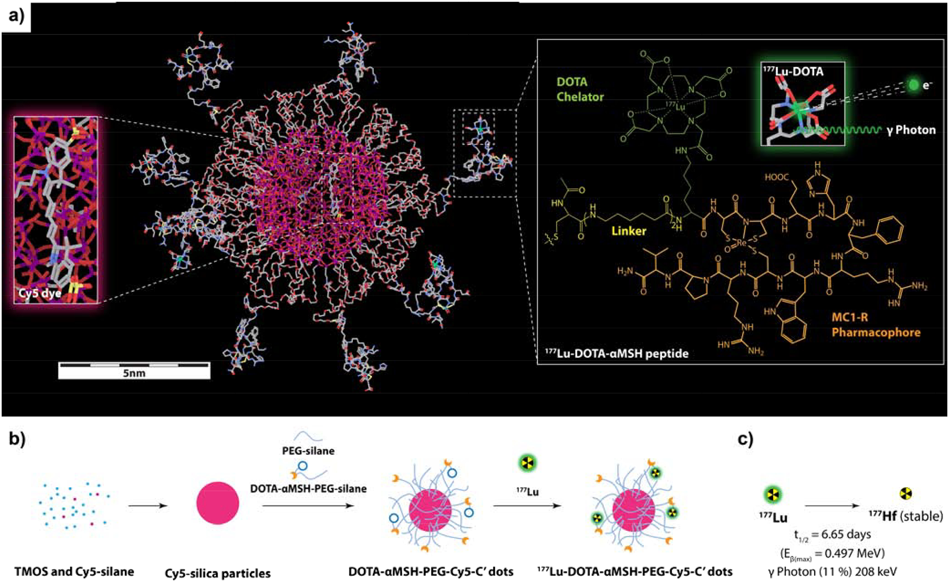
DOTA-aMSH-PEG-Cy5-C’ dots. (a) Molecular rendering of a 177Lu-labeled DOTA-αMSH-PEG-Cy5-C’ dot with Cy5 dye-encapsulating silica core (left insert) and the molecular structure of 177Lu-labeled MC1-R targeting cyclic DOTA-αMSH peptides on the right. (b) Schematic of the synthetic approach to DOTA-αMSH-PEG-Cy5-C’ dots and its radiolabeling with 177Lu. (c) Radioactive half-life and decay scheme of predominant beta-emitting particles and imageable gamma photons of 177Lu.